Article

Bacterial Community Composition and Presence of Plasmids in the Endosphere- and Rhizosphere-Associated Microbiota of Sea Fig (Carpobrotus aequilaterus)

by
Angela M. Sánchez-Salazar
1,2,
Jacquelinne J. Acuña
2,3,4,
Michael J. Sadowsky
5 and
Milko A. Jorquera
2,3,*
1
Programa de Doctorado en Ciencias de Recursos Naturales, Facultad de Ingeniería y Ciencia, Universidad de La Frontera, Av. Francisco Salazar 01145, Temuco 4811230, Chile
2
Laboratorio de Ecología Microbiana Aplicada (EMALAB), Departamento de Ciencias Químicas y Recurso Naturales, Universidad de La Frontera, Av. Francisco Salazar 01145, Temuco 4811230, Chile
3
The Network for Extreme Environment Research (NEXER), Scientific and Technological Bioresource Nucleus, Universidad de La Frontera, Av. Francisco Salazar 01145, Temuco 4811230, Chile
4
Millennium Institute Center for Genome Regulation (MI-CGR), Valenzuela Puelma 10207, La Reina 7800003, Chile
5
College of Agriculture, Food and Environmental Sciences, University of Minnesota, St. Paul, MN 55108, USA
*
Author to whom correspondence should be addressed.
Diversity 2023, 15(11), 1156; https://doi.org/10.3390/d15111156
Submission received: 10 October 2023 / Revised: 14 November 2023 / Accepted: 14 November 2023 / Published: 20 November 2023
(This article belongs to the Special Issue Diversity in 2023)

Abstract

:
The plant microbiome is one of the most important environments for ecological interactions between bacteria that impact the plant and the ecosystem. However, studies on the diversity of mobile genetic elements (such as plasmids) associated with the plant microbiome are very scarce. Here, we determined the bacterial community composition and the occurrence of plasmids in the microbiota associated with sea fig, Carpobrotus aequilaterus (N.E. Br.), a succulent species widely used as an ornamental plant in Chile. The abundance and composition of the endophytic and rhizospheric bacterial communities were determined by quantitative PCR (qPCR) and DNA metabarcoding analysis. Plasmid diversity in the plant microbiome was determined by plasmid DNA extraction and screened by endpoint PCR of backbone genes for four different incompatibility groups (Inc). The results showed about 106 copies of the 16S rRNA gene in the endosphere and rhizosphere, showing significant differences according to the diversity index. Proteobacteria (Pseudomonadota; 43.4%), Actinobacteria (Actinomycetota; 25.7%), and Bacteroidetes (Bacteroidota; 17.4%) were the most dominant taxa in both plant compartments, and chemoheterotrophy (30%) was the main predicted function assigned to the microbiota. Plasmid diversity analysis showed the presence of transferable plasmids in the endosphere and rhizosphere of C. aequilaterus, particularly among environmental plasmids belonging to the IncP and IncN incompatibility groups.

1. Introduction

Over the last several decades, conjugative plasmid-mediated horizontal gene transfer (HGT) occurring between bacteria in the environment has acquired a high relevance in public health and environmental sciences [1,2,3,4]. Conjugative plasmids are mobile genetic elements (MGEs), self-replicative and auto-transferable, that usually carry genes involved in antimicrobial resistance, virulence, heavy-metal resistance, and degradation of compounds. These MGEs confer adaptative traits to the bacterial host cells [5,6]. In this context, plasmid transfer events have been reported within complex bacterial communities in a wide variety of environments, such as soils, plants, and the animal gut [7,8,9,10]. Plasmid transfer between donor and recipient cells depends on many abiotic and biotic factors, including intrinsic plasmid incompatibility (Inc groups) [6]. The long-term prevalence of plasmids in an environment provides insight into how the bacterial community acquires new traits via HGT in the environment [11], where a single species or bacterial cell can act as a plasmid reservoir harboring one or more plasmid types [11]. In this context, niches that harbor a high diversity and richness of bacterial species might also be rich in exchangeable genes that may contribute to the competitiveness of bacteria in changing environments [12,13,14].
Many studies have now revealed that plants represent a complex environment harboring an enormous amount and diversity of microorganisms (also often called the plant microbiome) where plant–bacteria interactions are crucial for plant fitness and, therefore, for terrestrial ecosystem functioning [15,16]. Particularly, the rhizosphere (the soil directly influenced by plant roots) is widely recognized as one of the main hotspots of biological activity in many ecosystems. The rhizosphere often contains a great quantity and variety of nutrients for bacteria, which promote microbial activities and interactions [17], often associated with the concomitant gene exchange by conjugative plasmid events [18,19,20]. Recently, endophytic bacteria (those living in internal plant tissues) have also attracted the attention of microbiologists, plant physiologists, and agriculturists due to their close interactions with plant hosts, improving plant growth, stress tolerance, and disease control, particularly in agronomically relevant plant species [21]. However, since the role of bacteria as plasmid reservoirs promoting HGT in the plant microbiome is still poorly investigated and understood, major studies on plant–bacteria interactions mediated by plasmid transfer in natural vegetation or agroecosystems are required [22].
While several studies revealed plasmid transfer in compartments of model and agronomically relevant plant species [3,10,14], the presence, diversity, transfer rates, and prevalence of conjugative plasmids in the plant–soil continuum of urban areas or built environments remain an unexplored “black box” [17,22]. In this context, ornamental plants can acquire a special relevance because they are widely planted and grown in many outdoor (e.g., parks, gardens, squares, etc.) and indoor (e.g., houses, offices, hospitals, etc.) environments.
Carpobrotus aequilaterus (Haw.) N.E.Br. (Aizoaceae family, Magnoliopsida class) is a succulent plant species, whose common names include Chilean pigface, sea fig, ice plant, pigface, and angled pigface. This clonal plant species is native to South Africa and widely distributed in Australia, Mexico, the U.S.A., and Europe [23,24,25,26]. This plant was commonly introduced for ornamental use in residential areas, landscaping in coastal areas, and sand dune stabilization [23,24]. Its ability to resist drought and salt stress [23] makes this plant an aggressive invader in many coastal ecosystems [25,26], modifying soil properties such as nutrient content and water retention [24]. C. aequilaterus in Chile is popularly used as an ornamental plant in outdoor and indoor environments. Besides studies on its salt and drought tolerance or ornamental use, C. aequilaterus has also been studied in Chilean mining tailing due to its phytostabilization capacity of metals and sulfur and its abundant aerial biomass where organic matter is incorporated [27]. However, little is known about the microbiome of this urban plant species. Under this scenario, the present study was conducted to explore the composition of the bacterial community and the occurrence of plasmids in the rhizosphere and endosphere of C. aequilaterus collected in urban areas of Chile.

2. Materials and Methods

2.1. Plant Specimens

Specimens of C. aequilaterus were collected from an urbanized area in Calama city (22°27′45″ S 68°55′38″ W), located in the Atacama Desert in northern Chile. A cleaned spade was used to carefully remove intact specimens from the soil at a depth of 0–20 cm. The plants were placed in sterile polyethylene bags and transported to the Applied Microbial Ecology Laboratory (EMALAB) at Universidad de La Frontera. When the radicle emerged (after approximately 3 weeks), the specimens were planted and maintained individually in pots containing 600 g of a sand/soil substrate (3:1) under greenhouse conditions (temperature, 21 °C; humidity, 65%; natural light); they were watered once per week to field capacity for 60 days until sampling preparation.

2.2. Sampling and DNA Extraction

Six plants were independently used to isolate endosphere tissues rhizosphere soil samples. Samples of the endosphere from each plant specimen were processed as described by Govindasamy et al. [28], with a few modifications. The aerial parts of the plants were washed and surface-sterilized by repeated immersion in 70% (vol vol−1) ethanol for 3 min, followed by 2.5% (vol vol−1) sodium hypochlorite (NaClO) for 5 min. The plant tissues were exhaustively rinsed with sterile distilled water (SDW). Portions of the tissues (1–2 g) were aseptically cut, placed into sterile plastic tubes, stored at −80 °C, and then used for genomic DNA (gDNA) extraction. In parallel, samples of rhizosphere soil were processed as described by Lagos et al. [29]. Soil influenced by the roots was carefully collected in sterile polypropylene microtubes. Around 1–2 g of rhizosphere samples was stored at –80 °C and then used for gDNA extraction.
Total gDNA was extracted by using the E.Z.N.A.®Bacterial DNA Kit for Soil (Omega Bio-Tek, Norcross, GA, USA) according to the manufacturer’s instructions. Successful DNA isolation was confirmed by agarose gel electrophoresis using TAE 1× buffer, 1% agarose gels, and UV irradiation. The quality and quantity of the gDNA extracts were measured by the A280/A260 absorbance ratio using a microplate spectrophotometer (Multiskan GO, Thermo Fisher Scientific, Inc., Waltham, MA, USA). Appropriate dilutions of gDNA extracts were stored at −80 °C and used for further molecular analyses.

2.3. Bacterial Community Analyses

2.3.1. Quantification of the Bacteria

Dilutions of gDNA were used to estimate the abundance of bacteria (16S rRNA gene) in each plant compartment by quantitative PCR (qPCR) using the universal primer sets Bac1369F (5′–CGG TGA ATA CGT TCY CGG–3′) and Prok1492R (5′–GGW TAC CTT GTT ACG ACT–3′) with a StepOne Real-Time PCR System (ThermoFisher Scientific, Inc., Waltham, MA, USA) and PowerUpTM SYBRTM Green Master Mix (Applied BiosystemsTM, Foster City, CA, USA), using ~25 ng μL−1 of gDNA as described by Jorquera et al. [30]. The copy number of the 16S rRNA gene was calculated using Escherichia coli standards, built with dsDNA gBlock® Gene Fragments (Integrated DNA Technologies, Inc., Coralville, IA, USA), and the equation [concentration of the dsDNA gBlock® Gene Fragment in ng μL−1] × [molecular weight in fmol ng−1] × [Avogadro’s number] = copy number, following the method described by Whelan et al. [31]. Based on the standard curves, the absolute quantification (AQ) of the 16S rRNA gene was expressed as copy number per gram of dry soil or tissue (gene copy g−1 of soil or tissue) and later used to determine the relative quantification (RQ) of oriT genes in relation to the 16S rRNA gene, as described by Acuña et al. [32].

2.3.2. DNA Metabarcoding Analysis

The diversity, structure, and putative functions of the bacterial community in both plant compartments were investigated by using DNA metabarcoding analysis. The V4 region of the 16S rRNA gene was amplified by PCR using the primers 341F (5′–CCT ACG GGN GGC WGC AG–3′) and 805R (5′–GAC TAC HVG GGT ATC TAA TCC–3′) [33]. The PCR reactions were carried out with reagents supplied with GoTaq® Flexi DNA Polymerase (Promega Co., Madison, WI, USA) as follows: the denaturalization was performed at 95 °C for 10 min, the annealing temperature was initially set at 65 °C and then decreased by 0.5 °C every cycle until reaching 55 °C, a value that was maintained for 1 min, and the extension was performed at 72 °C for 3 min. The 16S rRNA gene libraries were gel-purified using the AxyPrep DNA Gel Extraction Kit (Axygen®, Corning, NY, USA), and library integrity was confirmed by the 4150 TapeStation System (Agilent, Santa Clara, CA, USA). Sequencing was conducted using barcode primers and the dual indexing method. The DNA fragments were pooled and paired-end sequenced on the Illumina MiSeq platform (Illumina, Inc., San Diego, CA, USA) at the Scientific and Technological Bioresources Nucleus of Universidad de La Frontera (BIOREN–UFRO), Temuco, Chile.

2.3.3. DNA Data Analyses

The data were analyzed using R (version 4.2.3). Firstly, QIIME 2 (Version 1.9.1) artifacts were loaded with the R package qiime2R into the R software and stored as a phyloseq-class object. The sequence data were rarefied to a sampling depth of 11,000 per sample. Chimeras were screened and removed with USEARCH using the UCHIME algorithm. Chimeric-free sequences were grouped into operational taxonomic units (OTUs) at 97% similarity by using UPARSE (version 7.0.1090) [34]. Further analyses were performed with the R package phyloseq (citation: R, qiime2R, phyloseq) for visualization and general data handling. OTUs with low prevalence (singletons) were filtered out (abundance <10 reads or prevalence <5% in all samples). Taxonomy was assigned using the DADA2 package (Version 1.16.0) [35], using the Silva taxonomy database (silva-132–99–nb–classifier.qza) and removing sequences identified as mitochondrial and chloroplast. For normalization, the feature table data (OTU counts) were transformed into relative abundance by dividing them by the total reads of each sample (total-sum scaling) and multiplying them by 1000 to achieve integer proportions. Taxonomy summaries were accomplished by merging OTUs of the same taxonomic rank and calculating their relative abundance in the samples. Richness (OTUs observed, Chao1) and alpha diversity indices (Shannon) were estimated within the phyloseq package [36]. Beta diversity was determined by calculating the Bray–Curtis dissimilarity between all samples and visualized in a non-metric multidimensional scaling (nMDS) plot with the R package vegan [37]. Global statistical significance of the differences between sample types was assessed by a permutational multivariate analysis of variance (PERMANOVA) using 999 permutations. The most correlated (R2 > 0.6) OTUs were added to the nMDS ordination plot. Finally, to explore the functional trait prediction of the bacterial communities, the functional annotation of prokaryotic taxa (FAPROTAX project) [38] database and scripts were used, assigning environmental functions as described by Zhang et al. [39].

2.4. Occurrence of Plasmids

2.4.1. Detection of Plasmids

The occurrence of conjugative plasmids in the C. aequilaterus-associated microbiota was determined by endpoint PCR as follows. First, plasmid DNA (pDNA) was extracted from endosphere and rhizosphere samples by using the QIAGEN Plasmid Mini Kit (QIAGEN, Hilden, Germany) according to methods described by Zhang et al. [40] and following the protocol of the manufacturer. Specific plasmid primer sets described by Gotz et al. [41] and Carattoli et al. [42] were used (Table 1) to amplify the pDNA fragments from genes of incompatibility (Inc) groups contained in plasmid “backbone” regions involved in the replication (IncP, trfA1/2, and korA; IncN, rep; IncW, oriV; and IncQ, oriV, and repB) and transfer (IncP, oriT, and traG; IncN, oriT; and IncW, oriT, and trwAB) of plasmids. The PCR reactions contained 1× PCR buffer, 1mM deoxynucleoside triphosphates (dNTPs), MgCl2 1.5 mM, 5U u Promega GoTaq® DNA polymerase (Promega Co., Madison, WI, USA), 5 ng μL−1 of pDNA, 0.5 mM solutions of each primer set, and nuclease-free water. Thirty-five cycles of amplification were performed, each consisting of 1 min of denaturation at 94 °C, 1 min of primer annealing at the annealing temperature as described in Table 1, and 1 min of primer extension at 72 °C, followed by a final extension step of 10 min at 72 °C. The PCR products were visualized under UV light following electrophoresis.

2.4.2. Quantification of IncP Plasmids

Since the endpoint PCR results indicated the occurrence of IncP plasmids in both plant compartments, the abundance of IncP plasmids was determined by qPCR using 5 ng μL−1 of pDNA and the primer set oriT1 (5′CAG CCT CGC AGA GCA GGA T 3′) and oriT2 (5′CAG CCG GGC AGG ATA GGT GAA GT 3′). The PCR conditions were as follows: enzyme activation step at 95 °C for 10 min, followed by 40 cycles of 15 s at 95 °C and 1 min of annealing plus extension at 60 °C. The PCR reactions were performed in triplicate with 20 μg L−1 of total plasmid DNA in a StepOne PlusTM Real-Time PCR System (Applied Biosystems, Waltham, MA, USA) using PowerUpTM SYBRTM Green Master Mix (Applied Biosystems) and following the manufacturer’s instructions. The abundance of IncP plasmids was estimated based on a standard curve built using pDNA from a derivative of the broad-host-range plasmid RK2 (IncP) [43].

2.5. Statistical Analysis

Assumption tests for normality (Shapiro–Wilk test) and equality of variance (Levene’s test) of data were initially performed. Statistical significance was calculated with either the two-sample t-test (normally distributed data with equal variances) or the Wilcoxon Rank Sum Exact Test (data with equal variances).

3. Results

3.1. Bacterial Community

The qPCR analysis revealed that the abundance of bacteria ranged from 6.9 × 10⁵ to 1.5 × 10⁶ copies of 16S rRNA gene g−1 of endosphere tissue and from 6.2 × 10⁵ to 2.2 × 10⁶ copies of 16S rRNA gene g−1 of rhizosphere soil (Figure 1A). Significant differences in copy number between the two plant compartments were not observed (p = 0.4894). In contrast, the alpha diversity analysis revealed significantly (p = 0.0005) greater richness in the endosphere samples compared with the rhizosphere samples, ranging from 440 to 568 of the observed OTUs and from 296 to 437 of the observed OTUs, respectively (Figure 1B). Similarly, a higher diversity was also observed in the endosphere samples than in the rhizosphere samples, as revealed by the Shannon index, with values from 5.8 to 6.1 and from 5.3 to 5.8, respectively. Statistical differences were observed in the Shannon (p = 0.0016) and Chao1 (p = 0.0011) indices between samples obtained from the endosphere and rhizosphere.
Differences between endophytic and rhizosphere bacterial communities were also revealed by beta diversity analyses (Figure 2). The nMDS analysis showed the presence of two clusters at 95% significance. In addition, the bacterial structure in the endosphere samples was mainly composed of members of the orders Myxococcales, Sapropirales, Gemmatimonadales, and Xanthomonadales, whereas the rhizosphere comprised two groups, one characterized by the Actinomycetales and Myxococcales orders, and the other by the Vibrionales and Rhodobacterales orders (Figure 2).
Taxonomic analyses of the bacterial community revealed that the phylum Proteobacteria showed a high relative abundance in the endosphere (43.4%) and rhizosphere (39.7%), followed by the Actinobacteria (25.7%) in the rhizosphere and the Bacteroidetes in the endosphere (17.4%) (Figure 3A). Other abundant phyla found in the endosphere and rhizosphere samples were the Acidobacteria (5.4% and 4.6%, respectively), Gemmatimonadetes (5.1% and 2.3%, respectively), and Verrumicrobia (3.6% and 1.9%, respectively). Moreover, assignments at the order level (Figure 3B) indicated that members of the Actinomycetales order were the most abundant (21.3%) in the rhizosphere samples, followed by members of the Xanthomonadales (5.5%), Burkholderiales (5%), and Sphingomonadales (4.8%) orders. In the endosphere samples, members of the Xanthomonadales order were the most abundant (11.4%), followed by members of the Burkholderiales (7%), Actinomycetales (6.9%), and Sphingomonadales (6.8%) orders.
Family-level analyses showed higher relative abundances of members of the Xanthomonadaceae (11%), Chitinophagaceae (10%), and Sphingomonadaceae (7%) families in the endosphere samples, whereas Flavobacteriaceae (6.8%) and Nocardioidaceae (6.7%) showed the higher relative abundances in the rhizosphere samples (Figure 3C). Lastly, genus-level analyses showed greater relative abundances of the genera Flavobacterium (9.6%) and Rhodanobacter (Xanthomonadales) (9.5%) in the endosphere samples, whereas members of the genus Streptomyces (5.8%) had the greatest relative abundance in the rhizosphere samples (Figure 3D). However, statistical differences (p < 0.05) between the endosphere and the rhizosphere microbiota were only found for the genera Dokdonella, Sphingomonas, and Opitutus (Supplementary Table S1).
Despite these taxonomic differences, functionality appeared conserved, and the FAPROTAX analysis showed chemoheterotrophy and aerobic chemoheterotrophy as the major functions assigned to the microbiota in both studied plant compartments, with values equal to or higher than 30% (Figure 4). Overall, similar functions were attributed to the microbiota in the two plant compartments, particularly, functions related to fermentation and nitrogen cycling, such as nitrate reduction, nitrate respiration, nitrogen respiration, nitrate denitrification, and nitrite denitrification, among others. Interestingly, greater relative abundances of sequences were found for microorganisms involved in the degradation of aromatic compounds and in oxygenic photoautotrophy and for photosynthetic cyanobacteria in the rhizosphere compared with the endosphere. In contrast, higher abundances of sequences were found for microorganisms involved in methylotrophy and methanol oxidation in the endosphere compared with the rhizosphere.

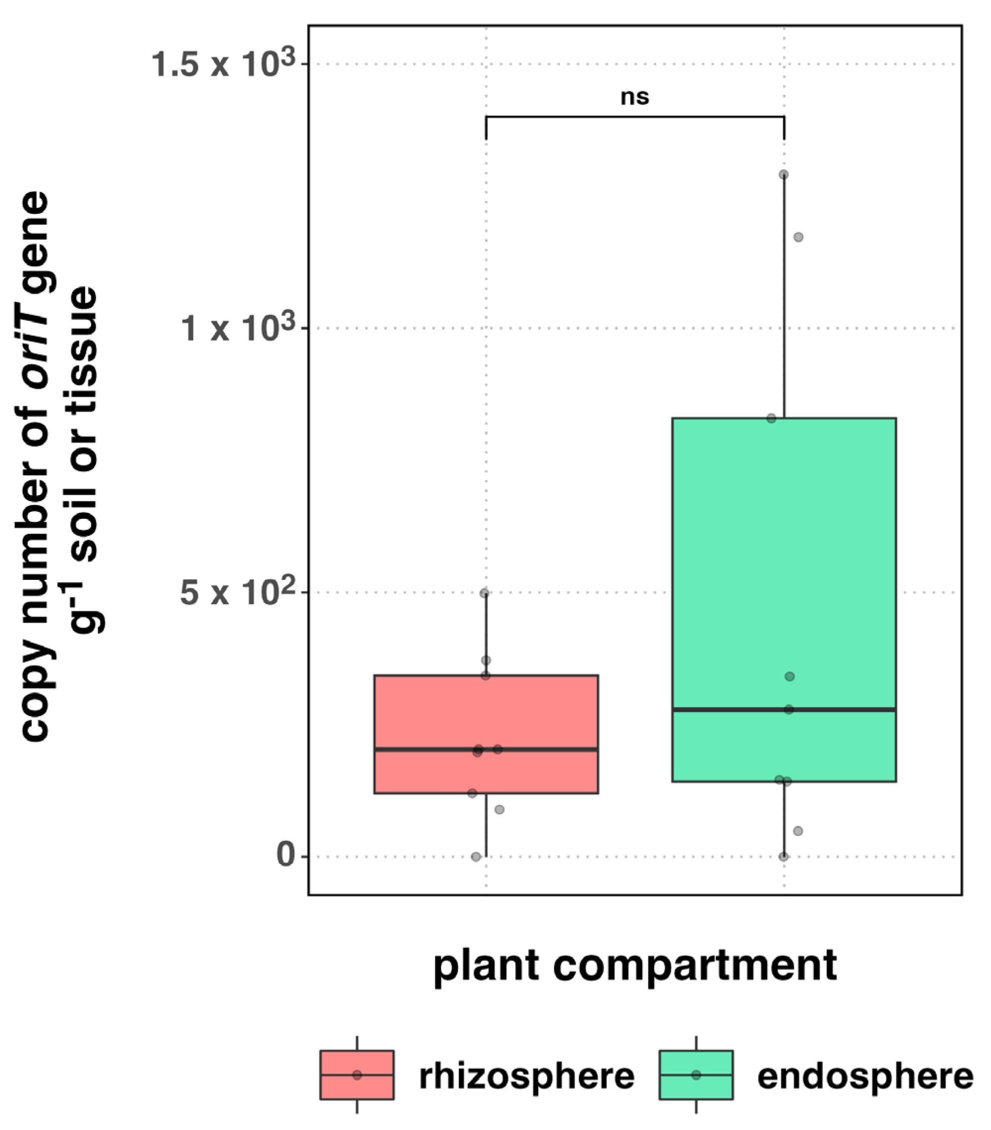
3.2. Occurrence of Plasmids

The use of specific endpoint PCR primer sets to identify genes involved in the replication and transfer of plasmids resulted in positive amplifications in both endosphere and rhizosphere samples. In the endosphere samples, amplicons were observed with the use of primer sets for the trfA1 (IncP) and oriT (IncN and IncP) genes, while amplicons for the oriV (IncW) and oriT (IncN and IncP) genes were observed in the rhizosphere samples (Table 2). In contrast to what observed for the endosphere, signals for the rep (IncN) and oriV (IncW) genes were only revealed in the rhizosphere by PCR.
We also specifically examined the abundance of IncP plasmids in both studied plant compartments by using a primer set for the oriT gene. The qPCR analysis showed from 0.49 to 12 × 102 copies of the oriT gene g−1 of endosphere tissue and from 1.2 to 8.9 × 10² copies of the oriT gene g−1 of rhizosphere soil (Figure 5). No statistical difference was found between the plant compartments (p = 0.175).

4. Discussion

4.1. Bacterial Communities

Here, we investigated the rhizospheric and endophytic bacterial communities, as well the plasmids in the C. aequilaterus (sea fig) microbiome. Our analysis of the bacterial communities showed that there were from 105 to 106 copies of the 16S rRNA gene g−1 of rhizosphere soil or endosphere tissue of C. aequilaterus. The endosphere values found here are lower than those observed for plants grown in desert ecosystems. Particularly, Zhang et al. [44] reported values ranging from 1010 to 1012 in the endosphere of Distichlis spicata and Pluchea absinthioides plants growing in the Atacama Desert of northern Chile. Similarly, greater values (from 107 to 1010 copies of the 16S rRNA gene g−1 of soil) were also reported in the rhizosphere of native plants (Atriplex spp.) from the Atacama Desert by Acuña et al. [32]. It appears that the 16S rRNA copy number is not consistent and likely varies by plant species and location. For example, Li et al. [45] reported similar values to ours in the rhizosphere of two species of Haloxylon growing in the desert and sand dunes, with about 106 copies of the 16S rRNA genes g−1 of soil. In this context, it was also indicated that limited nutrient and water in soils can influence the microbiome recruitment by plants, leading to the selection of specific soil bacterial communities with beneficial functions that might help plants under harsh environmental conditions [46,47]. Thus, plant roots can modulate positive and negative bacterial interactions, as observed across the altitudinal vegetation belt in the Atacama Desert by Mandakovic et al. [48]. Similarly, studies on two cactus species, Myrtillocactus geometrizans and Opuntia robusta, in arid ecosystems revealed that plant compartments, plant species, site, and season played a crucial role in the assembly of the microbiota [49].
Sequence analysis revealed that OTUs (440 to 568) and Shannon indices (5.8 to 6.1) were significantly higher for the endosphere of the examined plant than for the endosphere of plants from the Atacama Desert, determined by Zhang et al. [44], which ranged from 148 to 245 and from 2.28 to 4.41, respectively. Additionally, it is necessary to mention that C. aequilaterus is a succulent plant considered to be one of the most remarkable examples of convergent evolution, particularly because of the presence of large cells for water storage (90 to 95%) in the leaf endosphere [50]. The water content and particular endospheric characteristics of C. aequilaterus might regulate and promote higher richness and diversity compared with the rhizosphere in desert environments, as observed in our study.
Despite these results, our study also showed that the Shannon index values were significantly (p < 0.01) lower in the rhizosphere (5.3 to 5.8) compared with the endosphere and were much higher than those reported by Gao et al. [51], who observed values from 2.8 to 4.0 for the rhizosphere bacterial communities of the shrub Caragana microphylla grown in arid and semiarid environments. It should be noted, however, that while our results differed from those of other studies reporting that the rhizosphere harbors a major richness and diversity compared to the endosphere [39,52,53], some studies also highlighted that the endophytic bacteria of plants growing in dry environments represent an important niche of vital microbial functions for plant development, such as ACC deaminase production, nitrogen fixation, phosphorus solubilization, root biomass, among others [28,49,54]. Studies also revealed that endophytic characteristics are relevant for cactus seed germination and for the survival of plants growing in arid ecosystems [49,55]. Accordingly, the presence of a great diversity of microbes in the endosphere may be just as important as it is in the rhizosphere for plant growth under a variety of limiting conditions.
Our taxonomic analyses indicated that the phyla Proteobacteria (Pseudomonadota; from 39 to 43%), Actinobacteria (Actinomycetota; from 8 to 25%), and Bacteroidetes (Bacteroidota; from 13 to 17%) were the most abundant microbiota components in both studied compartments. Members of these phyla were observed as dominant taxa in the endosphere and rhizosphere of a great variety of plant species [24,56,57], including succulent plants such as Carpobrotus edulis, taxonomically related to sea fig, which showed relative abundances from 48 to 38% and from 29 to 27% for Proteobacteria and Actinobacteria in rhizosphere samples, respectively [24,58]. Moreover, Rodriguez-Caballero et al. [59] reported that Actinobacteria (43%) and Proteobacteria (42%) were dominant taxa in the rhizosphere of C. edulis. At the family level, members of the Sphingomonadaceae, Nocardioidaceae, Micrococcaceae, Chitinophagaceae, and Rhizobiaceae families were reported as the most representative in the C. edulis rhizosphere [58,59,60]. While our results similarly showed that the Chitinophagaceae and Sphingomonadaceae families were the most abundant taxa in the rhizosphere of C. aequilaterus, we also found that members of the families Flavobacteriaceae and Xanthomonadaceae were abundant.
Based on the FAPROTAX analysis, chemoheterotrophy and aerobic chemoheterotrophy were the most assigned predicted functions in the bacterial community, followed by functions involved in fermentation and nitrogen cycling (e.g., nitrate reduction). These results agree with several studies on plant–soil microbiomes, reporting that chemoheterotrophs are commonly observed and isolated from compartments of plants grown in arid ecosystems [56,61,62]. Similarly, according to Novoa et al. [24] and Rodriguez-Caballero et al. [59], Carpobrotus increases the soil organic content due to the decomposition of its recalcitrance tissue. This deposition may allow plant to selectively recruit bacteria taxa with specific functions, such as the degradation of organic compounds as a source of energy [24,59]. In agreement with this, we reported in both plant compartments the genus Flavobacterium, which is involved in nitrogen cycling and is a facultative anaerobic chemoheterotroph that uses organic compounds as electron donors and a source of cellular carbon [63].
Interestingly, aromatic compound degradation was another microbial function predicted to be present in the rhizosphere of C. aequilaterus. According to the taxonomic analysis, members of the family Sphingomonadaceae are commonly found in soil ecosystems and carry out a variety of metabolic processes such as the catabolism of various organic compounds, including aromatic compounds, e.g., chlorinated phenols, and xenobiotics. Moreover, some strains of Sphingobium spp. were shown to detoxify phenolic contaminants and pesticides (e.g., fenpropathrin, deltamethrin, fenvalerate, cypermethrin, and permethrin), suggesting that these microbes may be useful in treating sites with pesticide waste [64,65].

4.2. Occurrence of Plasmids

Our results also showed the occurrence of environmental plasmids in microbes present in both studied plant compartments. The presence of environmental plasmids was detected in the rhizosphere, endosphere, and phyllosphere of a variety of plants, such as wheat [66], pea [67], alfalfa [68], and barley [69], among others. In the Chilean terrestrial ecosystem, to our knowledge, studies on native plasmids in plant compartments are very limited. In this sense, Jorquera et al. [70] determined the occurrence and stability of plasmids encoding for Al tolerance in the rhizospheres of pasture and crop plants growing in acidic volcanic soils in southern Chile. Using the biparental mating assay and Al as a selective pressure, this study demonstrated the occurrence of Al tolerance and antibiotic resistance plasmids in bacterial populations in the rhizosphere. Interestingly, this study also revealed that independently of the presence of Al pressure, the plasmid pRPA21 was highly stable, whereas the plasmid pOPA21 was only stable in the presence of Al pressure. This study suggests that the presence of Al tolerance plasmids might play an important role in the adaptation of bacterial communities to Al toxicity in volcanic soils.
Of the plasmids examined, we observed positive PCR reactions for IncP and IncN plasmids in the endosphere and rhizosphere samples of C. aequilaterus. The genes encoded by the IncP and IncN plasmids are often associated with resistance to a wide variety of antibiotics and with tolerance to heavy metals and quaternary ammonium compounds used as disinfectants [6]. IncP and IncN plasmids were also observed in plant and human pathogenic strains with multiple antibiotic resistance traits and in some strains known to harbor pesticide-degrading genes encoded by plasmids [71,72,73]. Marcoleta et al. [74] also reported that antibiotic resistance genes found in desert ecosystems, such as the Antarctic soil, can be encoded in plasmids. Several studies demonstrated that plants can recruit plant- and compartment-specific bacterial communities, which can significantly differ in structure and activity depending on the plant compartment [39]. In each plant compartment, bacterial recruitment can be influenced and regulated by a wide battery of factors, including plant genotype, phenological stage, soil properties, soil fertilization, and environmental factors, among others [75]. Therefore, differentiation among bacterial communities could also influence the occurrence and/or copy number of plasmids in the endosphere and rhizosphere of C. aequilaterus. Another involved factor could be the incompatibility between environmental plasmids and the regulation of their prevalence and dissemination by the activity of each taxonomic group. However, major studies are still required to confirm plasmid differentiation in plant compartments of C. aequilaterus.

5. Conclusions

In this study, we found that C. aequilaterus harbors diverse bacteria living in the endosphere and rhizosphere. Our results also revealed a higher richness and diversity of the microbiota in the endosphere than that in the rhizosphere. While the total amount of 16S rRNA was not significantly different between the two compartments, a remarkable difference was observed in the alpha and beta diversity analysis, suggesting the relevance of the C. aequilaterus endosphere as an important plant niche. While previous studies showed that the plant rhizosphere is important for plant growth and survival in a variety of environments, our data suggest that the endosphere is likely equally important for plant growth. Our results also showed an important signal for oriT genes coding for transferable plasmids belonging to the IncN and IncP groups in both studied plant compartments. This study presents novel insights into the bacterial community associated with C. aequilaterus in urban environments. Thus, these ornamental plants might act as an important and unexplored reservoir and hotspot for the dissemination and exchange of bacterial genes with potential relevance for public health and environmental sciences.

Supplementary Materials

The following supporting information can be downloaded at: https://www.mdpi.com/1424-2818/15/11/1156/s1. Table S1. Differences in the dominant taxa between endosphere and rhizosphere samples from C. aequilaterus.

Author Contributions

Conceptualization, A.M.S.-S. and M.A.J.; formal analysis, A.M.S.-S., J.J.A. and M.A.J.; investigation, A.M.S.-S., J.J.A. and M.A.J.; writing—original draft preparation, A.M.S.-S. and M.A.J.; writing—review and editing, J.J.A. and M.J.S.; funding acquisition, M.A.J. and J.J.A. All authors have read and agreed to the published version of the manuscript.

Funding

This research was funded by “The National Fund for Scientific and Technological Development” (FONDECYT) project no. 1201386 and 1221228 (to M.A.J. and J.J.A.), by a Doctoral Scholarship provided by The National Agency for Research and Development of Chile (ANID) no. 21202218 (to A.M.S.-S.), by the Program “Apoyo a Profesores Patrocinantes de Alumnos de Pre y Postgrado”, code DI19-2016 from Vicerrectoría de Investigación y Postgrado of the La Frontera University (to A.M.S.-S.), and by the Millennium Science Initiative Program—ICN2021_044 (to J.J.A.).

Institutional Review Board Statement

Not applicable.

Data Availability Statement

The data presented in this study are available on request from the corresponding author (accession no. PRJNA1044468, NCBI).

Conflicts of Interest

The authors declare no conflict of interest.

References

  1. Gwenzi, W.; Musiyiwa, K.; Mangori, L. Sources, behavior and health risks of antimicrobial resistance genes in wastewaters: A hotspot reservoir. J. Environ. Chem. Eng. 2020, 8, 102220. [Google Scholar] [CrossRef]
  2. Marcoleta, A.E.; Varas, M.A.; Costa, J.; Rojas-Salgado, J.; Arros, P.; Berríos-Pastén, C.; Tapia, S.; Silva, D.; Fierro, J.; Canales, N.; et al. Mapping the microbial diversity and natural resistome of North Antarctica soils. bioRxiv 2021. [Google Scholar] [CrossRef]
  3. Riva, V.; Patania, G.; Riva, F.; Vergani, L.; Crotti, E.; Mapelli, F. Acinetobacter baylyi Strain BD413 Can Acquire an Antibiotic Resistance Gene by Natural Transformation on Lettuce Phylloplane and Enter the Endosphere. Antibiotics 2022, 11, 1231. [Google Scholar] [CrossRef] [PubMed]
  4. World Health Organization. Antimicrobial Resistance: Global Report on Surveillance; World Health Organization: Geneva, Switzerland, 2014. [Google Scholar]
  5. Chen, C.Y.; Fuqua, C.; Jackson, C.R.; Kadlec, K.; Top, E.M. Plasmid transfer-mechanisms, ecology, evolution and applications. Front. Microbiol. 2022, 13, 993628. [Google Scholar]
  6. Shintani, M.; Sanchez, Z.K.; Kimbara, K. Genomics of microbial plasmids: Classification and identification based on replication and transfer systems and host taxonomy. Front. Microbiol. 2015, 6, 242. [Google Scholar] [CrossRef]
  7. Li, X.; Stokholm, J.; Brejnrod, A.; Vestergaard, G.A.; Russel, J.; Trivedi, U.; Thorsen, J.; Gupta, S.; Hjelmsø, M.H.; Shah, S.A.; et al. The infant gut resistome associates with E. coli, environmental exposures, gut microbiome maturity, and asthma-associated bacterial composition. Cell Host Microbe 2021, 29, 975–987. [Google Scholar] [CrossRef] [PubMed]
  8. Macedo, G.; Olesen, A.K.; Maccario, L.; Hernandez Leal, L.; Maas, P.v.d.; Heederik, D.; Mevius, D.; Sørensen, S.J.; Schmitt, H. Horizontal gene transfer of an IncP1 plasmid to soil bacterial community introduced by Escherichia coli through manure amendment in soil microcosms. Environ. Sci. Technol. 2022, 56, 11398–11408. [Google Scholar] [CrossRef] [PubMed]
  9. Olesen, A.K.; Pinilla-Redondo, R.; Hansen, M.F.; Russel, J.; Dechesne, A.; Smets, B.F.; Madsen, J.S.; Nesme, J.; Sørensen, S.J. IncHI1A plasmids potentially facilitate horizontal flow of antibiotic resistance genes to pathogens in microbial communities of urban residential sewage. Mol. Ecol. 2022, 31, 1595–1608. [Google Scholar] [CrossRef]
  10. Xu, H.; Chen, Z.; Huang, R.; Cui, Y.; Li, Q.; Zhao, Y.; Wang, X.; Mao, D.; Luo, Y.; Ren, H. Antibiotic resistance gene-carrying plasmid spreads into the plant endophytic bacteria using soil bacteria as carriers. Environ. Sci. Technol. 2021, 55, 10462–10470. [Google Scholar] [CrossRef]
  11. Pilosof, S. Conceptualizing microbe–plasmid communities as complex adaptive systems. Trends Microbiol. 2023, 31, 672–680. [Google Scholar] [CrossRef]
  12. Taghavi, S.; Barac, T.; Greenberg, B.; Borremans, B.; Vangronsveld, J.; van der Lelie, D. Horizontal gene transfer to endogenous endophytic bacteria from poplar improves phytoremediation of toluene. Appl. Environ. Microbiol. 2005, 71, 8500–8505. [Google Scholar] [CrossRef]
  13. Wang, Y.; Xiao, M.; Geng, X.; Liu, J.; Chen, J. Horizontal transfer of genetic determinants for degradation of phenol between the bacteria living in plant and its rhizosphere. Appl. Microbiol. Biotechnol. 2007, 77, 733–739. [Google Scholar] [CrossRef]
  14. Wang, Y.; Li, H.; Zhao, W.; He, X.; Chen, J.; Geng, X.; Xiao, M. Induction of toluene degradation and growth promotion in corn and wheat by horizontal gene transfer within endophytic bacteria. Soil Biol. Biochem. 2010, 42, 1051–1057. [Google Scholar] [CrossRef]
  15. Berendsen, R.L.; Pieterse, C.M.; Bakker, P.A. The rhizosphere microbiome and plant health. Trends Plant Sci. 2012, 17, 478–486. [Google Scholar] [CrossRef] [PubMed]
  16. Haney, C.H.; Samuel, B.S.; Bush, J.; Ausubel, F.M. Associations with rhizosphere bacteria can confer an adaptive advantage to plants. Nat. Plants 2015, 1, 15051. [Google Scholar] [CrossRef] [PubMed]
  17. Van Elsas, J.D.; Turner, S.; Bailey, M.J. Horizontal gene transfer in the phytosphere. New Phytol. 2003, 157, 525–537. [Google Scholar] [CrossRef]
  18. Hassani, M.; Durán, P.; Hacquard, S. Microbial interactions within the plant holobiont. Microbiome 2018, 6, 1–17. [Google Scholar] [CrossRef] [PubMed]
  19. Mølbak, L.; Kroer, N. Root growth and exudate production define the frequency of horizontal plasmid transfer in the rhizosphere. FEMS Microbiol. Ecol. 2007, 59, 167–176. [Google Scholar] [CrossRef]
  20. Zhu, H.; Zhang, L.; Li, S.; Wang, Y.; Sun, S.; Chen, J.; Kou, S.; Jin, Q.; Xiao, M. The rhizosphere and root exudates of maize seedlings drive plasmid mobilization in soil. Appl. Soil Ecol. 2018, 124, 194–202. [Google Scholar] [CrossRef]
  21. Carrión, V.J.; Perez-Jaramillo, J.; Cordovez, V.; Tracanna, V.; de Hollander, M.; Ruiz-Buck, D.; Mendes, L.W.; van Ijcken, W.F.J.; Gomez-Exposito, R.; Elsayed, S.S.; et al. Pathogen-induced activation of disease-suppressive functions in the endophytic root microbiome. Science 2019, 366, 606–612. [Google Scholar] [CrossRef]
  22. Sánchez-Salazar, A.M.; Taparia, T.; Olesen, A.K.; Acuña, J.J.; Sørensen, S.J.; Jorquera, M.A. An overview of plasmid transfer in the plant microbiome. Plasmid 2023, 127, 102695. [Google Scholar] [CrossRef] [PubMed]
  23. Earnshaw, M.J.; Carver, K.A.; Charlton, W.A. Leaf anatomy, water relations and crassulacean acid metabolism in the chlorenchyma and colourless internal water-storage tissue of Carpobrotus edulis and Senecio? mandraliscae. Planta 1987, 170, 421–432. [Google Scholar] [CrossRef] [PubMed]
  24. Novoa, A.; Keet, J.H.; Lechuga-Lago, Y.; Pyšek, P.; Roux, J.J.L. Urbanization and Carpobrotus edulis invasion alter the diversity and composition of soil bacterial communities in coastal areas. FEMS Microbiol. Ecol. 2020, 96, fiaa106. [Google Scholar] [CrossRef]
  25. Campoy, J.G.; Acosta, A.T.R.; Affre, L.; Barreiro, R.; Brundu, G.; Buisson, E.; González, L.; Lema, M.; Novoa, A.; Retuerto, R.; et al. Monographs of invasive plants in Europe: Carpobrotus. Bot. Lett. 2018, 165, 440–475. [Google Scholar] [CrossRef]
  26. GISD. Global Invasive Species Database, ISSG, Species Profile: Carpobrotus edulis. 2018. Available online: http://www.iucngisd.org/gisd/ (accessed on 23 July 2023).
  27. Tapia, Y.; Bustos, P.; Salazar, O.; Casanova, M.; Castillo, B.; Acuña, E.; Masaguer, A. Phytostabilization of Cu in mine tailings using native plant Carpobrotus aequilaterus and the addition of potassium humates. J. Geochem. Explor. 2017, 183, 102–113. [Google Scholar] [CrossRef]
  28. Govindasamy, V.; George, P.; Ramesh, S.V.; Sureshkumar, P.; Rane, J.; Minhas, P.S. Characterization of root-endophytic actinobacteria from cactus (Opuntia ficus-indica) for plant growth promoting traits. Arch. Microbiol. 2022, 204, 150. [Google Scholar] [CrossRef] [PubMed]
  29. Lagos, L.M.; Navarrete, O.U.; Maruyama, F.; Crowley, D.E.; Cid, F.P.; Mora, M.L.; Jorquera, M.A. Bacterial community structures in rhizosphere microsites of ryegrass (Lolium perenne var. Nui) as revealed by pyrosequencing. Biol. Fertil. Soils 2014, 50, 1253–1266. [Google Scholar] [CrossRef]
  30. Jorquera, M.A.; Martínez, O.A.; Marileo, L.G.; Acuna, J.J.; Saggar, S.; Mora, M.L. Effect of nitrogen and phosphorus fertilization on the composition of rhizobacterial communities of two Chilean Andisol pastures. World J. Microbiol. Biotechnol. 2014, 30, 99–107. [Google Scholar] [CrossRef]
  31. Whelan, J.A.; Russell, N.B.; Whelan, M.A. A method for the absolute quantification of cDNA using real-time PCR. J. Immunol. Methods 2003, 278, 261–269. [Google Scholar] [CrossRef] [PubMed]
  32. Acuña, J.J.; Lagos, L.M.; Ogram, A.; Jorquera, M.A. Bacterial alkaline phosphatase in the rhizosphere of plants grown in Chilean extreme environments. Biol. Fertil. Soils 2016, 52, 763–773. [Google Scholar] [CrossRef]
  33. Herlemann, D.P.; Labrenz, M.; Jürgens, K.; Bertilsson, S.; Waniek, J.J.; Andersson, A.F. Transitions in bacterial communities along the 2000 km salinity gradient of the Baltic Sea. ISME J. 2011, 5, 1571–1579. [Google Scholar] [CrossRef]
  34. Edgar, R.C. UPARSE: Highly accurate OTU sequences from microbial amplicon reads. Nat. Methods 2013, 10, 996–998. [Google Scholar] [CrossRef] [PubMed]
  35. Callahan, B.J.; McMurdie, P.J.; Rosen, M.J.; Han, A.W.; Johnson, A.J.A.; Holmes, S.P. DADA2: High-resolution sample inference from Illumina amplicon data. Nat. Methods 2016, 13, 581–583. [Google Scholar] [CrossRef] [PubMed]
  36. McMurdie, P.J.; Holmes, S. phyloseq: An R package for reproducible interactive analysis and graphics of microbiome census data. PLoS ONE 2013, 8, e61217. [Google Scholar] [CrossRef] [PubMed]
  37. Oksanen, J.; Blanchet, F.G.; Friendly, M.; Kindt, R.; Legendre, P.; McGlinn, D.; Peter, R.; Minchin, P.R.; O’Hara, R.B.; Simpson, G.L.; et al. Vegan: Community Ecology Package (Version 2.5-6); The Comprehensive R Archive Network: Vienna, Austria, 2016. [Google Scholar]
  38. Louca, S.; Parfrey, L.W.; Doebeli, M. Decoupling function and taxonomy in the global ocean microbiome. Science 2016, 353, 1272–1277. [Google Scholar] [CrossRef] [PubMed]
  39. Zhang, Q.; Acuña, J.J.; Inostroza, N.G.; Duran, P.; Mora, M.L.; Sadowsky, M.J.; Jorquera, M.A. Niche differentiation in the composition, predicted function, and co-occurrence networks in bacterial communities associated with Antarctic vascular plants. Front. Microbiol. 2020, 11, 1036. [Google Scholar] [CrossRef]
  40. Zhang, T.; Zhang, X.X.; Ye, L. Plasmid metagenome reveals high levels of antibiotic resistance genes and mobile genetic elements in activated sludge. PLoS ONE 2011, 6, e26041. [Google Scholar] [CrossRef] [PubMed]
  41. Götz, A.; Pukall, R.; Smit, E.; Tietze, E.; Prager, R.; Tschäpe, H.; van Elsas, J.D.; Smalla, K. Detection and characterization of broad-host-range plasmids in environmental bacteria by PCR. Appl. Environ. Microbiol. 1996, 62, 2621–2628. [Google Scholar] [CrossRef]
  42. Carattoli, A.; Bertini, A.; Villa, L.; Falbo, V.; Hopkins, K.L.; Threlfall, E.J. Identification of plasmids by PCR-based replicon typing. J. Microbiol. Methods 2005, 63, 219–228. [Google Scholar] [CrossRef]
  43. Jorquera, M.; Yamaguchi, N.; Tani, K.; Nasu, M. A combination of direct viable counting, fluorescence in situ hybridization, and green fluorescent protein gene expression for estimating plasmid transfer at the single cell level. Microbes Environ. 2006, 21, 101–111. [Google Scholar] [CrossRef]
  44. Zhang, Q.; Acuña, J.J.; Inostroza, N.G.; Mora, M.L.; Radic, S.; Sadowsky, M.J.; Jorquera, M.A. Endophytic bacterial communities associated with roots and leaves of plants growing in Chilean extreme environments. Sci. Rep. 2019, 9, 4950. [Google Scholar] [CrossRef] [PubMed]
  45. Li, C.; Li, Y.; Tang, L.; Ikenaga, M.; Liu, R.; Xu, G. Soil microbial community shifts explain habitat heterogeneity in two Haloxylon species from a nutrient perspective. Ecol. Evol. 2023, 13, e9727. [Google Scholar] [CrossRef] [PubMed]
  46. Xing, R.; Gao, Q.B.; Zhang, F.Q.; Wang, J.L.; Chen, S.L. Large-scale distribution of bacterial communities in the Qaidam Basin of the Qinghai-Tibet Plateau. MicrobiologyOpen 2019, 8, e909. [Google Scholar] [CrossRef] [PubMed]
  47. Zhang, L.; Zhang, W.; Li, Q.; Cui, R.; Wang, Z.; Wang, Y.; Zhang, Y.-Z.; Ding, W.; Shen, X. Deciphering the root endosphere microbiome of the desert plant Alhagi sparsifolia for drought resistance-promoting bacteria. Appl. Environ. Microbiol. 2020, 86, e02863-19. [Google Scholar] [CrossRef] [PubMed]
  48. Mandakovic, D.; Aguado-Norese, C.; García-Jiménez, B.; Hodar, C.; Maldonado, J.E.; Gaete, A.; Latorre, M.; Wilkinson, M.D.; Gutiérrez, R.; Cavieres, L.A.; et al. Testing the stress gradient hypothesis in soil bacterial communities associated with vegetation belts in the Andean Atacama Desert. Environ. Microbiome 2023, 18, 1–17. [Google Scholar] [CrossRef]
  49. Fonseca-García, C.; Desgarennes, D.; Flores-Núñez, V.M.; Partida-Martínez, L.P. The Microbiome of Desert CAM Plants: Lessons from Amplicon Sequencing and Metagenomics; Elsevier Inc.: Amsterdam, The Netherlands, 2018. [Google Scholar] [CrossRef]
  50. Griffiths, H.; Males, J. Succulent plants. Curr. Biol. 2017, 27, R890–R896. [Google Scholar] [CrossRef]
  51. Gao, J.; Luo, Y.; Wei, Y.; Huang, Y.; Zhang, H.; He, W.; Sheng, H.; An, L. Effect of aridity and dune type on rhizosphere soil bacterial communities of Caragana microphylla in desert regions of northern China. PLoS ONE 2019, 14, e0224195. [Google Scholar] [CrossRef]
  52. Chen, J.; Xu, D.; Chao, L.; Liu, H.; Bao, Y. Microbial assemblages associated with the rhizosphere and endosphere of an herbage, Leymus chinensis. Microb. Biotechnol. 2020, 13, 1390–1402. [Google Scholar] [CrossRef]
  53. Dong, C.J.; Wang, L.L.; Li, Q.; Shang, Q.M. Bacterial communities in the rhizosphere, phyllosphere and endosphere of tomato plants. PLoS ONE 2019, 14, e0223847. [Google Scholar] [CrossRef]
  54. Zhang, Q.; White, J.F. Bioprospecting desert plants for endophytic and biostimulant microbes: A strategy for enhancing agricultural production in a Hotter, drier future. Biology 2021, 10, 961. [Google Scholar] [CrossRef] [PubMed]
  55. Karray, F.; Gargouri, M.; Chebaane, A.; Mhiri, N.; Mliki, A.; Sayadi, S. Climatic aridity gradient modulates the diversity of the rhizosphere and endosphere bacterial microbiomes of Opuntia ficus-indica. Front. Microbiol. 2020, 11, 1622. [Google Scholar] [CrossRef] [PubMed]
  56. Astorga-Eló, M.; Zhang, Q.; Larama, G.; Stoll, A.; Sadowsky, M.J.; Jorquera, M.A. Composition, predicted functions and co-occurrence networks of rhizobacterial communities impacting flowering desert events in the Atacama Desert, Chile. Front. Microbiol. 2020, 11, 571. [Google Scholar] [CrossRef]
  57. Zhang, W.; Bahadur, A.; Zhang, G.; Zhang, B.; Wu, X.; Chen, T.; Liu, G. Diverse Bacterial Communities from Qaidam Basin of the Qinghai–Tibet Plateau: Insights into Variations in Bacterial Diversity Across Different Regions. Front. Microbiol. 2020, 11, 554105. [Google Scholar] [CrossRef]
  58. Caravaca, F.; Torres, P.; Díaz, G.; Roldán, A. Elevated functional versatility of the soil microbial community associated with the invader Carpobrotus edulis across a broad geographical scale. Sci. Total Environ. 2022, 813, 152627. [Google Scholar] [CrossRef] [PubMed]
  59. Rodriguez-Caballero, G.; Caravaca, F.; Díaz, G.; Torres, P.; Roldán, A. The invader Carpobrotus edulis promotes a specific rhizosphere microbiome across globally distributed coastal ecosystems. Sci. Total Environ. 2020, 719, 137347. [Google Scholar] [CrossRef] [PubMed]
  60. Menicagli, V.; Balestri, E.; Giommoni, F.; Vannini, C.; Lardicci, C. Plastic litter changes the rhizosphere bacterial community of coastal dune plants. Sci. Total Environ. 2023, 880, 163293. [Google Scholar] [CrossRef]
  61. Liu, J.; Sun, X.; Zuo, Y.; Hu, Q.; He, X. Plant species shape the bacterial communities on the phyllosphere in a hyper-arid desert. Microbiol. Res. 2023, 269, 127314. [Google Scholar] [CrossRef]
  62. Zhang, M.; Li, X.; Shi, C.; Qiu, Z.; Han, J.; Wang, K.; Zheng, P.; Shi, F. Driving factors, co-occurrence networks, and metabolic profiles of soil bacterial communities within the root proximity of Amaranthus palmeri. J. Soil Sci. Plant Nutr. 2022, 22, 1928–1941. [Google Scholar] [CrossRef]
  63. Vymazal, J. Removal of nutrients in various types of constructed wetlands. Sci. Total Environ. 2007, 380, 48–65. [Google Scholar] [CrossRef]
  64. Balkwill, D.L.; Fredrickson, J.K.; Romine, M.F. Sphingomonas and related genera. In The Prokaryotes: Volume 7: Proteobacteria: Delta, Epsilon Subclass; Dworkin, M., Falkow, S., Rosenberg, E., Schleifer, K.-H., Stackebrandt, E., Eds.; Springer: New York, NY, USA, 2006; pp. 605–629. [Google Scholar]
  65. Stolz, A. Molecular characteristics of xenobiotic-degrading sphingomonads. Appl. Microbiol. Biotechnol. 2009, 81, 793–811. [Google Scholar] [CrossRef]
  66. Smit, E.; Wolters, A.; van Elsas, J.D. Self-transmissible mercury resistance plasmids with gene-mobilizing capacity in soil bacterial populations: Influence of wheat roots and mercury addition. Appl. Environ. Microbiol. 1998, 64, 1210–1219. [Google Scholar] [CrossRef] [PubMed]
  67. Kinkle, B.K.; Schmidt, E.L. Transfer of the pea symbiotic plasmid pJB5JI in nonsterile soil. Appl. Environ. Microbiol. 1991, 57, 3264–3269. [Google Scholar] [CrossRef]
  68. Mølbak, L.; Licht, T.R.; Kvist, T.; Kroer, N.; Andersen, S.R. Plasmid transfer from Pseudomonas putida to the indigenous bacteria on alfalfa sprouts: Characterization, direct quantification, and in situ location of transconjugant cells. Appl. Environ. Microbiol. 2003, 69, 5536–5542. [Google Scholar] [CrossRef]
  69. Musovic, S.; Oregaard, G.; Kroer, N.; Sørensen, S.J. Cultivation-independent examination of horizontal transfer and host range of an IncP-1 plasmid among gram-positive and gram-negative bacteria indigenous to the barley rhizosphere. Appl. Environ. Microbiol. 2006, 72, 6687–6692. [Google Scholar] [CrossRef] [PubMed]
  70. Jorquera, M.A.; Hernández, M.; Martínez, O.; Marschner, P.; de la Luz Mora, M. Detection of aluminum tolerance plasmids and microbial diversity in the rhizosphere of plants grown in acidic volcanic soil. Eur. J. Soil Biol. 2010, 46, 255–263. [Google Scholar] [CrossRef]
  71. Assinder, S.J.; Williams, P.A. The TOL Plasmids: Determinants of the Catabolism of Toluene and the Xylenes; Advances in Microbial Physiology; Academic Press: Cambridge, MA, USA, 1990; Volume 31, pp. 1–69. [Google Scholar]
  72. Dealtry, S.; Holmsgaard, P.N.; Dunon, V.; Jechalke, S.; Ding, G.-C.; Krögerrecklenfort, E.; Heuer, H.; Hansen, L.H.; Springael, D.; Zühlke, S.; et al. Shifts in abundance and diversity of mobile genetic elements after the introduction of diverse pesticides into an on-farm biopurification system over the course of a year. Appl. Environ. Microbiol. 2014, 80, 4012–4020. [Google Scholar] [CrossRef] [PubMed]
  73. Dunon, V.; Sniegowski, K.; Bers, K.; Lavigne, R.; Smalla, K.; Springael, D. High prevalence of IncP-1 plasmids and IS 1071 insertion sequences in on-farm biopurification systems and other pesticide-polluted environments. FEMS Microbiol. Ecol. 2013, 86, 415–431. [Google Scholar] [CrossRef]
  74. Marcoleta, A.E.; Arros, P.; Varas, M.A.; Costa, J.; Rojas-Salgado, J.; Berríos-Pastén, C.; Tapia-Fuentes, S.; Silva, D.; Fierro, J.; Canales, N.; et al. The highly diverse Antarctic Peninsula soil microbiota as a source of novel resistance genes. Sci. Total Environ. 2022, 810, 152003. [Google Scholar] [CrossRef]
  75. Compant, S.; Clément, C.; Sessitsch, A. Plant growth-promoting bacteria in the rhizo-and endosphere of plants: Their role, colonization, mechanisms involved and prospects for utilization. Soil Biol. Biochem. 2010, 42, 669–678. [Google Scholar] [CrossRef]
Figure 1. Abundance (A) and alpha diversity (B) of the bacterial community in the endosphere and rhizosphere of C. aequilaterus. Legend: ns, non-significant differences; ** denotes significant differences at p < 0.01 (n = 3); *** denotes significant differences at p < 0.001 (n = 3).
Diversity 15 01156 g001
Figure 2. Nonmetric multidimensional scaling (nMDS) analysis of the bacterial community found in the endosphere and rhizosphere of C. aequilaterus. The most abundant bacterial orders are highlighted. ** p < 0.01.
Diversity 15 01156 g002
Figure 3. Mean (n = 3) relative abundance of the taxonomic structure of the bacterial community in the endosphere and rhizosphere of C. aequilaterus at the phylum (A), order (B), family (C), and genus (D) levels.
Diversity 15 01156 g003
Figure 4. Mean (n = 3) relative abundances of predicted microbial functional groups of the bacterial community in the endosphere and rhizosphere of C. aequilaterus. Dots represent outlier values and bars represent the standard deviation.
Diversity 15 01156 g004
Figure 5. Quantification of IncP plasmids in the endosphere and rhizosphere of C. aequilaterus by quantitative PCR. Legend: ns, non-significant differences (p < 0.05; two-sample t-test). Dots represent outlier values and bars represent the standard deviation.
Diversity 15 01156 g005
Table 1. Primer sets used for PCR amplification in endosphere and rhizosphere samples of C. aequilaterus according to the different compatibility (Inc) groups.
Inc GroupRegion FunctionPrimer Sequence (5′-3′)Annealing Temperature (°C)
IncNrep 1ReplicationAGT TCA CCA CCT ACT CGC TCC G55
rep 2ReplicationCAA GTT CTT CTG TTG GGA TTC CG
oriT1TransferTTG GGC TTC ATA GTA CCC49
oriT2TransferGTG TGA TAG CGT GAT TTA TGC
IncPoriT1TransferCAG CCT CGC AGA GCA GGA T57
oriT2TransferCAG CCG GGC AGG ATA GGT GAA GT
trfA1 1ReplicationATG ACG ACC AAG AAG CG57
trfA1 2ReplicationAAC CCC CAG CCG GAA CTG
traG 1TransferCTG CGT CAC GAT GAA CAG GCT TAC C63
traG 2TransferACT TCC AGC GGC GTC TAT GTG G
IncQrepB 1ReplicationTCG TGG TCG CGT TCA AGG TAC G62
repB2ReplicationCTG TAA GTC GAT GAT CTG GGC GTT
IncWoriV 1ReplicationGAC CCG GAA AAC CAA AAA TA58
oriV2ReplicationGTG AGG GTG AGG GTG CTA TC
oriT 1TransferTCT GCA TCA TTG TAG CAC C51
oriT2TransferCCG TAG TGT TAC TGT AGT GG
Table 2. PCR amplification in endosphere and rhizosphere samples of C. aequilaterus revealed by specific primer sets for plasmids in different compatibility (Inc) groups.
ReplicationTransference
IncNIncPIncQIncWIncNIncPIncW
SamplereptrfA1 repBoriVoriToriTtraGoriT
EndosphereE1.1+++(+)
E1.2+++(+)
E1.3+++(+)
E2.1+++(+)
E2.2+++(+)
E2.3+++(+)
E3.1+++(+)
E3.2+++(+)
E3.3+++(+)
RhizosphereR1.1+(+)(+)+++(+)(+)
R1.2+(+)(+)+++(+)(+)
R1.3+(+)(+)+++(+)(+)
R2.1+(+)(+)+++(+)(+)
R2.2+(+)(+)+++(+)(+)
R2.3+(+)(+)+++(+)(+)
R3.1+(+)(+)+++(+)(+)
R3.2+(+)(+)+++(+)(+)
R3.3+(+)(+)+++(+)(+)
+: positive amplification; (+): weak signal of amplification; –: negative amplification.
Disclaimer/Publisher’s Note: The statements, opinions and data contained in all publications are solely those of the individual author(s) and contributor(s) and not of MDPI and/or the editor(s). MDPI and/or the editor(s) disclaim responsibility for any injury to people or property resulting from any ideas, methods, instructions or products referred to in the content.

