Article

Toxic Effect of Anionic Surfactants on Freshwater Sponge Lubomirskia baikalensis and Its Endosymbiotic Microalgae Chlorella sp.

by
Alyona Alexandrovna Nikonova
1,*,
Igor Borisovich Mizandrontsev
1,
Boris Nikolaevich Bazhenov
2,
Igor Veniaminovich Khanaev
1,
Olesya Viktorovna Shabalina
2,
Alexandra Alexandrovna Afanasyeva
1,
Tatiana Nikolaevna Avezova
1,
Anna Nikolaevna Chindyavskaya
1,2,
Alexander Nikolaevich Bityutsky
1,
Andrey Yurievich Kan
1,
Leonid Gennadievich Karikh
1,
Kristina Sergeevna Dubrova
1,
Svetlana Semyonovna Vorobyeva
1 and
Olga Yurievna Glyzina
1
1
Limnological Institute of the Siberian Branch of the Russian Academy of Sciences, 3, Ulan-Batorskaya Street, 664033 Irkutsk, Russia
2
Department of the General and Inorganic Chemistry, Chemical Faculty, Irkutsk State University of the Ministry of Science and Higher Education of the Russian Federation, 1, Karl Marx Street, 664003 Irkutsk, Russia
*
Author to whom correspondence should be addressed.
Diversity 2023, 15(1), 77; https://doi.org/10.3390/d15010077
Submission received: 4 October 2022 / Revised: 29 November 2022 / Accepted: 2 January 2023 / Published: 6 January 2023
(This article belongs to the Special Issue Diversity and Ecology of Freshwater Lake Microbial Communities)

Abstract

:
A number of reports on sponge diseases, including from Lake Baikal, have increased dramatically all over the world in recent years. Herewith, there are various hypotheses for sponge mortality. Lubomirskia baikalensis (phylum Porifera, order Spongillida) is a unique endemic freshwater sponge of Lake Baikal that contains a complex community of eukaryotic and prokaryotic endosymbiotic microorganisms. In this work, we present the first results for the effect of anionic surfactants viz. linear alkylbenzene sulphonates (LAS) at low 10 and 20 µg L−1 concentrations on Baikal sponge species and their symbiotic community as an experimental model. A new toxicity test protocol under conditions close to natural is proposed. It uses the sponge amoebocytes called as SA1-cells, which contain eukaryotic green microalgae Chlorella sp. These SA1-cells are shown to be representative indicator in assessing the impact of anionic surfactants. The acute toxic effect resulted in 97–100% sponge cell death in less than 48 h, as well as 100% symbiotic microalgae Chlorella sp. death over 72 h was noted under LAS solution (20 μg L−1) exposure during in vivo experiments. This includes the cell membrane fatty acid changes, change in the cell sizes, cell swelling, and lysis. Long term exposure to LAS solution (10 μg L−1) reflected in cellular stress (oxidative stress) and accompanied by malondialdehyde formation (0.16–2.0 μg g−1 of dry weight) during 14-day exposure was noted. Oxidative stress and mortality of L. baikalensis are associated with their low antioxidant activity. Trolox-equivalent antioxidant capacity (TEAC) found in the range from 0.00031 to 0.00077 Trolox equivalents for these freshwater sponges.

1. Introduction

The freshwater Lake Baikal is the deepest lake in the world, with a maximal depth of 1642 m [1,2]. The littoral zone contributes only ~1116 km2 (3.4%) to its total area of 32,822 km2 [3]. This is due to geomorphological peculiarities of the Baikal basin [4]. Nevertheless, at depths of up to 20 m, the Lake Baikal littoral zone contains more than 98% of all bottom-dweller diversity because of the minimal depths and maximal light intensity sufficient for photosynthesis. At depths up to 4 m, the biomass of bottom organisms achieves 620 kg per hectare, and at depths of 20–70 m it achieves 150 kg [5].
Freshwater sponges (Phylum: Porifera, Order: Spongillida) inhabit many lake ecosystems [6] including Tanganyika [7,8], Malawi [8], Michigan [9], Baikal, and others. Baikal sponges are one of the most abundant bottom invertebrates. According to modern taxonomy, family of endemic Lubomirskiidae and family of cosmopolitan Spongillidae represent sponges in Lake Baikal [6,10,11]. Endemic sponges Lubomirskia baikalensis (Pallas, 1776) (class Demospongiae, order Spongillida, family Lubomirskiidae), Baikalospongia bacillifera (Dybowsky, 1880) (class Demospongiae, order Spongillida, family Lubomirskiidae), and Baikalospongia intermedia (Dybowsky, 1880) (class Demospongiae, order Spongillida, family Lubomirskiidae) are the dominant sponge species in the lake. The cortical sponge Baikalospongia intermedia and the globular sponge Baikalospongia bacillifera cover the stony nearshore bottom from 1 m of depth. The branching sponge Lubomirskia baikalensis covers the stones from 1 m to the maximum depths where rocks exist. Some individuals were found at 1340 m of depth with deep-submergence vehicles Mir-1 and Mir-2 [12], but their abundance and species diversity are concentrated mainly at depths from 5 to 30–50 m [10,13].
The Lubomirskia baikalensis is the only branching sponge in Lake Baikal [14]. It contains a complex symbiotic assemblage. There are exosymbionts living on the sponge’s external surface (eukaryotic and prokaryotic microorganisms, protozoa, and crustacea, e.g., Brandtia parasitica and Brandtia latissima (order Amphipoda)), and endosymbiotic microorganisms inside the sponge cells and in intracellular space (microalgae, cyanobacteria, bacteria, archaea, fungi, and viruses). Photosynthetic green microalgae Chlorella sp. (synonyms: zoochlorellae, Zoochlorella parasitica) is the dominant (by biomass) symbiotic eukaryotic microorganism of L. baikalensis. According to recent publications, these algae might belong to genus Choricystis [15,16] or to genus Mychonastes [17]. Diatoms can occur as well [18].
Numerous picocyanobacteria developing in Lake Baikal plankton penetrate into healthy sponge body during water filtration and comprise an essential fraction of their microbiome [18]. A molecular approach based on the identification of the 16S rRNA and ribulose-1,5-bisphosphate carboxylase/oxygenase large subunit genes was applied to investigate diversity and phylogeny of bacterial phototrophs associated with four species of Lubomirskiidae in Lake Baikal. The phylogeny inferred from both genes represents three main clusters of Synechococcus associated with Baikal sponges [16]. Over 90% of the cyanobacterial molecular sequences for L. baikalensis revealed belonged to Synechococcus [18]. Sick sponge cyanobacteria fouling mainly represented by Synechococcales, Chroococcales, Oscillatoriales, and Nostocales [19]. Bacterial phyla (Bacteroidetes, Proteobacteria, Actinobacteria, Planctomycetes, Verrucomicrobia, Nitrospirae, OD1, and Chloroflexi) were identified in L. baikalensis as well.
The microbes living in sponges make up to 35% of the biomass of the symbiont community [20] and up to 40–60% of the sponge’s biomass [21]. Algal symbionts may comprise up to 75% of the cellular tissue in sponge-algal associations [21]. They are involved in photosynthesis which takes place even in deep cell layers of the sponge body, carbon fixation, nitrogen transformation, and anaerobic metabolism; they also perform protective functions [20].
The littoral zone is exposed to negative anthropogenic impact of biological and chemical pollutants, invasive organisms, and temperature changes to a greater extent than the pelagic zone. This can result in stress situations for different water organisms [3]. Over last years, changes in the structure of numerous benthic communities have been marked in the nearshore zone of Lake Baikal [22]. The oxidative stress of nearshore phytoplankton was revealed [23]. Widespread mortality of sponges including the endemic L. baikalensis along the Lake Baikal perimeter was noticed [10,24,25]. In recent years, the number of reports on sponge diseases have increased dramatically, with marine sponge populations decimated throughout the Mediterranean and Caribbean seas. The increased prevalence of marine sponge disease in Papua New Guinea, the Great Barrier Reef, and in the reefs of Cozumel, Mexico [26] has also been reported. The sponge population declines and, as a result, the filtration efficiency decreases, especially in freshwater ecosystems. This can lead to irreversible consequences regarding health both of aquatic organisms and the ecosystem as a whole.
At present, there are various hypotheses for the mass mortality of sponges in Lake Baikal, which are associated with global warming [10,26], anthropogenic impact [11,27], methane concentration in coastal water [10,28], and changes in viral assemblages [29]. The presence of anionic surfactants at 40 μg/L concentration in Tyya River, the tributary of Lake Baikal, was fixed using the methylene blue active substances (MBAS) method in 2019 [30]. A source of these pollutants is shown to be untreated wastewater with the surfactant concentration of 170 μg L−1. The structure of anionic surfactants in the samples was related to widespread linear alkylbenzene sulfonates (LAS) according to gas chromatography coupled with mass spectrometry [30]. For comparison, the concentrations of alkylbenzene sulfonates in freshwater and marine ecosystems of the world are estimated from 1–7 to 250–300 μg L−1 [31,32,33,34]. It has been reported that relatively low LAS concentrations (5–20 μg L−1) can cause toxic effects on aquatic microorganisms including oxidative stress, extreme growth of cyanobacteria, and cell lysis [35,36,37]. So, the effect of anionic surfactants on sponges of Lake Baikal may be one of the reasons for sponge disease.
Unfortunately, little attention has been paid to the determination of the mentioned toxicants in the environment and their effect on water organisms. This study aims to assess the impact of LAS on L. baikalensis and its endosymbiotic microalgae Chlorella sp. as a whole organism under conditions close to natural.

2. Materials and Methods

2.1. Sponge Sampling

2.1.1. Choosing the Sponge Target Species for the Experiments

The Lubomirskia baikalensis target species was choosing as more convenient experimental model in the contrary to Baikalospongia bacillifera and Baikalospongia intermedia due to the different structures of these sponges. The B. bacillifera is very hard sponge. A cell sampling from its body without cell damage is problematic. The B. intermedia consistency is too soft and friable. Sampling and transportation of these sponges to the laboratory are difficult. The L. baikalensis consistency is soft, elastic, and non-friable, which allows for taking its cells with a mechanical pipette to exclude serious sponge damage.

2.1.2. Lubomirskia baikalensis Sampling for In Vitro Aquarian Experiments

Branching sponges L. baikalensis were collected by self-contained underwater breathing apparatus (SCUBA) divers from the depths 5–20 m at nearshore stations of Lake Baikal. Sponges for in vitro toxicity test were collected in 2021 at sampling site No. 1 (N 51°52′01.2″, E 104°49′38.1″) in Listvennichnyi Bay from the depths of 10–20 m in the southern basin of Lake Baikal (Figure 1). This sampling site is located not far from Listvyanka Settlement, which is a large recreation center of the south-west coast of Lake Baikal. The L. baikalensis population has almost disappeared in Listvennichnyi Bay over two last decades. Nevertheless, epilithic, visually healthy, undamaged branching sponges of ≤12 cm branch length (n = 12) were taken from sampling site No. 1. This can be explained by the position of the site No. 1 with the underwater ridge in the source of the Angara River. This geomorphological peculiarity provides deep water motion from the depths up to 500 m toward the surface in this region. This water is suggested to be cleaner as opposed to surface water in Listvennichnyi Bay. After sampling sponges were put into oxygen aerated 12 L thermoses preventing the contact of sponges with atmospheric air, transported to the laboratory, and placed into 20 L volume aquariums.

2.1.3. Lubomirskia baikalensis Sampling for In Situ Biochemical Analysis

Sponge samples for in situ biochemical analysis were collected from south and north basins of the lake (Figure 1) from depths of 5–20 m. Sponges from sample site No. 2, located in the region of Kultuk Settlement (N 51°43′42.2″, E 103°44′01.7″) and No. 3 located in the region of Bolshiye Koty Settlement (N 51°54′08.7″, E 104°49′38.1″) were collected in September 2019 to reveal their oxidative stress (n = 20) (Figure 1). The stations No. 2 and 3 differed by the intensity of anthropogenic load. All samples were frozen at –70 °C.
Sponges from the sample sites No. 4 located in Ulanovo region (N 51°47′47.6″, E 104°31′34.1″), No. 5 located in the region of the inoperative Baikalsk City paper mill, No. 6 near Bolshoi Ushkanii Island, No. 7 in the region of Elokhin Cape (N 54°33′05.8″, E 108°39′55.0″), and No. 8 in Tompuda Bight (N 55°07′43.2″, E 109°43′42.6″) were collected in 2022 (June–July) from the depths of 5–20 m to estimate L. baikalensis antioxidant activity (n = 11, 2–3 samples were collected from each station) (Figure 1). All samples were also frozen.

2.2. Conditions for Cultivation of L. baikalensis

Near-natural conditions were provided for cultivation of L. baikalensis. The biomass of sponges in each aquarium did not exceed 300 g. The aquariums were filled with 10–12 L of bottled Lake Baikal water from a depth of 400 m and with 3–5 L of fresh pelagic surface Lake Baikal water. Pelagic water was collected from the offshore station located at 1 km from the shoreline (N 51°51.427′, E 104°50.020′) in Listvennichnyi Bay. The depth to bottom was 0.5 km. Surface water from 0.3 m depth was sampled using a plastic bottle, transported to the laboratory and poured into aquarium. The concentration of anionic surfactants in deep-bottled water did not exceed 3.0 μg L−1.
Zooplankton were collected at the same pelagic station once a week to feed the sponges. The sampling was carried out at the same pelagic station using an Apstein-type net with 100 μm mesh size. Live zooplankton (~0.2 g) were put into a bottle filled with pelagic water. We changed 10–30% of aquarium water once in 2–4 days. Water temperature was kept at +8 °C, as in Lake Baikal, during the sampling period. It is known that sponges of Lake Baikal can live in a wide range of temperatures from +2 to +24 °C in the nearshore zone. The light regime was a 12-hour light–dark cycle. Light intensity (illuminance) was 2 lux at the bottom of the aquarium. The light source provided 500–600 nm range in the electromagnetic spectrum. Light conditions were close to natural at depths of 10–15 m in Lake Baikal. The pH varied from 7.66 to 8.02 at 25 °C.
The dissolved oxygen analysis was carried out by iodometric titration according to Winkler [38]. The oxygen concentration of 10–20 mg L−1 in water was achieved by aeration with gaseous oxygen supplied from a high-pressure gas cylinder with a sprayer at the constant flow of ~3.2 ± 0.2 L per minute as well as with atmospheric air using an air compressor, which also saturated water with CO2 at the constant air flow of ~5.0 ± 0.3 L per minute. The oxygen concentration in Lake Baikal water varies from 10 to 16 mg L−1. The higher concentrations up to 20 mg L−1 are nontoxic and friendly for sponges. This can be explained by the oxygen concentration in photosynthetic organisms, which is higher than in the environment. The daily oxygen evasion into the atmosphere across the air–water interface in Lake Baikal confirms this fact [39,40]. Oxygen aeration allows us to prevent the parasitic water mold Saprolegnia sp.’s growth (order Saprolegniales, family Saprolegniaceae) and to provide better adaptation and survival for sponges and its internal and external symbionts. For example, it was shown that aeration with atmospheric air results only in gradual or mass mortality of symbiotic Brandtia parasitica, which reflects the unusual conditions for these organisms.

2.3. Cell Viability Test Procedure

Cell suspension from the sponge body of ~50 μL volume was taken under the water with a mechanical pipette. This allowed us to obtain sponge cells without damaging them. Suspension was placed into a plastic Eppendorf and stored in a thermos at +8 °C. Microscopy was carried out immediately after sampling. For this purpose, 10 µL of cell suspension was placed onto a microscope slide. Methylene blue (tetramethylthionine chloride, C16H18ClN3S) aqueous solution (3 µL, C = 0.007 g L−1, CM = 0.02 mM) was added to the sample. The cover glasses of 0.17 mm thickness and 15 × 15 mm size were used. The microscopic slides with samples were cooled to +8–12 °C during microscopy.
Live and dead cells of sponges and symbiotic Chlorella sp. were counted using an Axiovert inverted microscope (Carl Zeiss, Göttingen, Germany) at 1000× magnification or Olympus CX23 Binocular Microscope (Olympus, Tokyo, Japan) with 100× (oil) infinity plan objectives, 10× focusable eyepieces with 20 mm field of view, and the LED-transmitted illumination. Each sample was analyzed in 10–12 replicates (10–12 fields of view) and 1–70 cells in each field of view were analyzed in every replicate. The sum number of screened cells was up to 230. The number of live unstained cells and dead, blue-stained ones was counted within 1–3 min and percentages of live and dead cells were calculated.

2.4. Blank Samples–The “Control Sponges”

The blank samples were analyzed before the anionic surfactants toxicity assessment. To select sponge individuals that would reflect representatively the viability of L. baikalensis under LAS impact, we sampled visible, healthy, adult sponges in the lake. These sponges were cultivated for 10 days in clean water after sampling. Two parameters were controlled: the percentages of live/dead sponge amoebocytes containing endosymbiotic microalgae Clorella sp. (Division: Chlorophyta) and the presence of lipid peroxidation [41] and oxidation stress marker vis. MalonDiAldehyde (MDA) in sponge biomass. Sponges were considered to be “control sponges” if the percentages of live cells was ≥80% and MDA was not revealed. Fatty acid content and profiles were investigated before toxicity testing as well. Control sponges were used in the subsequent experiments.

2.5. Linear Alkylbenzene Sulfonate Toxicity Studies

The toxic effect of linear alkylbenzene sulfonates (LAS) on sponge cells was evaluated under near-natural conditions. The standard solution of sodium linear alkylbenzene sulfonates (GSO 8578-2004, C = 100 mg mL−1, Analytik-Him, Russia) containing 20 prevailing isomers related to C10, C11, C12, and C13 homologue groups was used in toxicity test experiments. To prepare LAS stock solution, 10 μL of standard solution were diluted in 10 mL of bi-distilled water to obtain 100 μg mL−1 concentration. The stock solution was added into aquariums in which the control sponges were cultivated. The LAS concentrations in aquarian water after LAS addition were 10 or 20 μg L−1 and were stable during the experiments. It should be said that two mentioned concentrations were chosen according to concentration range of LAS in Lake Baikal surface water from <10 μg L−1 (n = 70) to 10–20 μg L−1 (n = 32) [23]. The toxicity test protocol aimed to establish the LAS toxicity to freshwater sponge L. baikalensis and included live/dead cell counting, fatty acid and MDA analysis. Mentioned analyses were realized in the same time intervals of 12 h duration.

2.6. Fatty Acid Qualitative and Quantitative Analysis

2.6.1. Lipid Extraction and Fatty Acid Derivatization

The brilliant green sponge cell biomass was wrung out into glass. Biomass was mixed with a glass stick. To calculate the biomass humidity according to gravity measurements, ~0.4 g of sponge biomass was placed into a 1.5 mL plastic microcentrifuge tube and dried at 50 °C to achieve the constant weight. To extract lipids, ~0.2 g of the biomass was placed into a 2 mL plastic microcentrifuge tube and sonicated for 5 min with 1.2 mL of Folch mixture (chloroform–methanol, 2:1, by volume). Distilled water (~0.2 mL) was added to the extracts, shaken for 10 s and centrifuged at 13,000 rpm for 3 min. The extract (lower layer) was put into glass 10 mL vial. The solvent was evaporated using argon gas stream at 30 °C. Thereafter 4.5 mL of 2% H2SO4 solution in methanol was added immediately to dry extract. The obtained solution was heated during 1.5 h at 55 °C. Fatty acid methyl esters (FAMEs) were extracted with n-hexane (3 mL × 2 × 2 min). The extract was dried with anhydrous Na2SO4 and concentrated to ~0.5 mL using an argon stream. Note: 1 mL of water was added to the solutions before the second extraction. The di-n-decyl ether (C20H42O) solution in n-hexane was used as an internal standard for quantitative analysis.

2.6.2. Fatty Acid Analysis by Gas Chromatography Coupled with Mass-Spectrometry

The gas chromatography coupled with mass spectrometry (GC-MS) method was used to analyze FA composition of L. baikalensis samples. The extracts were analyzed using the gas chromatograph coupled to mass spectrometric detector “6890B GC System, 7000C GC/MS Triple Quad” (Agilent Technologies Inc., Wilmington, DE, USA) with Optima-17MS column (30 m × 0.25 mm, 0.25 μm, Macherey-Nagel GmbH and Co. KG., Düren, Germany). The injector temperature was 290 °C; the injection volume was 2 μL in splitless mode; the quadrupole temperature was 150 °C; the ion source temperature was 230 °C; the ionization energy was 70 eV. Chromatography of the extracts was carried out by heating the column from the initial temperature of 80 °C (0.5 min retention) up to 310 °C at a heating rate 2 °C/min (5 min retention). Chromatographic peaks were detected in the m/z range of 40–500.
The mass spectra of fatty acid methyl esters were identified using the NIST Mass Spectral Library. The calibration function was obtained in the range of concentrations of fatty acid sum from 40 to 540 μg in a sample using “F.A.M.E. Mix C4–C24” (Supelco, Bellefonte, PA, USA) and “Methyl cis-4,7,10,13,16,19-Docosahexaenoic ester” (10 mg mL−1 in heptane) (Supelco, St. Louis, MO, USA). The calibration coefficients for individual substances and for the groups of saturated fatty acids (SFA), monounsaturated fatty acids (MUFA), polyunsaturated fatty acids (PUFA), and demospongic fatty acids (DSFA) were calculated to quantitate FA that were absent in standard mixtures [42].

2.7. Malondialdehyde Content Determination

To reveal the lipid peroxidation processes in collected sponges, the malondialdehyde content in sponge biomass was analyzed. Sample preparation was carried out according to Al-Rashed et al.’s method [43] with modifications. Sponge biomass (~0.2 g at ~95–98% humidity) was placed into a 2 mL round-bottom plastic Eppendorf taste-tube. Trichloroacetic acid (TCA) 10% solution (1 mL) was added. Samples were sonicated for 5 min in 40 GHz ultrasonic bath and centrifuged at 13,000 rpm for 3 min. The supernatant was placed into a 1.5 mL conic-bottom Eppendorf taste-tube, centrifuged at 13,000 rpm for 3 min, and moved into a 5 mL glass vial with 2 mL of 0.5% thiobarbituric acid (TBA) solution in 10% TCA. The vial was capped with a plastic screw cap and kept for 20 min at 95 °C. Then, the vial was opened, and the solution was kept for 10 min at 95 °C. The solution volume was normalized to the initial volume. The obtained solutions were cooled and centrifuged at 13,000 rpm for 3 min. Absorbance of pink MDA-TBA complex was measured using a double-beam UV-Vis Cintra-20 spectrophotometer (GBC Scientific Equipment, Braeside, Australia) and standard quartz cuvettes of 1 cm path length. The MDA absorption was measured at 532 nm. Baseline absorption was measured at 532 nm using the solution obtained by mixing of 1 mL of 10% TCA with 2 mL of 0.5% TBA in 10% TCA.

2.8. Total Antioxidant Activity (AOA) of Lubomirskia baikalensis

2.8.1. Extraction of Antioxidants

To estimate L. baikalensis antioxidant capacity, the frozen sponges were used. Frozen sponges were cut to separate pieces. Sponge biomass was not wrung out and the whole sponge including the skeleton was prepared. Masses of wet samples varied from 0.4 to 2.0 g. The humidity was estimated gravimetrically, with values from 83.9 to 93.3%. Fractions of sponge antioxidants were extracted for 2 min with 1:1 by volume ethanol:acetone mixture (1.5 mL) using a mortar. The second extraction was carried out for 1 min with acetone (1.0 mL). The extracts were combined, centrifuged, and kept in the dark until the analysis. Each sample was analyzed in two replicates. The analysis of obtained crude extracts was carried out immediately.

2.8.2. Trolox Equivalent Antioxidant Capacity (TEAC) Assay

The Trolox-equivalent antioxidant capacity (TEAC) assay was used to determine the total antioxidant capacity (TAC) of freshwater sponge Lubomirskia baikalensis and its endosymbionts as a whole. TEAC assay is based on the ability of antioxidants to quench activity of long-lived stable radical cation 2,2′-Azino-Bis(3-ethylbenzoThiazoline-6-Sulfonic acid) (ABTS•+)-containing chromophore group responsible for its intensive blue color. To prepare the stable stock solution of ABTS•+ 14.5 mg of ammonia persulfate (NH4)2S2O8 were diluted in 25 mL of bi-distilled water to obtain 0.0025 M solution. Then, 10.6 mg of ABTS•+ diammonium salt (HPLC grade, Sigma-Aldrich, USA) was diluted in 2.6 mL of bi-distilled water and 2.6 mL of (NH4)2S2O8 solution was added. The stock solution was kept in the dark at room temperature for 12–16 h before use. At the beginning of the analysis day, the ABTS•+ working solution was obtained by the dilution in ethanol of the stock solution to an absorbance of 0.70 ± 0.02 AU at 730 nm [44]. The absorbance was measured using an SF-2000 spectrophotometer (OKB-Spectr, St. Petersburg, Russia) and PE-5300V spectrophotometer (ECROSKHIM Co. Ltd, St. Petersburg, Russia) at 37 °C and standard quartz cuvettes of 1 cm path length. Two mL of ABTS•+ working solution was placed into a cuvette. Then, 20 μL of sponge extract with a known concentration of dry substance was added to ABTS•+ working solution and the decreased value of absorbance was measured in 4 min. The next portions of the sponge extract were added into a cuvette every 4 min till absorbance value was ≤0.3 AU. The calibration function was obtained for ABTS•+ solutions. Baseline absorption was measured using distilled water in the parallel cuvette. Results of the measurements were recalculated to the sponge’s dry weight (d.w.).

2.8.3. TEAC Data Presentation

The obtained data were compared with that of Trolox (6-hydroxy-2,5,7,8-tetramethylchroman-2-carboxylic) (Sigma-Aldrich, St. Louis, MO, USA), a water-soluble vitamin E analog. Antioxidant capacity of Trolox was stated as 1.0. The values of ABTS•+ scavenging by the obtained crude extracts of L. baikalensis were expressed as Trolox-equivalent antioxidant capacity (TEAC) units. This abbreviation is the analogous with Trolox equivalents (TE). The inhibition was estimated according to the formula I (%) = (A0 − Ai)/A0 · 100%, where A0 and Ai are the initial optical absorbance of ABTS•+ working solution and the subsequent decreased absorbance value, correspondingly. The values of ABTS•+ inhibition (I) were presented as ABTS•+ percentages that were scavenged by 1 μg of Trolox standard solution (μg Trolox mL−1 ABTS•+) or 1 μg of dry sponge sample extract per mL ABTS•+ solution. The half maximal inhibitory concentration IC50 (μg mL−1) was determined as the concentration of Trolox or dry sponge extract, which scavenges 50% of ABTS•+ activity.

3. Results

3.1. Classification of L. baikalensis Dominant Cell Types

It was already noted that unicellular symbiotic microorganisms are located within the organelles viz. vacuoles of mesenchymal cells (archeocytes) from freshwater sponges [45] or located directly in sponge amoebocyte cytosol [21]. In this study, we classified Lubomirskia baikalensis (Figure 2A) amoebocytes into four types depending on the domain of endosymbiotic microorganisms inside. Microalgae of two domains of Chlorophyta and Cyanophyta were easily seen with a light microscope and were localized within colorless amoebocytes (Figure 2B–E). The amoebocytes containing eukaryotic symbiotic green microalgae Chlorella sp. are found to be the largest and dominant cells of the investigated sponges (Figure 2A). The list below illustrates a description of different amoebocytic cell types.
  • Sponge Amoebocytes of the first type named as SA1-cells (Figure 2B1) contained predominantly eukaryotic Chlorophyta (Figure 2B4). These spherical sponge cells were characterized by relatively large size from 10 to 25 μm and large central globular nuclei with prominent nucleoli of ~2.8 μm. The flexible cell membranes of L. baikalensis SA1-cells provide structural strength, regular spherical shape, as well as counteracting the osmotic pressure of the cytoplasm. Ectoplasm and endoplasm of different viscosity were found within the plasma membrane. The outside membrane was characterized by the singular and branching strands (Figure 2B) of different lengths. The cells of the similar structure are known for marine sponge Microciona prolifera (Ellis and Solander, 1786) (phylum Porifera, class Demospongiae, order Poecilosclerida, family Microcionidae) [46]. We suppose that the SA1-cells present nucleolar archeocytes, also called sponge stem-like cells or an intermediate cell type between the archeocytes and gray cells [46]. These cells might have different functions, including the function of phagocytes or amoebocytes [17], immunocytes [46], as well as potentially representing evolutionary precursors to a true nervous system [47,48], and allow the sponge cells to communicate with one another by passing electrical or chemical signals.
  • Amoebocytes of the second type (SA2-cells) (Figure 2D2) contained prokaryotic Cyanophyta (Figure 2C5,D5).
  • Amoebocytic single cells of the third type contained both prokaryotic symbionts and small amount of eukaryotic symbionts (SA3-cells) (Figure 2E8).
  • Amoebocytic cells with moving flagella containing predominantly prokaryotic symbionts were also noticed (SA4-cells) (Figure 2E3).

3.2. Lubomirskia baikalensis SA1-Cells as an Indicator of LAS Pollution of Water

In this investigation, we demonstrate staining effect of methylene blue dye on live and dead amoebocytes of L. baikalensis. Staining with methylene blue of SA1-cells is shown to depend on cell viability and the presence of LAS in aquarium water. Staining of SA2-, SA3-, and SA4-cells is nonselective and useless to revealing LAS pollution. The following variants of sponge cell staining took place:
  • The SA1-cells of the healthy control sponges L. baikalensis did not dye with methylene blue. Cells of symbiotic microalgae Chlorella sp. were not stained with methylene blue either. On the contrary, the amoebocytic cells of other types as well as endosymbiotic prokaryotes Cyanophyta stained with methylene blue. Instant staining of SA2-, SA3-, SA4-, and Cyanophyta cells occurs due to absence of enzymes that suppress the methylene blue effect.
  • The SA1-cells and Chlorella sp. cells exposed to 10 µg L−1 LAS solution for two weeks also did not stain as well as the control sponge cells. On the contrary, the amoebocytes of other types and blue–green algae cells stained immediately.
  • The SA1-cells and endosymbiotic Chlorella sp. cells exposed to 20 µg L−1 LAS solution for 48 and 72 h, accordingly, became blue in color. The SA2-, SA3-, SA4-, and Cyanophyta cells stained immediately. Flagellum motility of SA4-cells and endosymbiotic flagellates was noted during the first minutes after LAS being added into the aquariums.
Thus, the L. baikalensis SA1-cells, containing endosymbiotic green algae Chlorella sp. were selected as the indicator of LAS pollution of water and used to carry out the toxicity testing of synthetic anionic surfactants to evaluate their effect on the sponge.

3.3. Control Sponges Cell Viability

Using light microscopy, it was shown that healthy SA1-cells of the control sponges were not dyed with methylene blue (n = 12). Healthy symbiotic algae Chlorella sp. did not stain with methylene blue either. On the contrary, the amoebocytic cells of other types vis. SA2-, SA3-, SA4-cells as well as endosymbiotic prokaryotes Cyanophyta stained with methylene blue instantly.
The percentage of unstained live SA1-cells was estimated in the range from 82 to 95 (n = 3) and from 95 to 100 (n = 9) and did not decrease after 10-day cultivation in clean water. Control sponges were used for further experiments.

3.4. Cellular Stress as a Response to Toxic Effect of Linear Alkylbenzene Sulfonates

The acute toxicity of alkylbenzene sulfonates at 10 μg L−1 concentration on L. baikalensis was not revealed. The percentages of the live cells were the same in the beginning of the experiment and in its ending. The SA1-cells and Chlorella sp. cells stained with methylene blue like the control sponges in 1, 2, 3, 7, and 14 days of exposure. No significant changes in FA composition were noticed after two-week-term 10 µg L−1 LAS impact on L. baikalensis. The malondialdehyde was not revealed in L. baikalensis samples after 1, 2, 3, and 7 days of 10 μg L−1 LAS exposure.
Cellular stress as a response to the toxic effect of alkylbenzene sulfonates at 10 μg L−1 concentration on L. baikalensis was revealed after 14 days of anionic surfactant exposure. The oxidation stress was accompanied by malondialdehyde formation. Its content was rated from 0.16 ± 0.06 to 2.0 ± 0.7 μg g−1 of dry weight (d.w).

3.5. Acute Toxicity of Linear Alkylbenzene Sulfonates to Lubomirskia baikalensis

3.5.1. Cell Size and Shape Changes: Cell Viability

We revealed acute toxicity of LAS to Lubomirskia baikalensis at 20 μg L−1 concentration, which already appeared in the first hours of the influence. This toxic effect accompanied the drastic biochemical and physiological changes in L. baikalensis as a whole.
It was shown that healthy control sponge SA1-cell sizes (diameters) varied from 10 to 25 μm. These SA1-cell sizes increased up to 1.5 times after 48–72 h LAS exposure. Their values ranged from 15 to 35 μm. A lot of lysed SA1-cells–up to 97%–were found (Table 1). The increased number of larger symbiotic algae cells was found after LAS treatment. Their percentage increased from 17.5 to 32.1 (Table 1).
Changes in the shape of the cells from round to elongated were fixed (Figure 3C,D). The swelling and the subsequent lysis and death were observed. During 10 h of exposure, ~60% of the SA1-cells died and were stained with methylene blue. During 48–72 h of exposure ~97–100% of SA1-cells died (number of sponges n = 5) (Figure 3C,D and Figure 4A). When host cells disrupted, the symbiotic organisms moved out (see Figure 3D). Therefore, mass mortality of green algae was noted. During 72 h of exposure, 100% Chlorella sp. cells died (number of sponges n = 3) (Figure 4B). Death algae stained with methylene blue (Figure 4C).

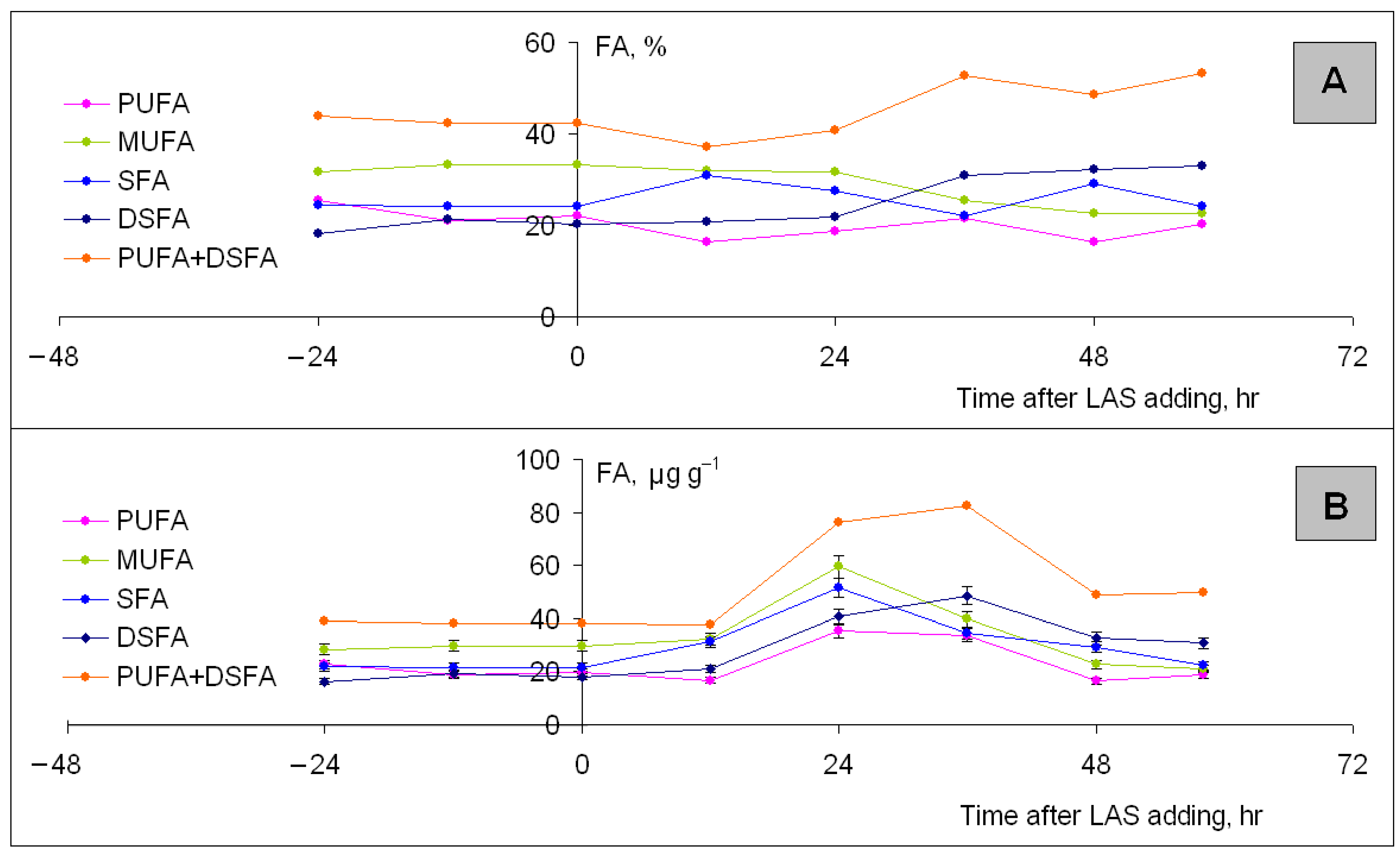
3.5.2. Lubomirskia baikalensis Fatty Acid Composition and Content Changes

Nineteen dominant FAs were identified for all individuals of control sponges. Percentages of saturated fatty acids (SFA) ranged from 24 to 25. Percentages of monounsaturated fatty acids (MUFA) and polyunsaturated fatty acids (PUFAs) including demospongic fatty acids (DSFA) ranged from 32 to 34% and from 43 to 44%, respectively (Figure 5A, left). These values are similar to the literature data for healthy individuals of L. baikalensis sampled in 1990 [49]. Total FA content of control sponge was ~90 ± 9 μg g−1 of the dry weight (of d.w.) (Figure 5B, left).
Significant changes in FA composition within 24–48 h after LAS addition (Figure 5A, right) into aquarian water were fixed. Percentages of SFA, MUFA, and the sum of PUFA and DSFA ranged within 22–30, 23–32, and 37–53, respectively (Figure 5A).
Quick increases in SFA, MUFA, and PUFA production including DSFA were also noticed (Figure 5B). The total FA content increased up to 187 ± 13. The sum content of PUFA and DSFA increased from 40 to 80 μg g−1 of d.w. within 24 and 36 h. Thereafter, it decreased to a level somewhat higher than the initial one. An increase in PUFA percentages was fixed within 24, 36, 48, and 58 h as well. An increase in SFA and MUFA percentages was fixed within 24 h.

3.6. In Situ Experiments: Analysis of the Environmental Samples

3.6.1. Oxidative Stress of Lubomirskia baikalensis in Lake Baikal

Two lipid peroxidation markers were analyzed viz. MDA concentration (CMDA) and PUFA percentages to reveal oxidative stress of frozen sponges collected at the sampling sites No. 2 and No. 3 in Lake Baikal (Table 2). The MDA was not revealed in a number of sponges (n = 9). Percentages of PUFA in these sponges were 42–43 (Table 2).
Concentration of MDA rated from 0.010 ± 0.004 to 0.53 ± 0.19 μg g−1 of d.w. in other sponges (n = 11) collected at the same stations (Table 2). The PUFA percentages for that were 1.3 times lower (Table 2).
MDA was not found in control sponges (n = 12) after 10 days cultivation in clean water. Percentages of PUFA in these individuals were the highest and achieved 55.

3.6.2. Antioxidant Capacity of Lubomirskia baikalensis

Low values of ABTS•+ inhibition (I) by crude extracts of frozen sponges collected at the sampling sites No. 4, 5, 6, 7, and 8 in southern and northern basins of Lake Baikal were noted. High IC50 values indicated low antioxidant capacity (AOC) as well (Table 3). The AOC values were ranged from 0.00031 to 0.00077 Trolox equivalents (TE). The lowest values of 0.00031 and 0.00040 were found in sponges collected at the site No. 4.
Low values of ABTS•+ inhibition and AOC by crude extracts of live control sponge that lived in the aquarium for six months at the moment of AOC analysis were also fixed (Table 2). Mentioned low values were close to that for the same sponge treated with 20 μg L−1 LAS for 72 h.

3.7. Statistical Data

To determine sponge humidity, each sample was analyzed in two replicates. To assess the ability of the method to generate similar results for multiple preparations, the repeatability was measured and characterized by variation coefficient V = 2%.
To determine the total fatty acid content, each sample was analyzed in two replicates as well. The repeatability of the technique was characterized by V = 6%.
To assess the antioxidant capacity, two pieces were cut from the sponge top and from the sponge foot. Pieces were extracted and analyzed. The coefficient of variation was V = 5%.
To determine MDA content, each sponge sample was analyzed in three replicates. The variation coefficient was characterized by the value of 25%.
To conduct the cell viability test and to count the number of live/dead cells, each sample of sponge cell suspension was analyzed in two–four replicates and 10–12 fields of view were screened. The total number of blue stained SA1-cells and symbiotic Chlorella sp. cells as well as unstained green cells achieved 230 for SA1-cells and 500 for symbiotic algae cells. The repeatability was characterized by V = 15%.

4. Discussion

4.1. Toxicity Test Protocol

In this study, we propose the toxicity test protocol to evaluate adverse effect of short-term exposure of linear alkylbenzene sulfonates at low concentrations (10 and 20 µg L−1) on freshwater sponge Lubomirskia baikalensis. The experiments were carried out with the use of live multicellular invertebrates exposed in aquariums under conditions close to natural in Lake Baikal. Symbiotic microalgae Chlorella sp. were studied, excluding their extraction from the host organism and cultivation in vitro. This allowed us to reflect the LAS effect on a whole sponge organism.
Sampling cell suspension of ~50 μL volume from live L. baikalensis was proposed to exclude serious damage to the target animal. This made multiple replicate analysis in short time intervals to evaluate the cell viability possible. Low-temperature (~8–12 °C) inverted microscopy with light-emitting diodes (LED)-transmitted illumination of live cells allowed us to elongate the duration of microscopy of a sample by up to 3 min. Incandescent lamps, on the contrary, increased the temperature with the following cell swelling during the first minute of analysis that led to errors of up to 60%.
The sponge cell classification suggested by the authors allows us to assign the nucleolar sponge amoebocytes of the first type (SA1-cells) containing symbiotic microalgae Chlorella sp. as indicator cells to evaluate the toxic effects of surfactants. Enzymes of live eukaryotic cells containing nuclei with nucleoli are known to oxidize the methylene blue and suppress its staining effect [50]. This explains the absence of staining of live L. baikalensis SA1-cells and symbiotic Chlorella sp. (see Figure 3A,B). In turn, the absence of methylene blue oxidizing enzymes in prokaryotic cells results in the blue staining of live sponge SA2-, SA3-, SA4-cells and symbiotic Cyanophyta cells. Therefore, live SA1-cells were not stained with methylene blue, and the dead ones were stained, which reflects the acute toxic LAS effect on L. baikalensis.
The large size of SA1-type-cells makes the viability test procedure easy, repeatable, and effective. Thus, L. baikalensis and symbiotic Chlorella sp. can be used as bioindicators or biomonitors to evaluate the ecosystem state of Lake Baikal. The proposed toxicity test protocol includes a cell viability test, fatty acid analysis, and probable oxidation stress estimation. It can be used to evaluate anthropogenic impact level at regional and global scales in the interest of ecological health and biodiversity conservation.

4.2. Linear Alkylbenzene Sulfonate Acute Toxicity Effect

This study presents the results of acute toxic 20 μg L−1 LAS effect on organisms of two different kingdoms: Animalia and Plantae. This effect caused 97–100% sponge L. baikalensis cell death in less than 48 h (see Figure 4A) and 100% symbiotic microalgae Chlorella sp. cell death over 72 h (see Figure 4B). Changes in sizes of the sponge amoebocytes (SA1-cells) containing the eucaryotic symbionts Chlorella sp. as well as symbiotic cells have been revealed. It was noted that SA1-cells’ volume increased up to 1.5 times and more. Therefore, cell lysis occurred as a result of cell swelling and osmotic imbalance at a critical volume (see Table 1).
A close toxic effect of 20 μg L−1 LAS has been found [51] on different marine microalgae such as Tetraselmis chuii (division Chlorophyta, class Chlorodendrophyceae, family Chlorodendroceae), Rhodomonas salina (division Cryptophyta, class Cryptophyceae, family Pyrenomonadaceae), Chaetoceros sp. (division Ochrophyta, class Bacillariophyceae, family Chaetocerotaceae), Isochrysis galbana (division Haptophyta, class Prymnesiophyceae, family Isochrysidaceae), and Nannochloropsis gaditana (division Ochrophyta, class Eustigmatophyceae, family Monodopsidaceae). The inhibition of cell growth and the general tendency to increasing cell volume/size were revealed [51].
A report on acute toxic influence of LAS solutions at the same concentration of 20 μg L−1 on Chlorella ellipsoidea (division Chlorophyta, class Trebouxiophyceae, family Chlorellaceae) was published [52]. The strongest ability of LAS to decrease lipid content in the thylakoid membranes was shown. Changes in cell membrane permeability, metabolism, and growth interruption were found [52]. The literature’s data testify that 20–100 μg L−1 LAS concentrations are the most harmful to aquatic organisms [36,37,51,52,53,54].
According to the results (see Figure 4A), the SA1-cells’ mortality curve exhibits steep slope due to cell death at the beginning of the experiment. The small plateau and the gentle slope on the microalgae mortality curve (see Figure 4B) until 24 h of LAS exposure corresponds to the moment of sponge SA1-cell death (see Figure 4A). The plateau and the gentle slope at the start of the experiment till 24 h reflects viability of microalgae inside the host cells, which protect them from a hazardous impact (see Figure 4B).
Nonspecific adaptation response of the cell and the whole organism is the response to a stress factor that is common for different organisms. This response aims to restore the homeostasis of a live system. A normal live cell surrounded by a liquid cell membrane with a lipid bilayer ~4–7 nm thick tends to its state of minimum free energy and minimum surface according to Le Chatelier’s principle. Aggregation of LAS on a cell surface disrupts the bilayer reducing surface tension. An imbalance occurs with the environment [3,55,56,57,58]. A prolonged, intensive, or recurrent effect results in adaptation malfunction and potential exhaustion of a cell membrane.
A visual example of a nonspecific adaptation response of L. baikalensis to LAS impact is the sharp change in fatty acid content within 24 h of exposure (see Figure 5B). Let us compare Figure 4B and Figure 5B. One can see the correlation between the beginning of symbiotic algae death (Figure 4B) and increase in FA content up to two times (Figure 5B). We assume that this increase is caused by Chlorella sp.’s ability to trigger the adaptive mechanism and to produce FA more intensively. As supposed, SFA and MUFA production decreases within 36 h of exposure due to nutrient deficit. The cell membrane potential is exhausted. PUFA production decreases within 48 h due to lack of SFA and MUFA as PUFA synthesis raw materials. Thus, 20 µg L−1 LAS concentrations found in water of the nearshore zone can cause the mass morbidity effect on Porifera in Lake Baikal.

4.3. Oxidative Stress as a Response to LAS Toxic Effect: In Situ and In Vitro Investigations

For the first time, we found the oxidative stress of some individuals of Lubomirskia baikalensis, collected in situ in Lake Baikal (see Table, Figure 1, sample sites No. 2 and 3), but for some others, the stress was not revealed. The pollution of aquatic ecosystems by xenobiotics and the absence of adaptation of water dwellers to their impact are acute problems for the 21st century. Some of the most persistent micropollutants in aquatic ecosystems such as polycyclic aromatic hydrocarbons (PAH) [59], heavy metals [60], and anionic surfactants including alkylbenzene sulfonates [35,36] are able to induce oxidative stress and hypoxia of the cell and organism as a whole. The oxidative stress of freshwater organisms can occur due to species sensitivity to these toxicants.
Our first results of in vitro experiments have shown the oxidative stress of L. baikalensis as a response to toxic effect of alkylbenzene sulfonates at 10 μg L−1 concentration after 14-day exposure. The oxidation stress is accompanied by malondialdehyde formation. Alkylbenzene sulfonates are the most widespread pollutants for water ecosystems. The mechanism of the effect of these substances is associated with the induction of a negative charge of the cell membrane. When redistributing the charge, the process of lipid peroxidation under free radical attack is activated [3,53]. One of the final products of the attack is MDA, which is a marker of oxidative stress [55,61,62]. The cell wall of L. baikalensis, as well as many other organisms of cold water oligotrophic ecosystems, contains a high percentage of unsaturated fatty acids (see Figure 3, suppl. Table S1), which are the most suitable target for the attack of free radicals. We observed the correlation of MDA presence in stressed sponges with the decrease of PUFA content (see Table 2). The same situation for Lake Baikal phytoplankton was found in previous work [23].
The high sensitivity of L. Baikalensis to the hazardous LAS exposure can be related to its very low antioxidant capacity (TEAC) from 0.00031 to 0.00077 Trolox equivalents (see Table 3) and low ability to adapt. These values are up to 10–110 times lower than the TEAC for marine species (see Table 4). The antioxidant capacity of L. Baikalensis dry crude extract is responsible for the sponge ability to scavenge free radical attack. The high values of IC50 in L. Baikalensis also confirm the low sponge ability to resist the oxidation stress factor and to adapt to the oxidant impact. Inattention to the problem of anionic surfactants in freshwater ecosystems and the incorrect choice of toxicity test protocol with the use of testing animals that have not highest responsiveness to the hazardous impact of toxicants can lead to dramatic situations for sponge populations and their associated flora and fauna in particular.

5. Conclusions

A new toxicity test protocol under conditions close to natural was proposed. It has good potential for reliable estimation of the anionic surfactant toxicity because of it using an in vivo assay and testing sponges of high responsibility, and it is applicable to a wide variety of water pollution problems. The sponge amoebocytes named SA1-cells that contain eukaryotic microalgae Chlorella sp. are shown to be the representative indicator in assessing the impact of anionic surfactants. Toxic effects of linear alkylbenzene sulfonates on L. baikalensis and its symbiotic microalgae Chlorella sp. was found at low concentrations in in vitro experiments. The acute toxic effect of LAS was noticed at 20 μg L−1. Oxidation stress of the sponges in response to the LAS effect at 10 μg L−1 was revealed. For the first time, we revealed the oxidative stress of L. baikalensis in situ in Lake Baikal. The stress may be a result of long-term pollutant effects coupled with the low-antioxidant activity of L. baikalensis.

Supplementary Materials

The following supporting information can be downloaded at: https://www.mdpi.com/article/10.3390/d15010077/s1, Table S1: Fatty acid (FA) profiles of controlled individual of Lubomirskia baikalensis as well as of the same sponge individual treated with 20 μg L−1 LAS solution.

Author Contributions

Conceptualization, methodology–A.A.N., I.B.M., B.N.B., I.V.K., O.V.S.; writing the text of the manuscript, translation– A.A.N., I.V.K., I.B.M.; discussion and editing–A.A.N., I.V.K., S.S.V., B.N.B., O.V.S., A.N.B.; investigation–A.A.N., S.S.V., K.S.D., B.N.B., O.V.S., A.A.A., T.N.A., A.N.C., O.Y.G.; validation–A.A.N., I.B.M., I.V.K., S.S.V., B.N.B.; visualization–A.A.N., T.N.A.; chemical analysis of FAs–A.A.N., T.N.A., A.A.A.; analysis of MDA–A.A.N.; TEAC analysis–B.N.B., O.V.S., A.N.C., A.A.N.; microscopy of sponge cells–A.A.N.; microscopy of algae cells–S.S.V., K.S.D., A.A.N.; cell viability test–A.A.N.; LAS toxicity studies–A.A.N., K.S.D.; sampling of sponges–I.V.K.; sampling of pelagic water and zooplankton–A.N.B., A.Y.K., L.G.K., A.A.N.; providing of conditions for cultivation of L. baikalensis–O.Y.G., A.Y.K., L.G.K., I.V.K., A.N.B., A.A.N.; statistical analysis–A.A.N., B.N.B. The investigation is the authors’ common cause. All authors have read and agreed to the published version of the manuscript.

Funding

The investigation was carried out within the State Projects of the Ministry of the Science and High Education of the Russian Federation (No. 0279-2021-0005 “Investigation of the transformation in the states of water bodies and watercourses in East Siberia…”) using the “Ultramicroanalysis” Instrumental Center equipment and the Unique Scientific Center “Experimental Freshwater Aquarium Complex of Lake Baikal water organisms” of Limnological Institute of SB RAS.

Acknowledgments

The authors thank Andrey Petrovich Fedotov for valuable advice and for sponge sampling in 2020, 2021, and 2022, Galina Afanasyevna Fedorova for sponge samples in 2019, Elena Valentinovna Likhoshway for the equipment provided by “Ultramicroanalysis” Instrumental Center, Mikhail Alexandrovich Grachev and Alexander Georgievich Gorshkov for helpful advices, Ivan Alexandrovitch Nebesnykh for Lake Baikal map-making, and Irina Viktorovna Tomberg for the determination of dissolved oxygen. We thank the crews of R/V “Vereshchagin”, “Papanin”, “Koptyug”, and “Titov” for fieldwork made on Lake Baikal. Alyona A. Nikonova expresses deep gratitude to her parents for supplying the aquariums with oxygen and pelagic Lake Baikal water and Marina Vyacheslavovna Nikonova for language proofreading.

Conflicts of Interest

The authors declare no conflict of interest.

References

  1. Schmid, M.; De Batist, M.; Granin, N.G.; Kapitanov, V.A.; McGinnis, D.F.; Mizandrontsev, I.B.; Obzhirov, A.I.; Wuest, A. Sources and sinks of methane in Lake Baikal: A synthesis of measurements and modeling. Limnol. Oceanogr. 2007, 52, 1824–1837. [Google Scholar] [CrossRef] [Green Version]
  2. De Batist, M.; Canals, M.; Sherstyankin, P.; Alekseev, S. A New Bathymetric Map of Lake Baikal. The INTAS Project 99-1669 Team. 2002. Available online: http://www.lin.irk.ru/intas/index.htm (accessed on 4 January 2023).
  3. Vadeboncoeur, Y.; McIntyre, P.B.; Vander Zanden, M.J. Borders of Biodiversity: Life at the Edge of the World’s Large Lakes. BioScience 2011, 61, 526–537. [Google Scholar] [CrossRef]
  4. Lut, B.F. Morfologiya i morfometriya Baikalskoi vpadiny [Morphology and Morphometry of Baikal Depression]. Chapter II. In The Way for Knowledge of Baikal; Afanasieva, E.L., Beckman, M.Y., Bezrukova, E.V., Verbolov, V.I., Votintsev, K.K., Galasii, G.I., Goldyrev, G.S., Granina, L.Z., Dryukker, V.V., Ladeischikov, N.P., et al., Eds.; Nauka Publ.: Novosibirsk, Russia, 1987; pp. 34–47. (In Russian) [Google Scholar]
  5. Votintsev, K.K.; Meshcheryakova, A.I.; Popovskya, G.I. Krugovorot Organicheskogo Veshchestva v Ozere Baikal [Organic Substance Cycle in Lake Baikal]; Nauka Publ.: Novosibirsk, Russia, 1975; 190p. (In Russian) [Google Scholar]
  6. Manconi, R.; Pronzato, R. Phylum Porifera Ecology and General Biology: Thorp and Covich’s Freshwater Invertebrates, 4th ed.; Thorp, J., Rogers, D.C., Eds.; Elsevier: Amsterdam, The Netherlands, 2015; pp. 133–157. [Google Scholar]
  7. Erpenbeck, D.; Weier, T.; de Voogd, J.N.; Wörheide, G.; Sutcliffe, P.; Todd, J.A.; Michel, E. Insights into the evolution of freshwater sponges (Porifera: Demospongiae: Spongillina): Barcoding and phylogenetic data from Lake Tanganyika endemics indicate multiple invasions and unsettle existing taxonomy. Mol. Phylogenet. Evol. 2011, 61, 231–236. [Google Scholar] [CrossRef] [PubMed]
  8. Itskovich, V.; Kaluzhnaya, O.; Ostrovsky, I.; McCormack, G. The number of endemic species of freshwater sponges (Malawispongiidae; Spongillina; Porifera) from Lake Kinneret is overestimated. J. Zool. Syst. Evol. Res. 2013, 51, 252–257. [Google Scholar] [CrossRef]
  9. Laurer, T.E.; Spacie, A. New Records of Freshwater Sponges (Porifera) for Southern Lake Michigan. J. Great Lakes Res. 1996, 22, 77–82. [Google Scholar] [CrossRef]
  10. Khanaev, I.V.; Kravtsova, L.S.; Maikova, O.O.; Bukshuk, N.A.; Sakirko, M.V.; Kulakova, N.V.; Butina, T.V.; Nebesnykh, I.A.; Belikov, D.I. Current state of the sponge fauna (Porifera: Lubomirskiidae) of Lake Baikal: Sponge disease and the problem of conservation of diversity. J. Great Lakes Res. 2018, 44, 77–85. [Google Scholar] [CrossRef]
  11. Maikova, O.; Bukshuk, N.; Kravtsova, L.; Nebesnyh, I.; Yakhnenko, A.; Butina, T.; Khanaev, I. Baikal endemic sponge disease and anthropogenic factor. IOP Conf. Ser. Earth Environ. Sci. 2021, 937, 022071IOP. [Google Scholar] [CrossRef]
  12. Itskovich, V.; Belikov, S.; Kaluzhnaya, O. Study of the biodiversity of deep-water sponges of Lake Baikal by molecular methods. Proceedings of The Materials of the VIII World Sponge Conference “Ancient animals, new challenges”, Girona, Spain, 20–24 September 2010; p. 222. [Google Scholar]
  13. Kozhova, O.M.; Izmest’eva, L.R. Lake Baikal. Evolution and Biodiversity; Backhuys Publishers: Leiden, The Netherlands, 1998; 447p. [Google Scholar]
  14. Rusinek, O.T.; Takhteev, V.V.; Khodzher, T.V.; Pleshanov, A.S.; Voronin, V.I.; Arov, I.V.; Azovskii, M.G.; Goryunova, O.I.; Dryukker, V.V.; Zadonina, N.V.; et al. Baikalogy. Book 2: In 2 Books; Nauka: Novosibirsk, Russia, 2012; 644p. (In Russian) [Google Scholar]
  15. Kulakova, N.V.; Kashin, S.A.; Bukin, Y.S. The genetic diversity and phylogeny of green microalgae in the genus Choricystis (Trebouxiophyceae, Chlorophyta) in Lake Baikal. Limnology 2020, 21, 15–24. [Google Scholar] [CrossRef]
  16. Kulakova, N.V.; Denikina, N.N.; Belikov, S.I. Diversity of bacterial photosymbionts in Lubomirskiidae sponges from Lake Baikal. Int. J. Biodivers. 2014, 2014, 152097. [Google Scholar] [CrossRef] [Green Version]
  17. Ereskovsky, A.V.; Chernogor, L.I.; Belikov, S.I. Ultrastructural description of development and cell composition of primmorphs in the endemic Baikal sponge Lubomirskia Baicalensis. Zoomorphology 2016, 135, 1–17. [Google Scholar] [CrossRef]
  18. Gladkikh, A.S.; Kalyuzhnaya, O.V.; Belykh, O.I.; Ahn, T.S.; Parfenova, V.V. Analysis of Bacterial Communities of Two Lake Baikal Endemic Sponge Species. Mikrobiologiya 2014, 83, 682–693. [Google Scholar] [CrossRef]
  19. Belykh, O.I.; Fedorova, G.A.; Kuzmin, A.V.; Tikhonova, I.V.; Timoshkin, O.A.; Sorokovikova, E.G. Microcystins in Cyanobacterial Biofilms from the Littoral Zone of Lake Baikal. Mosc. Univ. Biol. Sci. Bull. 2017, 72, 225–231. [Google Scholar] [CrossRef]
  20. Mohamed, N.M.; Rao, V.; Hamann, M.T.; Kelly, M.; Hill, R.T. Monitoring bacterial diversity of the marine sponge Ircinia strobilina upon transfer into aquaculture. Appl. Environ. Microbiol. 2008, 74, 4133–4143. [Google Scholar] [CrossRef] [PubMed] [Green Version]
  21. Chernogor, L.; Denikina, N.; Kondratov, I.; Solovarov, I.; Khanaev, I.; Belikov, S.; Ehrlich, H. Isolation and identification of the microalgal symbiont from primmorphs of the endemic freshwater sponge Lubomirskia baikalensis (Lubomirskiidae, Porifera). Eur. J. Phycol. 2013, 48, 497–508. [Google Scholar] [CrossRef]
  22. Timoshkin, O.A.; Samsonov, D.P.; Yamamuro, M.; Moore, M.V.; Belykh, O.I.; Malnik, V.V.; Sakirko, M.V.; Shirokaya, A.A.; Bondarenko, N.A.; Domysheva, V.M.; et al. Rapid ecological change in the coastal zone of Lake Baikal (East Siberia): Is the site of the world’s greatest freshwater biodiversity in danger? J. Great Lakes Res. 2006, 42, 487–497. [Google Scholar] [CrossRef] [Green Version]
  23. Nikonova, A.A.; Shishlyannikov, S.M.; Volokitina, N.A.; Galachyants, Y.P.; Bukin, Y.S.; Blinov, V.V.; Gnatovsky, R.Y.; Vorobyeva, S.S. Fatty Acid Changes in Nearshore Phytoplankton under Anthropogenic Impact as a Biodiversity Risk Factor for the World’s Deepest Lake Baikal. Diversity 2022, 14, 55. [Google Scholar] [CrossRef]
  24. Maikova, O.O.; Kravtsova, L.S.; Khanaev, I.V. Baikal endemic sponges in the system of ecological monitoring. Limnol. Freshw. Biol. 2020, 1, 364–367. [Google Scholar] [CrossRef] [Green Version]
  25. Belikov, S.I.; Feranchuk, S.I.; Butina, T.V.; Chernogor, L.I.; Khanaev, I.V.; Maikova, O.O. Mass disease and mortality of Baikal sponges. Limnol. Freshw. Biol. 2018, 1, 36–42. [Google Scholar] [CrossRef]
  26. Webster, N.S. Sponge disease: A global threat? Environ. Microbiol. 2007, 9, 1363–1375. [Google Scholar] [CrossRef]
  27. Denikina, N.N.; Dzyuba, E.V.; Bel’kova, N.L.; Khanaev, I.V.; Feranchuk, S.I.; Makarov, M.M.; Granin, N.G.; Belikov, S.I. Pervyi sluchai zabolevaniya gubki Lubomirskia baikalensis: Issledovanie microbioma [The first case of Lubomirskia baikalensis sponge decease. Microbiome investigation]. Izv. RAN Biol. Ser. Ecol. 2016, 3, 315–322. (In Russian) [Google Scholar]
  28. Granin, N.G.; Mizandrontsev, I.B.; Obzhirov, A.I.; Vereshchagina, O.F.; Gnatovskii, R.Y.; Zhdanov, A.A. Okislenie metana v vodnoi tolshe ozera Baikal [Oxidation of methane in water column of Lake Baikal]. Dokl. Akad. Nauk. Earth Sci. 2013, 451, 332–335. [Google Scholar]
  29. Butina, T.V.; Bukin, Y.S.; Petrushin, I.S.; Tupikin, A.E.; Kabilov, M.R.; Belikov, S.I. Extended evaluation of viral diversity in Lake Baikal. Microorganisms 2021, 9, 760. [Google Scholar] [CrossRef] [PubMed]
  30. Nikonova, A.A. Organic synthetic anionic surfactants as persistent organic pollutants of water ecosystems. Limnol. Freshw. Biol. 2020, 4, 620–621. [Google Scholar] [CrossRef]
  31. Belanger, S.E.; Bowling, J.W.; Lee, D.M.; Le Blank, E.M.; Kerr, K.M.; McAvoy, D.C.; Christman, S.C.; Davidson, D.H. Integration of aquatic fate and ecological responses to linear alkyl benzene sulfonate (LAS) in model stream ecosystems. Ecotoxicol. Environ. Saf. 2002, 52, 150–171. [Google Scholar] [CrossRef]
  32. Hon-Nami, H.; Hanya, T. Linear alkylbenzene sulfonates in river, estuary and bay water. Water Res. 1980, 14, 1251–1256. [Google Scholar] [CrossRef]
  33. Terzic, S.; Ahel, M. Input and behaviour of linear alkylbenzenesulphonates (LAS) in a stratified estuary. Mar. Pollut. Bull. 1994, 28, 735–740. [Google Scholar] [CrossRef]
  34. Okbah, M.A.; Ibrahim, A.M.A.; Gamal, M.N.M. Environmental monitoring of linear alkylbenzene sulfonates and physicochemical characteristics of seawater in El-Mex Bay (Alexandria, Egypt). Environ. Monit. Assess. 2013, 185, 3103–3115. [Google Scholar] [CrossRef] [Green Version]
  35. Lewis, M.A. Chronic and sublethal toxicities of surfactants to aquatic animals: A review and risk assessment. Water Res. 1991, 25, 101–113. [Google Scholar] [CrossRef]
  36. Jorgensen, E.; Christoffersen, K. Short-term effects of linear alkylbenzene sulfonate on freshwater Plankton studied under field conditions. Environ. Toxicol. Chem. 2000, 19, 904–911. [Google Scholar] [CrossRef]
  37. Stamatelatou, K.; Pakou, C.; Lyberatos, G. Linear alkylbenzene sulfonates (LAS). Occurrence, toxicity, and biodegradation of selected emerging priority pollutants in municipal sewage sludge. In Comprehensive Biotechnology, 2nd ed.; Moo-Young, M., Ed.; Pergamon: Oxford, UK, 2011; Volume 6, pp. 473–484. [Google Scholar]
  38. Wetzel, R.G. Limnology: Lake and River Ecosystems, 3rd ed.; Academic Press: San Diego, CA, USA; San Francisco, CA, USA; New York, NY, USA; Boston, MA, USA; London, UK; Sydney, Australia; Tokyo, Japan, 2001; 1006p. [Google Scholar]
  39. Mizandrontsev, I.B.; Mizandrontseva, K.N. Kislorodnyi obmen mezhdu vodnoi poverhnost’yu I atmosferoi nad Baikalom [The oxygen exchange between Lake Baikal water surface and the atmosphere]. Russ. Meteorol. Hydrol. 1993, 2, 108–113. (In Russian) [Google Scholar]
  40. Mizandrontsev, I.B.; Domysheva, V.M.; Mizandrontseva, K.N.; Tomas, K. O sutochnoi dinamike koncentracii svobodnoi uglekisloty I kisloroda v vode Baikala [About daily dynamics of free carbonic acid and oxygen in Baikal water]. Geogr. Nat. Resour. 2002, 1, 73–78. (In Russian) [Google Scholar]
  41. Rakita, S.M.; Colovic, D.S.; Levart, A.R.; Banjac, V.V.; Colovic, R.R.; Dragojlovic, D.M.; Duragic, O.M. Rapid spectrophotometric method for determination of thiobarbituric acid reactive substances in rainbow trout feed. Food Feed. Res. 2020, 47, 43–53. [Google Scholar] [CrossRef]
  42. Nikonova, A.A.; Shishlyannikov, S.M.; Shishlyannikova, T.A.; Avezova, T.N.; Babenko, T.A.; Belykh, O.I.; Glyzina, O.Y.; Obolkin, V.A.; Pavlova, O.N.; Smagunova, A.N.; et al. Determination of Free and Esterified Fatty Acids in Hydrocoles of Different Content of Polyunsaturated Fatty Acids by Gas–Liquid Chromatography. J. Anal. Chem. 2020, 75, 1310–1321. [Google Scholar] [CrossRef]
  43. Al-Rashed, S.A.; Ibrahim, M.M.; El-Gaaly, G.A.; Al-Shehri, S.; Mostafa, A. Evaluation of radical scavenging system in two microalgae in response to interactive stresses of UV-B radiation and nitrogen starvation. Saudi J. Biol. Sci. 2016, 23, 706–712. [Google Scholar] [CrossRef] [PubMed]
  44. Pellegrini, N.; Serafini, M.; Colombi, B.; Del Rio, D.; Salvatore, S.; Bianchi, M.; Brighenti, F. Total Antioxidant Capacity of Plant Foods, Beverages and Oils Consumed in Italy Assessed by Three Different In Vitro Assays. J. Nutr. 2003, 133, 2812–2819. [Google Scholar] [CrossRef] [Green Version]
  45. Potts, E. Freshwater sponges; A monograph. Proc. Acad. Nat. Sci. Phila. 1887, 1887, 157–279. [Google Scholar]
  46. Sabella, S.; Faszewski, E.; Himic, L.; Colpitts, K.M.; Kaltenbach, J.; Burger, M.M.; Fernàndez-Busquets, X. Cyclosporin A suspends transplantation reactions in the marine sponge Microciona prolifera1. J. Immunol. 2007, 179, 5927–5935. [Google Scholar] [CrossRef] [Green Version]
  47. Kozlov, M. Sponge cells hint at origins of nervous system. Synapse genes help cells to communicate in the digestive chambers of sponges. Nature 2021, 559, 193. [Google Scholar] [CrossRef]
  48. Musser, J.M.; Schippers, K.J.; Kohn, A.B.; Pape, C.; Ronchi, P.; Arendt, D. Profiling cellular diversity in sponges informs animal cell type and nervous system evolution. Science 2021, 374, 717–723. [Google Scholar] [CrossRef]
  49. Dembitsky, V.M.; Rezanka, T.; Kashin, A.G. Comparative study of the endemic freshwater fauna of Lake Baikal. Unusual lipid composition of two sponge species Baicalospongia bacillifera and Baicalospongia intermedia (family Lubomirskiidae, class Demospongiae). Comp. Biochem. Physiol. Part B Comp. Biochem. 1993, 106, 825–831. [Google Scholar] [CrossRef]
  50. Sharga, B.M.; Pylypiv, D.B.; Feketa, V. Cytology. Practical. 4. Differentiation between living and dead eukaryotic cells by staining. In Medical Biology Practicals; Uzhorod National University Publ.: Uzhorod, Ukraine, 2017. [Google Scholar]
  51. Debelius, B.; Forja, J.M.; Del Valls, A.; Lubian, L.M. Effect of linear alkylbenzene sulfonate (LAS) and atrazine on marine microalgae. Mar. Pollut. Bull. 2008, 57, 559–568. [Google Scholar] [CrossRef] [PubMed]
  52. Choi, E.A.; Suk, K.; Lee, C.S.; Lee, C.S. The effects of surfactants on the biosynthesis of galactolipid and the composition of fatty acids in chloroplast envelope and thylakoid membrane of Chlorella ellipsoidea. Korean J. Biol. Sci. 1998, 2, 341–349. [Google Scholar] [CrossRef]
  53. Lewis, M.A. Chronic Toxicities of Surfactants and Detergent Builders to Algae: A Review and Risk Assessment. Ecotoxicol. Environ. Saf. 1990, 20, 123–140. [Google Scholar] [CrossRef]
  54. Janero, D.R. Malondialdehyde and thiobarbituric acidreactivity as diagnostic indices of lipid peroxidation and peroxidative tissue injury. Free Radic. Biol. Med. 1990, 9, 515–540. [Google Scholar] [CrossRef] [PubMed]
  55. Marnett, L.J. Lipid peroxidation-DNA damage by malondialdehyde. Mutat. Res. Fundam. Mol. Mech. Mutagen. 1999, 424, 83–95. [Google Scholar] [CrossRef] [PubMed]
  56. Parshi, K. Interaction of detergent sclerosants with cell membranes. Phlebology 2015, 30, 306–315. [Google Scholar] [CrossRef] [PubMed]
  57. Ebnesajjad, S. Surface treatment of materials for adhesive bonding. In Surface Tension and Its Measurement, 2nd ed.; Elsevier: New York, NY, USA, 2014; pp. 7–24. [Google Scholar]
  58. Aguirre-Ramírez, M.; Silva-Jiménez, H.; Banat, I.M.; De Rienzo, M.A.D. Surfactants: Physicochemical interactions with biological macromolecules. Biotechnol. Lett. 2021, 43, 523–535. [Google Scholar] [CrossRef] [PubMed]
  59. Vega-López, A.; Ayala-López, G.; Posadas-Espadas, B.P.; Olivares-Rubio, H.F.; Dzul-Caamal, R. Relations of oxidative stress in freshwater phytoplankton with heavy metals and polycyclic aromatic hydrocarbon. Comp. Biochem. Physiol. Part A Mol. Integr. Physiol. 2013, 165, 498–507. [Google Scholar] [CrossRef]
  60. Srivastava, S.; Tripathi, G.; Mishra, S.; Gupta, G. Copper-induced oxidative stress and responses of antioxidants and phyto-chelatins in Hydrilla verticillata (L.f.). Royle Aquatic Toxicol. 2006, 8, 405–415. [Google Scholar] [CrossRef]
  61. Zhou, J.; Wu, Z.; Yu, D.; Pang, Y.; Cai, H.; Liu, Y. Toxicity of linear alkylbenzene sulfonate to aquatic plant Potamogeton perfoliatus L. Environ. Sci. Pollut. Res. 2018, 25, 32303–32311. [Google Scholar] [CrossRef]
  62. Liu, N.; Wu, Z. Toxic effects of linear alkylbenzene sulfonate on Chara vulgaris L. Environ. Sci. Pollut. Res. 2018, 25, 4934–4941. [Google Scholar] [CrossRef]
  63. Montaño-Castañeda, M.C.; Santafé-Patiño, G.G. Evaluation of antioxidant activity of marine sponges from the Colombian Caribbean. Actual Biol. 2011, 33, 173–181. [Google Scholar] [CrossRef]
  64. Quiroz-Lobo, Y.; Santafe-Patino, G.; Quiros-Rodriguez, J.-A. Bioactivity and identification of fatty acids of the marine sponge Tetilla rodriguesi (Tetractinellida: Tetillidae) in the Colombian Caribbean. Rev. Biol. Trop. 2022, 70, 20–29. [Google Scholar] [CrossRef]
Figure 1. Scheme of Lake Baikal and the sampling sites of Lubomirskia baikalensis (class Demospongiae, order Spongillida, family Lubomirskiidae). Live sponges from the site No. 1 were transported into aquariums, and sponges from the sites No. 2–8 were frozen immediately.
Diversity 15 00077 g001
Figure 2. Photo of Lubomirskia Baikalensis (class Demospongiae, order Spongillida, family Lubomirskiidae) in aquarium and the sponge dominant cell light microscope images. (A)–underwater image of epilithic sponge with exosymbionts (7) Brandtia parasitica (class malacostraca, order Amphipoda, family Acanthogammaridae); (B)–healthy sponge amoebocytes of the first type (SA1-cells) (1) and its eukaryotic endosymbionts Chlorella sp. (4); (C)—healthy amoebocytes of the second type (SA2-cells) (2) and its prokaryotic endosymbionts; (5); (D)–healthy amoebocytes (SA2-cells) (2) and its prokaryotic endosymbionts (5), stained with methylene blue; (E)–amoebocytes containing symbiont assemblage (SA3-cells) (8) including green algae (6) and a cell with flagellum (SA4-cell) containing prokaryotic symbionts (3).
Diversity 15 00077 g002
Figure 3. Lubomirskia baikalensis cells. (A)–normal SA1-cell of 21.7 μm size; (B)–normal SA1-cell of 16.1 μm size; (C)–swelling of SA4-cell with flagellum; a cell stained with methylene blue; (D)–SA1-cell of 27.8 × 23.9 μm size swelled and lysed due to linear alkybenzene sulfonate 72 h exposure (20 μg L−1); the cell stained with methylene blue; symbionts Chlorella sp. moving out of the host cell.
Diversity 15 00077 g003
Figure 4. Cell death curves. (A)–Lubomirskia baikalensis dead SA1-cell percentages increase in dependence on duration of 20 μg L−1 linear alkylbenzene sulfonates exposure; (B)–Chlorella sp. dead cell percentages increase in dependence on duration of 20 μg L−1 LAS exposure; (C)–dead symbiotic Chlorella sp. cells stained with methylene blue for 72 h exposure.
Diversity 15 00077 g004
Figure 5. Lubomirskia baikalensis fatty acid composition and content changes. (A)—fatty acid composition (%) including saturated, monounsaturated, polyunsaturated, and demospongic acids before anionic surfactant exposure (in the left of the vertical axis) and after linear alkylbenzene sulfonate (LAS) addition into aquarian water (in the right of the vertical axis) to achieve an LAS concentration of 20 μg L−1; (B)—fatty acid content (μg g−1 of d.w.) before and after LAS addition.
Diversity 15 00077 g005
Table 1. Sponges and symbiotic algae cell sizes before and after anionic surfactant exposure.
Organisms
L. baikalensis SA-1 CellsEndosymbiotic Algae Chlorella sp. Cells
Cell Size, μmCell Percentages (of the Sum)Cell Size, μmCell Percentages (of the Sum)
Control48 h LAS ExposureControl48 h LAS Exposure
10.0–15.017.90.02.0–2.527.53.6
15.0–20.038.50.52.5–3.020.035.7
20.0–25.038.51.03.0–3.530.028.6
25.0–30.05.10.83.5–4.017.532.1
30.0–35.00.00.54.0–4.55.00.0
35.0–40.00.00.24.5–5.00.00.0
Lysed cells0.097Lysed cells0.00.0
Table 2. Lipid peroxidation in Lubomirskia baikalensis collected in Lake Baikal from two sampling sites of different anthropogenic load in 2019.
Sample
Site No.
Depth of Sampling in Lake Baikal,
m
Lipid Peroxidation Markers
CMDA,
μg g−1 of d.w.
PUFA, %
25from 0.05 to 0.53
average 0.18 ± 0.06
33 (n = 7)
20not detected43 (n = 3)
35–15from 0.01 to 0.41
average 0.20 ± 0.07
32 (n = 4)
5–15not detected42 (n = 6)
Control sponges after 10 days cultivation in clean water5–15not detected42–55 (n = 12)
Table 3. Inhibition (I), half maximal inhibitory concentration (IC50), and antioxidant activity (AOA) of L. baikalensis crude extracts and standard (Trolox).
Sample Site No.Sampling SitesSample (Sponge) No.I, %IC50,
mg L−1
AOC,
TE
4Ulanovo Cape10.0064 ± 0.00037808 ± 3900.00040
20.0059 ± 0.00038542 ± 4270.00031
5Region of inoperative Baikalsk City paper mill30.0140 ± 0.00073574 ± 1790.00075
40.0133 ± 0.00073770 ± 1890.00064
50.0084 ± 0.00045972 ± 2990.00044
6Bolshoi Ushkanii Island60.0129 ± 0.00063889 ± 1940.00071
70.0110 ± 0.00064529 ± 2260.00065
7Elokhin Cape80.0149 ± 0.00073360 ± 1680.00077
90.0111 ±0.00064491 ± 2250.00062
8Tompuda Bight100.0140 ± 0.00073580 ± 1790.00071
110.0089 ±0.00045628 ± 2810.00046
2Control healthy sponge after 6 months exposure in clean aquarian water120.0160 ± 0.00083125 ± 1560.00062
The same sponge treated with 20 μg L−1 LAS for 72 h120.0122 ± 0.00064098 ± 2050.00081
Trolox (standard solution)19.7 ± 1.02.53 ± 0.131.00000
Table 4. ABTS•+ antioxidant activity for some marine sponges in comparison with freshwater Lake Baikal sponge Lubomirskia baikalensis and with trolox.
Sponge [Ref.]Type: Marine/FreshwaterIC50,
μg mL−1
I (%)TEAC
Lubomirskia baikalensis with maximal TEAC
(present investigation)
freshwater31250.0160very low
Lubomirskia baikalensis with minimal TEAC
(present investigation)
freshwater85420.0059very low
Tedania ignis [63]marineno data0.135 *moderate
Niphates erecta [63]marineno data0.184 *moderate
Callyspongia vaginalis [63]marineno data0.265 *moderate
Lissodendoryx carolinensis [63]marineno data0.325 *moderate
Tetilla rodriguesi [64]marine2970.327 *moderate
Amorphinopsis atlantica [63]marine880.531 *high
Ircinia felix [63]marine890.608 *high
Mycale microsigmatosa [63]marine600.669 *high
Standard (Trolox)2.519.733 *very high
* The data recalculated from 100 ppm to 1 ppm to universe them for different investigations.
Disclaimer/Publisher’s Note: The statements, opinions and data contained in all publications are solely those of the individual author(s) and contributor(s) and not of MDPI and/or the editor(s). MDPI and/or the editor(s) disclaim responsibility for any injury to people or property resulting from any ideas, methods, instructions or products referred to in the content.

