Article

DNA Barcoding and Distribution of Gastropods and Malacostracans in the Lower Danube Region

1
National Institute for Research and Development on Marine Geology and Geoecology-GeoEcoMar, 024053 Bucharest, Romania
2
Department of Microbiology, Institute of Biology Bucharest of the Romanian Academy, 060031 Bucharest, Romania
3
Facultad de Ciencias del Mar y Recursos Biológicos, Departamento de Biotecnología, Universidad de Antofagasta, Antofagasta 1240000, Chile
*
Author to whom correspondence should be addressed.
Diversity 2022, 14(7), 533; https://doi.org/10.3390/d14070533
Submission received: 31 May 2022 / Revised: 27 June 2022 / Accepted: 28 June 2022 / Published: 30 June 2022
(This article belongs to the Special Issue Aquatic Organisms Research with DNA Barcodes)

Abstract

:
This survey reports the spatial distribution of gastropods belonging to Caenogastropoda, Architaenioglossa, Littorinimorpha, Cycloneritida and Hygrophila orders, and malacostracans from Amphipoda and Mysida orders in the lower sector of the Danube River, Romania, using DNA barcoding based on the cytochrome C oxidase I (COI) gene sequence. Sampling was performed for eight locations of Danube Delta branches and Bechet area during three consecutive years (2019–2021). Molecular identification of sixteen gastropods and twelve crustacean individuals was confirmed to the species level, providing the first molecular identification of gastropods from the Lower Danube sector. Phylogenetic analysis showed that species of gastropods and crustaceans clustered in monophyletic groups. Among gastropods, Microcolpia daudebartii acicularis, Viviparus viviparus, Bithynia tentaculata, Physa fontinalis, Ampullaceana lagotis and Planorbarius corneus were identified in Chilia and Sulina branches; and the Bechet area was populated by Holandriana holandrii, Theodoxus transversalis and Gyraulus parvus. The amphipods and mysids were present along the three main Danube branches. The calculated density of these species revealed an abundant community of crustacean Chelicorophium robustum on Sulina branch, and Dikerogammarus haemobaphes and D. villosus in extended areas of the Danube Delta. The presence of these invertebrates along Danube River was reported in relation to the sediment type and water depth.

Graphical Abstract

1. Introduction

Danube is one of the most important inland waterways and the second-largest river in Europe. It has a length of 2857 km from the source (Black Forest, Germany) to the delta and the Black Sea, Romania [1]. The lower Danube course, between Baziaş to its mouth at the Black Sea, with a length of 1075 km, represents Romania’s natural borders with Serbia, Bulgaria, Ukraine and the Republic of Moldova [2]. Over a third of the river’s length is in Romania, covering almost a third of the surface area of the Basin [3]. With a hydrographic basin of 816,028 km2, covering 11% of the European continent [4], the river discharges into the Black Sea in a characteristic delta formed by three main branches.
The Danube Delta represents one of the continent’s most valuable habitats for wetland and is the largest remaining natural wetland. Its unique ecosystems consist of a labyrinthine network of river channels, shallow bays and hundreds of lakes. The three main channels flowing through the delta are represented by the Chilia branch, which carries 63% of the total flow; the Sulina branch, which accounts for 16%; and the St. George branch, which carries the remainder [3,5]. As the largest delta in the European Union covering about 5640 km2 (including the outer lagoons areas), of which 4400 km2 is in Romanian territory, the Danube Delta acts as a natural filter for about 7 to 10% of the total water, sediment and pollutant discharges of the river into the sea [6].
Benthic invertebrates are an important component of freshwater ecosystems; they contributing to accelerating detrital decomposition [7,8], material circulation and energy flow and supply food for both aquatic and terrestrial vertebrate consumers [9].
Gastropods are one of the largest benthic groups with regard to the number of species and their relative abundances in large lowland rivers [10,11]. They have a substantial function in riverine systems, controlling the growth of algal communities and grazed systems, resulting in decreased algal biomass [12], and provide an important food source for some fish species [13]. Danube’s gastropod fauna belongs to the richest in Europe, encompassing species with a wide European distribution, but also with unique Danubian and Ponto-Caspian elements [14,15,16].
Malacostracan crustacean groups represented by amphipods and mysids play key roles in water quality assessment and ecological [17] and ecotoxicological studies, being sensitive to some chemical contaminants at environmentally relevant concentrations [18]. Taking into consideration their large distribution, their ecological role in the food chain and their susceptibility to pollutants, these organisms are frequently used as bioindicators [19,20] and contribute to nutrient recycling and water purification, representing an important food source for a variety of animals [17]. Ponto–Caspian amphipods, isopods, mysids and cumaceans represent some of the most successful groups of aquatic invaders, comprising several high-impact species, such as Chelicorophium robustum, Dikerogammarus villosus and D. haemobaphes [21].
Owing to their sensitivity to water quality, hydrology and sediment conditions, benthic invertebrates are the most commonly used organisms for biological monitoring of freshwater ecosystems worldwide; they are frequently used in environmental assessment studies and as indicators of functional change [22,23]. However, monitoring functions depend, to a large extent, on the accuracy of the species identification [24,25].
For the last few decades, DNA barcoding based on mitochondrial cytochrome c oxidase 1 (COI) gene sequencing [26] was extensively used for efficient and accurate species identification, facilitating the discovery of cryptic and new species [27]. To date, this method has been successfully applied for the identification of gastropods [28], amphipods [29] and mysid crustaceans [30] to overcome the limitations of specimen identification based on morphological characters [31,32].
Several studies carried out in the Lower Danube region based on morphological identification aimed to assess the distribution and ecology of macroinvertebrates [33], including gastropod fauna [34,35,36,37]. Only limited data on Ponto-Caspian amphipods and mysids from this region have been provided [38,39,40,41,42]. Moreover, molecular identification of amphipods [43,44,45] and mysids [30,46] from the Lower Danube sector and Danube Delta targeted only a few species. Additionally, [30,45,46] conducted studies on specimens collected from unspecified locations of the Danube Delta. Meanwhile, no such investigations based on molecular identification were carried out so far regarding gastropod fauna.
In this context, the current report based on molecular identification by DNA barcoding provided new data on the distributions of several gastropod and Ponto-Caspian malacostracan amphipods and mysids species along the Lower Danube River sector in relation to the depth and substrate type of their habitat.

2. Materials and Methods

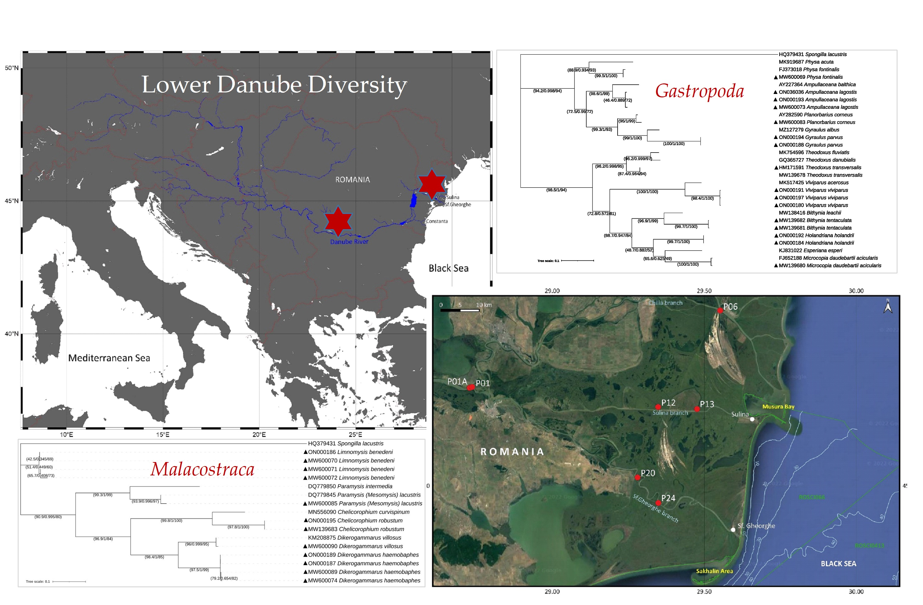
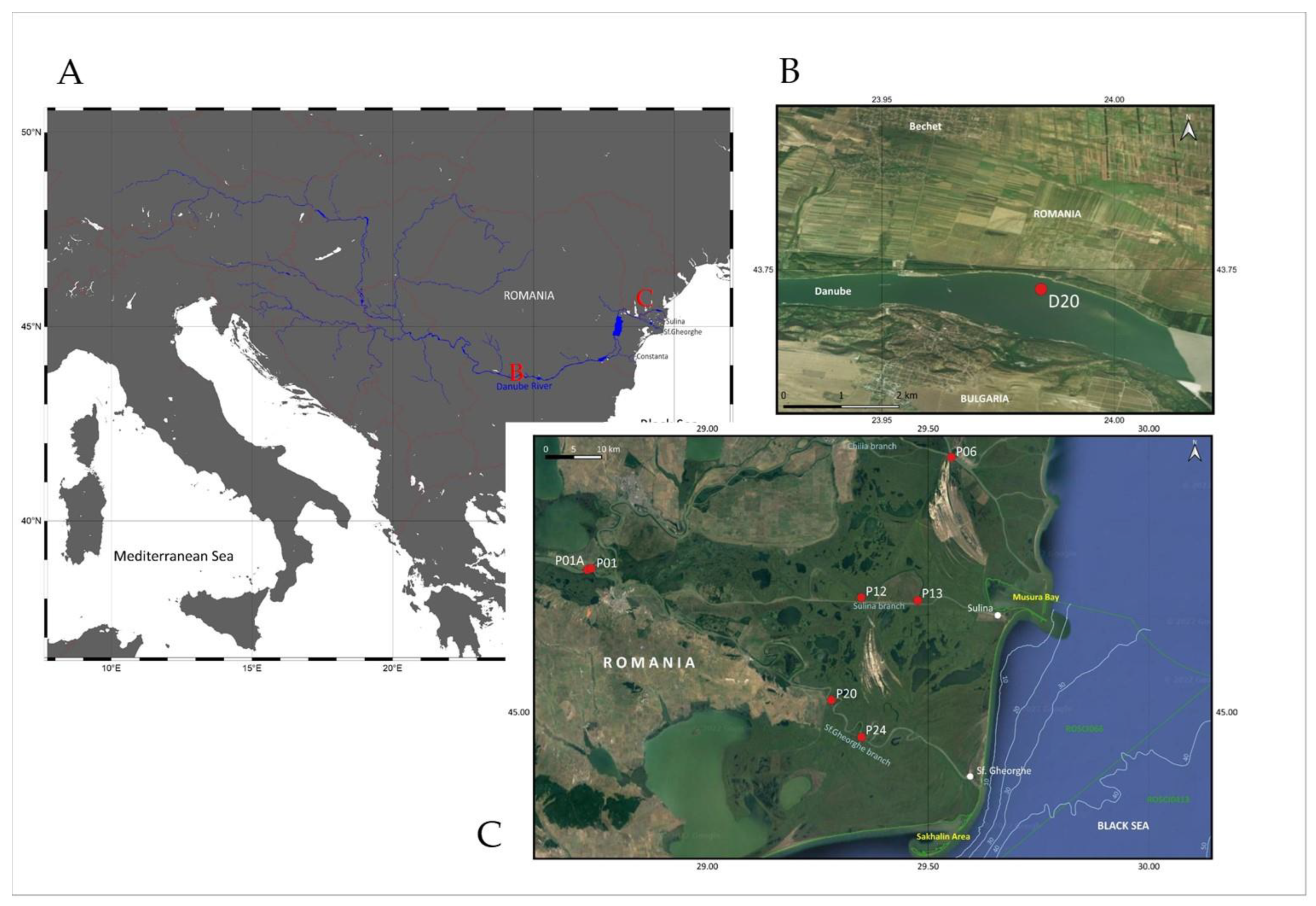
2.1. Study Area, Sampling and Sample Preparation

Sediment samples were collected from 8 sites along the Lower Danube sector in 2019, 2020 and 2021 during 3 field trip sessions in late spring (May–June) periods (Figure 1, Table 1). Among these, the sites P01 and P01A were located within the Ceatal Izmail area, the apex of the Danube Delta where the splitting of the river in Chilia and Tulcea distributaries occurs—P06 on Chilia branch, P12 and P13 on Sulina branch, P20 and P24 on the St. George branch and D20 in the Bechet area (km 676), respectively (Figure 1). Sampling was carried out at various depths, corresponding to different substrate composition (Table 1). The substrate from the Danube branches site was represented by mixed, sandy and muddy sediments, and in the Bechet area by submerged vegetation and a solid substrate. The water depth in the D20 station located in Bechet area was 3.7 m on average and ranged from 4.7 to 24 m along the Danube Delta branches.
Sediments were collected using a Van Veen grab with a surface of 0.1 m2 and a limnological net. Samples were washed immediately after collection using 250 and 125 μm mesh sieves to remove excess sediment particles and preserve macrofauna. For collecting the phytophilous organisms, the vegetation was swept by using a limnological net with 125 μm mesh size. Each specimen selected for genetic analyses was washed with sterile water and placed in 200 μL Tris-EDTA pH 8 buffer at −20 °C [47]. For samples collected in 2021 with the Van Veen grab, the quantitative distribution of each species was evaluated by counting all individuals and calculating their theoretical density per unit surface (1 m2) using a multiplication factor of 10 [48]. For the samples collected with the limnological net, the abundance was expressed as the total number of individuals collected.

2.2. Morphological Identification

In a first attempt, all collected species were morphologically assigned according to the identification keys for gastropods [49], amphipods [50], and mysids [51], and further submitted to DNA barcoding analysis.

2.3. DNA Extraction, PCR Amplification and COI Gene Sequencing

Total genomic DNA was extracted using the DNeasy Blood and Tissue Kit (Qiagen, Hilden, Germany), following an optimized protocol including an initial stage of cell disruption [52]. The specimens were introduced into Tris-EDTA pH 8 buffer and homogenized at 20 °C, 50 Hz, for 12 min, in a SpeedMill PLUS Cell Homogenizer (Analytik, Jena, Germany) in the presence of 5 ZR BashingBead lysis matrix 0.2 mm (Zymo Research, Irvine, CA, USA), and further processed following the manufacturer’s protocol. A partial region of mitochondrial COI gene was amplified using metazoan universal primers (CO1490 (5′-GGTCAACAAATCAAA-GATATTGG-3′) and HCO2198 (5′-TAAACTTCAGGGTGAC-CAAAAAATCA-3′)) [53]. PCR amplification was carried out in a 50 μL reaction volume containing 1 unit ofTaq DNA polymerase (ThermoFisher Scientific, Waltham, MA, USA), 1 μL genomic DNA, 1 μL each of LCO1490 and HCO2198 primer, 0.1 mM of dNTP (ThermoFisher Scientific), 1 × BSA (New England Biolab, BiolabIpswich, MA, USA) and 1 × Taq buffer containing 2.5 mM MgCl2 (ThermoFisher Scientific). The COI fragment was amplified after an initial incubation at 95 °C for 2 min, followed by 5 cycles of incubation at 94 °C for 30 s, annealing at 45 °C for 1.5 min and extension at 72 °C for 1 min; and 35 cycles of 94 °C for 30 s, 50 °C for 1.5 min and 72 °C for 1 min, with a final extension step of 5 min at 72 °C. The size and integrity of the amplified DNA were analyzed by electrophoresis in 1% agarose gel (Cleaver Scientific, Ltd., England). The amplicons were further purified using a QIAquick PCR Purification Kit (Qiagen) and sequenced on both strands using the amplification primers (Macrogen, Amsterdam, The Netherlands).
The resulting COI nucleotide sequences were edited using Sequence Assembly and Alignment—CodonCode Aligner Software (CodonCode Corporation 2003). Sequence identification was performed using the BLAST-NCBI platform [54]. Molecular identification of isolated gastropods and crustaceans collected from the Danube branches was based on the sequence identity of the COI amplicons using a combination of approaches that included the use of the R package INSECT with the database classifier version [55,56] and a 97% threshold for BLAST sequence screening of the NCBI GenBank database [26].
The COI sequence of all identified gastropod and crustacean specimens from the Lower Danube sector were deposited in GenBank (Supplementary Table S1).

2.4. Phylogenetic Analysis and Calculation of Intra- and Interspecific Genetic Distance

The alignment of gastropods and crustaceans’ COI sequences retrieved from NCBI GenBank (Supplementary Table S1) was performed using MUSCLE with default parameters [57]. Phylogenetic analysis for both gastropod and crustacean species based on COI sequences was performed via maximum likelihood statistical method using the IQ-TREE web server (Available online: http://iqtree.cibiv.univie.ac.at/ (accessed on 20 June 2022)). We used the ModelFinder to find the best substitution model according to BIC (K3Pu + F + I + G4), with ultrafast bootstrapping (1000 iterations), single branch test SH-aLRT (1000 iterations) and the Approximate Bayes test [58]. The resulting tree was visualized using Interactive Tree of Life (Available online: https://itol.embl.de/ (accessed on 20 June 2022)) [59]. Spongilla lacustris COI gene (HQ379431) was used as an outgroup for both gastropods and crustacean phylogenetic trees. Pairwise intraspecific and interspecific genetic distances were calculated using the Kimura two-parameter (K2P) model using the Molecular Evolutionary Genetics Analyses (MEGA) platform version 11 [60,61]. A 3% molecular threshold was taken into account as the most used cut-off value for species delimitation [26].

3. Results

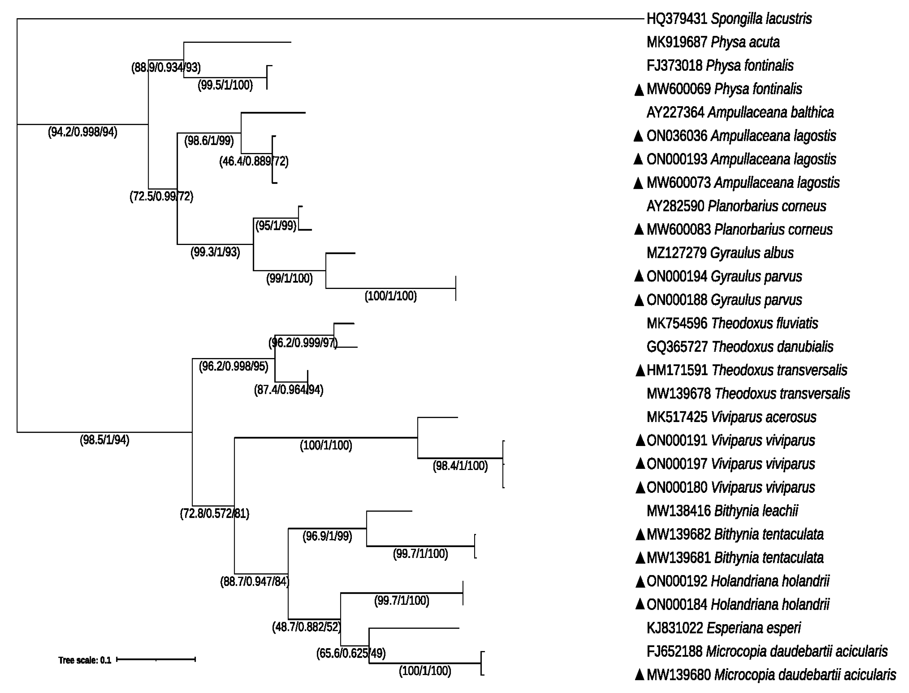
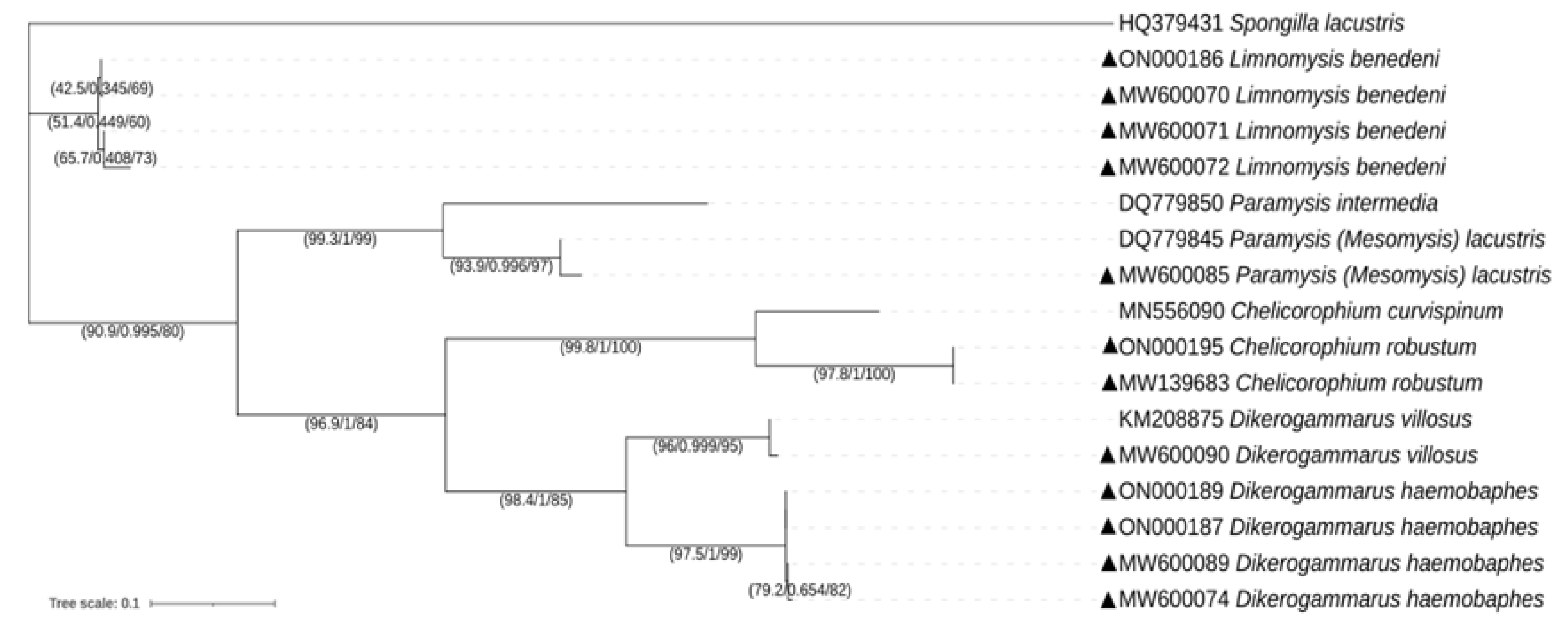
3.1. Molecular Identification and Phylogeny of Gastropods and Crustaceans

Following an initial morphological screening of the invertebrates collected from the eight stations of the Lower Danube sector (Figure 1), all specimens were successfully identified by DNA barcoding, including the 16 gastropods and 12 crustacean individuals collected (Supplementary Table S1). The taxonomic assignment of these new species using the R package INSECT analysis confirmed their affiliation (Supplementary Table S2).
The COI amplicons’ sizes varied between 513 and 654 bp with an average was 602 bp for gastropods, and between 539 and 651 bp with an average of 600 bp for crustaceans.
The nine gastropod species from Caenogastropoda, Architaenioglossa, Littorinimorpha, Cycloneritida and Hygrophila orders belonged to eight families (Amphimelaniidae, Melanopsidae, Viviparidae, Bithyniidae, Neritidae, Lymnaidae, Physidae and Planorbidae), and the five identified crustaceans from Amphipoda and Mysida orders were classified into three different families (Corophiidae, Gammaridae and Misidae) (Table 2).
A phylogenetic tree for gastropod species was constructed based on 28 individuals’ DNA barcode sequences, of which 16 were from the current study and 10 additional sequences were retrieved from the NCBI GenBank database (Figure 2). All individuals assigned to the same species belonged to monophyletic clusters, and all individuals of the same species formed a branch, each with high bootstrap support values (<90%) (Figure 2).
The intraspecific and interspecific distances were measured for the specimens assigned to the same species and same family, in order to validate the existence of the 3% molecular threshold. For gastropods, the intraspecific distance calculation was conducted for four species which were represented by more than one individual, as follows: H. holandrii, V. viviparus., B. tentaculata and A. lagotis. Intraspecific T3P distances of the COI sequences within species ranged from 0% to 0.7%, the highest distance being found in A. lagotis. Interspecific distances for gastropods ranged from 17.2% to 37.8%. In the case of Planorbidae being represented by two species, the average genetic distance within this family was 16.9% (Table 3).
For crustacea, the phylogenetic reconstruction was based on 16 COI sequences, of which 12 were from this study and 4 were retrieved from GenBank (Figure 3). All individuals belonging to the same species analyzed in the present study formed distinct clusters in the tree, with bootstrap support values <90% (Figure 3).
The calculated intraspecific distances for C. robustum, D. haemobaphes and L. benedeni showed the highest genetic distance (2.7%) for L. benedeni, in addition to the identified 3% threshold for different species. The lowest interspecific distance of 20.4% was obtained between D. haemobaphes and D. villosus, and the highest value was 38.9% between C. robustum and P. lacustris, and the same between D. haemobaphes and L. benedeni. Additionally, the interspecific distances between species belonging to the same family varied in the case of Gammaridae. For the genus Dikerogammarus, the interval was 19.9–20.5% and the average value was 20.2%. The values for the two Misidae species ranged from 30.5% to 33.8%, and the average value was 31.4% (Table 4).
Intra- and interspecific pairwise distances for crustacean species COI analyses showed that the obtained intraspecific and interspecific genetic distances between individuals of both gastropod and crustacean species do not overlap, further supporting the species identification.

3.2. Distribution and Ecology of Gastropod and Crustacean Species along the Lower Danube Region

Out of the eight investigated sites, gastropod species were identified in four locations along the Danube branches and in the Bechet area site (Figure 4) during different field trips. The crustacean species were found in all seven sampling sites located along the Danube branches (Figure 5), during the whole time interval (Table 2).
The gastropod species identified along the Danube branches (M. daudebartii acicularis, V. viviparus, B. tentaculata, P. fontinalis and P. corneus) were present only on Chilia and Sulina Danube branches (Figure 4). Meanwhile, only three phytophilous gastropods were retrieved from the Bechet area, H. holandrii, T. transversalis and G. parvus (Figure 4). These specimens were retrieved from different water depths ranging from 3.7 to 24 m (Table 2). The river-bed substrate below the isolated species was also variable. In this respect, M. daudebartii acicularis, V. viviparus, B. tentaculata, P. fontinalis and P. corneus species were associated with mixed sediments, whereas A. lagotis was detected in areas dominated by sandy and muddy sediments rich in submerged vegetation (Table 2).
The calculated density of gastropod species was relatively low, ranging from 10 to 20 individuals/m2 interval. There was a high presence (30 ind/m2) of P. corneus in site P12 (Sulina branch) (Table 5).
The amphipods and mysids were identified from the investigated sites located along the three main Danube channels (Chilia, Sulina and St. George). D. haemobaphes was detected in all three branches, D. villosus only in Sulina and St. George areas and C. robustum in Chilia and Sulina; the mysids were identified only in the Sulina branch (Figure 5). Their habitat was characterized by a variable water depth ranging from 4.7 to 25 m (Table 2). All investigated species were detected in substrates characterized by mixed sediments, but only representatives of Amphipoda order were encountered in sandy and muddy sediments (Table 2).
Overall, the amphipods recorded higher calculated density as compared to mysid species. The highest values were found for C. robustum and D. haemobaphes, reaching 420 and 300 ind./m2 at P12 and P13 stations, respectively. The two mysid species registered relatively low densities, the highest being recorded by P. lacustris with 30 ind./m2 in P13 station. (Table 5).

4. Discussion

For the last decades, the molecular approach based on DNA barcoding has become an important tool for biodiversity assessment worldwide, being suitable for the identification of species from different life stages and species with sexual dimorphism, or for putative cryptic species, from both fresh and preserved materials [62]. In the current study, DNA barcoding was proven to be an effective instrument for identifying gastropods and crustaceans.
Traditionally, the method for validating presumptive species using DNA barcoding analysis is based on the comparison between intraspecific and interspecific genetic nucleotide divergence enabling the inference of a molecular threshold to help taxonomic identification [26,63]. There are many debates in the scientific literature about the most appropriate similarity threshold. It can vary in the 2–4% interval depending on the taxonomic group of macroinvertebrate species [64]. The variation between species needs to exceed the variation within species, which allows clear genetic differentiation of species by the existence of the barcoding gap [65]. Here, for performing pairwise genetic distances, the 3% molecular threshold was used. The calculated intraspecific and interspecific divergence values for gastropods (Table 3) were comparable to those reported for species retrieved from the Portuguese coast, Vaal River and Adriatic Sea, varying in the 8.44–74.67% and 0–2.9% intervals, respectively [66,67,68]; and for amphipods and mysids collected from the Pacific coast of Canada, the Black Sea, the Caspian Sea, Danube River and Don and Rhine river systems, these values were in 0%–4.3% and 4.92–34.2% intervals [69,70,71]. These findings support the efficacy of DNA barcoding based on COI gene sequencing in species delineation. Moreover, our results showed no overlap between intra- and interspecific genetic divergence for both gastropod and crustacean taxa.
COI represent a better target, having major advantages: the universal LCO1490 and HCO2198 primers for this gene are very robust, allowing the recovery of the 5′ end from the majority of the representatives of animal phyla [53,72]. The evolution of this gene was fast enough to enable the discrimination of closely related species and phylogeographic groups within a single species [73,74]. For both gastropod and crustacean taxa, the ML tree showed distinctness of all the studied species. For instance, although sequences belonging to the specimens identified as V. viviparus grouped closely with the GenBank retrieved sequences belonging to V. acerosus, the species formed distinct clusters in the COI phylogenetic reconstruction. Our results are consistent with those other studies showing that COI-based phylogeny could confirm the genetic differentiation between Viviparus species [75]. Moreover, [76] reported morphological similarities between the aforementioned species, but a later revision and molecular analyses confirmed their delimitation. Another example is given by the representatives of the Planorbidae family. The phylogenetic reconstruction showed that the species were grouped in distinct branches and sequences belonging to the same species clustered together. Recently, phylogenetic analyses based on mitochondrial and nuclear DNA sequences [77] showed that G. laevis and G. parvus are in fact part of the same species-level clade, the latter having nomenclatural priority. Our data indicated similar results for both amphipods and mysids collected from the lower sector of the Danube River. While members of the same family (e.g., Gammaridae, Misidae) or the same genus (Dikerogammarus) did not cluster together, sequences of same species grouped together, suggesting the efficacy of COI sequences in species delineation. Previous debates related to the taxonomic status of Dikerogammarus species considered that D. villosus and D. bispinosus are synonyms of D. haemobaphes [78]. However, the taxonomical revision performed by [79] and the analyses based on mitochondrial genomes performed by [80] revealed genetic distinctions among these taxa. Furthermore, a COI gene analysis performed on Chelicorophium revealed that the specimens clustered in two separate groups corresponding to C. curvispinum and to C. robustum [81], which is consistent with our data obtained in the case of the Lower Danube specimens.
Mollusca represent the most abundant organisms of the Danube River in terms of biomass. Owing to their size, Bivalvia make up to 80% of the total biomass, followed by Gastropoda, covering 10% to 35% of the community [82].
The types of substrate associated with invertebrates could also vary. Along the Danube branches, the mixed sediments dominated by mud with aquatic vegetation were populated by all species detected in this area, except for A. lagotis, which was identified in sandy mud. The mixed sediments were associated with M. daudebartii acicularis, V. viviparus and B. tentaculata. A recent investigation [83] reported that V. viviparus, B. tentaculata, M. daudebartii acicularis and P. fontinalis could also inhabit several types of substrate, such as gravelly, muddy and sandy river bottoms, and areas with aquatic vegetation. Although gastropods can populate areas with sandy and muddy river bottoms, these organisms are frequently associated with solid substrata (boulders, stones, plant parts) [84]. Along the Danube branches, the substrate was represented by mixed sediments, mud and sand, explaining the low density of the species identified in this area. Both A. lagotis and P. corneus were identified in only one substrate each, in mixed sediments and sandy mud, respectively. Previous studies reported that these species are pelophilous and phytophilous, characteristic of stagnant waters [85,86]; this may explain their absence from the majority of the investigated samples. B. tentaculata and M. daudebartii acicularis were previously reported in the Danube Delta area, along the St. George branch [42]. In addition, our study showed that these species also populated Ceatal Izmail and Sulina branch, whereas no individuals were found in the St. George sites. H. holandrii, T. trasversalis and G. albus were identified only in the Bechet area, which is characterized by the presence of submerged vegetation and a solid substrate. The Ponto-Caspian snail T. transversalis, listed as Endangered in the IUCN Red List, is nowadays found in the Danube River in a very restricted area only in the lower stretch [82,87,88], and H. holandrii is known as one of the Balkanian fauna of the Danube River [82,89]. Both species are known to populate river bottoms with hard substrates [82,87,88,89], and our data confirm these ecological preferences of the species.
In terms of abundance, the fauna of the Danube River was dominated by crustaceans. Amphipoda was reported to be the dominant group in al Danube branches, representing up to 75% of the total abundance [82]. While previous reports indicated that D. villosus, D. haemobaphes and C. robustum are associated with gravelly substrates [90], the current data revealed the presence of the amphipod species in several types of substrates. Although the two representatives of Dikerogammarus showed a strong preference for large cobble and artificial substrate, the species are adapted to various ecological conditions [91,92,93], as confirmed by the current study where these species were identified in mixed sediments, sand and mud. A previous report on the macroinvertebrate communities from the Danube Delta [42] also showed the occurrence of D. haemobaphes and D. villosus in several locations along the St. George branch, similarly to our data, in support of the resilience of these species for more than 5 years. Furthermore, C. robustum, which is reported to inhabit gravelly and muddy substrates [94,95], was also found in the current survey to populate areas dominated by mixed sediments and sandy mud. Our data revealed the association of both L. benedeni and P. lacustris with mixed sediments dominated by mud, in accordance with initial reports regarding their preferred habitat being characterized by fine sediments (sand and mud) with standing water or slow to moderate currents [51]. L. benedeni is often found in great densities on the shore at depths of only 0–0.5 m, although they can occur at a depth of 6 m [51]. D. haemobaphes was identified in the littoral zone at 50–70 cm depth [96].

5. Conclusions

The current findings regarding the distributions of several gastropod and Ponto-Caspian amphipods and mysids populating the Lower Danube region extended the knowledge on the presence and density of these benthic invertebrates based on molecular identification by DNA barcoding using COI gene sequencing, and complementary meta-data regarding their habitats (substrate type and river depth), thereby adding to the ecological profile of these fauna populating the Danube Delta sector. The accuracy of species identification by this method was highlighted in the cases of several specimens belonging to same species of gastropods or crustaceans clustered together in monophyletic groups. Moreover, this survey contributed to the first gastropod barcode dataset for the Romanian Danube sector.

Supplementary Materials

The following is available online at https://www.mdpi.com/article/10.3390/d14070533/s1. Table S1. Specimen taxonomy and accession number, sampling period and location, best match COI gene sequence for gastropod, amphipod and mysid species isolated from the Lower Danube sector and Table S2. Taxonomy assignation by the INSECT R package for all COI sequences used on the present research (including sequences recovered from NCBI).

Author Contributions

S.M. and C.P. wrote the manuscript; S.M. isolated the DNA, performed DNA barcoding, performed phylogenetic analysis and contributed to experimental design and data interpretation; T.B. performed the statistical analyses; A.T. contributed to map construction and species identification; M.M. contributed to map construction and sample collection; P.L. performed the phylogenetic tree construction and R package INSECT analyses; C.P. performed the experimental design and coordinated the study. All authors have read and agreed to the published version of the manuscript.

Funding

The study was financially supported by the Romanian Ministry of Research as part of the CORE Programme projects PN19200401, PN19200204 and PN19200302; by the Romanian Academy RO1567-IBB05/2021 project; by the European Union’s Horizon 2020 DOORS project, grant agreement number 101000518; and by the Program Development of the National R&D system—AMBI AQUA—No. 23PFE. T.

Institutional Review Board Statement

Not applicable.

Data Availability Statement

All data supporting the conclusions of this article are included in the manuscript.

Acknowledgments

The authors thank Ana Bianca Pavel from GeoEcoMar for assisting in collecting and sorting the benthic material, and Lavinia Iancu for helping with the COI barcoding experiment.

Conflicts of Interest

The authors declare no conflict of interest.

References

  1. Helmer, R.; Hespanhol, I. Water Pollution Control—A Guide to the Use of Water Quality Management Principles; United Nations Environment Programme, the Water Supply & Sanitation Collaborative Council and the World Health Organization WHO/UNEP: New York, NY, USA, 1997; Available online: https://www.who.int/water_sanitation_health/resourcesquality/wpcbegin.pdf (accessed on 25 April 2022).
  2. Gâştescu, P.; Ţuchiu, E. The Danube River in the Pontic Sector—Hydrologycal Regime. Water Resources and Wetlands. In Proceedings of the 1st Water and Wetlands Resources Conference, Tulcea, Romania, 14–16 September 2012; Gâştescu, P., Lewis, W., Jr., Breţcan, P., Eds.; Riscuri si Catastrofe: Cluj-Napoca, Romania, 2012; Volume 12, pp. 13–26, ISBN 978-606-605-038-8. [Google Scholar]
  3. ICPDR. Danube River Basin District Management Plan; ICPDR: Vienna, Austria, 2009. [Google Scholar]
  4. Oaie, G.; Secrieru, D.; Bondar, C.; Szobotka, Ş.; Duţu, L.; Manta, T. Lower Danube River: Characterization of sediments and pollutants. GeoEcoMarina 2015, 21, 19–34. [Google Scholar]
  5. Hanganu, J.; Grigoras, I.; Stefan, N.; Sarbu, I.; Zhmud, E.; Dubyna, D.; Menke, U.; Drost, H. Transboundary Vegetation Map of the Biosphere Reserve Danube Delta; Danube Delta Institute DDI, Institute for Inland Water Management and Waste Water Treatment RIZA, Danube Biosphere Reserve of the National Academy of Sciences of Ukraine DPA, I&KIB: Tulcea, Romania, 2002. [Google Scholar]
  6. Mee, L.D. The Black Sea in crisis: A need for concerted international action. Ambio 1992, 21, 278–286. [Google Scholar]
  7. Hutchinson, G.E. A Treatise on Limnology. In The Zoobenthos; John Wiley & Sons: New York, NY, USA, 1993; Volume 4, p. 944. [Google Scholar]
  8. Wallace, J.B.; Webster, J.R. The role of macroinvertebrates in stream ecosystem function. Annu. Rev. Entomol. 1996, 41, 115–139. [Google Scholar] [CrossRef]
  9. Clarke, K.D.; Knoechel, R.; Ryan, P.M. Influence of trophic role and life-cycle duration on timing and magnitude of benthic macroinvertebrate response to whole-lake enrichment. Can. J. Fish. Aquat. Sci. 1997, 54, 89–95. [Google Scholar] [CrossRef]
  10. Gomes, M.A.; Novelli, R.; Zalmon, I.R.; Souza, C.M. Malacological assemblages in sediments of eastern Brazilian continental shelf, coordinates 108 and 208 S, between Bahia and Espírito Santo State. Bios 2004, 12, 11–24. [Google Scholar]
  11. Tubić, B.; Simić, V.; Zorić, K.; Gačić, Z.; Atanacković, Z.; Csányi, B.; Paunović, M. Stream section types of the Danube River in Serbia according to the distribution of macroinvertebrates. Biologia 2013, 68, 294–302. [Google Scholar] [CrossRef]
  12. Rosemond, A.D.; Mullholland, P.J.; Ellwood, J.W. Top-down and bottom-up control of stream periphyton: Effects of nutrients and herbivores. Ecology 1993, 74, 1264–1280. [Google Scholar] [CrossRef]
  13. Brown, K.M.; Alexander, J.E.; Thorp, J.H. Differences in the ecology and distribution of lotic pulmonate and prosobranch gastropods. Am. Malacol. Bull. 1998, 14, 91–101. [Google Scholar]
  14. Frank, C.; Jungbluth, J.; Richnovszky, A. Die Mollusken der Donau vom Schwarzwald bis zum Schwarzen Meer; Akaprint: Budapest, Hungary, 1990; pp. 1–142. [Google Scholar]
  15. Moog, O.; Humpesch, U.H.; Konar, M. The distribution of benthic invertebrates along the Austrian stretch of the River Danube and its relevance as an indicator of zoogeographical and water quality patterns—Part 1. Arch. Hydroboil. Robiol. 1995, 9 Suppl. 101, 121–213. [Google Scholar] [CrossRef]
  16. Wesselingh, F.P.; Neubauer, T.A.; Anistratenko, V.V.; Vinarski, M.V.; Yanina, T.; ter Poorten, J.J.; Kijashko, P.; Albrecht, C.; Anistratenko, O.Y.; D’Hont, A.; et al. Mollusc species from the Pontocaspian region—An expert opinion list. ZooKeys 2019, 827, 31–124. [Google Scholar] [CrossRef] [Green Version]
  17. Glazier, D.S. Amphipoda. Reference Module in Earth Systems and Environmental Sciences; Elsevier: Amsterdam, The Netherlands, 2014; p. 49. [Google Scholar] [CrossRef]
  18. Roast, S.D.; Thompson, R.S.; Widdows, J.; Jones, M.B. Mysids and environmental monitoring: A case for their use in estuaries. Mar. Freshw. Res. 1998, 49, 827–832. [Google Scholar] [CrossRef]
  19. Alonso, Á.; De Lange, H.J.; Peeters, E.T.H.M. Development of a feeding behavioural bioassay using the freshwater amphipod Gammarus pulex and the Multispecies Freshwater Biomonitor. Chemosphere 2009, 75, 341–346. [Google Scholar] [CrossRef] [PubMed]
  20. Grabowski, M.; Bacela-Spychalska, K.; Pešić, V. Reproductive traits and conservation needs of the endemic gammarid Laurogammarus scutarensis (Schäferna, 1922) from the Skadar Lake system, Balkan Peninsula. Limnologica 2014, 47, 44–51. [Google Scholar] [CrossRef]
  21. Borza, P.; Huber, T.; Leitner, P.; Remund, N.; Graf, W. Success factors and future prospects of Ponto-Caspian peracarid (Crustacea: Malacostraca) invasions: Is “the worst over”? Biol. Invasions 2017, 19, 1517–1532. [Google Scholar] [CrossRef] [Green Version]
  22. Heino, J.; Louhi, P.; Muotka, T. Identifying the scales of variability in stream macroinvertebrate abundance, functional composition and assemblage structure. Freshw. Biol. 2004, 49, 1230–1239. [Google Scholar] [CrossRef]
  23. Seymour, M.; Deiner, K.; Altermatt, F. Scale and scope matter when explaining varying patterns of community diversity in riverine metacommunities. Basic Appl. Ecol. 2016, 17, 134–144. [Google Scholar] [CrossRef] [Green Version]
  24. Frézal, L.; Leblois, R. Four years of DNA barcoding: Current advances and prospects. Infect. Genet. Evol. 2008, 8, 727–736. [Google Scholar] [CrossRef] [Green Version]
  25. Macher, J.N.; Salis, R.K.; Blakemore, K.S.; Tollrian, R.; Matthaei, C.D.; Leese, F. Multiple-stressor effects on stream invertebrates: DNA barcoding reveals contrasting responses of cryptic mayfly species. Ecol. Indic. 2016, 61, 159–169. [Google Scholar] [CrossRef]
  26. Hebert, P.D.N.; Cywinska, A.; Ball, S.L. Biological identifications through DNA barcodes. Proc. R. Soc. Lond. B 2003, 270, 313–321. [Google Scholar] [CrossRef] [Green Version]
  27. Puillandre, N.; Cruaud, C.; Kantor, Y.I. Cryptic species in Gemmuloborsonia (Gastropoda: Conoidea). J. Mollus. Stud. 2009, 76, 11–23. [Google Scholar] [CrossRef] [Green Version]
  28. Sands, A.F.; Glöer, P.; Gürlek, M.E.; Albrecht, C.; Neubauer, T.A. A revision of the extant species of Theodoxus (Gastropoda, Neritidae) in Asia, with the description of three new species. Zoosyst. Evol. 2020, 96, 25–66. [Google Scholar] [CrossRef] [Green Version]
  29. Lipinskaya, T.; Radulovici, A.E. DNA barcoding of alien Ponto-Caspian amphipods from the Belarusian part of the Central European invasion corridor. Genome 2017, 60, 963–964. [Google Scholar] [CrossRef] [Green Version]
  30. Audzijonyte, A.; Daneliya, M.; Väinölä, R. Comparative phylogeography of Ponto-Caspian mysid crustaceans: Isolation and exchange among dynamic inland sea basins. Mol. Ecol. 2006, 15, 2969–2984. [Google Scholar] [CrossRef] [PubMed]
  31. Barco, A.; Claremont, M.; Reid, D.G.; Houart, R.; Bouchet, P.; Williams, S.T.; Cruaud, C.; Couloux, A.; Oliverio, M. A molecular phylogenetic framework for the Muricidae, a diverse family of carnivorous gastropods. Mol. Phylogenet. Evol. 2010, 56, 1025–1039. [Google Scholar] [CrossRef] [PubMed]
  32. Layton, K.K.; Martel, A.L.; Hebert, P.D.N. Patterns of DNA Barcode Variation in Canadian Marine Molluscs. PLoS ONE 2014, 9, e95003. [Google Scholar] [CrossRef] [Green Version]
  33. Graf, W.; Csányi, B.; Leitner, P.; Paunović, M.; Huber, T.; Szekeres, J.; Nagy, C.; Borza, P. Joint Danube Survey 3, Full Report on Macroinvertebrates; ICPDR, International Commission for the Protection of the Danube River: Sofia, Bulgaria, 2014; p. 87. Available online: www.icpdr.org (accessed on 20 May 2022).
  34. Glöer, P.; Sȋrbu, I. New freshwater molluscs species found in the Romanian fauna. Heldia 2005, 6, 229–238. [Google Scholar]
  35. Gomoiu, M.T.; Begun, T.; Opreanu, P.; Teaca, A. Present state of benthic ecosystem in Razelm-Sinoie Lagoon Complex (RSLC). In Proceedings of the 37th IAD Conference, The Danube River Basin in a Changing World, Chişinău, Moldova, 29 October–1 November 2008; pp. 108–112. [Google Scholar]
  36. Paraschiv, G.M.; Begun, T.; Teaca, A.; Bucur, M.; Tofan, L. New data about benthal populations of the Golovita and Zmeica lakes. J. Environ. Prot. Ecol. 2010, 11, 253–260. [Google Scholar]
  37. Begun, T.; Teacă, A.; Mureşan, M.; Pavel, A.B. Current state of the mollusc populations in the Razim-Sinoe Lagoon System. Anim. Sci. 2020, 63, 553–561. [Google Scholar]
  38. Begun, T. Complex Ecological Studies of Crustaceans Populations (Cumaceans and Mysidaceans) of the North Western Black Sea Coast. Ph.D. Thesis, University “Ovidius” Constanta, Constanța, Romania, 2006; p. 276. [Google Scholar]
  39. Paraschiv, G.M.; Schroder, V.; Samargiu, M.D.; Sava, D. Ecological study of zoobenthos communities from the Matita and Merhei lakes (Danube Delta). Res. J. Agric. Sci. 2007, 39, 489–498. [Google Scholar]
  40. Graf, W.; Csányi, B.; Leitner, P.; Paunović, M.; Janeček, B.; Šporka, F.; Chiriac, G.; Stubauer, I.; Ofenböck, T. Joint Danube Survey 2, Report on Macroinvertebrates; ICPDR International Commission for the Protection of the Danube River: Vienna, Austria, 2008; p. 87. [Google Scholar]
  41. Borza, P.; Csányi, B.; Paunović, M. Corophiids (Amphipoda, Corophioidea) of the river Danube—The results of a longitudinal urvey. Crustaceana 2010, 83, 839–849. [Google Scholar]
  42. Stoica, C.; Gheorghe, S.; Petre, J.; Lucaciu, I.; Nita-Lazar, M. Tools for assessing Danube Delta systems with macro invertebrates. Environ. Eng. Manag. J. 2014, 13, 2243–2252. [Google Scholar] [CrossRef]
  43. Cristescu, M.E.; Hebert, P.D.; Onciu, T.M. Phylogeography of Ponto-Caspian crustaceans: A benthic–planktonic comparison. Mol. Ecol. 2003, 12, 985–996. [Google Scholar] [CrossRef] [PubMed]
  44. Rewicz, T.; Wattier, R.; Grabowski, M.; Rigaud, T.; Bącela-Spychalska, K. Out of the Black Sea: Phylogeography of the invasive killer shrimp Dikerogammarus villosus across Europe. PLoS ONE 2015, 10, e0118121. [Google Scholar] [CrossRef] [PubMed] [Green Version]
  45. Jażdżewska, A.M.; Rewicz, T.; Mamos, T.; Wattier, R.; Bącela-Spychalska, K.; Grabowski, M. Cryptic diversity and mtDNA phylogeography of the invasive demon shrimp, Dikerogammarus haemobaphes (Eichwald, 1841), in Europe. NeoBiota 2020, 57, 53–86. [Google Scholar] [CrossRef]
  46. Cristescu, M.E.A.; Hebert, P.D.N. The ‘Crustacean Seas’—An evolutionary perspective on the Ponto-Caspian peracarids. Can. J. Fish. Aquat. Sci. 2005, 62, 505–551. [Google Scholar] [CrossRef]
  47. Ross, K.S.; Haites, N.E.; Kelly, K.F. Repeated freezing and thawing of peripheral blood and DNA in suspension: Effects on DNA yield and integrity. J. Med. Genet. 1990, 27, 569–570. [Google Scholar] [CrossRef] [Green Version]
  48. SR EN ISO 10870; Water Quality—Guidelines for the Selection of Sampling Methods and Devices for Benthic Macroinvertebrates in Fresh Waters. ISO: Geneva, Switzerland, 2012.
  49. Grossu, A.V. Mollusca—Gastropoda Prosobranchia si Opistobranchia; Fauna Republicii Populare Române: Bucharest, Romana, 1956; Volume 3. [Google Scholar]
  50. Cărăuşu, S.; Dobreanu, E.; Manolache, C. Fauna Republicii Populare Romȋne. Crustacea. Volumul IV. Fascicula 4. Amphipoda. Forme Salmastre şi de apă Dulce; Editura Acedemiei Republicii Populare Romȋne: Bucharest, Romana, 1955; pp. 1–401. [Google Scholar]
  51. Băcescu, M. Crustacea: Mysidacea; Editura Academiei Republicii Populare Romîne: Bucureşti, Romania, 1954; Volume 126, p. 52. [Google Scholar]
  52. Iancu, L.; Carter, D.O.; Junkins, E.N.; Purcarea, C. Using bacterial and necrophagous insect dynamics for postmortem interval estimation during cold season: Novel case study in Romania. Forensic Sci. Int. 2015, 254, 106–117. [Google Scholar] [CrossRef]
  53. Folmer, O.; Black, M.; Hoeh, W.; Lutz, R.; Vrijenhoek, R. DNA primers for amplification of mitochondrial cytochrome c oxidase subunit I from diverse metazoan invertebrates. Mol. Mar. Biol. Biotechnol. 1994, 3, 294–299. [Google Scholar]
  54. Available online: https://blast.ncbi.nlm.nih.gov (accessed on 28 April 2022).
  55. Leray, M.; Yang, J.Y.; Meyer, C.P.; Mills, S.C.; Agudelo, N.; Ranwez, V.; Machida, R.J. A new versatile primer set targeting a short fragment of the mitochondrial COI region for metabarcoding metazoan diversity: Application for characterizing coral reef fish gut contents. Front. Zool. 2013, 10, 34. [Google Scholar] [CrossRef] [Green Version]
  56. Wilkinson, S.P.; Davy, S.K.; Bunce, M.; Stat, M. Taxonomic identification of environmental DNA with informatic sequence classification trees. PeerJ Prepr. 2018, 6, e26812v1. [Google Scholar] [CrossRef]
  57. Edgar, R.C. MUSCLE: Multiple sequence alignment with high accuracy and high throughput. Nucleic Acids Res. 2004, 32, 1792–1797. [Google Scholar] [CrossRef] [PubMed] [Green Version]
  58. Trifinopoulos, J.; Nguyen, L.T.; von Haeseler, A.; Minh, B.Q. W-IQ-TREE: A fast online phylogenetic tool for maximum likelihood analysis. Nucleic Acids Res. 2016, 44, W232–W235. [Google Scholar] [CrossRef] [PubMed] [Green Version]
  59. Letunic, I.; Bork, P. Interactive Tree of Life (iTOL) v5: An online tool for phylogenetic tree display and annotation. Nucleic Acids Res. 2021, 49, W293–W296. [Google Scholar] [CrossRef] [PubMed]
  60. Kimura, M. A simple method for estimating evolutionary rates of base substitutions through comparative studies of nucleotide sequences. J. Mol. Evol. 1980, 16, 111–120. [Google Scholar] [CrossRef]
  61. Tamura, K.; Stecher, G.; Kumar, S. MEGA 11: Molecular Evolutionary Genetics Analysis Version 11. Mol. Biol. Evol. 2021, 38, 3022–3027. [Google Scholar] [CrossRef]
  62. Radulovici, A.E.; Archambault, P.; Dufresne, F. DNA Barcodes for Marine Biodiversity: Moving Fast Forward? Diversity 2010, 2, 450–472. [Google Scholar] [CrossRef] [Green Version]
  63. Hebert, P.D.N.; Penton, E.H.; Burns, J.M.; Janzen, D.H.; Hallwachs, W. Ten Species in One: DNA Barcoding Reveals Cryptic Species in the Neotropical Skipper Butterfly Astraptes fulgerator. Proc. Natl. Acad. Sci. USA 2004, 101, 14812–14817. [Google Scholar] [CrossRef] [Green Version]
  64. Sweeney, B.W.; Battle, J.M.; Jackson, J.K.; Dapkey, T. Can DNA Barcodes of Stream Macroinvertebrates Improve Descriptions of Community Structure and Water Quality? J. N. Am. Benthol. Soc. 2011, 30, 195–216. [Google Scholar] [CrossRef]
  65. Hebert, P.D.N.; Stoeckle, M.Y.; Zemlak, T.S.; Francis, C.M. Identification of Birds through DNA Barcodes. PLoS Biol. 2004, 2, e312. [Google Scholar] [CrossRef] [Green Version]
  66. Borges, L.; Hollatz, C.; Lobo, J.; Cunha, A.M.; Vilela, A.P.; Calado, G.; Coelho, R.; Costa, A.C.; Ferreira, M.S.G.; Costa, M.H.; et al. With a little help from DNA barcoding: Investigating the diversity of Gastropoda from the Portuguese coast. Sci. Rep. 2016, 6, 20226. [Google Scholar] [CrossRef] [Green Version]
  67. Lawton, S.P.; Allan, F.; Hayes, P.M.; Smit, N.J. DNA barcoding of the medically important freshwater snail Physa acuta reveals multiple invasion events into Africa. Acta Trop. 2018, 188, 86–92. [Google Scholar] [CrossRef] [PubMed] [Green Version]
  68. Buršić, M.; Iveša, L.; Jaklin, A.; Arko Pijevac, M.; Kučinić, M.; Štifanić, M.; Neal, L.; Bruvo Mađarić, B. DNA Barcoding of Marine Mollusks Associated with Corallina officinalis Turfs in Southern Istria (Adriatic Sea). Diversity 2021, 13, 196. [Google Scholar] [CrossRef]
  69. Costa, F.; deWaard, J.R.; Boutillier, J.; Ratnasingham, S.; Dooh, R.T.; Hajibabaei, M.; Hebert, P.D.N. Biological identifications through DNA barcodes: The case of the Crustacea. Can. J. Fish. Aquat. Sci. 2007, 64, 272–295. [Google Scholar] [CrossRef]
  70. Costa, F.; Henzler, C.; Lunt, D.; Whiteley, N.M.; Rock, J. Probing marine Gammarus (Amphipoda) taxonomy with DNA barcodes. Syst. Biodivers. 2009, 7, 365–379. [Google Scholar] [CrossRef]
  71. Audzijonyte, A.; Wittmann, K.J.; Ovcarenko, I.; Väinölä, R. Invasion phylogeography of the Ponto-Caspian crustacean Limnomysis benedeni dispersing across Europe. Divers. Distrib. 2009, 15, 346–355. [Google Scholar] [CrossRef]
  72. Zhang, D.-X.; Hewitt, G.M. Assessment of the universality and utility of a set of conserved mitochondrial primers in insects. Insect Mol. Biol. 1997, 6, 143–150. [Google Scholar] [CrossRef]
  73. Cox, A.J.; Hebert, P.D.N. Colonization, extinction and phylogeographic patterning in a freshwater crustacean. Mol. Ecol. 2001, 10, 371–386. [Google Scholar] [CrossRef]
  74. Wares, J.P.; Cunningham, C.W. Phylogeography and historical ecology of the North Atlantic intertidal. Evolution 2001, 12, 2455–2469. [Google Scholar] [CrossRef]
  75. Rysiewska, A.; Hofman, S.; Osikowski, A.; Beran, L.; Pešić, V.; Falniowski, A. Viviparus mamillatus (Küster, 1852), and partial congruence between the morphology-, allozyme- and DNA-based phylogeny in European Viviparidae (Caenogastropoda: Architaenioglossa). Folia Malacol. 2019, 27, 43–51. [Google Scholar] [CrossRef] [Green Version]
  76. Beran, L.; Horsák, M.; Hofman, S. First records of Viviparus acerosus (Bourguignat, 1862) (Gastropoda: Viviparidae) from the Czech Republic outside its native range. Folia Malacol. 2019, 27, 223–229. [Google Scholar] [CrossRef] [Green Version]
  77. Lorencová, E.; Beran, L.; Nováková, M.; Horsáková, V.; Rowson, B.; Jaroslav, Č.; Hlaváč, J.Č.; Nekola, J.C.; Horsák, M. Invasion at the population level: A story of the freshwater snails Gyraulus parvus and G. laevis. Hydrobiol. 2021, 848, 4661–4671. [Google Scholar] [CrossRef]
  78. Pjatakova, G.M.; Tarasov, A.G. Caspian Sea amphipods: Biodiversity, systematic position and ecological peculiarities of some species. Int. J. Salt Lake Res. 1996, 5, 63–79. [Google Scholar] [CrossRef]
  79. Müller, J.C.; Schramm, S.; Seitz, A. Genetic and morphological differentiation of Dikerogammarus invaders and their invasion history in Central Europe. Freshw. Biol. 2002, 47, 2039–2048. [Google Scholar] [CrossRef]
  80. Mamos, T.; Grabowski, M.; Rewicz, T.; Bojko, J.; Strapagiel, D.; Burzyński, A. Mitochondrial Genomes, Phylogenetic Associations, and SNP Recovery for the Key Invasive Ponto-Caspian Amphipods in Europe. Int. J. Mol. Sci. 2021, 22, 10300. [Google Scholar] [CrossRef] [PubMed]
  81. Marescaux, J.; Latli, A.; Lorquet, J.; Virgo, J.; Van Doninck, K.; Beisel, J.N. Benthic macro-invertebrate fauna associated with Dreissena mussels in the Meuse River: From incapacitating relationships to facilitation. Aquat. Ecol. 2016, 50, 15–28. [Google Scholar] [CrossRef]
  82. ICPDR. Joint Danube Survey 2. Final Scientific Report; ICPDR: Vienna, Austria, 2008. [Google Scholar]
  83. Martinovic-Vitanovic, V.; Raković, M.; Popović, N.; Kalafatic, V. Qualitative study of Mollusca communities in the Serbian Danube stretch (river km 1260–863.4). Biologia 2013, 68, 112–130. [Google Scholar] [CrossRef] [Green Version]
  84. ICPDR. Joint Danube Survey 1. Ecological Status Characterization. Macrozoobenthos; ICPDR: Vienna, Austria, 2002. [Google Scholar]
  85. Surugiu, V.; Mustaţă, G.; Hârţăscu, M. Contributions to the qualitative and quantitative study of the macrozoobenthos from the Danube—Black Sea Canal. Stud. Şi Cercet. Biol. Univ. Din Bacău 2004, 9, 75–80. [Google Scholar]
  86. Georgiev, D.; Hubenov, Z. Freshwater snails (Mollusca: Gastropoda) of Bulgaria: An updated annotated checklist. Folia Malacol. 2013, 21, 237–263. [Google Scholar] [CrossRef] [Green Version]
  87. Sȋrbu, I.; Sȋrbu, M.; Benedek, A. The freshwater molluskfauna from Banat (Romania). Trav. du Mus. Natl. d’Hist. Nat. Grigore Antipa 2010, 53, 21–43. [Google Scholar] [CrossRef]
  88. Solymos, P.; Feher, Z. Theodoxus transversalis (C. Pfeiffer1828). In IUCN 2011. IUCN Red List of Threatened Species. Version 2011. 2011. Available online: www.iucnredlist.org (accessed on 18 May 2022).
  89. Novakovic, B.; Markovic, V.; Tomović, J. Distribution of the snail Amphimelania holandrii Pfeiffer, 1828 (Melanopsidae; Gastropoda) in Serbia in the 2009–2012 period. Water Res. Manag. J. Belgrade 2013, 3, 21–26. [Google Scholar]
  90. ICPDR. Joint Danube Survey 3. Full Report on Macroinvertebrates; ICPDR: Vienna, Austria, 2014. [Google Scholar]
  91. Muskó, I.B. The life history of Dikerogammarus haemobaphes (Eichw.) (Crustacea: Amphipoda) living on macrophytes in Lake Balaton (Hungary). Arch. Für Hydrobiol. 1993, 127, 227–238. [Google Scholar] [CrossRef]
  92. Boets, P.; Lock, K.; Messiaen, M.; Goethals, P.L.M. Combining data-driven methods and lab studies to analyse the ecology of Dikerogammarus villosus. Ecol. Inform. 2010, 5, 133–139. [Google Scholar] [CrossRef]
  93. Clinton, K.E.; Mathers, K.L.; Constable, D.; Gerrard, C.; Wood, P.J. Substrate preferences of coexisting invasive amphipods, Dikerogammarus villosus and Dikerogammarus haemobap-hes, under field and laboratory conditions. Biol. Invasions 2018, 20, 2187–2196. [Google Scholar] [CrossRef] [Green Version]
  94. Lipták, B.; Šporka, F.; Necpálová, K.; Stloukal, E. First record of Ponto-Caspian amphipod Corophium curvispinum in Slovakside of the Danube River. Folia Faun. Slovaca 2012, 17, 183–186. [Google Scholar]
  95. Lipták, B. Non-indigenous invasivefreshwater crustaceans (Crustacea: Malacostraca) in Slovakia. Water Res. Manag. 2013, 3, 21–31. [Google Scholar]
  96. Lipinskaya, T.; Makaranka, A.; Razlutskij, V.; Semenchenko, V. First records of the alien amphipod Dikerogammarus haemobaphes (Eichwald, 1841) in the Neman River basin (Belarus). BioInvasions Rec. 2021, 10, 319–325. [Google Scholar] [CrossRef]
Figure 1. Sampling sites of the Lower Danube River sector. (A) Overview map of the Danube River; (B) sampling location in Bechet area; (C) sampling locations along the Danube Delta river branches.
Diversity 14 00533 g001
Figure 2. Phylogenetic tree of gastropod species based on cytochrome oxidase subunit I (COI). ▲: sequences from the current study; Spongilla lacustris COI (HQ379431) was used as an outgroup; numbers in parentheses are SH-aLRT support (%)/aBayes support/ultrafast bootstrap support (%).
Diversity 14 00533 g002
Figure 3. Phylogenetic tree of amphipod and mysid species based on cytochrome oxidase subunit I (COI); (▲): sequences from the current study; Spongilla lacustris (HQ379431) was used as an outgroup; numbers in parentheses are SH-aLRT support (%)/aBayes support/ultrafast bootstrap support (%).
Diversity 14 00533 g003
Figure 4. Distribution of gastropod species along Danube branches and in the Bechet area (inset).
Diversity 14 00533 g004
Figure 5. Distribution of crustacean species along Danube branches.
Diversity 14 00533 g005
Table 1. Study area. Sampling sites of the lower sector of the Danube River (Figure 1), year of collection, water depth and substrate type. MS: mixed sediments; MSM: mixed sediments dominated by mud with aquatic vegetation; SM: sandy mud with aquatic vegetation; S sand; M: mud; AV: aquatic vegetation with solid substrate.
StationYear of SamplingCoordinatesDepth (m)Substrate Type
Lat. (α)Long. (λ)
P012019, 2020, 202145°13′36.23′′28°43′57.49′′24.0MS
P01A2019, 2020, 202145°13′36.23′′28°43′57.49′′24.0MS
P062019, 2020, 202145°24′19.16′′29°33′12.71′′6.8SM
P122019, 2020, 202145°10′53.65′′29.20′47.84′′4.7MSM
P132019, 2020, 202145°10′35.34′′29°28′28.66′′5.1MSM
P202019, 2020, 202145°01′10.2′′29°16′36.64′′13.8S
P242019, 2020, 202144°57′33.80′′29°20′48.80′′19.5M
D212019, 202143°44′13.0′′23°59′4.3′′3.7AV
Table 2. Occurrences of gastropods and malacostracans according to the year of sampling, location, water depth and type of substrate. Sampling sites P01, P01A, P06, P12, P13, P20, P24, D20 (Figure 1).
Species/OrdersSampling SitesDepth (m)Type of Substrate
P01P01AP06P12P13P20P24D20
Gastropoda
Caenogastropoda
Holandriana holandrii 20213.7AV
Microcolpia daudebartii acicularis2019 2019 4.7MS
2021202124MSM
Architaenioglossa
Viviparus viviparus2020 2020 4.7MS
2021202124MSM
Littorinimorpha
Bithynia tentaculata2020 2020 5.1MS
2021202124MSM
Cycloneritida
Theodoxus transversalis 20193.7AV
2021
Hygrophila
Ampullaceana lagotis 2020 6.8SM
2021
Physa fontinalis 2020 5.1MSM
2021
Planorbarius corneus 2020 4.7MSM
2021
Gyraulus parvus 20213.7AV
Malacostraca
Amphipoda
Chelicorophium robustum2019201920192019 4.7MS
20212021202120216.8MSM
24
Dikerogammarus haemobaphes20192019201920192019 4.7MS
202020212020202020205.1MSM
2021 2021202120216.8
24
Dikerogammarus villosus20202021 2020202020202020 4.7MS
2021 20212021202120215.1MSM
13.8
19.5
24
Mysida
Limnomysis benedeni 2020 5.1MSM
2021
Paramysis (Mesomysis) lacustris 20202020 4.7MSM
202120215.1
Table 3. Intraspecific and interspecific K2P genetic pairwise distances for gastropod species. The values calculated for Danube River specimens are represented in bold.
1.2.3.4.5.6. 7. 8. 9. 10.11.12.13.14.15.16.
1. ON000184 H. holandrii-
2. ON000192 H. holandrii0.000-
3.MW139680 M. daudebartii acicularis0.2050.207-
4. ON000180 V. viviparus0.2810.2820.241-
5. ON000191 V. viviparus0.2750.2760.2410.003-
6. ON000197 V. viviparus0.2770.2780.2400.0030.003-
7. MW139681 B. tentaculata0.2350.2360.2400.2740.2680.271-
8. MW139682 B. tentaculata0.2420.2410.2430.2780.2680.2780.003-
9. MW139678 T. transversalis0.2400.2380.2110.2630.2590.2610.2240.225-
10. ON000193 A. lagotis0.3270.3170.3270.3470.3370.3450.3190.3210.277-
11. ON036036 A. lagotis0.2990.2940.3070.3320.3290.3290.3070.3070.2570.004-
12. MW600073 A. lagotis0.3150.3050.3060.3330.3300.3320.3080.3060.2580.0050.007-
13. MW600069 P. fontinalis0.3650.3520.3550.3150.3120.3120.2980.2980.2720.1800.1800.185-
14. MW600083 P. corneus0.3450.3290.3020.3050.3020.3040.3210.3180.2730.1730.1720.1750.202-
15. ON000188 G. parvus0.3620.3450.3330.3560.3470.3530.2990.3070.2950.2150.2070.2080.2180.164-
16. ON000194 G. parvus0.3620.3620.3500.3780.3720.3780.3160.3200.3160.2320.2190.2260.2300.1750.000-
Table 4. Intra- and interspecific K2P pairwise distances for crustacean species. The values calculated for Danube River specimens are represented in bold.
1.2.3.4.5.67.8.9.10.11.12.
1. MW139683 C.robustum-
2. ON000195 C. robustum0.000-
3. MW600074 D. haemobaphes0.3320.342-
4. MW600089 D. haemobaphes0.3310.3360.003-
5. ON000187 D. haemobaphes0.3370.3320.0040.002-
6. ON000189 D. haemobaphes0.3380.3310.0020.0020.000-
7. MW600090 D. villosus0.3220.3320.2040.1990.2050.203-
8. MW600070 L. benedeni0.3500.3730.3500.3470.3580.3490.326-
9. MW600071 L. benedeni0.3480.3700.3530.3500.3610.3530.3330.008-
10. MW600072 L. benedeni0.3740.4000.3800.3830.3890.3810.3550.0250.020-
11. ON000186 L. benedeni0.3500.3700.3480.3440.3550.3460.3330.0020.0060.027-
12. MW600085 P. lacustris0.3660.3890.3360.3330.3470.3480.3460.3100.3060.3380.305-
Table 5. Calculated densities of gastropod and crustacean species for the sampling sites of the Lower Danube River. Locations of sampling sites P01, P01A, P06, P12, P13, P20, P24 and D20 are indicated in Figure 1.
SpeciesDensity (Individuals/m2)
P01 *P01A *P06 *P12 *P13 *P20 *P24 *D20 **
Gastropoda
M. daudebartii acicularis 20 20
V. viviparus 20 20
B. tentaculata 10 10
A. lagotis 10
P. fontinalis 10
P. corneus 30
H. holandrii 2
T. transversalis 3
G. parvus 2
Malacostraca
C. robustum103020420
D. haemobaphes1013021020300 30
D. villosus40110 10301020
L. benedeni 10
P. lacustris 1030
* Samples collected with VV grab (density: expressed as individuals/m2, ** samples collected with the limnological net (abundance: total number of individuals collected).
Publisher’s Note: MDPI stays neutral with regard to jurisdictional claims in published maps and institutional affiliations.

