Article

Silica-Scaled Chrysophytes of Teletskoye Lake and Adjacent Area with a Description of a New Species from the Genus Mallomonas

A.N. Severtsov Institute of Ecology and Evolution, Russian Academy of Sciences, Leninsky Prospect 33, 119071 Moscow, Russia
*
Author to whom correspondence should be addressed.
Diversity 2022, 14(12), 1040; https://doi.org/10.3390/d14121040
Submission received: 23 October 2022 / Revised: 20 November 2022 / Accepted: 25 November 2022 / Published: 27 November 2022

Abstract

:
The silica-scaled chrysophyte flora of some lakes and rivers of the Altai Mountains was investigated by means of electron microscopy. Teletskoye Lake; the Biya, Chulyshman, and Katun Rivers; and small lakes around the area were studied. A total of sixteen taxa were recorded, including eight belonging to Mallomonas, five to Synura, and three to Paraphysomonas. A revision of previously reported taxa of scaled chrysophytes in this region was carried out. One species new to science from the genus Mallomonas was found and described. Mallomonas altaica sp. nov. belongs to the section Striatae and is characterised by scales with ribs covering only the anterior part of the shield and a posterior flange almost completely covered with the secondary siliceous layer. This species was discovered only at Teletskoye Lake.

1. Introduction

Chrysophytes are a large, widespread group of predominantly freshwater planktonic, heterokont protists with diverse morphology [1]. A special group of chrysophytes consists of species whose cells are covered with siliceous structures (scales, bristles, and spines). These taxa are represented in different phylogenetic lineages in the orders Paraphysomonadales, Chromulinales, and Synurales [2]. The identification of most taxa of silica-scaled chrysophytes is based on studying the ultrastructure of scales, bristles, and spines using electron microscopy (SEM and TEM). For this group, a species concept based on the ultrastructural features of silica structures is well developed and in good agreement with the molecular data [2,3,4]. This allows the use of chrysophyte algae with siliceous covers in palaeoecology, biomonitoring, and biogeographical studies [5,6,7].
Chrysophyte diversity has been studied intensely, but predominantly in certain regions of Europe and North America. Information about chrysophytes in northern Asia is insufficient [8,9,10]. Most water bodies in these northern areas are difficult to access. Any information about algae from these regions with extreme climatic conditions is very valuable. In Asian Russia, investigations have been conducted in the Taymyr Peninsula [8], the Polar Ural [11,12], the Yenisei River basin [13,14], the Yakutia and Magadanskaya oblasts [9,10,15,16,17], and the Baikal Lake area [18,19,20]. Several new taxa of Mallomonas and Synura have been described in this region, e.g., Mallomonas striata Asmund var. getseniae Voloshko and M. striata var. balonovii Voloshko [11]; M. kuzminii Gusev and Kulikovskiy [21]; M. voloshkoae Gusev, Němcová, and Kulikovskiy [22]; Synura petersenii Korshikov emend. Škaloud and Kynclová f. taymyrensis Kristiansen [8]; and S. tiksiensis Bessudova, Gabyshev, and Likhoshway [23]. Thus, this region potentially contains many interesting, rare and new species that could be discovered by a detailed study of the water bodies. Mountain lakes are particularly interesting due to their unique flora, which includes rare and endemic species [24,25].
Here, we describe a new species, M. altaica sp. nov., from a mountain lake in Altai and report the associated flora of silica-scaled chrysophytes in this area.

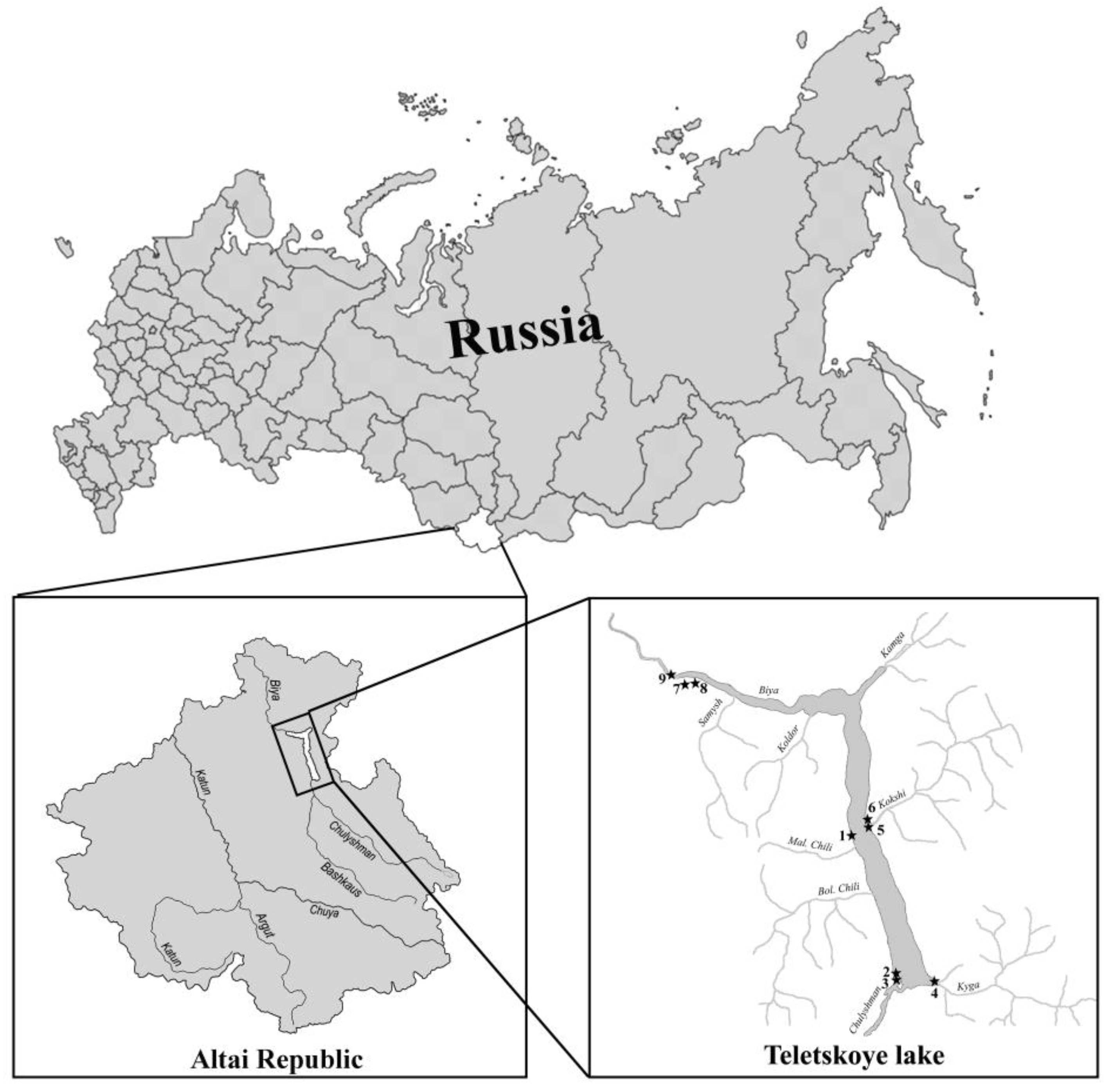
2. Materials and Methods

Samples from the Altai (Altai Republic) were included in this study (Figure 1, Table 1). The Altai samples were collected from Teletskoye Lake and the adjacent area on 27–29 May 2015. Teletskoye Lake is the deepest freshwater body in the southwest of Siberia. It is located in the northeast of the Altai Mountains at an altitude of 434 m above sea level. Part of the water body and its eastern coast are incorporated among the UNESCO World Heritage Sites as part of the “Golden Mountains of Altai” complex [26]. The lake has an elongated shape in the meridional direction, and is 78 km long with a maximum width of 5.2 km. The lake is quite large; its water area is 223 km2, and its volume is 40 km3. One of the unique characteristics of this lake is the sparse presence of shallow areas. The littoral area with depths up to 10 m is only 7.8% of the lake area, and the maximum depth is 325 m [27]. The catchment area is 19,500 km2, and most of this area has an average elevation of 1940 m, which ensures low mineralisation and organic matter concentrations in the lake. Among the 70 permanent tributaries, the Chulyshman River is the largest, accounting for 75% of the total inflow to the lake. The Biya River is almost the only outflow of Teletskoye Lake (98% of total water runoff) [28]. Teletskoye Lake is characterised by increased external water exchange, in which a complete change of water occurs every 5.81 years [29].
Samples were collected using a plankton net with a 20 μm mesh. For electron microscopy studies, an aliquot of each sample was washed three times by repeated centrifugation with deionised water. Drops of each washed sample were dried directly onto stubs for scanning electron microscopy (SEM) or grids for transmission electron microscopy (TEM) or digested for 4–5 min in sulphuric acid with potassium dichromate before mounting. For SEM studies, samples were dried onto aluminium stubs, coated with gold for 10 min with a JEE-4X (JEOL) sputter coater, and observed with a JEOL 6510 LV scanning electron microscope. For TEM studies, samples were dried onto formvar-coated grids (EMS FF200-Cu-50, Electron Microscopy Sciences, Hatfield, USA) and observed with a JEM-1011 transmission electron microscope. Specific conductance, pH, and temperature were measured using a Hanna HI 9828 (Hanna Instruments, Inc., Smithfield, RI, USA), and are represented in Table 1.

3. Results

During studies of Teletskoye Lake, adjacent small water bodies, and the Biya, Chulyshman, and Katun Rivers in the Altai Mountains, 16 taxa were observed (Table 2, Figure 2, Figure 3 and Figure 4). Eight taxa belong to the genus Mallomonas, while the others are species of the genera Synura (five) and Paraphysomonas (three). In Teletskoye Lake, 11 taxa were observed. In other lakes and rivers, between two and five taxa of silica-scaled chrysophytes were found.
During our investigation, we found scales of Mallomonas that differed in ultrastructure from the known species. Below, we give a formal description of this species.
Mallomonas altaica sp. nov. Gusev and Martynenko (Figure 2).
Description: Scales are 3.6–4.2 × 1.9–2.2 μm in size, oblong and oval in shape, with weak lateral incurvings. Scales possess a posterior rim, a posterior flange, a V-rib, anterior ribs and flanges, and a dome. The dome is smooth and subcircular, or it has several weakly developed ribs. The shield is marked with 9–17 regularly spaced transverse ribs in the anterior part. The posterior part of the shield near the angle of the V-rib is smooth. Base plate pores are irregularly distributed on the shield. Three to four large pores are located in the angle of the V-rib. The anterior flanges are narrow, with five to eight closely spaced struts on each side. The anterior submarginal ribs are well developed. The V-rib on the scales is rounded, slightly hooded, and bears internal struts. The posterior rim is smooth. The posterior flange is covered with a secondary siliceous layer and bears numerous struts (usually more than 30, well visible on SEM images) and scattered base-plate pores. Bristles and cysts were not observed.
Holotype specimen: Portion of a single cluster of cells on SEM stub No. A20 deposited at the Herbarium of the Papanin Institute for Biology of Inland Waters, RAS, Borok (IBIW). E.S. Gusev collected material from Teletskoye Lake in the Republic of Altai, Russia, on 27 May 2016. Figure 2A illustrates a representative scale from the holotype specimen.
Type Locality: Teletskoye Lake, Republic of Altai, Russia. Latitude/Longitude: N51° 21.336′, E87° 50.870′.
Epithet: The species name is derived from “Altai”, the area from which it was described.
Distribution: This species was found in the type locality only. The pH was 7.3 at the time of collection, the temperature was 11 °C, and the specific conductance was 109 μS cm−1.
Among other Mallomonas taxa, M. crassisquama (Figure 3f,g) was found in all studied localities except the Biya River. Mallomonas elongata (Figure 3h–j) was also frequently found but was absent only in the Katun River and unnamed lake 2. Other taxa were registered rarely (Table 2). Mallomonas annulata (Figure 3e), M. altaica, and M. pechlaneri (Figure 3k,l) were observed in one locality only.
Several morphotypes of the Synura petersenii sensu lato species complex were observed. We provide descriptions of morphotypes observed in our samples.
Synurapetersenii Korshikov emend. Škaloud and Kynčlová (Figure 3o,p): Body scales elongated, 4.5–4.6 × 2.3–2.6 μm. The keel is ornamented by rather small pores (diameter 46–69 nm). The ratio between scale and keel width varies from 3.0 to 3.6. The basal plate is ornamented by numerous small pores (diameter 21–29 nm). The diameter of the large pores in the anterior part varies from 0.36 to 0.43 μm. Numerous struts (28–29), often interconnected by transverse folds, regularly extend from the keel to the edge of the scales. The species is a widely distributed taxon and has been confirmed in many regions of the world [30], including localities in Russia [31,32,33].
Synura sp. 1 (Figure 4a,b): Body scales are elongated, 3.2–3.6 × 1.8–2.6 μm. The keel is ornamented by pores with a diameter of 53–72 nm. The ratio between scale and keel width varies from 3.4 to 3.8. The basal plate is ornamented by numerous small pores (diameter 16–29 nm). The diameter of the large pores in the anterior part ranges from 0.18 to 0.35 μm. Struts (20–22) regularly extend from the keel to the edge of the scales, with no well-developed interconnections by transverse folds.
This morphotype corresponds to Synura americana Kynčlová and Škaloud, but we are cautious to make this identification without confirmation by molecular data for a population so remote from its confirmed habitats.
Synura sp. 2 (Figure 4c–f): Body scales are elongated, 4.2–5.1 × 2.2–2.7 μm. The keel is ornamented by pores with a diameter of 59–80 nm. The ratio between scale and keel width varies from 2.8 to 4.3. The basal plate is ornamented by numerous pores with a diameter of 30–48 nm. The diameter of the large pores in the anterior part varies from 0.30 to 0.43 μm. Numerous struts (26–30) regularly extend from the keel to the edge of the scales and are rarely interconnected by transverse folds. This morphotype has large body scales. Based on scale size, the number of struts, and the diameter of the base plate and keel pores, it can be compared with S. borealis Škaloud and Škaloudová and S. hibbernica Škaloud and Škaloudová. However, S. hibbernica has elongated body scales and specific apical scales with a long spine. The body scales of S. borealis usually have an anteriorly widened keel, while on most body scales of Synura sp. 2, the keel is narrow.
Synura sp. 3 (Figure 4g): Body scales are elongated, 4.4 × 2.3 μm. The keel is wide in the anterior part and ornamented by pores with a diameter of 57–80 nm. The ratio between scale and keel width is 2.6. The basal plate is ornamented by numerous pores with a diameter of 36–48 nm. The diameter of the large pores in the anterior part is 0.38 μm. Numerous struts (26), interconnected by transverse folds, regularly extend from the keel to the edge of the scale. These scales are similar to those of S. petersenii sensu stricto, but there are larger base plate pores and a wider keel.
Synura sp. 4 (Figure 4h): Elongated apical scale, wide oval shape, 3.8 × 2.3 μm. The keel is wide and ornamented by large pores with a diameter of 70–96 nm. The ratio between scale and keel width is 2.6. The basal plate is ornamented by numerous small pores with a diameter of 28–31 nm. The diameter of the large pores in the anterior part of the scales is 0.40 μm. Numerous struts (23) regularly extend from the keel to the edge of the scales and are not interconnected by transverse folds. This morphotype differs from those described above in terms of the diameter of the pores of the basal plate and keel; however, due to the presence of only one scale, it is difficult to determine the group of species to which it may belong.
Several morphotypes of Paraphysomonas were observed.
Only a single scale of Paraphysomonas sp. 1 (Figure 4i) was found. The base plate is round, 1.4 µm in diameter, with a dense rim. The spine is 3.4 µm long and tapering to an attenuated blunt tip. This specimen is most similar in morphology and size to Paraphysomonas variosa Scoble and Cavalier-Smith, which is known from the tropics [4,34]. However, one scale is not enough to accurately identify this morphotype. Paraphysomonas sp. 1 is also similar to P. cambrispina Scoble and Cavalier-Smith and P. stylata var. limnetica Scoble and Cavalier-Smith. However, unlike Paraphysomonas sp. 1, P. cambrispina has shorter spines tapering to an oblique dull tip, while Paraphysomonas stylata var. limnetica has longer spines gently tapering completely to a tip.
Paraphysomonas sp. 2 (Figure 4j,k) has one type of spine scale. The spine is 4.2–5.8 µm long, gently tapering from a wide base to the tip, and there is a round base plate with a thickened margin of 2.1–2.8 µm. The scales resemble those of P. vulgaris Scoble and Cavalier-Smith but are larger (2.1–2.8 vs. 1.8–2.2 µm) and have a longer spine (4.2–5.8 vs. 3.1–4.5 µm). It is very likely our scales represent an undescribed Paraphysomonas species.
Paraphysomonas sp. 3 (Figure 4l) has one type of spine scale. The spine is 4.8–5.8 µm long, gently tapering from an inflated base to the tip, and there is a round base plate with a thickened margin of 2.4–2.5 µm. This morphotype is similar to Paraphysomonas sp. 2 but distinctly distinguished by the inflated base of the spine.

4. Discussion

The study of the phytoplankton in Teletskoye Lake started at the beginning of the 20th century, and the plankton have been described as extremely poor, except for diatoms [35]. Subsequent studies made it possible to identify in the lake flora 492 species from Bacillariophyta, Chlorophyta, Chrysophyta, Dinophyta, Euglenophyta, Cryptophyta, Xanthophyta, and Cyanobacteria [36]. Among chrysophytes several genera were reported, including Dinobryon Ehrenberg, Chrysococcus Klebs, Kephyrion Pascher, Mallomonas Perty, and Pseudokephyrion Pascher [36,37]. Silica-scaled chrysophytes, according to previous studies, were represented by nine species and one variety: Mallomonas globosa J. Schiller, M. producta (Zacharias) Iwanoff, M. elegans Lemmermann, M. elongata, M. ploesslii Perty, M. crassisquama, M. alpina, M. caudata Iwanoff, M. vannigera Asmund, and M. striata var. serrata K. Harris and D.E. Bradley [36,37]. These records need to be revised. Mallomonas globosa, M. producta, M. elegans, and M. ploesslii were identified by light microscopy only [38,39]. They are dubious taxa due to the lack of electron microscopy studies, thus a lack of species descriptions of the ultrastructure of the scales and bristles. Moreover, Mallomonas globosa is considered a member of the other chrysophycean genus, Spiniferomonas [1]. M. crassisquama was identified only with a type of stomatocyst found in Teletskoye Lake [40] and was not confirmed with illustrations of scales. The rest of the known species were identified based on electron microscopy: M. alpina, M. caudata, M. elongata, M. striata var. serrata, and M. vannigera [36,37]. The identification of M. striata var. serrata (Figure 2b in [36]) should be changed to M. pechlaneri due to the presence of smooth bristles with bifurcated tips and scale ultrastructure. Recently, Mallomonas pechlaneri was recorded in Teletskoye Lake by other researchers [22]. In addition, more than 70 morphotypes of chrysophycean stomatocysts were found in the phytoplankton of Teletskoye Lake [36,37,40]. This suggests that there are many more chrysophycean species in the phytoplankton of the lake than previously thought.
Our data made it possible to supplement the list of silica-scaled chrysophytes in Teletskoye Lake and the adjacent area with four Mallomonas and one Synura species. The presence of M. crassisquama in the lake phytoplankton was confirmed. In addition, four morphotypes from the genus Synura (Synura petersenii species complex) and three from the genus Paraphysomonas were found. Synura petersenii is a complex of closely related cryptic and pseudocryptic taxa that requires molecular methods for correct identification [2,30,41,42,43]. We could not correctly identify these morphotypes down to the species level without molecular data, but we consider it important to publish their images for further analysis of the diversity of chrysophytes in this region. Correct identification of Paraphysomonas also requires molecular data [4]. Thus, we provide images of the morphotypes found with comments on their similarity to known morphotypes. This is the first record of taxa from this genus in Teletskoye Lake.
Some interesting species from the genus Mallomonas were found in our study, primarily from the section Striatae. The most interesting record is a new species to science, Mallomonas altaica, which belongs in this section due to the presence of ribs on the shield [44]. This section encompasses 15 taxa, according to Kristiansen and Preisig [1], and seven more taxa have been described recently [11,22,25,45,46,47]. Many taxa in this section, such as Mallomonas striata var. striata Asmund, M. striata var. serrata Harris and Bradley, M. flora Harris and Bradley, M. cratis Harris and Bradley, M. pseudocratis Dürrschmidt, and M. asmundiae (Wujek and van der Veer) Nicholls, are widely distributed [1]. However, there are some very rare taxa with limited distribution in this section. In this study, we describe another very rare species from a mountain lake.
The main distinctive features of Mallomonas altaica are the ribs, which cover only the anterior part of the shield, and the posterior flange, which is almost completely covered with a secondary siliceous layer. Within the section Striatae, Mallomonas altaica is most similar to M. cratis and M. marina Jeong, (Kim, Jo, Kim, Siver, and Shin) by the oblong oval scales. However, the shield on the scales of M. cratis and M. marina is completely covered with numerous curved transverse ribs, anterior ribs are absent or short, and the dome is covered by concentric U-shaped ribs. Other species are well distinguished by the shape of the scales, and the shield on the scales is completely covered with transverse ribs.
Mallomonas pechlaneri is another rare taxon from the section Striatae. It was recently discovered in two North Tyrol alpine lakes [25] and in highland water bodies of the East Sayan Mountains [22]. It seems that this species is restricted to oligotrophic mountain lakes and has a wide distribution across Eurasia.
It is noteworthy that several taxa from this section have recently been described in other mountain water bodies of northern Asia. Mallomonas striata var. getseniae and M. striata var. balonovii from the Polar Urals were described [11]. The rank of these taxa should be raised to the species level. M. voloshkoae, from a high-altitude lake in the Barguzin Mountains (Transbaikal area), was described. It can be concluded that many rare or endemic taxa from the section Striatae have been found in oligotrophic mountain water bodies with low mineralization, temperature, and nutrient concentration.
The low diversity of silica-scaled chrysophytes is comparable with similar estimates made in studies of oligotrophic mountain lakes. Studies of high-altitude lakes in North Tyrol (Austria) revealed 27 taxa at 12 sites, with species richness varying from one to nine [25]. The diversity of silica-scaled chrysophytes in water bodies located at lower altitudes in the same area revealed 46 taxa in 22 localities, with species richness varying from four to thirty, but for most localities, the number of taxa found did not exceed ten [24]. A small number of taxa of scaled chrysophytes were also observed in ultra-oligotrophic lakes in North America [6] and Newfoundland [48]. In other cold-water oligotrophic mountain lakes in Siberia, the diversity of silica-scaled chrysophytes was also low. In the lakes of the Polar Urals, between one and sixteen taxa were recorded [12]. When studying the mountain lakes of Yakutia, quite a small number of taxa were also found: in Labynkyr Lake–twenty-three, in Vorota Lake–fifteen [17], and in Toko Lake–six [9]. Only 10 taxa were found in Lake Frolikha (Transbaikal area) [49]. Even in such a large lake as Baikal, only 25 taxa were found in plankton in the open part [18,19].
Mountain lakes are habitats for many rare species of chrysophytes. Recently, a number of new species to science of chrysophytes have been described from mountain water bodies of different origins in Eurasia. In addition to the previously mentioned species from the genus Mallomonas from the section Striatae, also described were Mallomonas tirolensis Pichrtová, Němcová, Škaloud, and Rott from the Alps in Europe [25]; M. kuzminii from the Barguzin Mountains in the Transbaikal area, Russia [21]; M. alpestrina Němcová and Zeisek from Haba Snow Mountain, China [50]; and Synura morusimila Pang and Wang from the Great Xing’an Mountains [51]. In the mountainous regions of China, a rich flora of chrysophycean cysts has also been identified, including many morphotypes new to science [52]. Thus, studies of the mountain lakes of Asia and, in particular, the eastern territories of Russia are very promising in this regard and should be intensified.

Author Contributions

Sampling, manuscript—writing, drafting, and editing, TEM observations, identification, SEM observations, identification, E.G.; sample preparation, TEM observations, measurements, manuscript—editing, N.M. All authors have read and agreed to the published version of the manuscript.

Funding

The research was supported by the state assignment of the Ministry of Science and Higher Education of the Russian Federation (theme no. AAAA-A18-118042490059-5).

Institutional Review Board Statement

Not applicable.

Data Availability Statement

Not applicable.

Acknowledgments

The authors are grateful to the staff of the Centre for Electron Microscopy at Papanin’s Institute for Biology of Inland Waters, RAS, for technical assistance.

Conflicts of Interest

The authors declare no conflict of interest.

References

  1. Kristiansen, J.; Preisig, H.R. Chrysophyta and Haptophyta Algae, 2nd part: Synurophyceae. In Süsswasserflora Von Mitteleuropa (Freshwater Flora of Central Europe); Büdel, B., Gärtner, G., Krienitz, L., Preisig, H.R., Schagerl, M., Eds.; Springer: Berlin, Germany, 2007; pp. 1–252. [Google Scholar]
  2. Škaloud, P.; Kristiansen, J.; Škaloudová, M. Developments in The Taxonomy of Silica-Scaled Chrysophytes—From Morphological and Ultrastructural to Molecular Approaches. Nord. J. Bot. 2013, 31, 385–402. [Google Scholar] [CrossRef]
  3. Siver, P.A.; Jo, B.Y.; Kim, J.I.; Shin, W.; Lott, A.M.; Wolfe, A.P. Assessing the Evolutionary History of The Class Synurophyceae (Heterokonta) Using Molecular, Morphometric, and Paleobiological Approaches. Am. J. Bot. 2015, 102, 921–941. [Google Scholar] [CrossRef] [Green Version]
  4. Scoble, J.M.; Cavalier-Smith, T. Scale Evolution in Paraphysomonadida (Chrysophyceae): Sequence Phylogeny and Revised Taxonomy of Paraphysomonas, New Genus Clathromonas, and 25 New Species. Eur. J. Protistol. 2014, 50, 551–592. [Google Scholar] [CrossRef] [PubMed] [Green Version]
  5. Smol, J.P. Applications of chrysophytes to problems in paleoecology. In Chrysophyte Algae: Ecology, Phylogeny and Development; Sandgren, C., Smol, J., Kristiansen, J., Eds.; Cambridge University Press: Cambridge, UK, 1995; pp. 303–330. [Google Scholar] [CrossRef]
  6. Siver, P.A. The distribution of chrysophytes along environmental gradients: Their use as biological indicators. In Chrysophyte Algae: Ecology, Phylogeny and Development; Sandgren, C., Smol, J., Kristiansen, J., Eds.; Cambridge University Press: Cambridge, UK, 1995; pp. 232–268. [Google Scholar] [CrossRef]
  7. Kristiansen, J. Biogeography of silica-scaled chrysophytes. Nova Hedwig. 2001, 122, 23–39. [Google Scholar]
  8. Kristiansen, J.; Düwel, L.; Wegeberg, S. Silica-scaled chrysophytes from the Taymyr Peninsula, Northern Siberia. Nova Hedwig. 1997, 65, 337–351. [Google Scholar] [CrossRef]
  9. Gusev, E.S.; Guseva, E.E.; Gabyshev, V.A. Taxonomic composition of silica-scaled chrysophytes in rivers and lakes of Yakutia and Magadanskaya oblast (Russia). Nova Hedwig. 2018, 147, 105–117. [Google Scholar]
  10. Bessudova, A.; Bukin, Y.; Likhoshway, Y. Dispersal of Silica-Scaled Chrysophytes in Northern Water Bodies. Diversity 2021, 13, 284. [Google Scholar] [CrossRef]
  11. Voloshko, L.N. New taxa of the genus Mallomonas (Chrysophyta, Synurophyceae) from lakes of the Polar Ural. Russ. Bot. J. 2009, 94, 1068–1076. [Google Scholar]
  12. Voloshko, L.N. The chrysophycean algae from glacial lakes of Polar Ural (Russia). Nova Hedwig. 2010, 136, 191–211. [Google Scholar] [CrossRef]
  13. Balonov, I.M.; Kuzmina, A.E. Chrysophyta. In Hydrochemical and Hydrobiological Studies in on the Khantay Reservoir; Votinzev, K.K., Ed.; Nauka: Novosibirsk, USSR, 1986; p. 119. [Google Scholar]
  14. Bessudova, A.; Bukin, Y.S.; Sorokovikova, L.M.; Firsova, A.D.; Tomberg, I.V. Silica-scaled Chrysophytes in Small Lakes of the Lower Yenisei Basin, the Arctic. Nova Hedwig. 2018, 107, 315–336. [Google Scholar] [CrossRef]
  15. Kuzmin, G.V.; Kuzmina, V.A. Species of the genus Mallomonas (Chrysophyta) from waterbodies of the Magadanskaya oblast. Russ. Bot. J. 1986, 71, 805–807. [Google Scholar]
  16. Kuzmin, G.V.; Kuzmina, V.A. Silica-scaled chrysophytes from Magadanskaya oblast. News Taxon. Low. Plants 1987, 24, 40–42. [Google Scholar]
  17. Bessudova, A.; Tomberg, I.V.; Firsova, A.D.; Kopyrina, L.I.; Likhoshway, Y.V. Silica-scaled chrysophytes in lakes Labynkyr and Vorota of the Sakha (Yakutia) Republic, Russia. Nova Hedwig. 2019, 148, 35–48. [Google Scholar] [CrossRef]
  18. Vorobyova, S.S.; Bondarenko, N.A.; Karpova, S.A.; Pomazkina, G.V.; Tanichev, A.I. To studies of Chrysophyta species composition of Lake Baikal. Algologia 1992, 2, 68–72. [Google Scholar]
  19. Bessudova, A.; Domysheva, V.M.; Firsova, A.D.; Likhoshway, Y. V Silica-scaled chrysophytes of Lake Baikal. Acta Biol. Sib. 2017, 3, 47–56. [Google Scholar] [CrossRef] [Green Version]
  20. Bessudova, A.; Sorokovikova, L.M.; Tomberg, I.V.; Likhoshway, Y.V. Silica-scaled chrysophytes in large tributaries of Lake Baikal. Cryptogam. Algol. 2018, 39, 145–165. [Google Scholar] [CrossRef]
  21. Gusev, E.S.; Kulikovskiy, M.S. A new species of the genus Mallomonas (Chrysophyceae: Synurales), Mallomonas kuzminii sp. nov., from lake Frolikha (Russia, Baikal region). Phytotaxa 2013, 155, 66–70. [Google Scholar] [CrossRef] [Green Version]
  22. Gusev, E.; Němcová, Y.; Kulikovskiy, M. Mallomonas voloshkoae sp. nov. (Synurales, Chrysophyceae) and distribution of M. pechlaneri in mountain lakes of Siberia. Phytotaxa 2022, 530, 221–229. [Google Scholar] [CrossRef]
  23. Bessudova, A.; Gabyshev, V.; Likhoshway, Y.V. Record of two rare taxa from Synura genus (Chrysophyceae) with a description of a new species (Synura tiksiensis sp. nov.) near the arctic settlement of Tiksi, Yakutia, Russia. Phytotaxa 2022, 560, 247–253. [Google Scholar] [CrossRef]
  24. Pichrtová, M.; Nemcová, Y.; Škaloud, P.; Rott, E. Silica-scaled chrysophytes from North Tyrol (Austria) including a description of Mallomonas tirolensis sp. nov. Nova Hedwig. 2013, 142, 69–85. [Google Scholar]
  25. Nemcová, Y.; Rott, E. Diversity of Silica-Scaled Chrysophytes in High-Altitude Alpine Sites (North Tyrol, Austria) Including a Description of Mallomonas pechlaneri sp. nov. Cryptogam. Algol. 2018, 39, 63–83. [Google Scholar] [CrossRef]
  26. Puzanov, A.V.; Bezmaternykh, D.M.; Kirillov, V.V.; Mitrofanova, E.Y.; Yanygina, L.V. Ecosystem features and environmental problems of lake Teletskoye (Republic of Altai). Limnol. Freshw. Biol. 2020, 4, 624–625. [Google Scholar] [CrossRef]
  27. Yanygina, L.V.; Koveshnikov, M.I.; Krylova, E.N.; Marusin, K.V. Spatial distribution of zoobenthos in Lake Teletskoye. In Proceedings of the Lake Ecosystems: Biological Processes, Anthropogenic Transformation, and Water Quality, Minsk, Belarus, 17–22 September 2007. [Google Scholar]
  28. Selegei, V.V.; Selegei, T.S. Lake Teletskoye; Gidrometeoizdat: Leningrad, USSR, 1978; pp. 1–143. [Google Scholar]
  29. Selegei, V.V. Lake Teletskoye: History Essays; Book I; Ofset: Novosibirsk, Russia, 2009; pp. 1–119. [Google Scholar]
  30. Škaloud, P.; Kynčlová, A.; Benada, O.; Kofroňová, O.; Škaloudová, M. Toward a Revision of the Genus Synura, Section Petersenianae (Synurophyceae, Heterokontophyta): Morphological Characterization of Six Pseudo-Cryptic Species. Phycologia 2012, 51, 303–329. [Google Scholar] [CrossRef] [Green Version]
  31. Gusev, E.S.; Kapustin, D.A.; Martynenko, N.A. Morphological and molecular studies of the genus Synura Ehrenb. (Chrysophyceae) from the algae culture collection of IBIW RAS. Tr. Inst. Biol. Vnutr. Vod Ross. Akad. Nauk 2016, 73, 5–11. [Google Scholar]
  32. Gusev, E.S.; Perminova, O.S.; Startseva, N.A.; Okhapkin, A.G. Genus Synura (Synurales, Synurophyceae) in small urban rivers of Nizhny Novgorod. Nov. Sist. Nizshikh Rast. 2017, 51, 57–70. [Google Scholar] [CrossRef]
  33. Kulizin, P.V.; Gusev, E.S.; Vodeneeva, E.L.; Okhapkin, A.G. Taxonomic Composition and Morphology of Silica-Scaled Chrysophytes of Some Left-Bank Volga Tributaries. Inland Water Biol. 2021, 14, 357–367. [Google Scholar] [CrossRef]
  34. Gusev, E.; Kapustin, D.; Martynenko, N.; Kulikovskiy, M. Diversity of Silica-Scaled Chrysophytes (Stramenopiles: Chrysophyceae) from Indonesian Papua. Diversity 2022, 14, 726. [Google Scholar] [CrossRef]
  35. Poretskiy, V.S.; Sheshukova, V.S. Diatoms in Lake Teletskoye and rivers connected with it. In Diatomovyi Sbornik (Diatom Book); Proshkina-Lavrenko, A.I., Sheshukova, V.S., Eds.; Leningr. Gos. Univ. Publishing: Leningrad, USSR, 1953; pp. 107–173. [Google Scholar]
  36. Mitrofanova, E.Y. Phytoplankton of Lake Teletskoye (Altai, Russia): Features of Development and LongTerm Dynamics. Russ. J. Ecol. 2018, 49, 180–185. [Google Scholar] [CrossRef]
  37. Mitrofanova, E.Y. Diversity of chrysophycean stomatocysts in the lake Teletskoye plankton. Probl. Bot. Yuzhnoi Sib. I Mong. 2012, 11, 139–141. [Google Scholar]
  38. Safonova, T.A.; Mitrofanova, E.Y. A study of phytoplankton of Teletskoye Lake (Altai Mountains, Russia). Int. J. Algae 2003, 5, 59–67. [Google Scholar] [CrossRef]
  39. Mitrofanova, E.Y.; Vorob’ev, R.I. Composition and structure of Lake Teletskoye phytoplankton (Altai Republic) during the winter period. Probl. Bot. Yuzhnoi Sib. I Mong. 2012, 17, 103–106. [Google Scholar]
  40. Bazhenova, O.P.; Mitrofanova, E.Y.; Shakhoval, V.E. Stomatocysts of chrysophyte algae from bodies of water in territory near Irtysh river in Omsk Region and Lake Teletskoye in Gorny Altai, Russia. Contemp. Probl. Ecol. 2012, 5, 423–429. [Google Scholar] [CrossRef]
  41. Škaloud, P.; Škaloudová, M.; Procházková, A.; Němcová, Y. Morphological Delineation and Distribution Patterns of Four Newly Described Species within the Synura petersenii Species Complex (Chrysophyceae, Stramenopiles). Eur. J. Phycol. 2014, 49, 213–229. [Google Scholar] [CrossRef]
  42. Škaloud, P.; Škaloudová, M.; Jadrná, I.; Bestová, H.; Pusztai, M.; Kapustin, D.; Siver, P.A. Comparing Morphological and Molecular Estimates of Species Diversity in the Freshwater Genus Synura (Stramenopiles): A Model for Understanding Diversity of Eukaryotic Microorganisms. J. Phycol. 2020, 56, 574–591. [Google Scholar] [CrossRef]
  43. Jo, B.Y.; Kim, J.I.; Škaloud, P.; Siver, P.A.; Shin, W. Multigene Phylogeny of Synura (Synurophyceae) and Descriptions of Four New Species Based on Morphological and DNA Evidence. Eur. J. Phycol. 2016, 51, 413–430. [Google Scholar] [CrossRef] [Green Version]
  44. Asmund, B.; Kristiansen, J. The Genus Mallomonas (Chrysophyceae). A Taxonomic Survey Based on The Ultrastructure of Silica Scales and Bristles. Opera Bot. 1986, 85, 1–128. [Google Scholar]
  45. Němcová, Y.; Bulant, P.; Kristiansen, J. Mallomonas solea-ferrea and Mallomonas siveri (Chrysophyceae/Synurophyceae): Two new taxa from the Western Cape (South Africa). Nova Hedwig. 2011, 93, 375–384. [Google Scholar] [CrossRef]
  46. Jeong, M.; Kim, J.I.; Jo, B.Y.; Kim, H.S.; Siver, P.A.; Shin, W. Surviving the marine environment: Two new species of Mallomonas (Synurophyceae). Phycologia 2019, 58, 276–286. [Google Scholar] [CrossRef]
  47. Gusev, E.S.; Kulikovskiy, M.S. Two New Species of Genus Mallomonas from Swamp Localities in Vietnam. Phytotaxa 2020, 468, 121–129. [Google Scholar] [CrossRef]
  48. Kristiansen, J. Golden Algae: A Biology of Chrysophytes; A.R.G. Gantner Verlag: Königstein, Germany, 2005; p. 167. ISBN 3-906166-23-6. [Google Scholar]
  49. Gusev, E.S. Taxonomic composition of silica-scaled chrysophytes in Frolikha Lake. In Ecology, Morphology and Systematics of Aquatic Plants; Bobrov, A.A., Ed.; Filigran: Yaroslavl, Russia, 2016; pp. 25–30. [Google Scholar]
  50. Němcová, Y.; Pichrtová, M.; Zeisek, V. Mallomonas alpestrina sp. nov. (Synurales, Chrysophyceae, Stramenopiles) and its spineless relatives - Mallomonas alata group. Phytotaxa 2015, 222, 111–120. [Google Scholar] [CrossRef] [Green Version]
  51. Pang, W.; Wang, Q. A new species, Synura morusimila sp. nov. (Chrysophyta), from Great Xing’an Mountains, China. Phytotaxa 2013, 88, 55–60. [Google Scholar] [CrossRef]
  52. Pang, W.; Van de Vijver, B.; Wu, H.; Li, Y. New chrysophyte stomatocysts from high mountain lakes in Three Parallel Rivers of Yunnan Protected Areas, China. Fottea 2022, 22, 228–237. [Google Scholar] [CrossRef]
Figure 1. Geographical position of the studied area. Sample sites are marked by asterisks. (1) Teletskoye Lake, station 1; (2) Teletskoye Lake, station 2; (3) Chulyshman River; (4) Teletskoye Lake, station 3, Kyga Bay; (5) Teletskoye Lake, station 4, mouth of the Kokshi River; (6) Teletskoye Lake, station 5; (7) Unnamed lake 1 near the Biya River; (8) Unnamed lake 2 near the Biya River; and (9) Biya River.
Diversity 14 01040 g001
Figure 2. Mallomonasaltaicasp. nov. (A) Body scale, SEM; (BD) body scales, TEM; (E) group of scales, illustrating an upper surface and bottom surface view, SEM; (F) group of body scales at higher magnification, SEM; and (G) group of body scales, TEM. Scale bars: (BD,G): 2 μm; (A,E,F): 1 μm.
Diversity 14 01040 g002
Figure 3. Mallomonas and Synura taxa from Teletskoye Lake and adjacent area, TEM. (A,B) Mallomonas acaroides, body scales; (C,D) Mallomonas alpina, body scale (C) and apical scale (D); (E) Mallomonas annulata body scale; (F,G) Mallomonas crassisquama, body scale (F) and apical scale (G); (HJ) Mallomonas elongata, domed body scale; (H), domeless body scale (I) and bristle (J); (K,L) Mallomonas pechlaneri, body scales; (M,N) Mallomonas tonsurata, domeless body scale (M) and domed scale with bristle (N); and (O,P) Synura petersenii, body scales. Scale bars: (J): 10 μm; (A): 5 μm; (BI,K,MP): 2 μm; and (L): 1 μm.
Diversity 14 01040 g003
Figure 4. Synura and Paraphysomonas taxa from Teletskoye Lake and adjacent area, TEM. (A,B) Synura sp. 1, body scales; (CF) Synura sp. 2, body scales; (G) Synura sp. 3, body scale; (H) Synura sp. 4, body scale; (I) Paraphysomonas sp. 1; (J,K) Paraphysomonas sp. 2; and (L) Paraphysomonas sp. 3. Scale bars: (K): 5 μm; (AJ,L): 2 μm.
Diversity 14 01040 g004
Table 1. Basic characteristics of the localities (Cond.—specific conductance, μS cm−1, T—temperature, °C).
NameCoordinatespHCond.T
1Teletskoye Lake, station 1N51° 33.691′, E87° 38.921′7.3985
2Teletskoye Lake, station 2N51° 21.900′, E87° 44.951′n/an/an/a
3Chulyshman RiverN51° 21.884′, E87° 44.964′7.819610
4Teletskoye Lake, station 3, Kyga BayN51° 21.336′, E87° 50.870′7.310911
5Teletskoye Lake, station 4, mouth of Kokshi RiverN51° 34.518′, E87° 41.146′7.3917
6Teletskoye Lake, station 5N51° 34.660′, E87° 41.069′8.39113
7Unnamed lake 1 near the Biya RiverN51° 46.917′, E87° 15.528′7.97023
8Unnamed lake 2 near the Biya RiverN51° 47.045′, E87° 16.120′7.33323.4
9Biya RiverN51° 47.154′, E87° 14.938′7.59110
10Katun RiverN50° 59.409′, E86° 15.634′7.910810
Table 2. List of silica-scaled chrysophytes found in this study; “+” indicates the presence of a taxon.
TaxonSample Sites *
12345678910
Mallomonas acaroides Perty emend. Iwanoff + ++
M. alpina Pascher and Ruttner emend.
Asmund and Kristiansen
++++ +
M. altaica sp. nov. +
M. annulata (D.E. Bradley) K. Harris +
M. crassisquama (Asmund) Fott + ++++++ +
M. elongata Reverdin ++ ++++ +
M. pechlaneri Nemcová and Rott +
M. tonsurata Teiling emend W. Krieger + ++
Synura petersenii Korshikov emend.
Škaloud and Kynčlová
+ ++
S. sp. 1 +
S. sp. 2 +
S. sp. 3 +
S. sp. 4 +
Paraphysomonas sp. 1 +
P. sp.2 +
P. sp.3 + +
* Sample site designations are presented in Table 1.
Publisher’s Note: MDPI stays neutral with regard to jurisdictional claims in published maps and institutional affiliations.

