Article

A New Species of the Genus Niphargus Schiödte, 1849 (Crustacea: Amphipoda: Niphargidae) from Groundwater Habitats of the Tarkhankut Upland, Crimean Peninsula

by
Ivan N. Marin
1,*,
Ilya S. Turbanov
2,3,
Gregoryi A. Prokopov
4 and
Dmitry M. Palatov
1
1
A.N. Severtsov Institute of Ecology and Evolution, Russian Academy of Sciences, 119071 Moscow, Russia
2
I.D. Papanin Institute of the Biology of Inland Waters, Russian Academy of Sciences, 152742 Borok, Russia
3
Eco-Analytical Laboratory, Cherepovets State University, 162600 Cherepovets, Russia
4
Department of Geoecology, V.I. Vernadsky Crimean Federal University, 295007 Simferopol, Russia
*
Author to whom correspondence should be addressed.
Diversity 2022, 14(12), 1010; https://doi.org/10.3390/d14121010
Submission received: 6 October 2022 / Revised: 13 November 2022 / Accepted: 14 November 2022 / Published: 22 November 2022
(This article belongs to the Special Issue Aquatic Biodiversity: Evolution, Taxonomy and Conservation)

Abstract

:
A new species of the genus Niphargus Schiödte, 1849 (Crustacea: Amphipoda), co-occurring with water louse Asellus cf. aquaticus (Linnaeus, 1758) (Crustacea: Isopoda) in deep wells, is described from the Tarkhankut Upland, located in the northwestern part of the Crimean Peninsula. Niphargus tarkhankuticus sp. nov. corresponds to a separate phylogenetic lineage (the “tarkhankuticus” ingroup), also including several undescribed species from the coastal habitats of the Black Sea (the Crimean Peninsula, the southern Caucasus and the northern coast of Turkey), which is related to the paraphyletic European “stygius-longicaudatus” group. The divergence of the “tarkhankuticus” ingroup from the related European species probably appeared in the Late Miocene age, about 11–10 Mya, related to the separation of the Eastern Paratethys for different basins (Euxinian, Alpine and Pannopian). At the same time, the speciation within the ingroup started in Pliocene, about 5.76–3.6 Mya, and correlated with the Black Sea leveling and the drainage of coastal marine carbonate accumulations, including the Tarkhankut Upland. Intraspecific values of COI mtDNA gene markers (p-distances) for N. tarkhankuticus sp. nov. are about 2%, showing that the division into a number of isolated subpopulations, probably associated with local tectonic movements, the active formation of the river network, and further karst processes in the Tarkhankut Upland occurred during the Pleistocene (since 2.58 Mya). Analysis of stable isotopes (δ13C/δ15N) revealed that only discovered macrocrustaceans in the studied wells of the Tarkhankut Upland have non-overlapping trophic niches, with A. cf. aquaticus possibly feeding on algae/periphyton, while the trophic position of N. tarkhankuticus sp. nov. is close to predators.

1. Introduction

The Crimean Peninsula (Crimea) has a peculiar “island” position and is almost completely surrounded by the waters of the Black and Azov Seas [1,2]. Starting from the Pliocene, as the sea regressed, Crimea repeatedly became part of the continental shelf, and during transgressions it was again isolated as “an island” [3,4]. A significant part of the Peninsula (84% of the territory) is represented by water-soluble carbonate rocks, mainly limestones (karst) with different genesis, composition, structure and age [5], divided into numerous karst regions, which are part of two large karst provinces: mountainous Crimea (southern coastal part) and the Scythian Platform (northern part) [6]. As a zone of insufficient moisture, the Crimea has about 1000–1200 mm of precipitation per a year in its southwestern mountainous region and about 250 mm in its northern flat arid region. The westernmost part of the Peninsula, the Tarkhankut Upland (Chernomorskoe, 45°30′11″ N 32°42′18″ E), has about 315–320 mm (average) of precipitation per year [7], most of which falls during the autumn–spring period. The groundwater in this region is isolated from mountainous Crimea and directly depends on meteorological conditions [8].
The subterranean (hypogeic) fauna of the Crimea is characterized by a high level of endemism and consists of a variety of troglo- and stygobiotic invertebrates, including about 50 presently known species of crustaceans, pseudoscorpions, millipedes, diplurans, springtails, beetles, etc. (e.g., [9,10,11]). At the same time, to date, only four species of the genus Niphargus Schiödte, 1849 have been described from the territory of Crimea, namely N. pliginskii Martynov, 1931, N. dimorphus Birštein, 1961, N. vadimi Birštein, 1961 and N. tauricus Birštein, 1964 [12,13,14], as well as a number of undescribed species [15,16], phylogenetically related to other Black Sea regions, such as the southern Caucasus and Romania ([17,18]; unpublished). However, the diversity of Niphargus across this region with such significant karst territories is very low compared to species diversity seen in the Eastern Mediterranean (Balkans, Caucasus), where several hundred species are presently known [17]. Thus, one has the impression of a significant depletion of the stygobiotic amphipod fauna in the underground waters of the Crimea.
Until now, the presence of large stygobiotic animals in the northwestern part of the Peninsula, on the Tarkhankut Upland, was doubtful, since there were no known large caves or karst cavities. This article provides the first insight into the biological assemblages of deep artificial wells, some of which have existed since the ancient Greek colonization of the Crimea. All the studied sites (wells) are confined to the first aquifer with a total mineralization from 0.2 to 3.0 mg/L. The bottom of the wells are covered by pebbles, and clay and fine sandy areas are covered with deposits of loose detritus. The temperature of the groundwater is between 12–18 °C. The nutrition and water content of these aquifers completely depend on meteorological conditions [8]. The flow in the wells occurs largely during the springtime, when, accordingly, most of the precipitation occurs.
The aim of this article is to describe the new species and its ecology in as much detail as possible. The discovery of a representative of the genus Niphargus in such isolated habitats and close to the northern border of the distribution of the genus sheds light on the zoogeographic relationships of the genus, and new molecular data will help in the reconstruction of its phylogeographic relationships.

2. Materials and Methods

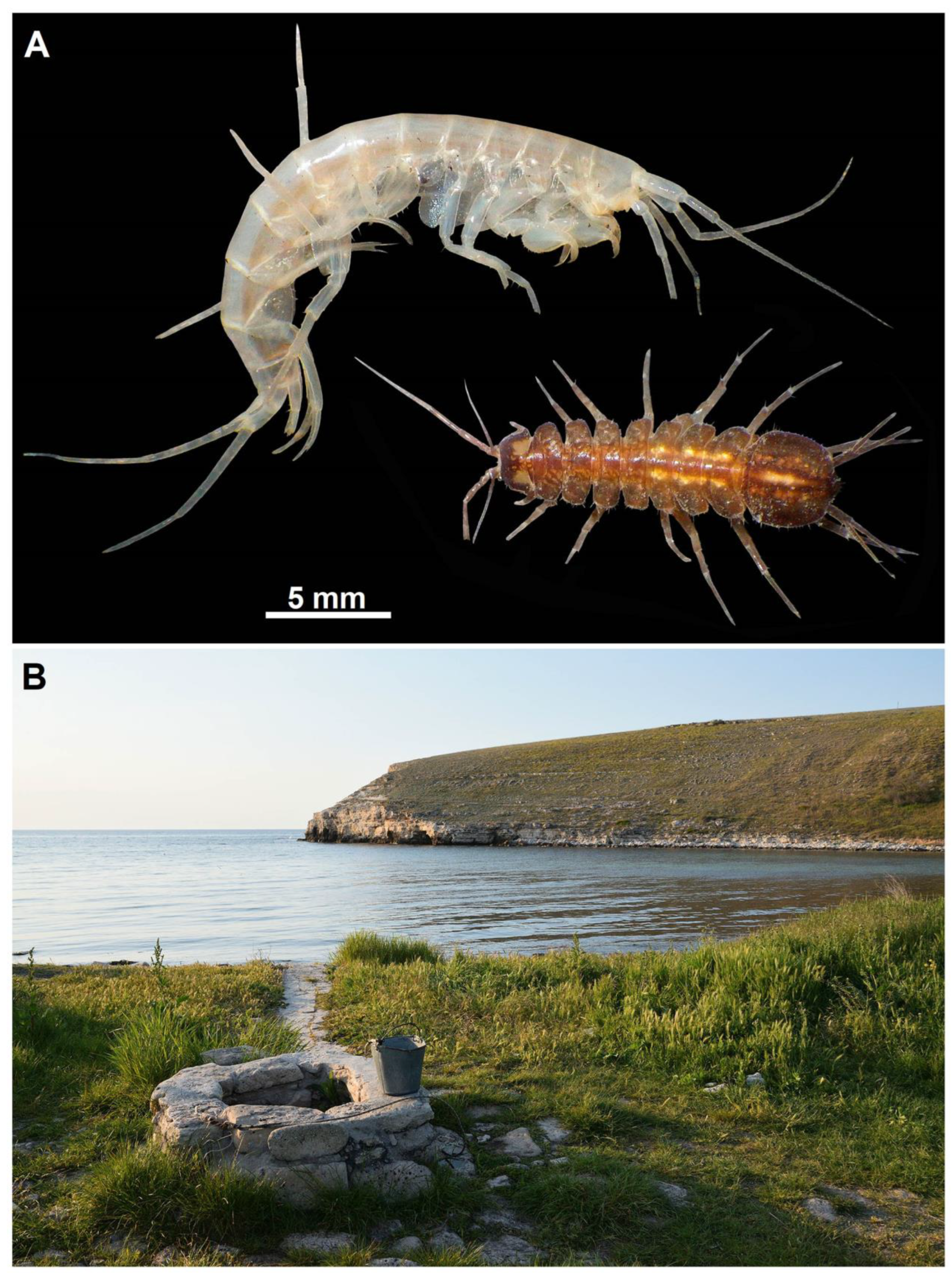
2.1. Specimen Sampling

Amphipods were collected in wells and springs of the western part of the Crimean Peninsula on the Tarkhankut Upland (see Figure 1 and Figure 2). Traps made from plastic bottles were used to collect crustaceans in the wells; boiled sausage or pellets of dry pet food were used as a bait. Some animals were collected by hand net from the bottom of the wells. After sampling, all specimens were fixed in 96% solution of ethanol. The type material was deposited in the collection of Zoological Museum of Moscow State University, Moscow (ZMMU); additional material was deposited in the private collection of the second author (IT). The distribution map was created using Google Earth Pro (ver. 7.3.4.8248) (Google LLC, Mountain View, CA, USA) and Adobe Photoshop CS6 (ver. 13.0.1.3) (Adobe Systems Incorporated, San Jose, CA, USA). The final images were processed with Adobe Photoshop CS6.

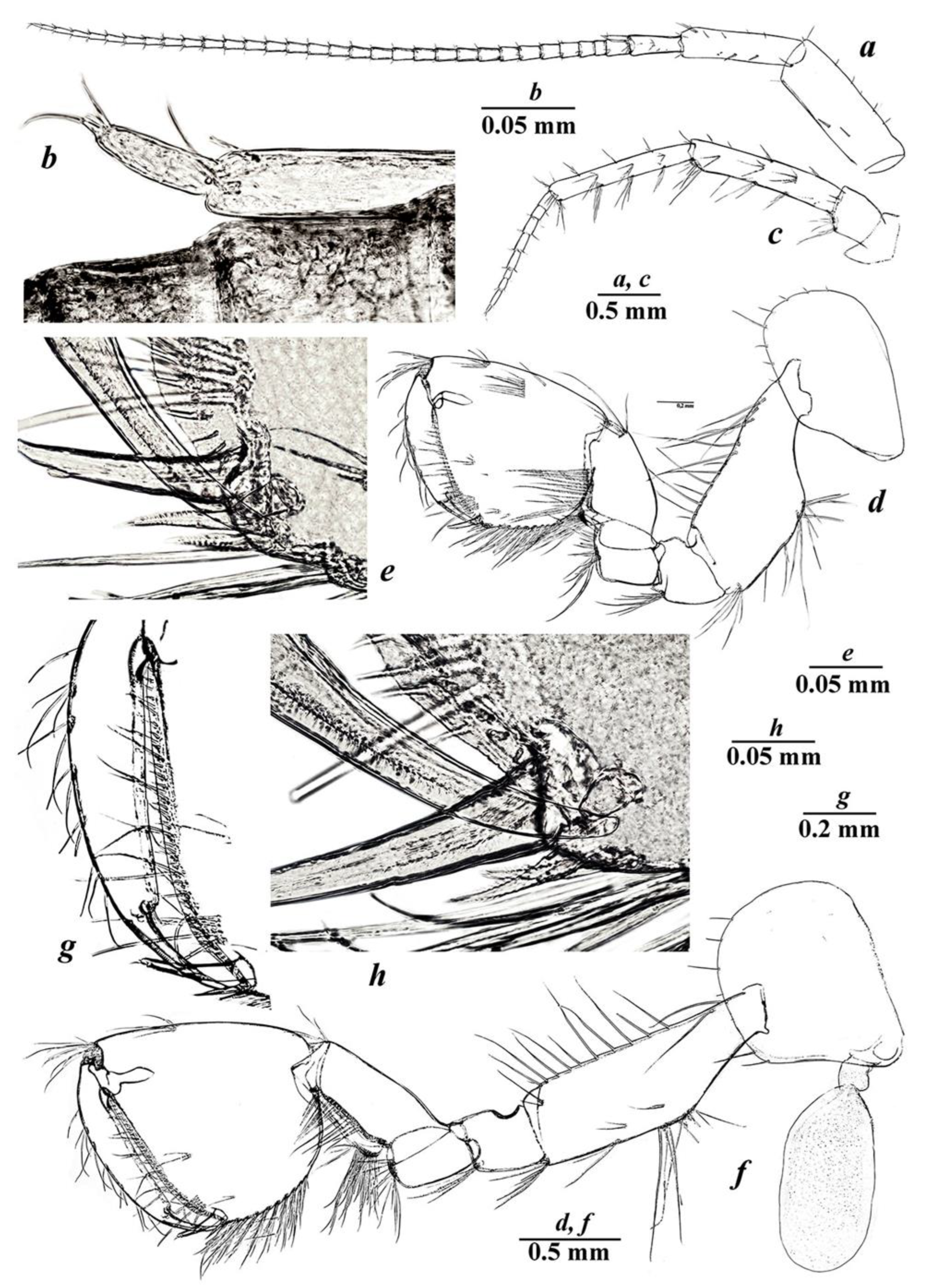
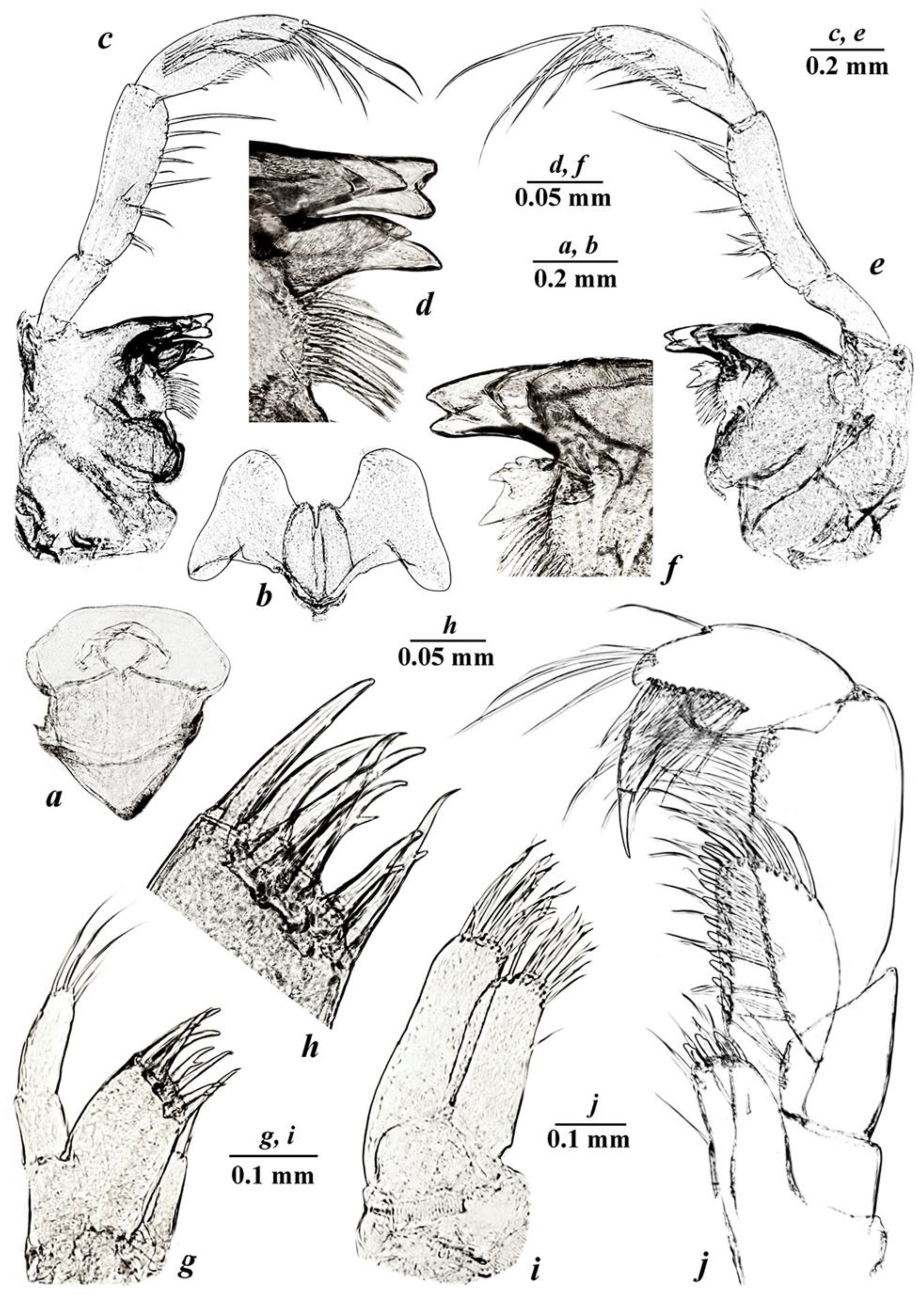
2.2. Morphological Studies

Scanning electron microscope (SEM) micrographs were made with standard methods using a CamScan S2 microscope in the Electronic Microscopy Laboratory of the Biological Faculty of the Moscow State University. The body length (bl., in mm)—the dorsal length from distal margin of head to the posterior margin of telson, without the length of uropod III and antennas—was used as a standard measurement.

2.3. Molecular and Phylogenetic Study

A fragment of cytochrome oxidase C subunit I (COI mtDNA) was used to study the cryptic diversity and phylogenetic relationships of a new species. Total genomic DNA was extracted from abdominal and pereopod muscle tissue using the innuPREP DNA Micro Kit (Analitik Jena, Jena, Germany) following the manufacturer’s protocol. The gene marker was amplified with the help of the universal primers LCO1490 and HC02198 [18] using a T100 amplificator (Bio-Rad, Hercules, CA, USA) under the standard protocol conditions.
The genetic divergences (p-distances) were calculated with MEGA 7.0 (University of Kent, NZ) using the Kimura 2-Parameter (K2P) model of evolution [19] with the sequences of the relative species deposited in the GenBank (NCBI) database.
Consensus of complementary sequences of the COI mtDNA gene marker, 646 base pairs in length, and all available data from the GenBank (NCBI) database was obtained with MEGA 7.0. The final dataset for the analysis included 41 sequences, displaying 244 variable (polymorphic) sites, of which 234 were parsimony-informative. Phylogenetic tree topologies were congruent between Bayesian and maximum likelihood analyses. The best evolutionary substitution model was determined using MEGA 7.0. (University of Kent, NZ, Canterbury, UK) and jModeltest2.1.141 (Diego Darriba, Universidade da Coruña as part of the Computer Architecture Group (GAC), Coruña, Spain) on XSEDE via the CIPRES (Cyber Infrastructure for Phylogenetic Research) Science Gateway V. 3.3 (http://www.phylo.org/, accessed on 10 November 2022). Phylogenetic analysis was performed on a single gene marker dataset (COI mtDNA) using PhyML 3.0 (Montpellier Bioinformatics, Montpellier, France) (http://www.atgc-montpellier.fr/phyml/, accessed on 10 November 2022) with the GTR+G+I model for maximum likelihood analysis (ML). The obtained phylogenetic trees were compared with the general phylogenetic tree of the genus Niphargus, presented by Copilaş-Ciocianu et al. [20] and Fišer et al. [21,22]. Phylogenetic analysis was used to search for related species and is not visualized in the article.
MOLECULAR CLOCK ANALYSIS was performed based on Bayesian inference trees with the BEAST2 package [23]. A maximum clade credibility tree was obtained using TreeAnnotator v2.5.1 [23], with 10% burn-in and selected mean node height [23]. The resulting trees were visualized with FigTree v1.4.3 (Andrew Rambaut, University of Edinburgh, Edinburgh, UK). Calibration points were chosen based on the adapted time-scale [24] and the analysis of historical events.

2.4. Stable Isotope Analysis

The organic matter and animals for the stable isotope analysis were collected in the wells of the Bolshoy Kastel Bay and Gnilaya Bay of the Tarkhankut Upland. The muscle tissue of collected animals were oven-dried at 50 °C for 4–5 days, and then were wrapped in tin foil (1200–1500 µg and 400–600 µg, respectively). The composition of stable isotope (δ13C/δ15N) was determined using a Thermo-Finnigan Delta V Plus continuous-flow mass spectrometer (Thermo Electron GmbH, Bremen, Germany) coupled with an elemental analyzer (Thermo Flash 1112, Thermo Electron) at the Joint Usage Center at the A.N. Severtsov Institute of Ecology and Evolution of the Russian Academy of Sciences. The isotopic composition of N and C was expressed in the δ-notation relative to the international standard (atmospheric nitrogen or VPDB): δX(‰) = [(Rsample/Rstandard) − 1] × 1000, where R is the ratio of the heavier isotope to the lighter isotope. Samples were analyzed with reference gases calibrated against IAEA (Vienna, Austria), reference materials USGS 40 and USGS 41 (glutamic acid). The drift was corrected using internal laboratory standards (acetanilide, casein). The standard deviation of δ15N/δ13C values in our laboratory standards (n = 8) was <0.15‰. A bivariate plot of δ15N/δ13C ± SD values (‰) is presented for visualization of the results. Unfortunately, due to small local diversity of macrozoobenthos, it was impossible to select enough data for food web analysis from only wells (localities), where the new species was sampled; therefore, we used a general plot that included data from neighboring water sources, for example the well in the Gnilaya Bukhta (45°19′19.9″ N 32°37′19.7″ E) that allowed us to present a more reliable and complete vision (biplot) of trophic interactions.

3. Results

3.1. Biological Assemblage of the Studied Wells

In all studied localities (wells), only two permanently living large species of macrozoobenthos were found—the macrocrustaceans Niphargus tarkhankuticus sp. nov. (Crustacea: Amphipoda: Niphargidae) (see below) and Asellus cf. aquaticus (Linnaeus, 1758) (Crustacea: Isopoda: Asellidae). Additionally, detrital remains of grass (vascular plants), filamentous algae Cladophora sp. (Chlorophyta: Cladophorales), larvae of Chironomus sp. (Diptera: Chironomidae) and imago Sigara stagnalis pontica Jaczewski, 1961 (Hemiptera: Corixidae) were also sampled from the bottom of the well of Bolshoy Kastel (see Figure 1).
At the same time, during the warm summer season, other animals are likely to fall into the wells and die, as their remains were also collected. Chitinous remains of beetles found in detritus belonged to Tentyria nomas taurica Tauscher, 1812, Blaps lethifera lethifera Marsham, 1802, Gonocephalum granulatum pusillum (Fabricius, 1792 (Coleoptera: Tenebrionidae), Amara aenea De Geer, 1774, Harpalus spp., Ophonus subquadratus Dejean, 1829, Acupalpus spp., Carabus hungaricus scythus Motschulsky, 1847 (Coleoptera: Carabidae) and Otiorhynchus sp. (Coleoptera: Curculionidae). It has been noted by local people that some vertebrates have also fallen into these wells, including Pelophylax ridibundus (Pallas, 1771), Bufotes viridis (Laurenti, 1768) (Amphibians: Anura), Dolichophis caspius (Gmelin, 1789) (Reptiles: Squamata), Apodemus witherbyi Thomas, 1902 (Mammals: Rodentia) and different bats (Mammalia: Chiroptera). However, such remains were not found during our studies.

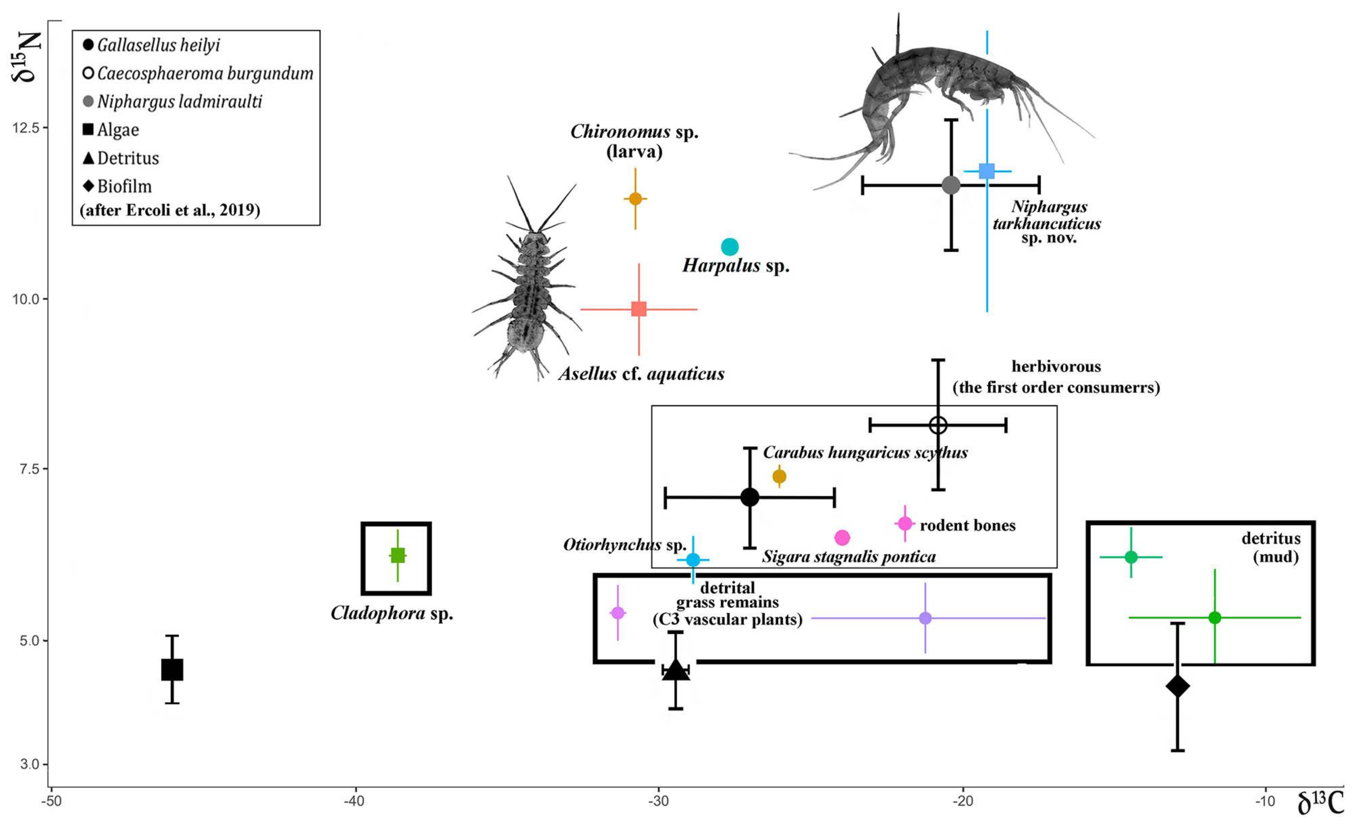
3.2. Trophic Relationships with the Studied Wells

The obtained results of the composition of the stable isotope (δ13C/δ15N) showed that two macrocrustaceans (Niphargus and Asellus) permanently living in the wells have non-overlapping trophic niches and no trophic relationships. Asellus cf. aquaticus (−30.65 ± 1.92 for δ13C and 9.84 ± 0.67 for δ15N) probably feeds on algae, while the trophic position of N. tarkhankuticus sp. nov. (−19.31 ± 0.78 for δ13C and 11.86 ± 2.05 for δ15N) is very high and close to predators (the second level consumers) (see Table 1).

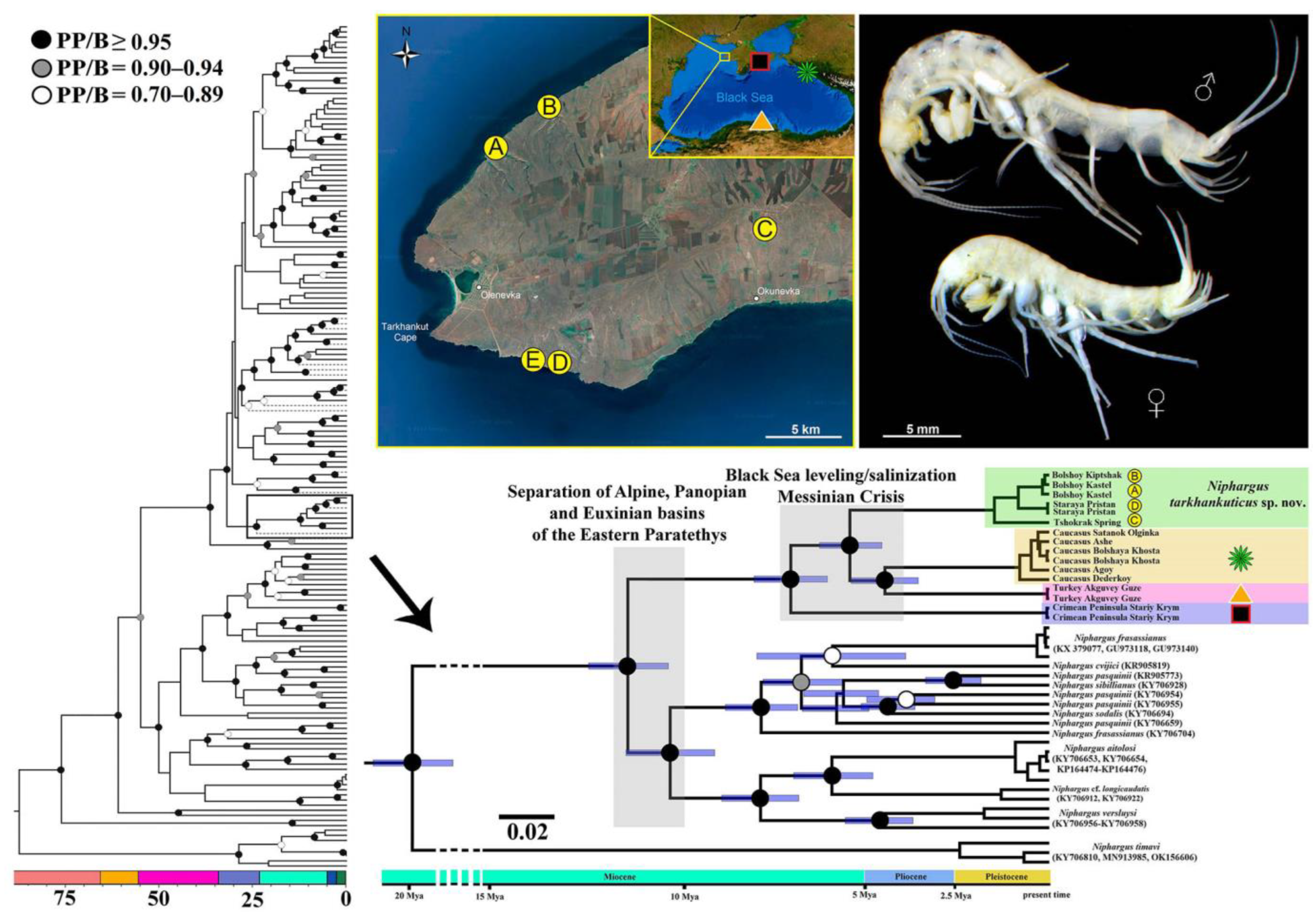
3.3. Phylogenetic Study of Newly Discovered Niphargus
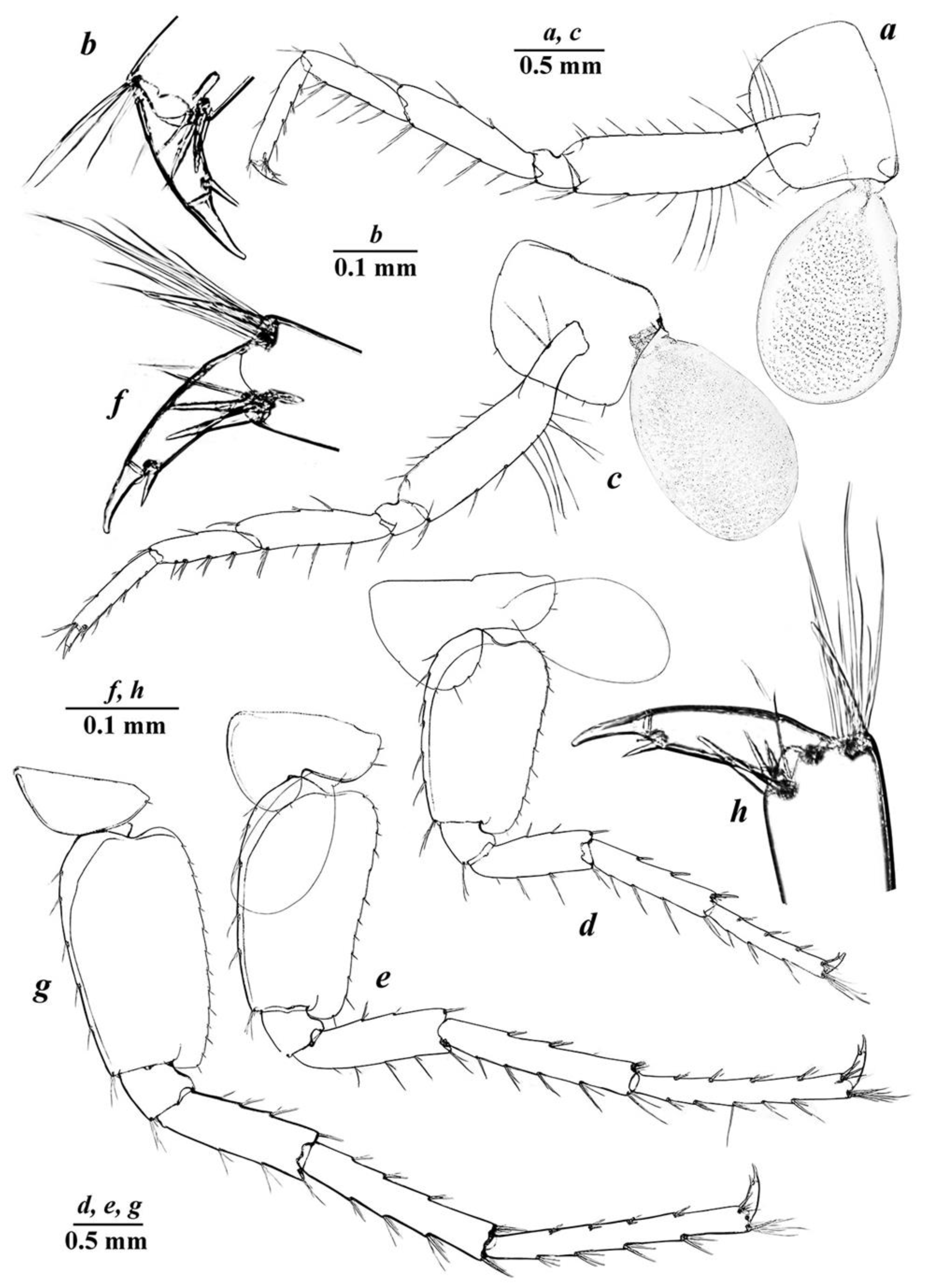
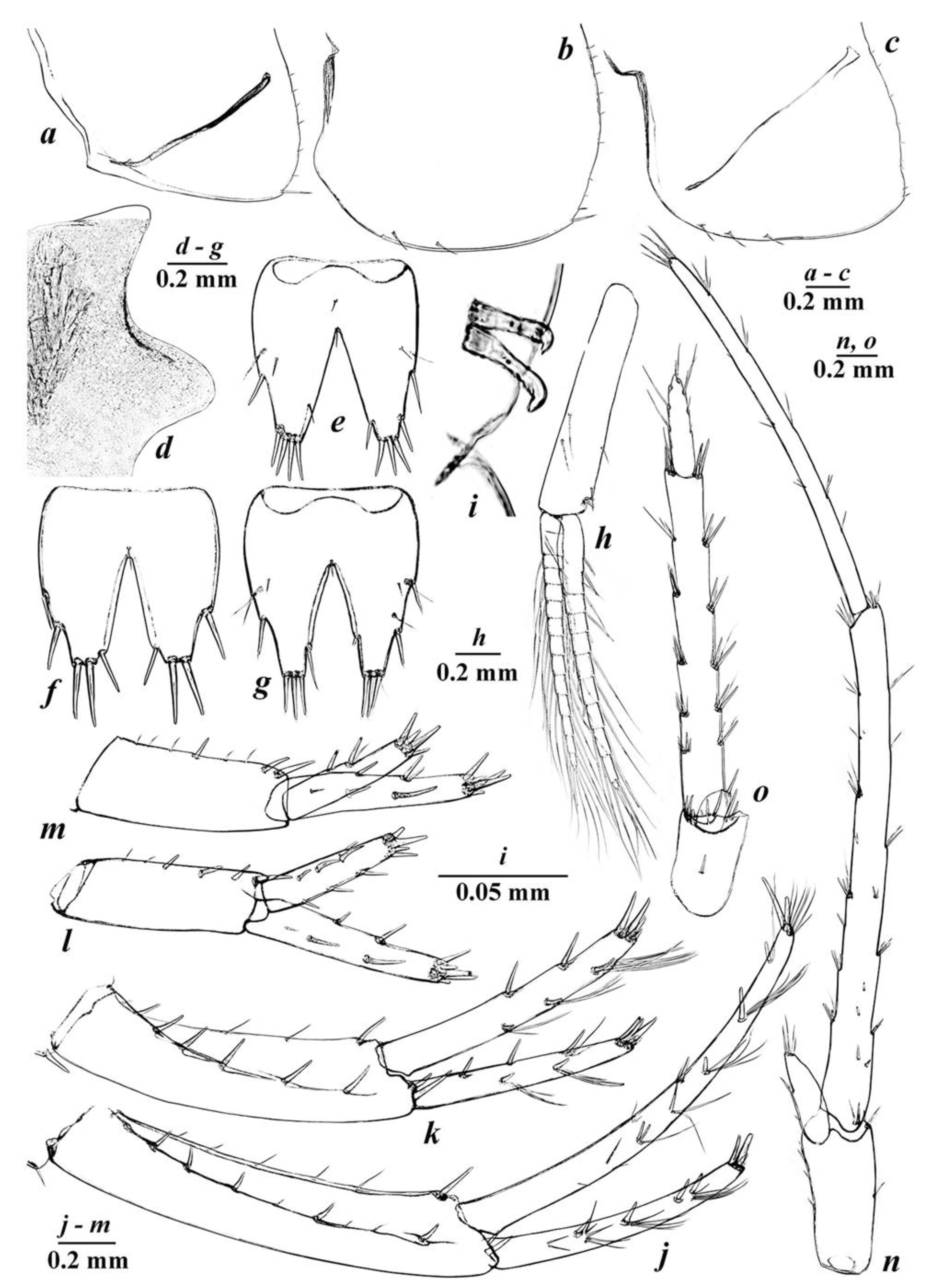
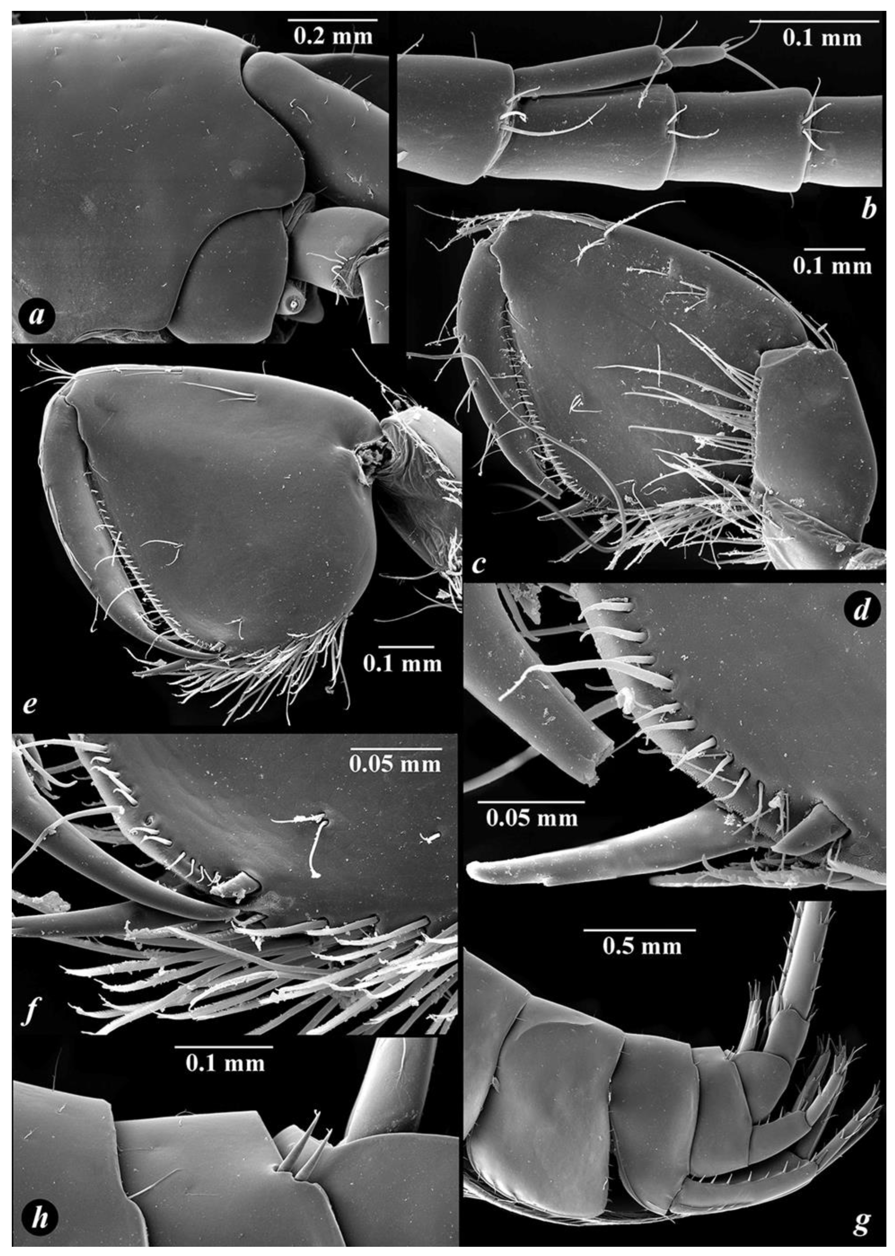
The molecular genetic analysis (Figure 1) clearly confirmed the monophyly (Bayesian-PP = 1.00; ML-BS = 95%) of the studied lineage (called the “tarkhankuticus” ingroup), including Niphargus tarkhankuticus sp. nov. and three undescribed species from the coastal habitats of the Black Sea (Turkey: Akguvey Guze; Caucasus: Satanok, Ashe, Bolshaya Khosta, Agoy, Dederkoy; and the Crimean Peninsula: Staryi Krym) (see Figure 1). The phylogenetic analysis (ML) also supports the relationship of this ingroup with some representatives of the paraphyletic European “stygius-longicaudatus” group, such as N. frasassianus Karaman, Borowsky et Dattagupta, 2010, N. cvijici S. Karaman, 1950, N. pasquinii Vigna-Taglianti, 1966, N. versluysi S. Karaman, 1950 and N. longicaudatus (A. Costa, 1851) sensu lato (see Taxonomic remarks for details). The latter group of species (Figure 1) is well-separated and -supported within the “stygius-longicaudatus” group (e.g., [19,20,21]). The genetic divergence between these two species groups is about 0.205 ± 0.016 substitution per 100 nucleotides (about 20%). Based on this data, the origin of the “tarkhankuticus” ingroup and its separation from the related species of the “stygius-longicaudatus” group was estimated in the Late Miocene age, about 11–10 Mya (95% HPD: 26.62–3.97) (after [25,26,27,28]), which may correlate with the time of separation of the Eastern Paratethys for different basins (Euxinian, Alpine and Pannopian) (see Figure 1).
Figure 1. The map of collection sites and the time-calibrated phylogenetic tree (reconstruction) of phylogenetic relationships of the Niphargus tarkhankuticus sp. nov. from the Tarkhankut Upland, in the western part of the Crimean Peninsula. Sites are indicated as (A)—well in the Bolshoy Kastel Bay (type locality); (B)—well in the Bolshoy Kiptshak Bay; (C)—Tshokrak Spring 3.6 km north of the Okunevka; (D)—well in the Staraya Pristan (Ubezhishche) Bay; (E)—well near to the Tshashcha Lyubvi. Posterior probabilities of the nodes are reported. Blue horizontal bars show the 95% HPD (highest posterior density) of node ages on an arbitrary time scale. The general phylogenetic tree is taken from Copilaş-Ciocianu et al. [19] with the adapted time-scale after McInerney et al. [24].
Diversity 14 01010 g001
Figure 2. General view and live coloration of Niphargus tarkhankuticus sp. nov. and Asellus cf. aquaticus (Linnaeus, 1758) (A). The well in the Bolshoy Kastel Bay (type locality), Tarkhankut Upland, in the northwestern part of the Crimean Peninsula (B).
Diversity 14 01010 g002
The interspecific uncorrected pairwise genetic distances (p-distances) between the populations of Niphargus tarkhankuticus sp. nov. is about 0.0197 ± 0.003 substitutions per 100 nucleotide positions (about 1.97%) that correspond to other species from the area (e.g., N. bzhidik Marin, Krylenko et Palatov 2021 [29]). The population divergence corresponds well with their geographic localities (see Figure 1).
The intraspecific uncorrected pairwise genetic differences (p-distances of COI mtDNA gene marker) between the species within the “tarkhankuticus” ingroup range from 13 to 17% (see Table 2), which also justifies the long-time isolation of new species from the congeners, as well their isolation from one another. The divergent time of the separation between Niphargus tarkhankuticus sp. nov. and other species of the “tarkhankuticus” ingroup was estimated using p-distances as 0.144 ± 0.013 substitution per 100 nucleotides (about 14%), which could be interpreted as about 5.76 Mya (after [25,27]).

3.4. Taxonomic Part

  • Phylum Arthropoda von Siebold, 1848
  • Class Malacostraca Latreille, 1802
  • Order Amphipoda Latreille, 1816
  • Family Niphargidae Bousfield, 1977
  • Genus Niphargus Schiödte, 1849
  • Niphargus tarkhankuticus sp. nov.
  • urn:lsid:zoobank.org:pub:878BE10A-27B7-45B2-A9A6-0362E12FA552
Material examined. Holotype, male (bl. 13.0 mm) (ZMMU Mb-1241), Crimean Peninsula, Chernomorskiy Region, Tarkhankut Upland, well in the Bolshoy Kastel Bay, 45°27′22.3′′ N 32°32′53.0′′ E, 24.04.2015, coll. G.A. Prokopov.
Paratypes, 2 males, 2 females (bl. 9.0–13.5 mm) (ZMMU Mb-1242), same locality and data as holotype.
Additional material. Crimean Peninsula, Chernomorskiy Region, Tarkhankut Upland: 8 males, 7 females (bl. 9.5–14.0 mm) (IT), same locality as holotype, 16 May 2022, I.S. Turbanov leg.; 52 males, 97 females (bl. 9.0–13.5 mm) (IT), well in the of Bolshoy Kiptshak Bay, 45°28′46.3″N 32°35′32.9″E, 2 April 2022, G.A. Prokopov leg.; 1 male (bl. 11.5 mm) (IT), Tshokrak Spring 3.6 km north of the Okunevka, 45°24′23.4″ N 32°46′05.7″ E, 1 April 2022, G.A. Prokopov leg.; 5 males, 7 females (bl. 9.5–13.0 mm) (IT), well in the Staraya Pristan (=Ubezhishche) Bay, 45°19′49.0″ N 32°35′59.5″ E, 5 September 2015, G.A. Prokopov leg.; 8 males, 11 females (bl. 10.0–13.5 mm) (IT), same locality and collector, 28–31 May 2015; 1 female (bl. 12.5 mm) (IT), well near to the Tshashcha Lyubvi, 45°19′53.2″ N 32°34′38.0″ E, 16 May 2022, I.S. Turbanov leg.
Etymology. The new species is named after the Tarkhankut Upland, located in the northwestern part of the Crimean Peninsula, where it was discovered.
Diagnosis. Head without eyes/pigmented spots on anterior lobe. Spines of maxilla I with one strong lateral tooth each. Posteroventral corners of epimeral plates I–II rounded, and bluntly produced in epimeral plate III. Urosomite I with one long simple seta on each side dorsolaterally; urosomite II with two simple strong spines each side dorsolaterally; urosomite III unarmed. Dactyli of pereopods III–VII with one small additional median spine and one median short plumose seta at outer margin. Palm of gnathopods I–II close to triangular, with its width shorter than depth; dactylus with numerous simple setae along outer margin. Rami of uropod I unequal in size: endopodite about 1.8 times longer than exopodite in males and slightly longer in females; rami of uropod II with tufts of marked long curved setae in both sexes; rami of uropod III differs in males and females; significantly longer in males. Retinacula of pleopods with two hooks, without additional setae. Telson with 2–4 medium-sized distal spines on each lobe and 1–2 lateral spines, accompanied by 2 small plumose setae, and with 0–1 small or medium submarginal spine on each side and 0–1 small mesial setae on its dorsal surface.
Description
BODY (Figure 2A): unpigmented (Figure 2A), moderately slender. The largest collected male had bl. 25.0 mm; the largest collected female had bl. 15.5 mm.
HEAD (Figure 2A, Figure 3D and Figure 7A): length was approximately 7.5% and 9% of body length in males and females, respectively; rostrum, eyes or pigmented spots on anterior lobe absent, with bluntly produced anteroventral lobe and excavated anteroventral sinus.
PEREON: pereonites I–VII smooth, without spines or teeth, with tiny setae.
PLEOSOMA: pleonites I–III with several short marginal setae on each posterodorsal margin.
EPIMERAL PLATES. Posteroventral corner of epimeral plates I–II rounded, and at epimeral plate III nearly right-angled, slightly rounded (Figure 6A–C). Epimeral plate I (Figure 6A): posterior margin convex, ventral margin slightly convex; without spines along ventral margin; with six setae along posterior margin; posteroventral angle with one strong seta. Epimeral plate II (Figure 6B): posterior and ventral margin convex; with two spiniform setae along ventral margin; nine setae along posterior margin; posteroventral angle with one strong seta. Epimeral plate III (Figure 6C): posterior margin almost straight slightly convex, ventral margin slightly convex; with three spiniform setae along ventral margin; nine setae along posterior margin, two of which are close to posteroventral angle.
UROSOMITES (Figure 7G,H): urosomite I with one long simple seta on each side dorsolaterally, with one posteroventral long simple seta near basis of uropod I dorsolaterally; urosomite II with two simple strong spines each side dorsolaterally; urosomite III unarmed.
COXAE (Figure 3D,F and Figure 5A,C–E,G): coxal plate I (Figure 3D) irregular oblong oval, with rounded anteroventral margin, armed with eight setae; width/depth ratio is 1/1.67; width/depth ratio of coxal plates II–IV (Figure 3F and Figure 5A,C) are 1/1.36, 1/1.31 and 1/1.29, respectively; anterior and ventral margins of coxal plate II with seven setae and coxal plates III–IV with eight setae each; with rounded anteroventral corners; coxal plates V–VI (Figure 5D,E) with large lobes anteriorly, with four and two setae, respectively; posterior margins with two setae each; coxal plate VII (Figure 5G) semicircular and with one seta on posterior lobe; coxal gills II–VI ovoid of which II and VI are more elongated, length ratio of gills/bases of pereopods are 0.70/1, 0.83/1, 0.91/1, 0.86/1 and 0.61/1, respectively.
ANTENNA I (Figure 3A): slender, about 48–50% of body length in males and 57–58% in females, respectively; peduncular articles moderately slender, ratio is 1/0.80/0.35; flagellum in males with 27–33 articles and 24–27 in females, most of them with two short aesthetascs each; accessory flagellum short, two-articulated (Figure 3B and Figure 7B); length ratio of antennas I/II is in range of 0.85–1/0.60–0.65 in males and 0.95–1/0.35–0.37 in females.
Figure 3. Niphargus tarkhankuticus sp. nov., male: (a) antenna I; (b) accessory flagellum of antenna I; (c) antenna II; (d) gnathopod I; (e) distoventral corner of GnI; (f) gnathopod II; (g) dactylus and distoventral palmar margin of GnII; (h) distoventral corner of chela of GnII.
Diversity 14 01010 g003
Figure 4. Niphargus tarkhankuticus sp. nov., male: (a) labrum (upper lip); (b) labium (lower lip); (c,e) mandible; (d,f) incisor process and pars incisiva of mandibles; (g) maxilla I; (h) same, distal margin of outer lobe; (i) maxilla II; (j) maxilliped.
Diversity 14 01010 g004
ANTENNA II (Figure 3C): peduncular articles moderately stout, with several long setae along ventral margin, dorsal setae shorter than inner ones; flagellum relatively short, consisting of 9–10 articles in males and 8–9 in females, respectively, covered with tiny setae; length ratio of peduncular articles 4/5 is 0.98–1/0.90–0.95; flagellum is 0.45–0.48 of length of peduncular articles 4 + 5.
LABRUM (upper lip) (Figure 4A) typical.
LABIUM (lower lip) (Figure 4B): with entire, oblong oval outer lobes and well-developed smaller inner lobes.
MANDIBLE (Figure 4C–F): Left mandible: incisor process with five teeth, lacinia mobilis with four teeth; with row of nine serrated setae between lacinia and molar process (Figure 4D); mandibular palp article 2/3 (distal) ratio is 1/0.98–1; proximal article of palp without setae; article 2 with 12 setae; distal article with group of 6 A-setae; 3 groups of B-setae; 24 D-setae and 6 E-setae (Figure 4C). Right mandible: incisor process with four teeth, lacinia mobilis bifurcate, with row of eight serrated setae between lacinia and molar process (Figure 4F).
MAXILLA I (Figure 4G): inner lobe with three distal setae, outer lobe with seven robust spines (three spines with one strong lateral tooth each (0–0–0–1–0–1–1) (Figure 4H)); palp 2-articulated, distal article with five simple setae distally.
MAXILLA II (Figure 4I): both plates with numerous long distal simple setae, outer lobe with row of fine setae along outer margin.
MAXILLIPED (Figure 4J): inner plate short, with three distal robust setae intermixed with five distal simple setae; outer plate reaching half of palpal article 2 and bearing row of 20–21 distolateral spines and distal setae; palpal article 3 with one median and one distal bunch of setae at outer margin; palpal article 4 with one median seta at outer margin; nail shorter than pedestal, with seta near basis.
GNATHOPOD I (Figure 3D and Figure 7C): basis elongated, with distal part greatly expanded, with long simple setae along anteriorly, posterior and posterodistal margins; ischium with group of six posterodistal setae; merus subquadrate, equal to ischium; carpus is 0.27 of length of basis and 0.48 of length of propodus, with single distal group of setae anteriorly, with transverse rows of setae along posterior margin and row of setae posterolaterally; propodus subtrapezoidal, setose, with 8–9 rows of setae at posterior margin, anterior surface with 2 groups of total 5–6 setae each and one single seta behind them in addition to anterodistal group of 5–6 setae, several groups of short setae on inner surface, palmar corner armed with long spiniform palmar seta, two serrated spiniform setae, single supporting spiniform seta on inner surface (Figure 3E and Figure 7D); dactylus with nine setae along anterior margin, some of which are grouped in pairs, and with a row of short setae along inner surface; length of nail is 0.27 of total length of dactylus.
GNATHOPOD II (Figure 3F and Figure 7E): basis width/length is 0.34/1, with 12 dorsolateral setae; ischium with five posterodistal setae; merus subquadrate, equal to ischium; carpus is 0.35 of length of basis and 0.61 of length of propodus, with distal group of setae anteriorly, few transverse rows of setae along posterior margin and row of setae posterolaterally; propodus subtrapezoidal, setose, larger than propodus (palm) of GnI (GnI/II as 0.87/1), posterior margin with 10–11 rows of setae, anterior surface with 1 group setae in addition to 8–9 anterodistal setae, with several groups of setae on inner surface, palmar corner with one strong palmar spiniform seta, single supporting spiniform seta on inner surface and two denticulated thick spiniform setae on outer side (Figure 3H); dactylus with 13 setae along anterior surface, some of which are grouped in pairs and a few short setae along inner surface; length of nail is about 0.25 of total length of dactylus (Figure 3G and Figure 7F).
Figure 5. Niphargus tarkhankuticus sp. nov., male: (a) pereopod III; (b) dactylus of PIII; (c) pereopod IV; (d) pereopod V; (e) pereopod VI; (f) dactylus of pereopod VI; (g) pereopod VII; (h) dactylus of pereopod VII.
Diversity 14 01010 g005
Figure 6. Niphargus tarkhankuticus sp. nov., male (ae,gj,l,n) and female (f,k,m,o): (ac) epimeral plates I–III; (d) head; (eg) telson; (h) pleopod III; (i) retinacula of pleopod III; (j,k) uropod I; (l,m) uropod II; (n,o) uropod III.
Diversity 14 01010 g006
PEREOPODS III–IV (Figure 5A,C) almost similar in size and shape; basis is 4.3–4.5 times as long as wide, with posterior margin bearing long marginal setae, with distoventral group of setae; ischium short, subquadrate, with distoventral group of setae; merus with slender simple setae along anterior and posterior surfaces; carpus/propodus ratio is 0.93–0.96/1; propodus with 4–5 groups of spines along ventral margin; dactylus (Figure 5B) relatively stout, curved, sharp distally, with one small posterior median spine and one median short plumose seta at outer margin; length of nail is 0.37 of total length of dactylus.
PEREOPODS V–VII (Figure 5D,E,G): length ratio of pereopod V/VI/VII is 1/1.40/1.52; length of pereopod VII is about 40% of total body length in males and about 54% in females, respectively.
PEREOPOD V (Figure 5D): length/width ratio of basis is 1/0.58, almost rectangular, with explicit posteroventral lobe; with facial setae; posterior margin almost straight with row of 14 slender marginal setae; anterior margin convex, with row of six slender marginal setae, which are distinctly longer than posterior ones, and group of setae in distal part; ischium subquadrate; merus with three bunches of slender spines along anterior surface and with two spines on posterior surface; carpus about as long as merus; propodus slender, 6.5–6.8 times as long as wide, with several bunches of short spines; dactylus with one small additional posterior median spine and one median short plumose seta at outer margin.
PEREOPOD VI (Figure 5E): length/width ratio of basis is 1/0.53, with distinct posteroventral lobe and straight posterior margin bearing row of 12–13 short marginal setae, anterior margin convex, with a row of five longer marginal setae, and group of setae in distal part; ischium subquadrate; merus with several bunches of short spines along anterior and posterior surfaces; carpus with group of spines intermixed with single short setae; propodus slender, about 7.5–7.6 times as long as wide, with several groups of short spines; dactylus (Figure 5F) slender, with one small additional posterior median spine and one short median plumose seta at outer margin.
PEREOPOD VII (Figure 5G): length/width ratio of basis is 1/0.56, with distinct posteroventral lobe and convex posterior margin bearing row of 14 short marginal setae; with facial setae; anterior margin convex, with row of five longer marginal setae; ischium about as long as wide; merus with several bunches of short spines along anterior and posterior margins; carpus with three groups of short spines along anterior and three groups along posterior margins; propodus slender, about 8.3–8.5 times as long as wide, with several groups of short spines; dactylus (Figure 5H) with one median spine and one seta at inner margin, and one short median plumose seta at outer margin.
PLEOPODS (Figure 6H): pleopods I and II with basal segments without setae, with two coupling hooks in retinacula each (Figure 6I); outer and inner rami with 15 and 17 segments, respectively; pleopod III with basal segment with 2–3 long simple setae, with 2 coupling hooks accompanied by short simple seta in retinacula; outer and inner rami with 10 and 14 segments, respectively.
UROPOD I (Figure 6J,K and Figure 7G): length/width ratio of protopodite is about 5.5 times in males and 4.3 in females, respectively; length ratio of protopodite/endopodite/exopodite is 1/0.58/0.93 in males and 1/0.61/0.75 in females, respectively; protopodite with dorsoexternal row of 7–8 spines or spiniform setae in males and 6 spines in females, and dorsointernal row of 6 thin spines in males and 3 in females; endopodite elongated, not paddle-like, with 1 single dorsal spine, 4–5 dorsolateral spines accompanied by groups of long bristles and 2 apical spines accompanied by group of 7–8 bristles in males, and 2 single dorsal spines, 3 dorsolateral spines accompanied by groups of long bristles and 4 apical spines in females, respectively; exopodite with 2 dorsal singles spines, 2 dorsolateral spines, accompanied by groups of long bristles and 4 apical spines in males, and 3 dorsal spines, 2 dorsolateral spines, accompanied by groups of long bristles and 5 apical spines in females, respectively.
Figure 7. Niphargus tarkhankuticus sp. nov., male: (a) head; (b) accessory flagellum of antenna I; (c) gnathopod I; (d) distoventral corner of GnI; (e) gnathopod II; (f) distoventral palmar margin of GnII; (g) urosomal segments and uropods; (h) dorsal surface of urosomal segments.
Diversity 14 01010 g007
UROPOD II (Figure 6M,L and Figure 7G): protopodite 3.35 times as long as wide in males and 2.7 in females, respectively; length ratio of protopodite/endopodite/exopodite is 1/0.61–0.65/0.98; rami with dorsal, lateral and apical slender spines: endopodite with three dorsal, three dorsolateral and five apical spines; exopodite with 2 dorsal, 0–3 dorsolateral and 5 apical spines.
UROPOD III (Figure 6N,O): different in males and females, about 0.36 of body length in males and 0.30 in females. Male: protopodite 2.2 times as long as wide, with 1 small external seta, 3–4 small internal setae, and 1–2 apical spiniform setae; rami unequal, endopodite short, about 9.9–10.0 times shorter than exopodite, with 2 small setae laterally and 4 setae apically; distal article is 0.76 of length of proximal article, with 3–4 groups of thin-flexible setae along each margin and group of simple setae apically; proximal article with seven groups of thin-flexible, plumose and spiniform setae along inner and outer margins. Female: protopodite 1.7 times as long as wide, with 1 spiniform seta laterally and 5–6 spiniform setae apically; rami unequal, endopodite short, about 9.4 times shorter than exopodite, without seta laterally and 1 spiniform seta apically; distal article is 0.31 of length of proximal article, with 4 groups of thin–flexible setae along each margin and group of simple setae apically; proximal article with 5–6 groups of spiniform setae along outer margin and 5–6 groups of thin–flexible and spiniform setae along inner margin.
TELSON (Figure 6E–G): length/width ratio is 1/0.90–0.93 in males and 1/1.05 in females; cleft is 0.58–0.62 of length of telson in males and 0.56 in females; margins weakly rounded, narrowing apically; with variable armature, including 2–4 medium-sized distal spines on each lobe and 1–2 lateral spines, accompanied by 2 plumose setae on each side; dorsal surface with 0–1 small or medium submarginal spine on each side and 0–1 small mesial setae; apical spiniform setae are 0.16–0.23 of length of telson in males and 0.43 in females.
SIZE. Males are slightly larger than females. The largest male reached 14.0 mm; the largest female was 10.5 mm.
GENBANK (NCBI) ACCESSION NUMBERS. OP737452, OP737453 (well in the Bolshoy Kastel Bay), OP737504 (well in the Bolshoy Kiptshak Bay), OP737505 (Tshokrak Spring in Okunevka), OP737454, OP737455 (well in the Staraya Pristan (=Ubezhishche) Bay).
DISTRIBUTION. An endemic species that lives in the underground waters of Tarkhankut Upland (northwestern part of the Crimean Peninsula), which are confined as a separate West Tarkhankut karst region [6]. Niphargus tarkhankuticus sp. nov. was found in wells located along the seashore at altitudes of 1–10 m a.s.l. and an epicarst spring located near the central part of the Tarkhankut Upland at an altitude of ~100 m a.s.l. (see Figure 1 and Figure 2). The most distant localities are separated by 18 km.
TAXONOMIC REMARKS. According to the presented molecular genetic analysis (Figure 1), Niphargus tarkhankuticus sp. nov. is closely related to N. longicaudatus (A. Costa, 1851) sensu lato, N. aitolosi Ntakis, Anastasiadou, Zakšek et Fišer, 2015, N. cvijici S. Karaman, 1950, N. frasassianus G. Karaman, Borowsky et Dattagupta, 2010, N. pasquinii Vigna-Taglianti, 1966, N. sibillianus G. Karaman, 1984, N. sodalis G. Karaman, 1984, N. timavi S. Karaman, 1954, and N. versluysi S. Karaman, 1950, known in the Apennine and Balkan Peninsulas (after [30,31]); common morphological features of these species are distally bluntly produced anterodistal lobe of the head, the presence of two dorsolateral spines on urosomite II, numerous setae along dorsal margin of dactyli of gnathopod I–II, uropod I with unequal rami (inner ramus is usually longer than outer), the presence of thin–flexible setae on rami of uropod I, two hooks in retinacula of pleopods, and rather rounded shape of posteroventral corner of all epimeral plates. Morphological comparison with some phylogenetically related species from the “stygius-longicaudatus” group (see above; Figure 1) is presented in Table S1.
The new species can be clearly separated from N. longicaudatus (A. Costa, 1851), originally described from the subterranean waters of Napoli, Italy (sensu [30]), by 1) the more trapezoid shape of palm of gnathopods I–II, with inner margin significantly shorter than dorsal; 2) the coxal plate of pereiopods I–IV are deeper than they are wide; 3) the singular spine on the outer margin of the dactyli of pereiopods III–VII; 4) the shorter inner rami of uropod I, which is about two times longer than outer (vs. three times); and 5) the longer telson with only several setae on its dorsal surface. Currently, it is known that N. longicaudatus represents a complex of species, and the new species obviously has no directly relation to N. longicaudatus sensu stricto (see Figure 1).
From N. versluysi S. Karaman, 1950 from the Skophos Spring, Zakynthos Island, Greece [32,33], the new species can be separated by (1) the outer plate of maxilliped with 20–21 setae (vs. 9–12 setae); (2) the more trapezoid shape of palm of gnathopods I–II, with inner margin significantly shorter than dorsal; (3) the coxal plate of pereiopod I–IV being deeper than wide; (4) the double setae along the dorsal margin of dactylus of gnathopod I–II (vs. single); (5) the shorter inner rami of uropod I, which is about two times longer than the outer (vs. three times); (6) the shorter inner ramus of uropod III, which is about 1/5 of the antepenultimate article of uropod III in males (vs. 1/3); and (7) less armed telson with only 1 spine-like setae along the inner margin of lobes, and 1–2 small simple setae on its dorsal surface.
From N. aitolosi Ntakis, Anastasiadou, Zakšek & Fišer, 2015 from the Lysimachia and Trichonida lakes, Greece [34], the new species can be clearly separated by (1) the smaller inner lobe of lower lip; (2) more trapezoid shape of palm of gnathopods I–II, with inner margin significantly shorter than dorsal; (3) longer outer ramus of uropod I, which is about twice longer than inner one; (4) significantly longer and slender uropod III, especially in males; (5) coxal plate of pereiopod I–IV are deeper than wide; and (6) epimeral plate III bluntly produced posteroventrally (vs. sharpening posteriorly).
From N. frasassianus G. Karaman, Borowsky & Dattagupta, 2010 from the Frassasi caves system, Italy [35], the new species can be clearly separated by (1) longer antennas I and II; (2) the absence of dorsal subdistal spines on teeth of maxilla I; (3) the more trapezoid shape of palm of gnathopods I–II, with inner margin significantly shorter than dorsal; (4) thinner and elongated propodus of pereiopods V–VII with a length/width ratio 6.5–6.8, 7.5–7.6 and 8.3–8.5 times (vs. 4.6–4.7, 4.4–4.6 and 6.0–6.2 times, respectively); (5) the longer outer ramus of uropod I, which is about twice as long than the inner one; (6) significantly longer and slender uropod III, especially in males; (7) the coxal plate of pereiopod I–IV are deeper than wide; (8) the epimeral plate III is less produced posteroventrally; and (9) the longer telson, which is about 1.3 times deeper than wide, with only 1–2 small simple setae on its dorsal surface.
From N. cvijici S. Karaman, 1950 from a small spring near the Vjetrenica Cave, Bosnia and Herzegovina [36], the new species can be clearly separated by (1) the more trapezoid shape of palm of gnathopods I–II, with the inner margin significantly shorter than dorsal; (2) the length/width ratio protopodite of uropod III is 1.7 times in males and 2.2 times in females (vs. 3.05 in males and 1.85 times in females); (3) epimeral plate III is less produced posteroventrally; and (4) longer telson, which is about 1.3 times deeper than wide, with distally tapering lobes, armed with only 1–2 small setae dorsally.
Three species, phylogenetically related to the new species (see Figure 1), namely N. pasquinii Vigna-Taglianti, 1966 from the “Sorgenti di San Vittorino” Spring, Province of Rieti, Italy [37], N. sodalis G. Karaman, 1984 from the subterranean waters of Teramo, Italy [38], and N. sibillianus G. Karaman, 1984 from the “Fonte del’Aso” Spring, Sibillini Mountains, Italy [38], were described as subspecies and their status is questionable at the present time. At the same time, the new species can be clearly separated from all of these by (1) the more trapezoid shape of palm of gnathopods I–II, with the inner margin significantly shorter than dorsal; (2) the longer outer ramus of uropod I, which is about twice as long as the inner one; (3) significantly longer and slender uropod III, especially in males; (4) the epimeral plate III is less produced posteroventrally; and (5) the elongated telson, which is about 1.3 times deeper than wide, with only 1–2 small setae on its dorsal surface.
From N. timavi S. Karamam, 1954, described from the Timavo River, Italy [39,40], the new species can be clearly separated by (1) the larger accessory flagellum of antenna I; (2) the absence of setation on the basal segment of mandibular palp; (3) the well-marked spine on the outer margin of dactyli of pereopod III–VII; (4) the different armature of urosome (one setae on urosomite I and two spines on urosomite II vs. three setae on urosomite I and two spines and two setae on urosomite II); (5) the longer distal article of uropod III in males, which is equal to the penultimate article; and (6) the dorsal armature of telson with 0–1 small submarginal dorsal spine on each side (vs. 1 small or medium submarginal dorsal spine and 2–3 medium-sized dorsal spines).
Niphargus tarkhankuticus sp. nov. is not phylogenetically related to other Crimean species [41,42] and can be easily separated by morphology. From Niphargus tauricus known only from several nearby springs near Shaitan-Merdven pass on the southern coast of Crimea [14,42] by (1) the different armature of urosome (see above); (2) the suboval posteroventral margins of epimeral plates I and III; (3) the different shape of telson, which is about as long as it is wide; (4) the numerous setae along the dorsal margin of dactyli of gnathopod I–II; (5) the absence of strong simple setae in retinacula of pleopods; (6) the presence of thin–flexible setae on the rami of uropod I; and (7) the shorter distal article of uropod III in females.
From both Niphargus dimorphus from wells in Sorokino (now Perevalnoe), located in the upper reaches of the Salgir River valley near Simferopol and Niphargus vadimi from the Skelskaya, Ognenniy Grifon and Tshernaya caves, located in the Baydar Valley on the western spurs of Ai-Petri Karst Massif [13], by (1) the more trapezoid shape of palm of gnathopods I–II, with the inner margin significantly shorter than dorsal; (2) the different armature of urosome (see above); (3) the numerous setae along the dorsal margin of dactyli of gnathopod I–II; (4) the presence of thin–flexible setae on rami of uropod I; and (5) the different shape of telson, which is about as long as it is wide.
From Niphargus pliginskii described from the Ful-Koba (Tuakskaya) Cave, located on the southeastern cliffs of Karabi Karst Massif near the Tshigenitra Pass [12], by (1) the more trapezoid shape of palm of gnathopods I–II, with the inner margin significantly shorter than dorsal; (2) numerous setae along the dorsal margin of dactyli of gnathopod I–II; (3) the presence of thin–flexible setae on rami of uropod I; and (4) different shape of telson, which is about as long as it is wide.
Comparison with undescribed related species from the Crimean Peninsula, the Caucasus and Turkey, presented in the phylogenetic analysis (Figure 1), is not given in the article, since these species are still not described. This will be carried out during further research.

4. Discussion

Biodiversity studies of wells in the Crimea are very rare. Only the copepods Eucyclops persistens tauricus Monchenko et Sopova, 1984, Acanthocyclops venustus (Norman et Scott, 1906) (Cyclopoida: Cyclopidae) [43] and the amphipod Niphargus dimorphus [13] have been indicated in wells located in the upper reaches of the Salgir River (central Crimea), while water loose Asellus aquaticus forma cavernicola Racovitza, 1925 were found in wells located along the southern coast of the peninsula [44]. Therefore, our data is the first insight into the fauna of wells in the northwestern part of the Crimean Peninsula.
The genus Niphargus is currently the largest among freshwater amphipods, including more than 460 valid species in the Western Palearctic, where it inhabits a wide range of underground and aboveground aquatic habitats [17]. The genus has a complex and long history of distribution/settlement associated with geological and paleogeographic events. The ancestor of the genus probably originated in northwestern Europe in the Upper Cretaceous/Paleogene (according to various estimates about 45–87 Mya) [24,45,46] where it survived the Cretaceous–Paleogene mass extinctions, probably due to its underground lifestyle [24]. The putatively most ancient and basal living representatives of the family Niphargidae are considered to be Niphargus irlandicus Schellenberg, 1932 and N. glenniei (Spooner, 1952), living in the territory of Great Britain and Ireland since at least the Miocene (ca. 19.5 Mya) [24]. Niphargus-like amphipods were also found in Eocene Baltic amber (ca. 44–49 Mya) [47,48]. The retreating Tethys Sea from the Eocene up to the Oligocene (approx. 25 Mya) provided the ancestors with the opportunity to spread to the southeast, occupying the territory of the modern Balkan and Appennine Peninsulas, and then further east to Western Asia (Eastern Turkey, Transcaucasia, Iran, etc.) [24,49].
Such a history of the genus suggests that current Niphargus species in the Black Sea region were most likely formed from Western European ancestors. The species currently living in the Azov–Black Sea region probably diverged in the Late Miocene (about 7.2–5.3 Mya) with the collapse of the Paratethys into isolated basins [29,50,51]. Subsequent specialization occurring in the Pleistocene [29,41] was probably determined by geomorphological events (the growth of the Caucasus Mountains) and the fragmentation of karst, rather than sea level fluctuations in the Black Sea basin [42,51].
Estimation of the divergence time of Niphargus tarkhankuticus sp. nov. in relation to the three closest related undescribed species “tarkhankuticus” ingroup from the coastal habitats of the Black Sea is approximately 3–6 Mya (see above), which correlates well with the history of this part of Crimea. The modern territory of the Tarkhankut Upland is composed of limestones, formed in the period from Middle Miocene (ca. 15.9 Mya) to Late Pliocene (ca. 3.6 Mya) [1,52]. At the same time, karst degradation processes began to develop after they were raised above sea level in the Late Pliocene (ca. 3.6–2.58 Mya). The beginning of the Pleistocene (after 2.58 Mya) was marked by the revival of tectonic activity here, which led to the restructuring of the relief of the Tarkhankut Upland and the dismemberment of the karst. Thus, suitable conditions for the introduction of stygobiotic fauna into the underground waters of the Tarkhankut Upland appeared immediately after the rise of the limestones above sea level Paratethys, starting with Late Pliocene (ca. 3.6 Ma); these data correspond to the divergence time we calculated (see Figure 1). However, intraspecific value between the studied populations (wells) in N. tarkhankuticus sp. nov. is up to 1.97%, which coincides with their division during the Pleistocene (2.58–0.0117 Mya) (see above). Additionally, the profile of river valleys deepened into the thickness of carbonate rocks (karst) and orographic separation of karst occurred [8], which likely produced geographical allopatric division of the populations in N. tarkhankuticus sp. nov.
Various types of nutrition are known for the representatives of the genus Niphargus: bacterio- (biofilm nutrition), detritus and phytophagy, feeding on carrion of animal origin, predation on oligochaetes and arthropods, and even cannibalism (e.g., [53,54,55,56,57,58]). At the same time, it was believed that the feeding strategy in groundwater habitats is severely limited by the lack of trophic resources and the animals inhabiting them should, if possible, feed on a wide range of available food sources (trophic generalists) [54,59,60]. However, studies based on the analysis of the stable isotope (δ13C/δ15N) showed different and specialized feeding strategies (trophic specialists) for co-occuring animals in groundwater habitats [60,61]. For example, some Niphargus have been shown to feed on sympatrically living asellid isopods, which are typically primarily detrito- or phylophagous [29,62]. The values of δ15N indicate a high trophic level of Niphargus tarkhankuticus sp. nov., whose diet is possibly based on herbivorous animals (any stygobiotic crustaceans, e.g., harpacticoids or smaller isopods, overlooked in the present study) (Figure 8); predation on other non-stygobiotic macrozoobenthos, including feeding on organic substances of terrestrial animal origin, trapped in wells (beetles, etc.), is also possible (e.g., [57]). At the same time, considering the δ13C/δ15N values in Asellus cf. aquaticus, it is reasonable to assume that the main resources in its diet are periphyton algae (obviously, filamentous Cladophora sp., found in the well of the Bolshoy Kastel Bay). Our data are very similar to the stable isotope biplot for stygobiotoic animals from Roches-Prémarie-Andillé, France [63] (see Figure 8).
Figure 8. Bivariate plot of δ13C and δ15N isotope values (±S.D.) of organic matter and macrozoobenthos from wells in the Tarkhankut Upland (multicolored icons) in comparison with the δ13C/δ15N biplot (black/white icons) from stygobiotic habitats of the Roches-Prémarie-Andillé, France [63].
Diversity 14 01010 g008
These data clearly show that the trophic niches of the two species of large crustaceans within the studied wells do not intersect, and these animals probably do not have any trophic relationships (such as predator–prey). In addition, it is very likely that Asellus, not a true stygobiotic species, live exclusively in the water mass of the well, without penetrating into the groundwater habitats, whereas Niphargus tarkhankuticus sp. nov., a true stygobiont, possibly washes into wells during floods.

Supplementary Materials

The following supporting information can be downloaded at: https://www.mdpi.com/article/10.3390/d14121010/s1. Table S1: Morphological comparison of the species of Niphargusstygius-longicaudatus” species group.

Author Contributions

Conceptualization, I.N.M., I.S.T., G.A.P. and D.M.P.; data curation, I.N.M., I.S.T. and D.M.P.; formal analysis, I.N.M. and I.S.T.; funding acquisition, I.S.T.; investigation, I.N.M. and I.S.T.; methodology, I.N.M., I.S.T., G.A.P. and D.M.P.; project administration, I.N.M. and I.S.T.; resources, I.N.M., I.S.T., G.A.P. and D.M.P.; supervision, I.S.T.; validation, I.N.M. and I.S.T.; visualization, I.S.T., G.A.P. and D.M.P.; writing—original draft, I.N.M., I.S.T. and D.M.P.; writing—review and editing, I.N.M., I.S.T. and D.M.P. All authors have read and agreed to the published version of the manuscript.

Funding

The research of I.S.T. was carried out within the framework of State Assignment No. 121051100109-1.

Institutional Review Board Statement

Not applicable.

Data Availability Statement

The data presented in this study are available on request from the corresponding author.

Acknowledgments

The authors are very grateful to: Oleg Artaev (Saransk, Russia) for their assistance in collecting samples in 2022; to Alexey Tiunov, Sergey Tsurikov, Andrey Zuev (IPEE, Moscow, Russia) and Aleksandr Gandlin (Borok, Russia) for the assistance and processing data of the analysis of stable isotopes; to Cene Fišer (Ljubljana, Slovenia), Fabio Stoch (Brussels, Belgium), Denis Copilaş-Ciocianu (Vilnius, Lithuania), Vera Naumenko, Sergey Tokarev (Simferopol, Crimea) and Vladimir Tchougounov (Borok, Russia) for bibliographic assistance.

Conflicts of Interest

The authors declare no conflict of interest.

References

  1. Muratov, V.V. A Brief Sketch of the Geological Structure of the Crimean Peninsula; State Scientific and Technical Publishing House of Literature on the Geology and Conservation of Mineral Resources: Moscow, Russia, 1960; pp. 1–208. (In Russian) [Google Scholar]
  2. Kostrytsky, M.E. Physical and geographical description. In Geology of the USSR. Vol. 8. Crimea. Part 1. Geological Description; Nedra: Moscow, Russia, 1969; pp. 18–31. (In Russian) [Google Scholar]
  3. Ryan, W.B.F.; Pitman, W.C.; Major, C.O.; Shimkus, K.; Moskalenko, V.; Jones, G.A.; Dimitrov, P.; Gorür, N.; Sakinç, M. An abrupt drowning of the Black Sea shelf. Mar. Geol. 1997, 138, 119–126. [Google Scholar] [CrossRef]
  4. Ryan, W.B.; Major, C.O.; Lericolais, G.; Goldstein, S.L. Catastrophic flooding of the Black Sea. Ann. Rev. Earth Planet. Sci. 2003, 31, 525–554. [Google Scholar] [CrossRef] [Green Version]
  5. Dublyansky, V.N.; Dublyanskaya, G.N. Karst of Crimea and Its Problems; Crimean Academy of Sciences: Simferopol, Russia, 1996; pp. 1–88. (In Russian) [Google Scholar]
  6. Vakhrushev, B.A. Regionalization of karst of the Crimean Peninsula. Speleol. Karstol. 2009, 3, 39–46. (In Russian) [Google Scholar]
  7. Ved, I.P. Climate Atlas of Crimea; Tavria Plus: Simferopol, Ukraine, 2000; p. 120. (In Russian) [Google Scholar]
  8. Lvova, E.V. Plain Crimea. Geological Structure, Hydrogeology, Nature Conservation; Naukova Dumka: Kyiv, Russia, 1978; p. 188. (In Russian) [Google Scholar]
  9. Turbanov, I.S.; Palatov, D.M.; Golovatch, S.I. The present state of the art of biospeleology in Russia and the countries of the former Soviet Union: A review of the cave (endogean) invertebrate fauna. 1. Introduction—Crustacea. Zool. Zh. 2016, 95, 1136–1159. (In Russian) [Google Scholar] [CrossRef]
  10. Turbanov, I.S.; Palatov, D.M.; Golovatch, S.I. The present state of the art of biospeleology in Russia and the countries of the former Soviet Union: A review of the cave (endogean) invertebrate fauna. 2. Arachnida—Acknowledgments. Zool. Zh. 2016, 95, 1283–1304. (In Russian) [Google Scholar] [CrossRef]
  11. Turbanov, I.S.; Palatov, D.M.; Golovatch, S.I. The present state of the art of biospeleology in Russia and the countries of the former Soviet Union: A review of the cave (endogean) invertebrate fauna. 3. References. Zool. Zh. 2016, 95, 1399–1426. (In Russian) [Google Scholar] [CrossRef]
  12. Martynov, A.B. Zur Kenntnis der Amphipoden der Krim. Zool. Jahrb. Abt. Syst. Ökol. Geog. Tiere. 1931, 60, 573–606. [Google Scholar]
  13. Birštein, J.A. The subterranean amphipods of the Crimea. Bull. Mosc. Soc. Nat. Biol. Ser. 1961, 66, 126–144. (In Russian) [Google Scholar]
  14. Birštein, J.A. A third species of the underground genus Niphargus (Crustacea Amphipoda) from the Crimea. Bull. Mosc. Soc. Nat. Biol. Ser. 1964, 69, 119–121. (In Russian) [Google Scholar]
  15. Vargovich, R. Crustaceans (Crustacea). In Cave fauna of Ukraine; Zagorodniuk, I., Ed.; National Science and Natural History Museum of the National Academy of Sciences of Ukraine: Kiev, Ukraine, 2004; pp. 47–52. (In Ukrainian) [Google Scholar]
  16. Zagorodniuk, I.; Vargovich, R. Checklist of cave fauna of Ukraine. In Cave Fauna of Ukraine; Zagorodniuk, I., Ed.; National Science and Natural History Museum of the National Academy of Sciences of Ukraine: Kiev, Ukraine, 2004; pp. 191–216. (In Ukrainian) [Google Scholar]
  17. Horton, T.; Lowry, J.; De Broyer, C.; Bellan-Santini, D.; Coleman, C.O.; Corbari, L.; Costello, M.J.; Daneliya, M.; Dauvin, J.-C.; Fišer, C.; et al. World Amphipoda Database. Niphargus Schiödte. 1849. Available online: https://www.marinespecies.org/amphipoda/aphia.php?p=taxdetails&id=545672 (accessed on 3 October 2022).
  18. Folmer, O.; Black, M.; Hoeh, W.; Lutz, R.; Vrijenhoek, R. DNA primers for amplification of mitochondrial cytochrome c oxidase subunit I from diverse metazoan invertebrates. Mol. Mar. Biol. Biotechnol. 1994, 3, 294–299. [Google Scholar]
  19. Kimura, M. A simple method for estimating evolutionary rates of base substitutions through comparative studies of nucleotide sequences. J. Mol. Evol. 1980, 16, 111–120. [Google Scholar] [CrossRef] [PubMed]
  20. Copilaş-Ciocianu, D.; Fišer, C.; Borza, P.; Petrusek, A. Is subterranean lifestyle reversible? Independent and recent large-scale dispersal into surface waters by two species of the groundwater amphipod genus Niphargus. Mol. Phylogen. Evol. 2018, 119, 37–49. [Google Scholar] [CrossRef] [PubMed] [Green Version]
  21. Fišer, C.; Alther, R.; Zakšek, V.; Borko, Š.; Fuchs, A. Altermatt, F. Translating Niphargus barcodes from Switzerland into taxonomy with a description of two new species (Amphipoda, Niphargidae). Zookeys 2018, 760, 113–141. [Google Scholar] [CrossRef] [PubMed] [Green Version]
  22. Fišer, C.; Delić, T.; Luštrik, R.; Zagmajster, M.; Altermatt, F. Niches within a niche: Ecological differentiation of subterranean amphipods across Europe’s interstitial waters. Ecography 2019, 42, 1212–1223. [Google Scholar] [CrossRef] [Green Version]
  23. Bouckaert, R.; Heled, J.; Kühnert, D.; Vaughan, T.; Wu, C.; Xie, D.; Suchard, M.A.; Rambaut, A.; Drummond, A.J. Beast 2: A software platform for Bayesian evolutionary analysis. PLoS Comput. Biol. 2014, 10, e1003537. [Google Scholar] [CrossRef]
  24. McInerney, C.E.; Maurice, L.; Robertson, A.L.; Knight, L.R.F.D.; Arnscheidt, J.; Venditti, C.; Dooley, J.S.G.; Mathers, T.; Matthijs, S.; Eriksson, K.; et al. The ancient Britons: Groundwater fauna survived extreme climate change over tens of millions of years across NW Europe. Mol. Ecol. 2014, 23, 1153–1166. [Google Scholar] [CrossRef] [Green Version]
  25. Lefébure, T.; Douady, C.J.; Gouy, M.; Trontelj, P.; Briolay, J.; Gibert, J. Phylogeography of a subterranean amphipod reveals cryptic diversity and dynamic evolution in extreme environments. Mol. Ecol. 2006, 15, 1797–1806. [Google Scholar] [CrossRef]
  26. Lefébure, T.; Douady, C.J.; Malard, F.; Gibert, J. Testing dispersal and cryptic diversity in a widely distributed groundwater amphipod (Niphargus rhenorhodanensis). Mol. Phylogen. Evol. 2007, 42, 676–686. [Google Scholar] [CrossRef]
  27. Copilaş-Ciocianu, D.; Petrusek, A. The southwestern Carpathians as an ancient centre of diversity of freshwater gammarid amphipods: Insights from the Gammarus fossarum species complex. Mol. Ecol. 2015, 24, 3980–3992. [Google Scholar] [CrossRef]
  28. Copilaș-Ciocianu, D.; Fišer, C.; Borza, P.; Balázs, G.; Angyal, D.; Petrusek, A. Low intraspecific genetic divergence and weak niche differentiation despite wide ranges and extensive sympatry in two epigean Niphargus species (Crustacea: Amphipoda). Zool. J. Linn. Soc. 2017, 181, 485–499. [Google Scholar] [CrossRef] [Green Version]
  29. Marin, I.; Krylenko, S.; Palatov, D. The Caucasian relicts: A new species of the genus Niphargus (Crustacea: Amphipoda: Niphargidae) from the Gelendzhik–Tuapse area of the Russian southwestern Caucasus. Zootaxa 2021, 4963, 483–504. [Google Scholar] [CrossRef] [PubMed]
  30. Karaman, G.S. Redescription of subterranean gammaridean species Niphargus longicaudatus (Costa 1851) based on topotypic material (Contribution to the Knowledge of the Amphipoda 161). Frag. Balc. 1986, 13, 27–42. [Google Scholar]
  31. Karaman, G.S. New species of the family Niphargidae and new localities of some other subterranean Gammaridean species from Yugoslavia (Contribution to the knowledge of the Amphipoda 166). Glas. Repub. Zav. Zaš. Prirod. Muz. Titograd. 1989, 19, 15–32. [Google Scholar]
  32. Karaman, S. Novi amfipodi podzemne faune Grčke. Rad Jugos. Akad. Znan. Umjet. 1950, 280, 106–114. [Google Scholar]
  33. Karaman, G.S. On the endemic subterranean amphipod Niphargus versluysi S. Karaman, 1950 (Fam. Niphargidae) in Greece (Contribution to the Knowledge of the Amphipoda 297). Biol. Serb. 2017, 39, 52–67. [Google Scholar] [CrossRef]
  34. Ntakis, A.; Anastasiadou, C.; Zakšek, V.; Fišer, C. Phylogeny and biogeography of three new species of Niphargus (Crustacea: Amphipoda) from Greece. Zool. Anz. 2015, 255, 32–46. [Google Scholar] [CrossRef]
  35. Karaman, G.S.; Borowsky, B.; Dattagupta, S. Two new species of the genus Niphargus Schiödte, 1849 (Amphipoda, fam. Niphargidae) from the Frasassi cave system in Central Italy. Zootaxa 2010, 2439, 35–52. [Google Scholar] [CrossRef] [Green Version]
  36. Karaman, S. Dve nove vrste podzemnih amfipoda Popova Polja u Hercegovini. O nekim amfipodima—izopodima Balkana i o njihovoj sistematici [CLXIII]. In Posebna Izdanja. Odjeljenje Prirodno-Matematičkih Nauka; Srpska Akademija Nauka: Belgrade, Yugoslavia, 1950; pp. 101–118. [Google Scholar]
  37. Vigna Taglianti, A. Sulla presenza di Niphargus del gruppo elegans nell’Italia appenninica (Amphipoda, Gammaridae). Arch. Zool. Ital. 1966, 51, 863–876. [Google Scholar]
  38. Karaman, G.S. Contribution to the knowledge of the Amphipoda. Description of several new Niphargus species (Gammaridea, Niphargidae) from Southern Europe. Poljop. I Sumar. Titograd. 1984, 30, 39–64. [Google Scholar]
  39. Karaman, S. Die Niphargiden des Slovenischen Karstes, Istriens sowie des benachb. Italiens. Acta Mus. Maced. Sci. Nat. 1954, 2, 159–180. [Google Scholar]
  40. Karaman, G.S. Contribution to the knowledge of the Amphipoda 145. Two subterranean species of the family Niphargidae, Niphargus lunaris, n. sp. and N. timavi S. Kar. 1954. Glas. Repub. Zav. Zaš. Prirod. Muz. Titograd. 1984, 17, 29–46. [Google Scholar]
  41. Marin, I.N. The Quaternary speciation in the Caucasus: A new cryptic species of stygobiotic amphipod of the genus Niphargus (Crustacea: Amphipoda: Niphargidae) from the Kumistavi (Prometheus) Cave, Western Georgia. Arth. Sel. 2020, 29, 419–432. [Google Scholar] [CrossRef]
  42. Marin, I.N.; Krylenko, S.V.; Palatov, D.M. Euxinian relict amphipods of the Eastern Paratethys in the subterranean fauna of coastal habitats of the Northern Black Sea region. Inverteb. Zool. 2021, 18, 247–320. [Google Scholar] [CrossRef]
  43. Monchenko, V.I.; Sopova, E.M. New cyclops (Crustacea, Cyclopidae) for the fauna of the Ukraine. Vest. Zool. 1984, 5, 10–16. (In Russian) [Google Scholar]
  44. Birštein, J.A. Freshwater Isopods (Asellota). Fauna of USSR. Crustacea. Vol. 7. No. 5; Izdatelstvo Akademii Nauk SSSR: Moskva, Russia; Leningrad, Russia, 1951; pp. 1–143. (In Russian) [Google Scholar]
  45. Borko, Š.; Trontelj, P.; Seehausen, O.; Moškrič, A.; Fišer, C. A subterranean adaptive radiation of amphipods in Europe. Nat. Commun. 2022, 12, 3688. [Google Scholar] [CrossRef]
  46. Borko, S.; Altermatt, F.; Zagmajster, M.; Fišer, C. A hotspot of groundwater amphipod diversity on a crossroad of evolutionary radiations. Divers. Distrib. 2022, 00, 1–13. [Google Scholar] [CrossRef]
  47. Coleman, C.O.; Myers, A.A. New Amphipoda from Baltic amber. Polsk. Arch. Hydrobiol. 2000, 47, 457–464. [Google Scholar]
  48. Jażdżewski, K.; Kupryjanowicz, J. One more fossil niphargid (Malacostraca: Amphipoda) from Baltic amber. J. Crust. Biol. 2010, 30, 413–416. [Google Scholar] [CrossRef] [Green Version]
  49. Palatov, D.M.; Marin, I.N. Epigean (pond-dwelling) species of the genus Niphargus Schiödte, 1849 (Crustacea: Amphipoda: Niphargidae) from the coastal plains of the Black and Azov seas of the north- and south-western Caucasus. Invert. Zool. 2021, 18, 105–151. [Google Scholar] [CrossRef]
  50. Marin, I.; Palatov, D. A new species of the genus Niphargus (Crustacea: Amphipoda: Niphargidae) from the southwestern part of the North Caucasus. Zool. Mid. East 2019, 65, 1–11. [Google Scholar] [CrossRef]
  51. Marin, I.; Palatov, D. Cryptic refugee on the northern slope of the Greater Caucasian Ridge: Discovery of Niphargus (Crustacea: Amphipoda: Niphargidae) in the North Ossetia-Alania, North Caucasus, separated from its relatives in the late Miocene. Zool. Anz. 2021, 292, 163–183. [Google Scholar] [CrossRef]
  52. Yudin, V.V. Geology of Crimea. Photoatlas; Publishing House Printing House Arial: Simferopol, Crimea, 2017; pp. 1–160. (In Russian) [Google Scholar]
  53. Sket, B. Prispevek k poznavanju naših amfipodov. Biol. Vest. 1958, 6, 67–75. [Google Scholar]
  54. Ginet, R. Écologie, éthologie et biologie de Niphargus (Amphipodes Gammaridés hypogés). Ann. Spéléo. 1960, 15, 127–237. [Google Scholar]
  55. Mathieu, J.; Debouzie, D.; Martin, D. Influence des conditions hydrologiques sur la dynamique dʼune population phreatique de Niphargus rhenorhodanensis (Amphipode souterrain). Vie et Milieu 1987, 37, 193–200. [Google Scholar]
  56. Rumm, P. Zur Ökologie des Grundwasseramphipoden Niphargus fontans (Bate 1859). In Deutsche Gesellschaft für Limnologie e. V. Tagungbericht; Deutsche Gesellschaft für Limnologie e. V.: Karlsruhe, Germany, 2000; pp. 384–388. [Google Scholar]
  57. Fišer, C.; Kovačec, Ž.; Pustovrh, M.; Trontelj, P. The role of predation in the diet of Niphargus (Amphipoda: Niphargidae. Speleobiol. Not. 2010, 2, 4–6. [Google Scholar]
  58. Knight, L.R.; Johns, T. Auto-ecological studies on Niphargus aquilex (Schiödte, 1855) and Niphargus glenniei (Spooner, 1952) (Crustacea: Amphipoda: Niphargidae). Cave Karst Sci. 2015, 42, 63–77. [Google Scholar]
  59. Magniez, G. Observations sur la biologie de Stenasellus virei (Crustacea Isopoda Asellota) des eaux souterraines. Int. J. Speleo. 1975, 7, 79–228. [Google Scholar] [CrossRef]
  60. Gibert, J.; Deharveng, L. Subterranean ecosystems: A truncated functional biodiversity. BioScience 2002, 52, 473–481. [Google Scholar] [CrossRef]
  61. Hutchins, B.T.; Schwartz, B.F.; Nowlin, W.H. Morphological and trophic specialization in a subterranean amphipod assemblage. Freshwat. Biol. 2014, 59, 2447–2461. [Google Scholar] [CrossRef]
  62. Francois, C.M.; Mermillod-Blondin, F.; Malard, F.; Fourel, F.; Lecuyer, C.; Douady, C.J.; Simon, L. Trophic ecology of groundwater species reveals specialization in a low-productivity environment. Funct. Ecol. 2016, 30, 262–273. [Google Scholar] [CrossRef]
  63. Ercoli, F.; Lefebvre, F.; Delangle, M.; Godé, N.; Caillon, M.; Raimon, R.; Souty-Grosset, C. Differing trophic niches of three French stygobionts and their implications for conservation of endemic stygofauna. Aqua. Cons. Mar. Freshwat. Ecosys. 2019, 29, 2193–2203. [Google Scholar] [CrossRef]
Table 1. Stable isotope (δ13C/δ15N ± SD) values (‰) for macrocrustaceans collected in the well of Bolshoy Kastel, 45°27′22.3″ N 32°32′53.0″ E.
Sampleδ13Cδ15NMean ± SD (δ13C)Mean ± SD (δ15N)Number
Niphargus tarkhankuticus sp. nov.−18.6312.86−19.31 ± 0.7811.86 ± 2.055
−18.3611.72
−20.3110.65
−19.1614.71
−19.619.36
Asellus cf. aquaticus−27.6510.63−30.65 ± 1.929.84 ± 0.675
−30.2610.36
−31.019.51
−32.848.94
−31.489.78
Table 2. Uncorrected pairwise genetic (COI mtDNA) distances (p-distances) (substitutions per 100 nucleotides) (±S.E.) between Niphargus tarkhankuticus sp. nov. and other relative congeners.
Species (or Locality for Undescribed Species)p-Distances ± S.E.
Caucasus: Satanok, Ashe, Bolshaya Khosta, Agoy, Dederkoy (n = 6)0.135 ± 0.017
Turkey: Akguvey Guze (n = 2)0.142 ± 0.019
Crimean Peninsula: Staryi Krym (n = 2)0.172 ± 0.020
N. frasassinus (n = 3)0.193 ± 0.022
N. cf. longicaudatus (n = 2)0.199 ± 0.019
N. aitolosi (n = 3)0.200 ± 0.021
N. pasquini (n = 4)0.204 ± 0.021
N. cvijici (n = 1)0.213 ± 0.024
N. versuysi (n = 3)0.233 ± 0.021
Publisher’s Note: MDPI stays neutral with regard to jurisdictional claims in published maps and institutional affiliations.

