Article

Morphology of Three Sphyrion (Copepoda: Siphonostomatoida: Sphyriidae) Species Infecting Teleost Fishes off South Africa with the First Description of Males of Two Species

by
Susan M. Dippenaar
* and
Makwena M. Sebone
Department of Biodiversity, University of Limpopo, Private Bag X1106, Sovenga 0727, South Africa
*
Author to whom correspondence should be addressed.
Diversity 2022, 14(11), 929; https://doi.org/10.3390/d14110929
Submission received: 10 October 2022 / Revised: 21 October 2022 / Accepted: 28 October 2022 / Published: 29 October 2022
(This article belongs to the Special Issue Diversity in 2022)

Abstract

:
Sphyrion is a member of the family Sphyriidae and currently consists of three valid species, namely S. laevigatum, S. lumpi and S. quadricornis. Species of Sphyrion are mesoparasites of a variety of bony fishes and occur worldwide. The bodies of the post-metamorphosis females are highly transformed with an elongated cephalothorax bearing conspicuous lateral processes. The structure of the cephalothorax is mostly used in the identification of the different species. The morphology of the adult males resembles those of the lernaeopodid males. Species of Sphyrion were collected from various fish hosts off the Indian and Atlantic Oceans off South Africa. Selected specimens were studied using dissection and compound light microscopy as well as scanning electron microscopy. Similar to previous reports, the morphology of specimens of the same species infecting the same host species is highly variable; thus, the structure of the cephalothorax should be considered together with the rest of the body features for identification. Additional information is provided regarding the morphology and armature of the appendages of the post-metamorphosis females of all three species, which resemble previous descriptions, although no detailed comparisons could be done. This is the first collection of descriptions and illustrations of the males of S. laevigatum and S. quadricornis. Male appendages of all three species are very similar with small differences in the armature. New geographical and host reports of the Sphyrion species are provided.

1. Introduction

Sphyrion Cuvier, 1830 is one of the eight genera of the family Sphyriidae (Wilson C.B., 1919) [1] and one of the four genera infecting teleosts [2]. Currently there are three accepted species, namely S. laevigatum (Quoy and Gaimard, 1824), S. lumpi (Krøyer, 1845) and S. quadricornis (Gaevskaya and Kovaleva, 1984) [1]. Of the three species, S. quadricornis and S. laevigatum seem to be the most cosmopolitan species reported from both the Atlantic and Pacific Oceans, while S. lumpi was only reported from the Atlantic Ocean [3]. However, S. lumpi is a generalist, infecting fishes representing at least 14 families grouped in three orders [3,4,5], while the other two species were reported from only four families (two orders) and one family, respectively for S. laevigatum and S. quadricornis [3,6]. In general, species of Sphyrion are regarded as mesoparasites of deep-water fishes [7].
Similar to the other post-metamorphosis females of the family Sphyriidae, the body of the female Sphyrion is highly transformed, comprising of the cephalothorax, neck and trunk. The abdomen is greatly reduced while the trunk bears the genital complex posteriorly and a pair of posterior processes [8]. Post-metamorphosis females of Sphyrion species can be distinguished from the other sphyriids by having a transversely elongated cephalothorax due to conspicuous lateral processes or protrusions (giving it a hammer-shape), embedded inside the host, a cylindrical neck without processes and posterior processes that branch close to the base [8,9]. All sphyriids are sexually dimorphic with a dwarf male being sometimes found attached to the female [8]. The body of the male is indistinctly divided into the cephalothorax and genito-abdominal trunk resembling those of the Lernaeopodidae [9].
The females of species of Sphyrion are distinguished mostly by the length of the neck and the shape and structure of the lateral edges of the cephalothorax with S. laevigatum with a neck shorter than the trunk, while the necks of both S. lumpi and S. quadricornis are longer than the trunk. The lateral edges of the cephalothorax of S. lumpi are smooth and not bifurcated while that of S. quadricornis are bifurcated [5]. However, variations in the structure of the cephalothorax and the length of the neck have been reported for specimens of the same species [7,10]. Only a few descriptions and illustrations exist of the female appendages, currently only including those of S. lumpi [4,9,11]. The males of the Sphyrion species are not often found [7], and the only available original descriptions of Sphyrion male are those of S. lumpi provided by Wilson [12], Squires [13], and Moran and Piasecki [4].
Currently, reports of Sphyrion species off South Africa include S. laevigatum from Genypterus spp. (mainly G. capensis (Smith, 1847)) representing the family Ophidiidae (Ophidiiformes), macrurid fishes and Coelorinchus fasciatus (Günther, 1878) representing the family Gadidae (Gadiformes) (all from the Atlantic Ocean off South Africa but G. capensis also from the Indian Ocean [14]) as well as Atractoscion aequidens (Cuvier, 1830) (see Payne [14]) representing the family Sciaenidae (Perciformes sedis mutabilis) (also from the Indian Ocean off South Africa). Reports of S. lumpi include the hosts Antimora rostrata (Günther, 1878) representing the family Gadidae (Gadiformes), Psychrolutes macrocephalus (Gilchrist, 1904) and Cottunculus sp. representing the family Psychrolutidae (Perciformes) all from the Atlantic Ocean off South Africa [15].
Sphyrion species were collected from hosts collected from both the Atlantic and Indian Oceans off South Africa. Thus, the present paper describes and illustrates the females of S. laevigatum, S. lumpi and S. quadricornis including the morphology of the appendages and the first descriptions of the males of S. laevigatum and S. quadricornis. Additionally, new hosts and geographical records are reported.

2. Materials and Methods
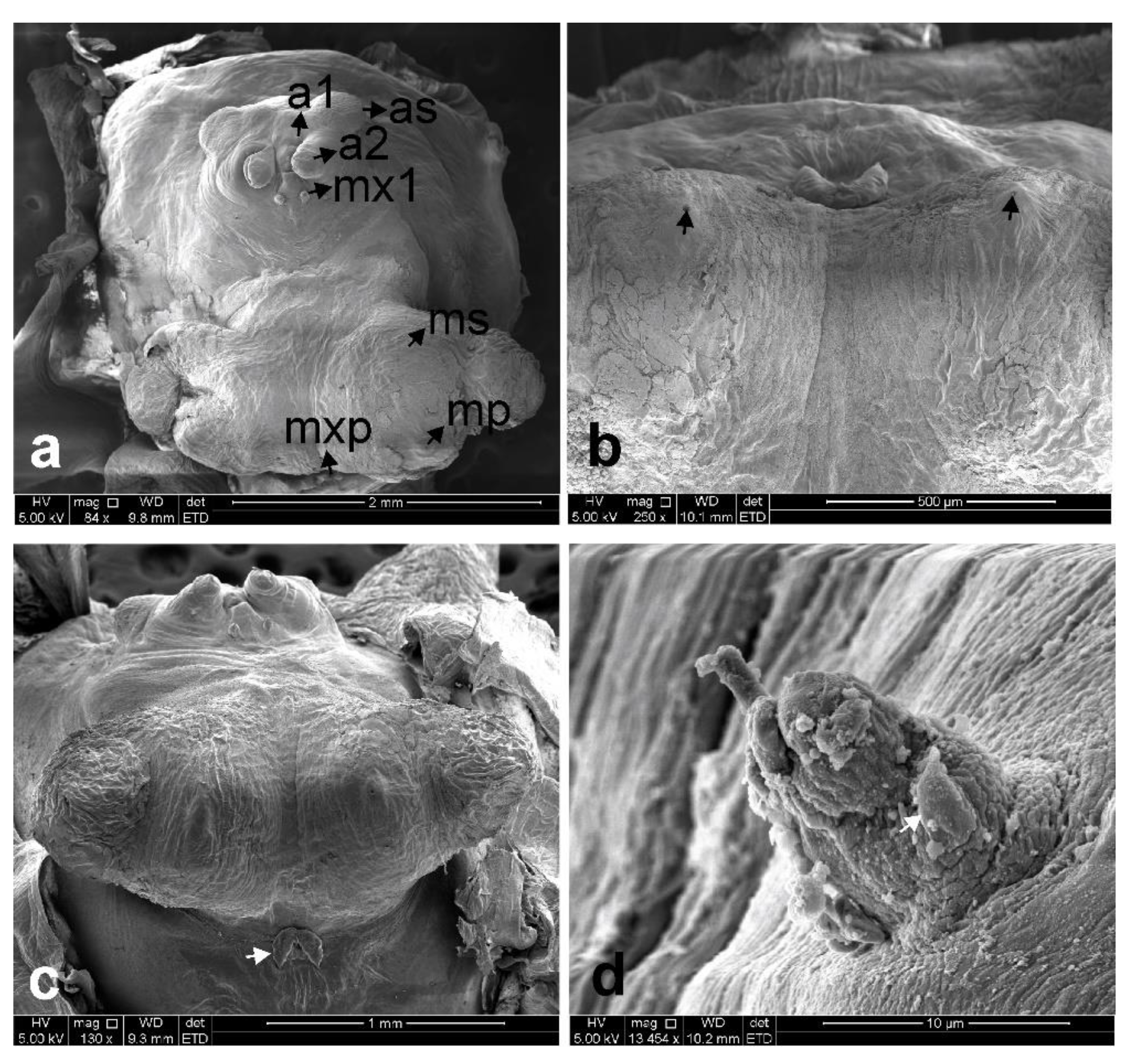
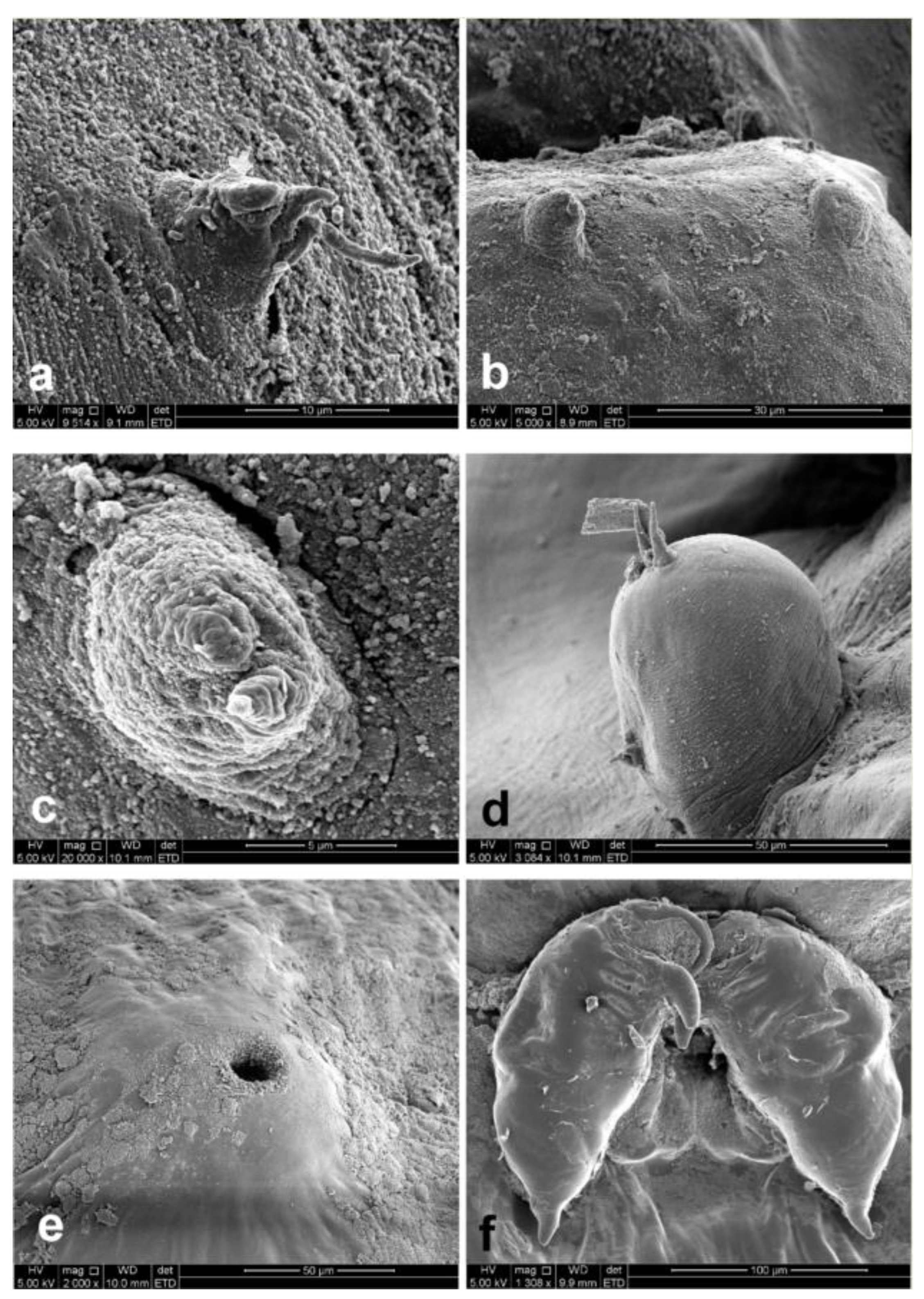
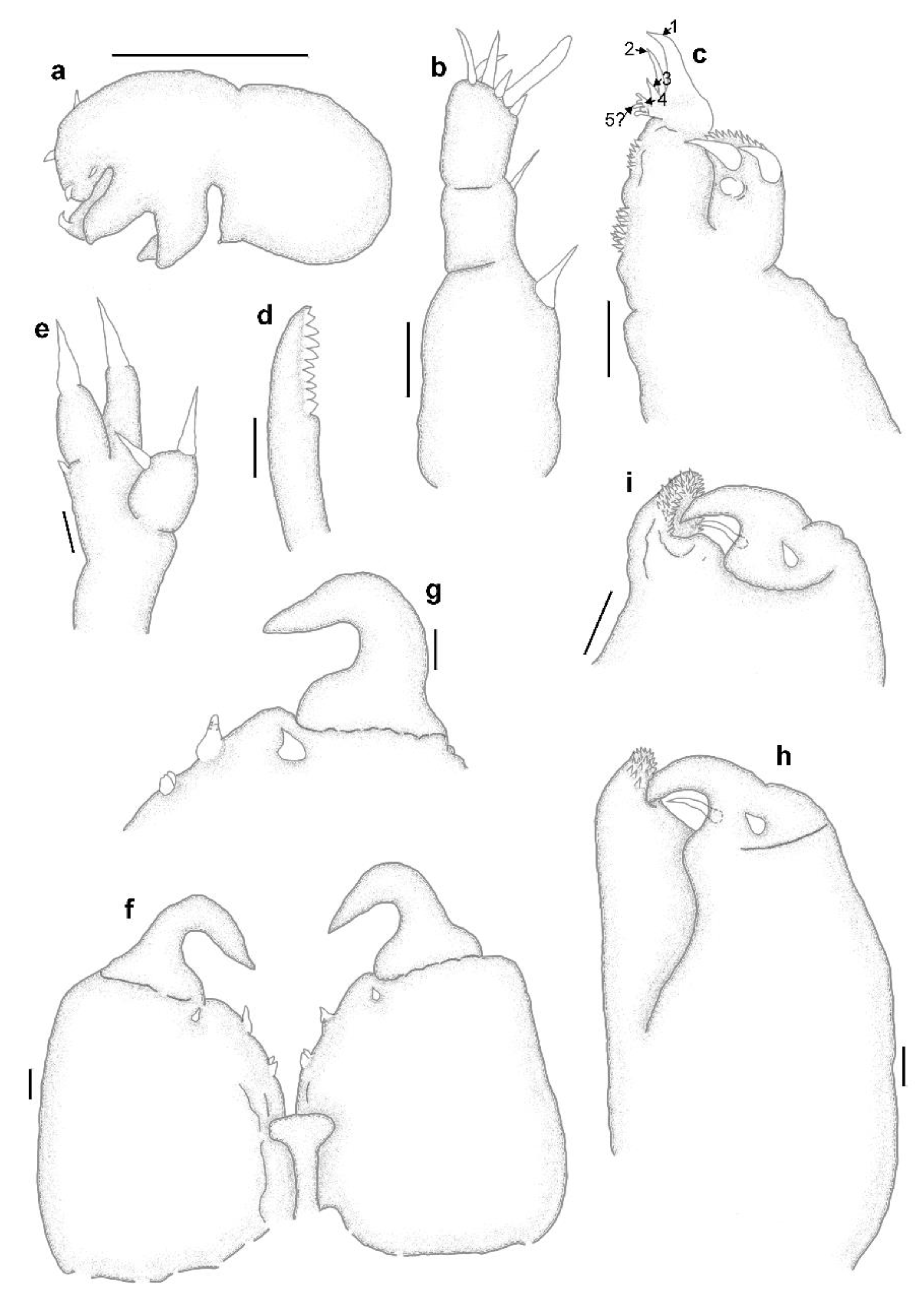
The copepod specimens were collected from fishes caught as by-catch during hake assessment demersal cruises off the south and west coasts of South Africa on board the research vessel R/V Africana (Department of Agriculture, Forestry and Fisheries; DAFF) during 2007, 2008, 2009, 2011 and 2012 as well as during demersal survey trawls off the South African east coast (Indian Ocean) during 2006 and 2010 (ACEP African Coelacanth Ecosystem Programme). The fish hosts were identified by researchers on board the vessels. Collected specimens were fixed and preserved in 70% ethanol. Selected specimens were cleared and stained in lactic acid with a small amount of dissolved lignin pink. These specimens were dissected and studied under both stereo- and light microscopes using the wooden slide technique [16]. Drawings were made with the aid of drawing tubes. Measurements were done with a stage micrometer (in mm) and presented as the mean (range). Selected specimens were prepared for scanning electron microscopy (SEM) by dehydrating them through a series of ethanol (70, 80, 90, 100, 100% for about 30 min each) followed by immersion in hexamethyldisilazane for about an hour. Excess hexamethyldisilizane, not evaporated, was removed and the specimens were allowed to dry completely before being sputter-coated with gold-palladium and carbon. Scanning electron microscopy was performed with a FEI Quanta 250 FEG SEM. Host names were verified using Eschmeyer’s catalog of fishes [17]. Morphological nomenclature mostly follows Kabata [9] and Huys and Boxshall [18].

3. Results

Family Sphyriidae Wilson C.B., 1919
Genus Sphyrion Cuvier, 1830

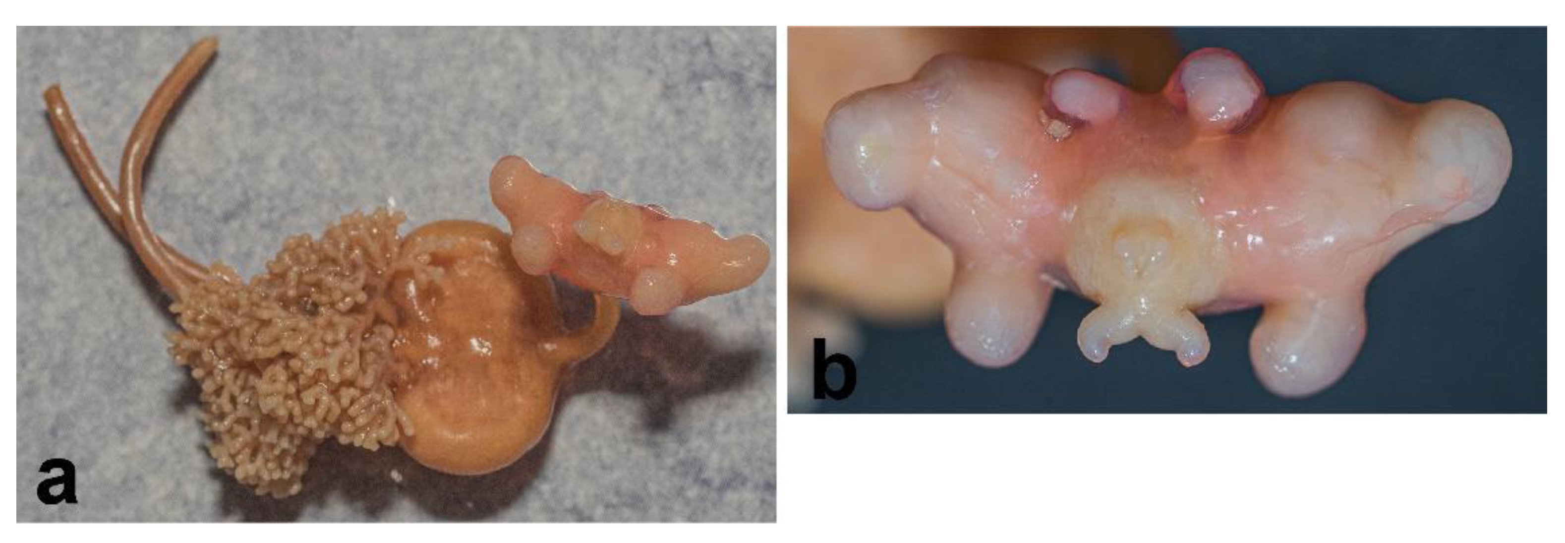
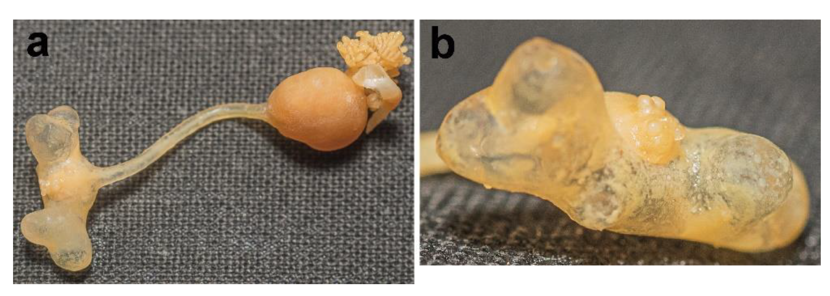
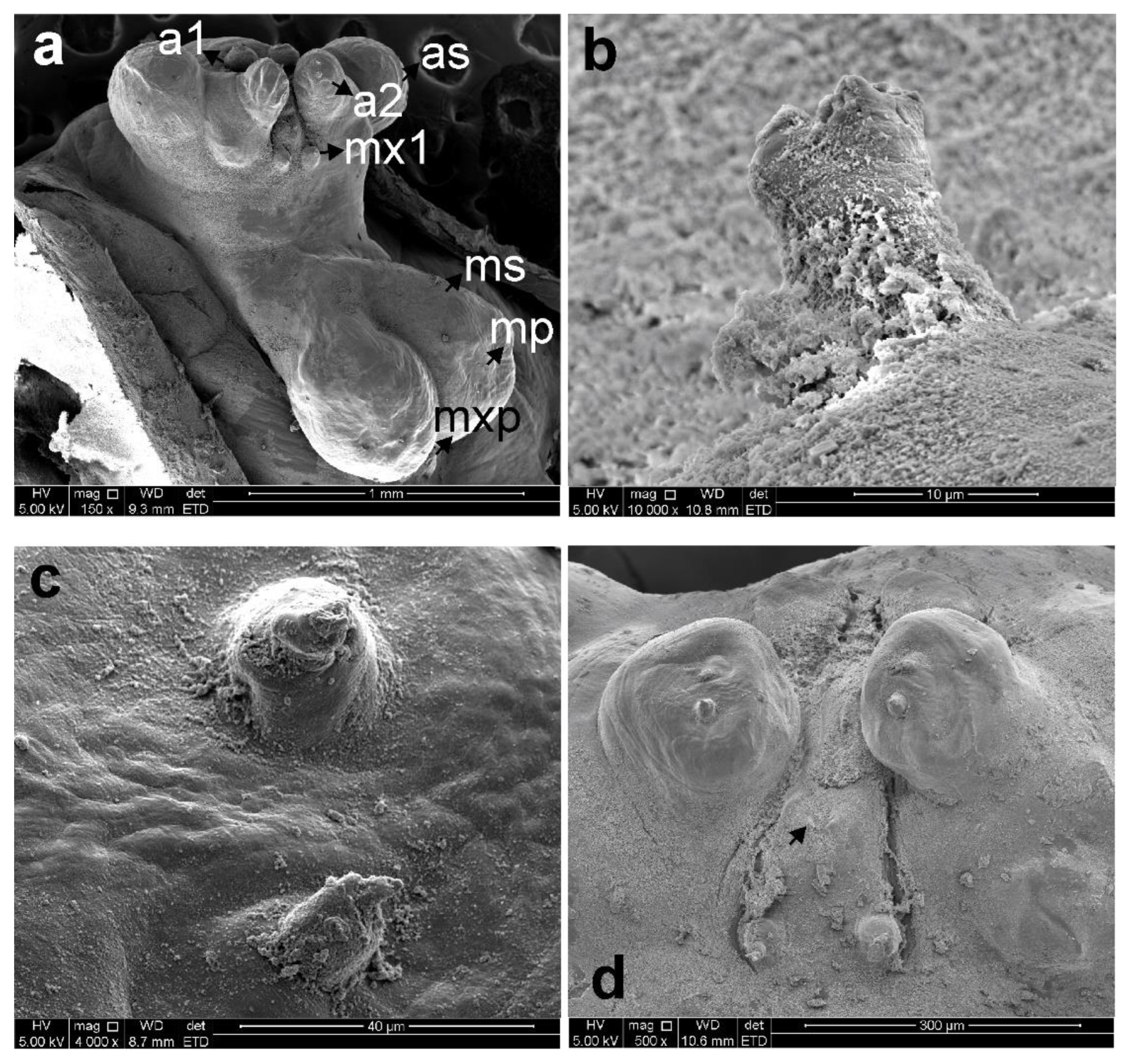
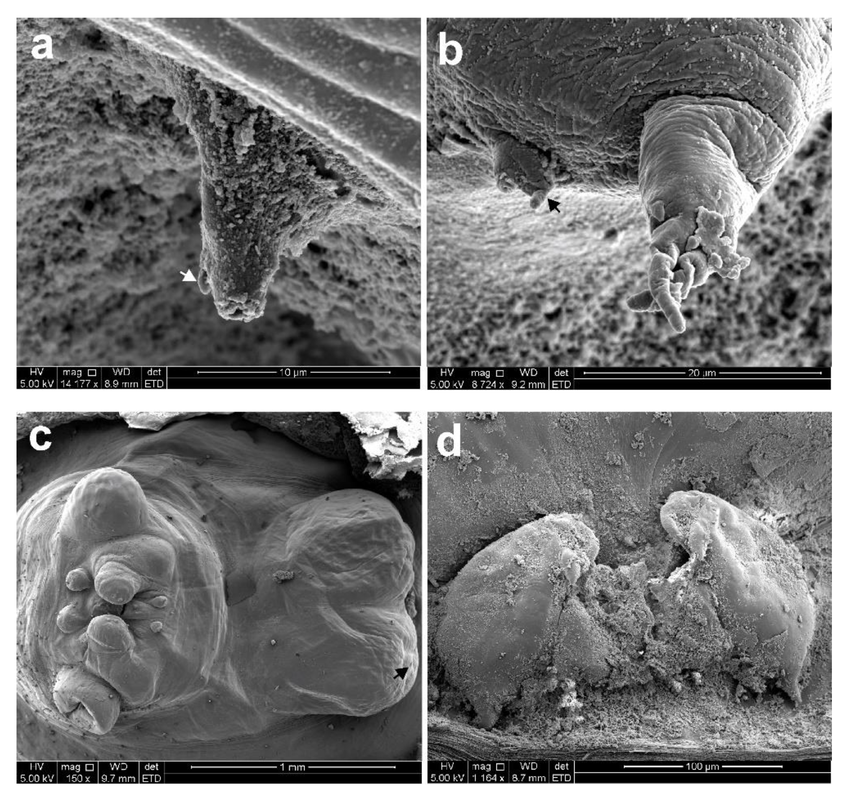
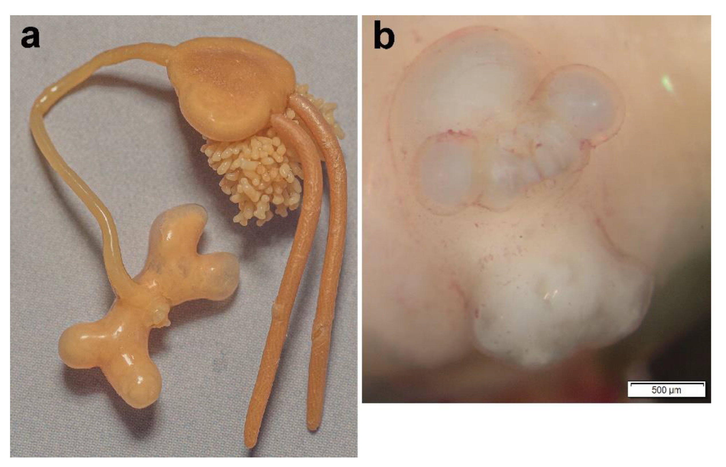
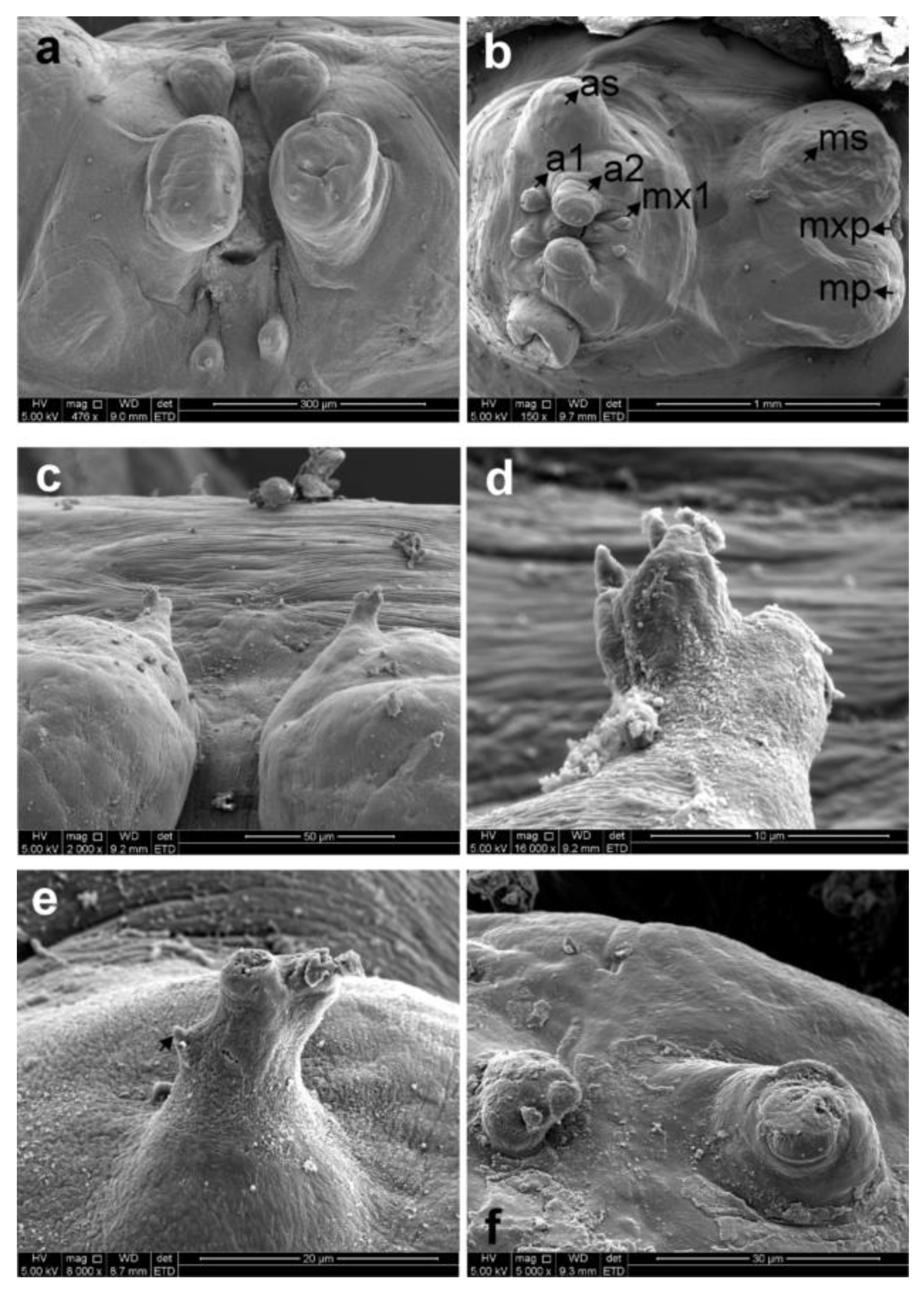
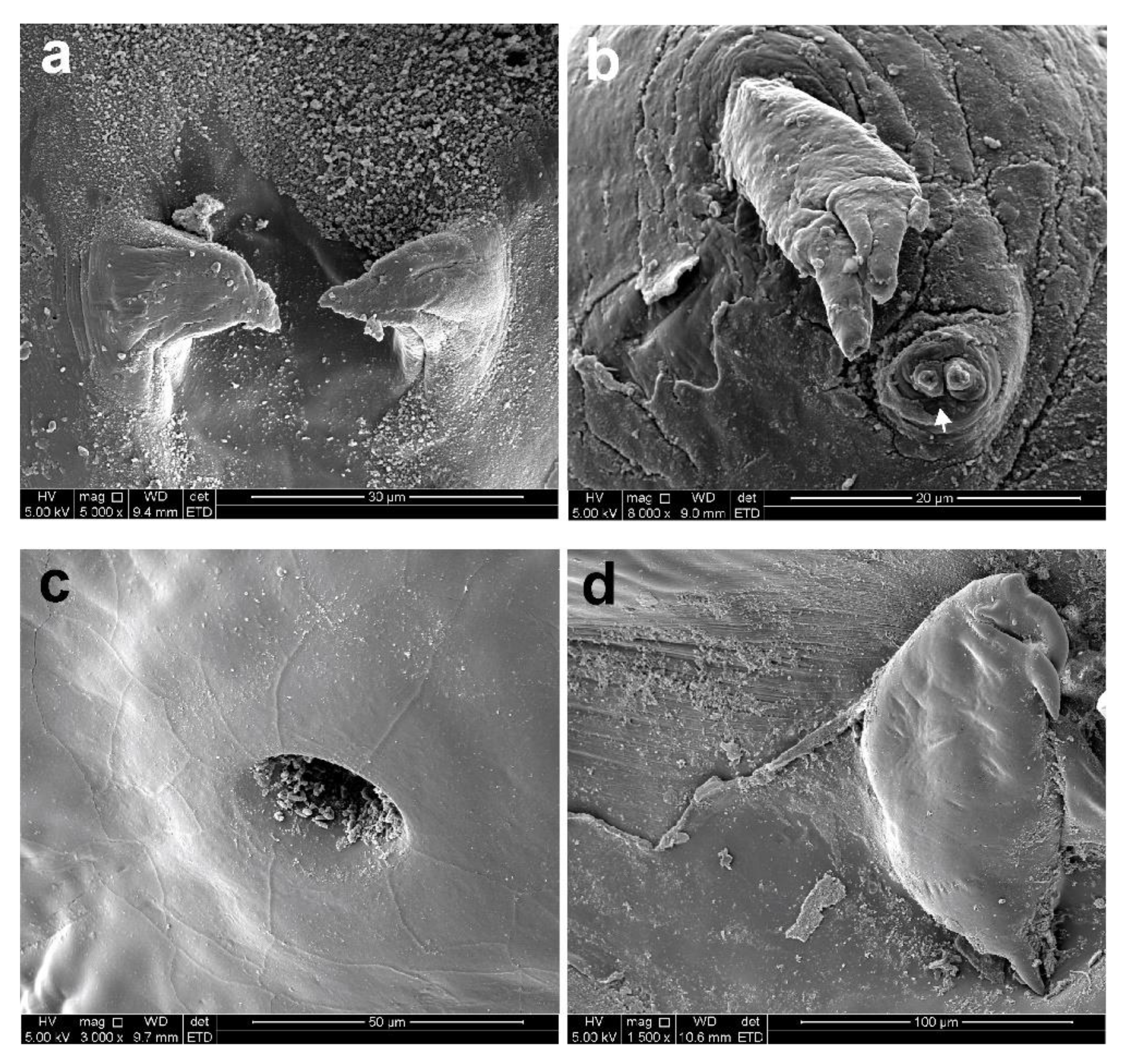
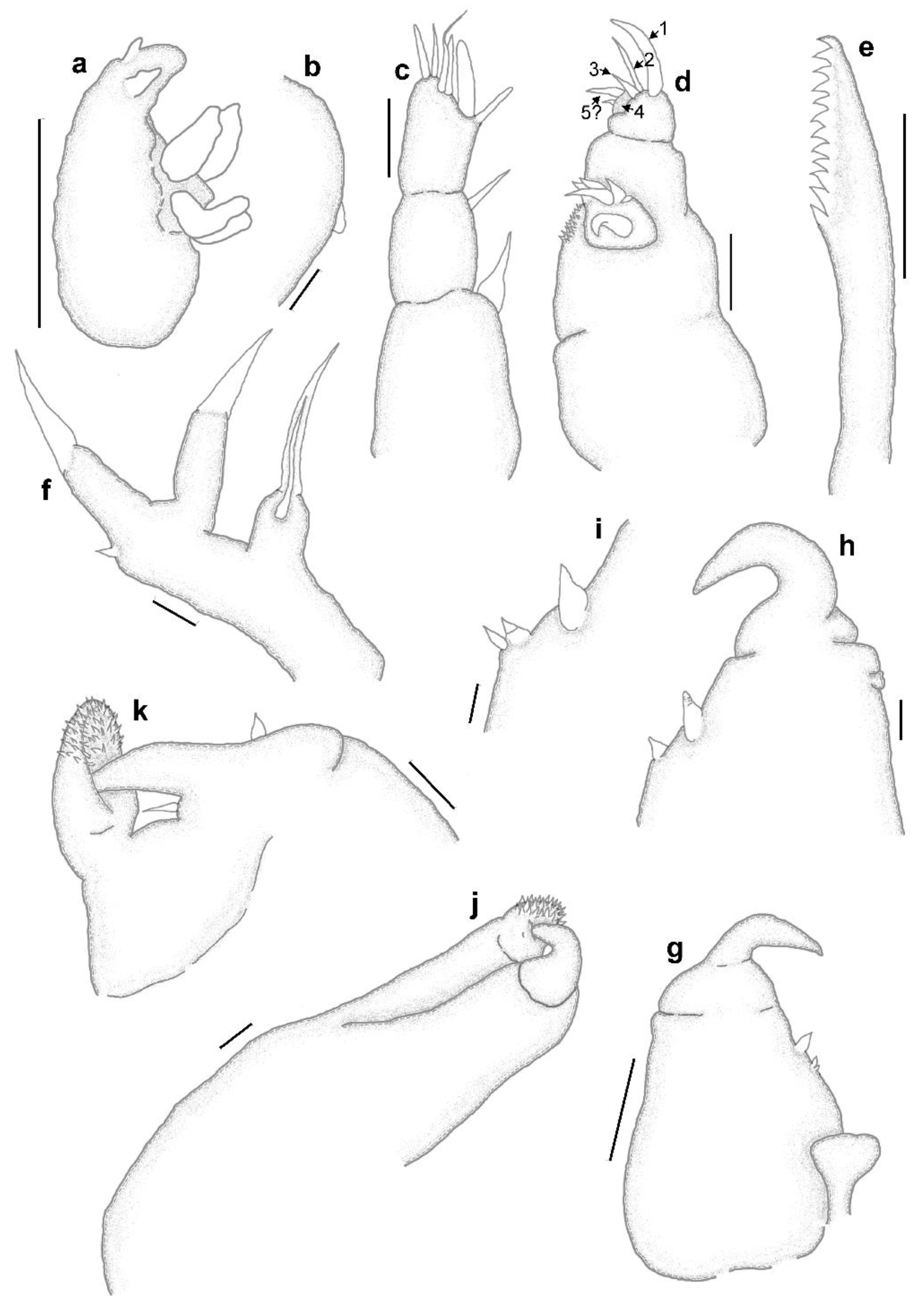
3.1. Sphyrion laevigatum (Quoy and Gaimard, 1824)

Host: Genypterus capensis (Smith, 1847) (Ophidiiformes: Ophidiidae)
Locality: Off the south coast (Atlantic Ocean), South Africa
Material examined: 4♀♀ from three host specimens and 1♂
Material collected: 32♀♀ and 3♂♂ all from G. capensis off the south and west coast (Atlantic Ocean)
Voucher material: Two adult female specimens (SAMC-A94722) and one male (SAMC-A94723) deposited in the Iziko South African Museum, Cape Town, South Africa.
Description
Post-metamorphosis female [Based on 15 specimens; Figure 1, Figure 2 and Figure 3] Body with cephalothorax, neck, trunk and small abdomen with posterior processes. Body length from tip of cephalothorax to tip of abdomen (excluding posterior processes) 27.6 mm (22.3–34.3); cephalothorax length 7.9 mm (6.3–9.8), width 20.4 mm (15.4–25.9); neck length 9.1 mm (7–14), width 2 mm (1.4–2.8); trunk length 10.7 mm (8.4–12.6), width 14.9 mm (11.3–19.6); posterior processes length 13.4 mm (8.4–17.5), width 14.5 mm (11.5–22.4 mm); egg-sac length 36.6 mm (n = 3; 35–39.2), width 1 mm (n = 10; 0.8–1.8). Cephalothorax (Figure 1a) transversely elongated, slightly compressed in dorsoventral aspect, with extended lateral protuberances and several protuberances on ventral surface: one pair of posteroventral protuberances, two pairs of anteroventral protuberances (medial pair larger than lateral pair) (Figure 1b), ventro-medially with raised area bearing antennary and maxillary swellings and cephalothoracic appendages (i.e., “head” according to Wilson [12]) (Figure 1b). Neck (Figure 1a) cylindrical, slightly increasing in width posteriorly, slightly shorter than trunk. Trunk (Figure 1a) slightly dorsoventrally flattened, pyriform, quickly expanding after connection with neck, wider than long, with abdomen (Figure 1a) situated posteriorly as small bifid tubercle with repeatedly branched posterior processes, first branching close to base. Egg-sacs (Figure 1a) cylindrical, longer than body; eggs multiseriate.
Cephalothoracic appendages (Figure 2a) situated on raised area ventro-medially on cephalothorax. Raised area bears two small swellings anteriorly, the antennary swellings (see Dippenaar [19]) with antennules medially at their posterior bases. Posterior to antennules, antennae on bulbous swellings, with maxillules situated at base of labium. On posterior margin of raised area, maxillary swellings consisting of four swellings, inner two bigger with small pores (possibly maxillary gland openings?) (Figure 2b), with maxillipeds medially below inner swellings (Figure 2c). Antennule (Figure 2d) slightly protruding area apparently bifid at tip with thump-like seta protruding distolaterally and smaller seta medially close to base (arrowed). Antenna (Figure 2a) posterolateral to antennule, consisting of bulbous sympod armed with two small tubercles; exopod details not observed, endopod (Figure 3a) with robust outer hook, one stout seta, inner extremity of margin slightly inflated. Mandible not observed. Labium with very small tubercles (arrowed) (Figure 3b) with two pointed tips (Figure 3c). Maxillule (Figure 3d) raised tubercle (endite) bearing two setae with small palp (arrowed) bearing two setae. Maxilla possibly represented by large thump-like maxillary swellings (Figure 2c) ventrally on raised area with maxillary gland pores (Figure 3e) on slightly protruded patch. Maxillipeds (Figure 3f) medially at base of maxillary swellings, subchelate, robust corpus with prominent pointed spine at base and similar curved spine distomedially, subchela not divided into shaft and claw, sharply curved at midlength and tapering claw.
Male [Based on three specimens; Figure 4] Body length from tip of cephalothorax (including mouth tube) to tip of posterior end about 2 mm (1.9–2.2). Cephalothorax (Figure 4a) almost half of total length, in line with ventral surface. Trunk inflated without distinct segmentation, posterior region curved towards cephalothorax, ending with caudal rami posteriorly close to maxillipeds.
Antennule (Figure 4b) 3-segmented; first segment with distomedial whip; second with distal solus and last segment with armature consisting of two short and three longer setae on apex. Antenna (Figure 4c) biramous, sympod indistinctly segmented, ornamented with patch of denticles distoventrally on last segment; exopod 1-segmented, shorter than endopod, apex ornamented with denticles and two subapical strong setae with smaller tubercle posteriorly; endopod 2-segmented, proximal segment with patch of denticles on ventral margin, distal segment with strong hook 1, spiniform seta 2, stout seta 3, process 4 with 4–5 small finger-like projections (5?) (according to Kabata [9] lernaeopodid structure). Mandible (Figure 4d) with 10 equally sized teeth. Maxillule (Figure 4e) biramous; palp with two apical setae; endite armed with two long truncated and one small apical setae. Maxilla (Figure 4f) broad and stout, linked to opposite maxilla by small tympanum; subchelate, corpus with two small closely related tubercles and digitiform seta distally on medial margin and stout seta close to base of subchela (Figure 4g); subchela indistinctly separated from claw, tip sharply curved, tapering. Maxilliped (Figure 4h) subchelate, corpus with myxal area extended into denticulated tip to accommodate tip of claw; subchela short and broad with small seta near base and longer seta medially at base of claw (Figure 4i); tip tapering into claw. Caudal ramus not observed.

3.2. Sphyrion lumpi (Krøyer, 1845)

Hosts: Coelorinchus trunovi Iwamoto and Anderson, 1994 (Gadiformes: Macrouridae)
Locality: Off the east coast (Indian Ocean), South Africa
Material examined: 2♀♀ from C. trunovi off the east coast (Indian Ocean), 1♀ from Allocyttus verrucosus (Gilchrist, 1906) (Zeiformes: Oreosomatidae) off the south coast (Atlantic Ocean).
Material collected: 11♀♀ from C. trunovi, 1♀ from Ventrifossa nasuta (Smith, 1935) off the east coast (Indian Ocean); 3♀♀ from A. verrucosus and 6♀♀ from C. simorhynchus Iwamoto and Anderson, 1994 both off the west coast (Atlantic Ocean) and 2♀♀ from Mesovagus antipodum (Hubbs and Iwamoto, 1977) off the south coast (Atlantic Ocean)
Voucher material: Two adult female specimens (SAMC-A94724) deposited in the Iziko South African Museum, Cape Town, South Africa.
Description
Post-metamorphosis female
Post-metamorphosis female [Based on three specimens; Figure 5 and Figure 6] Body length from tip of cephalothorax to tip of abdomen (excluding posterior processes) 21.8 mm (16.8–26.7); cephalothorax length 5.5 mm (3.6–7.3), width 11.3 mm (8.4–12.9); neck length 10.2 mm (7.8–12.6 mm), width 1.8 mm (1.5–2); trunk length 8.1 mm (5.6–10.1), width 10.1 mm (6.4–12.6); posterior processes length 6.3 mm (4.2–9.7), width 9.7 mm (7–10.2); egg-sac length 19.6 mm (n = 1), width 1.6 mm (n = 1). Cephalothorax and trunk (Figure 4a) as described and illustrated by Kabata (Figures 1440–1449) [9].
All appendages are situated on ventral part of cephalothorax (Figure 5b and Figure 6a) (see Moran and Piasecki [4] Figures 14 and 16). Antennule (Figure 6a) situated anteromedially between antennary swellings on small rounded swelling, apparently bifid at tip with reduced armature (Figure 6b) (cf Kabata [9] Figure 1446 and Moran and Piasecki [4] Figure 17). Antenna (Figure 6c) posterior to antennule, sympod bulbous armed with two small tubercles (cf Figure 1446 in Kabata [9] and Figure 23 in Moran and Piasecki [4]), armature of exo- and endopod not observed. Mandible not observed. Labium with two small tubercles (Figure 6d) with small subapical “thumb” (Figure 7a, arrowed). Maxillule (Figure 7b) posterior of mouth tube, small tubercle, endite with two longer and one small setae, palp (arrowed) small with two setae. Maxillae (Figure 6a) represented by maxillary swellings with maxillary gland pore (arrowed) on small raised patch (Figure 7c). Maxilliped (Figure 7d) posterior of maxillary swellings, like those of Kabata [9] Figure 1449 and Moran and Piasecki [4] Figure 26 with small seta on claw (cf Kabata [9] Figure 1449) not observed.

3.3. Sphyrion quadricornis Gaevskaya and Kovaleva, 1984

Host: Coelorinchus simorhynchus Iwamoto and Anderson, 1994 (Gadiformes: Macrouridae).
Locality: Off the west coast (Atlantic Ocean), South Africa.
Material examined: 3♀♀ from and 2♂♂.
Material collected: 25♀♀ and 4♂♂ all from C. simorhynchus off the west coast (Atlantic Ocean), 17♀♀ from C. trunovi off the east coast (Indian Ocean) and 1♀ from Saurida cf. undosquamis (Richardson, 1848) (see Russell et al. [20]) off the east coast (Indian Ocean).
Voucher material: Two adult female specimens (SAMC-A94725) and one male (SAMC-A94726) deposited in the Iziko South African Museum, Cape Town, South Africa.
Description
Post-metamorphosis female
Post-metamorphosis female [Based on six specimens; Figure 8, Figure 9 and Figure 10]. Body length from tip of cephalothorax to tip of abdomen (excluding posterior processes) 28.5 mm (25.8–35.8); cephalothorax length 4.9 mm (3.6–5.6), width 11 mm (8.4–12.6); neck length 16.6 mm (1–22,4), width 1.1 mm (0.84–1.26); trunk length 7 mm (6.2–8.4), width 8.1 mm (5.6–9.8); posterior processes length 7.9 mm (6.58–9.8), width 8.8 mm (7.4–10.1); egg-sac length 19.4 mm (n = 3; 14–23.1), width 1.3 mm (n = 6; 1.1–1.5). Cephalothorax (Figure 8a) transversely elongated, with bifid extended lateral protuberances, dorsal surface smooth, ventro-medially with raised area bearing antennary and maxillary swellings and cephalothoracic appendages (Figure 8b). Neck (Figure 8a) cylindrical, longer than trunk. Trunk (Figure 8a) slightly dorsoventrally flattened, pyriform, quickly expanding after connection with neck, slightly wider than long, with small abdomen situated posteriorly as small bifid tubercle with repeatedly branched posterior processes. Egg-sacs (Figure 8a) cylindrical, shorter than body; eggs multiseriate.
Cephalothoracic appendages (Figure 8b and Figure 9a) situated on raised area ventro-medially on cephalothorax. Raised area bears two small knobs anteriorly, the antennary swellings, with antennules antero-medially. Posterior to antennules, antennae on bulbous sympod, with maxillules situated posterior of mouth tube. On posterior margin of raised area, maxillary swellings consisting of two swellings (i.e., maxillae) with maxillipeds medially below medial separation of swellings (Figure 9b). Antennule (Figure 9a) rounded tubercle bifid at tip (Figure 9c) with strong setae on one tip (Figure 9d) and smaller seta close to base (Figure 9e, arrowed). Antenna (Figure 9f) posterolateral to antennule, consisting of bulbous sympod armed with two small tubercles; armature of rami not clear. Mandible not observed. Labium with small pointed tubercles (Figure 10a). Maxillule (Figure 10b) raised tubercle, endite bearing two big and one smaller setae, palp (arrowed) with two setae. Maxilla (Figure 9b) large maxillary swellings ventral on raised area with maxillary gland pores (Figure 10c) on small raised patch. Maxillipeds (Figure 10d) medially at base of maxillary swellings (Figure 9b), subchelate, robust corpus with prominent pointed spine at base and similar curved spine distomedially, subchela indistinctly divided into shaft and claw, shaft with small rounded tubercle midlength; claw sharply curved and tapering.
Male [Based on three specimens; Figure 11] Body length from tip of cephalothorax (including mouth tube) to tip of posterior end about 2 mm (1.9–2.2). Cephalothorax (Figure 11a) more than half of total length, in line with ventral surface. Trunk inflated without distinct segmentation, posterior region curved towards cephalothorax, ending with caudal rami (Figure 11b) posteriorly close to maxillipeds.
Antennule (Figure 11c) 3-segmented; first segment with distomedial whip; second with long distal solus and last segment with armature consisting of six setae, of almost equal lengths (one seta digitiform and one with elongated whip-like tip), on apex. Antenna (Figure 11d) biramous, sympod indistinctly segmented; exopod 1-segmented, shorter than endopod, apex ornamented with pronounced denticles and two strong setae (one apical, one subapical); endopod 2-segmented, proximal segment with patch of denticles on ventral margin, distal segment with strong hook 1, spiniform seta 2, stout seta 3, process 4 with two spine-like projections (5?). Mandible (Figure 11e) with 12 equally sized teeth. Maxillule (Figure 11f) biramous; palp with two long unequal apical setae; endite armed with two long truncated and one small apical setae. Maxilla (Figure 11g), broad and stout, linked to opposite by small tympanum; subchelate, corpus with two small closely related setae (sometimes seems like only one (Figure 11h)) and digitiform seta distally on medial margin (Figure 11i), subchela indistinctly separated from claw, tip sharply curved, tapering. Maxilliped (Figure 11j) subchelate, corpus with myxal area extended into denticulated tip to accommodate tip of claw; subchela short and broad with small seta near base and longer seta medially at base of claw (Figure 11k); tip tapering into claw. Caudal ramus small tubercles on posterior margin (Figure 11b).

4. Discussion

Even though variations exist in the morphology of the post-metamorphosis females [7,10], especially with age [9], as well as in the structure of the lateral protuberances of the cephalothorax which may sometimes be bifid e.g., in S. lumpi [9] or divided into three to five knobs in S. lumpi (current study), it still seems to be the best way to distinguish amongst the three current species. However, the structure of the cephalothorax should be considered together with the rest of the body features i.e., the relative length of the neck, relative size of the trunk and the relative length of posterior processes. None of these features should be considered independently as many variations exist in the same species e.g., the length of the neck [9], but also other morphological variations as also seen in the current study for specimens of S. laevigatum collected from the same host species.
The modified appendages of the post-metamorphosis females are very small and difficult to illustrate even with scanning electron microscopy due to attached host mucus and probable destruction of armature. However, Moran and Piasecki [4] provided micrographs of the appendages of S. lumpi indicating the clear resemblance between those of the male (also resembling the Lernaeopodidae males, see Kabata [9]) and the female. The armature of most of the appendages of the females in the current study is not clear but show resemblance to those in the micrographs in Moran and Piasecki [4]; detailed comparisons could not be done. However, there seems to be a resemblance between the armature of the female and male of S. laevigatum and S. quadricornis respectively, especially regarding the armature of the maxillule, and thus we agree with Moran and Piasecki [4] about the identification of the maxillule and the maxilla which differs from those of Kabata [9] and Ho and Kim [11]. The maxillae are identified as the maxillary swellings (see Figure 2c, Figure 6a and Figure 9b, respectively) in all three species due to the presence of pores that probably represent the maxillary gland openings (see Figure 3e, Figure 7c and Figure 10c, respectively). The maxillae of Tripaphylus species were also identified as being situated on the maxillary swellings [19,21], which may indicate that this is a feature common to the Sphyriidae. The presence of a “cuticular knob” on the maxillary swelling of S. lumpi (cf. Figure 22 in Moran and Piasecki [4]) was not observed in any of the three species. However, this knob was seen in an “intermediate female” which may have disappeared in adult post-metamorphosis females. Additionally, small tubercles were observed on the labium of all three species (see Figure 3b, Figure 6d and Figure 10a, respectively and Figure 18 of Moran and Piasecki [4]), identified as the maxillules by Kabata [9] and Ho and Kim [11], while the mandible (as indicated by Moran and Piasecki [4]) was not observed.
The morphology and armature of the appendages of the males of Sphyrion species are mostly similar. The antennule is 3-segmented in both S. laevigatum and S. quadricornis while that of S. lumpi is apparently 4-segmented [4]. The distal segment of the antennule is armed with six setae in all three species without clear separation between tubercles and setae (cf. Kabata [9], text Figure 66) and the gibber in S. lumpi (according to Moran and Piasecki [4]) was not observed in either S. laevigatum or S. quadricornis. The antennae are mostly similar among the three species with small differences in the ornamentation of the sympod and the structure of the armature on the ventral margin of the endopod (process 4). The mandible of all species is ornamented with a series of equally sized teeth variable in number. The maxillule is armed with an endite with two truncated and one small seta and a palp with two setae (only one seta in S. lumpi [4]). The maxillae of the three species seem to vary in small details regarding the ornamentation on the medial margins whereas the maxillipeds are mostly similar.
The current study provides the first detailed descriptions and illustrations of the males of S. laevigatum and S. quadricornis as well as some additional information regarding the appendages of the females of all three species. Additionally, this is the first report of S. lumpi from C. trunovi and V. nasuta off the east coast (Indian Ocean), A. verrucosus and C. simorhynchus both off the west coast (Atlantic Ocean) and M. antipodum off the south coast (Atlantic Ocean), South Africa. This report of S. lumpi infecting A. verrucosus constitutes a new host record (new family and new order of infected fish) for the species. Furthermore, this is the first report of S. quadricornis from South Africa which constitutes a new geographical record as well as new host records for C. simorhynchus, C. trunovi and S. cf. undosquamis (Aulopiformes: Synodontidae).

Author Contributions

S.M.D.: Conceptualization, resources, writing—original draft preparation, writing—review and editing, illustrations, supervision, project administration; M.M.S.: investigation, writing—review and editing. All authors have read and agreed to the published version of the manuscript.

Funding

This research received funding from the NRF as a research grant that enabled the sampling of copepods from by-catch on the Africana during 2008.

Institutional Review Board Statement

Not applicable.

Data Availability Statement

Voucher specimens are deposited in the Iziko South African Museum, Cape Town, South Africa.

Acknowledgments

We would like to thank the University of Limpopo (UL) for laboratory support and BP Jordaan for assistance during sampling on board the Africana. Additionally, we also thank R Leslie (then DAFF) and D Vaughan (now Central Queensland University) for collecting specimens during demersal cruises on board the Africana. Additionally, for fishes obtained during the east coast surveys of KZN marine waters, in 2006 and the KZN bight Ecosystem Functioning Project in 2010 funded by the ACEP African Coelacanth Ecosystem Programme, we thank Sean Fennessy of the Oceanographic Research Institute (ORI). We also acknowledge A Jordaan (NWU) for assistance with SEM and A Pretorius for assistance with photos. Lastly, we thank the NRF for financial assistance to MMS.

Conflicts of Interest

The authors declare no conflict of interest.

References

  1. Walter, T.C.; Boxshall, G.A. World of Copepods Database. Sphyrion Cuvier, 1830. 2021. Available online: https://www.marinespecies.org/copepoda/aphia.php?p=taxdetails&id=135656 (accessed on 2 June 2022).
  2. Dojiri, M.; Deets, G.B. Norkus cladocephalus, new genus, new species (Siphonostomatoida: Sphyriidae), a copepod parasitic on an elasmobranch from southern California waters, with a phylogenetic analysis of the Sphyriidae. J. Crustacean Biol. 1988, 8, 679–687. [Google Scholar] [CrossRef]
  3. Ho, J.-S. Does Sphyrion lumpi (Kroyer) (Copepoda, Sphyriidae) occur in the Sea of Japan? With discussion on the origin and dispersal of Sphyrion Cuvier, 1830. Rep. Sado Mar. Biol. Stn. Niigata Univ. 1992, 22, 37–48. [Google Scholar]
  4. Moran, J.; Piasecki, W. External morphology of the male and female of Sphyrion lumpi (Krøyer,1845) (Copepoda; Siphonostomatoida; Sphyriidae). Hydrobiologia 1994, 292, 171–178. [Google Scholar] [CrossRef]
  5. Kazachenko, V.N. Nahodki predstavitelei roda Sphyrion (Crustacea: Copepoda) u yugo-zapadnyh beregov Afriki. Findings of representatives of the genus Sphyrion (Crustacea: Copepoda) off the south-western coasts of Africa. Nauchnye Tr. Dalrybvtuza 2017, 40, 13–16. [Google Scholar]
  6. Kensley, B.; Grindley, J.R. South African parasitic Copepoda. Ann. South Afr. Mus. 1973, 62, 69–130. [Google Scholar]
  7. Gaevskaya, A.V.; Kovaleva, A.A. Rachki roda Sphyrion (Copepoda, Sphyridae) u ryb Atlantiki. Crustacea of the genus Sphyrion (Copepoda, Sphyriidae) in Atlantic fishes. Gidrobiol. Zhurnal Saratov 1984, 20, 41–45. [Google Scholar]
  8. Boxshall, G.A.; Halsey, S. An Introduction to Copepod Diversity; The Ray Society: London, UK, 2004; p. 966. [Google Scholar]
  9. Kabata, Z. Parasitic Copepoda of British Fishes; The Ray Society: London, UK, 1979; p. 468. [Google Scholar]
  10. Barnard, K.H. South African Parasitic Copepoda. Ann. South Afr. Mus. 1955, 41, 223–312. [Google Scholar]
  11. Ho, J.-S.; Kim, I. Lophoura (Copepoda: Sphyriidae) parasitic on the rattails (Pisces: Macrouridae) in the Pacific, with note on Sphyrion lumpi from the Sea of Japan. Publ. Seto Mar. Biol. Lab. 1989, 34, 37–54. [Google Scholar] [CrossRef] [Green Version]
  12. Wilson, C.B. North American parasitic copepods belonging to the new family Sphyriidae. Proc. United States Natl. Mus. 1919, 55, 549–604. [Google Scholar] [CrossRef]
  13. Squires, H.J. Reproduction in Sphyrion lumpi, a copepod parasitic on redfish (Sebastes spp.). J. Fish. Res. Board Can. 1966, 23, 521–526. [Google Scholar] [CrossRef]
  14. Payne, A.I.L. Observations on some conspicuous parasites of the southern African kingklip Genypterus capensis. S. Afr. J. Mar. Sci. 1986, 4, 163–168. [Google Scholar] [CrossRef] [Green Version]
  15. Dippenaar, S.M. Reported siphonostomatoid copepods parasitic on marine fishes of southern Africa. Crustaceana 2004, 77, 1281–1328. [Google Scholar] [CrossRef]
  16. Humes, A.G.; Gooding, R.U. A method for studying the external anatomy of copepods. Crustaceana 1964, 6, 238–240. [Google Scholar] [CrossRef] [Green Version]
  17. Fricke, R.; Eschmeyer, W.N.; Fong, J.D. Eschmeyer’s Catalog of Fishes: Genera/Species by Family/Subfamily. 2022. Available online: http://researcharchive.calacademy.org/research/ichthyology/catalog/SpeciesByFamily.asp (accessed on 8 March 2022).
  18. Huys, R.; Boxshall, G.A. Copepod Evolution; The Ray Society: London, UK, 1991; p. 468. [Google Scholar]
  19. Dippenaar, S.M. Description of four new species and a revision of the genus Tripaphylus Richiardi in Anonymous, 1878 (Copepoda: Siphonostomatoida: Sphyriidae). Syst. Parasitol. 2018, 95, 173–200. [Google Scholar] [CrossRef] [PubMed]
  20. Russell, B.C.; Golani, D.; Tikochinski, Y. Saurida lessepsianus a new species of lizardfish (Pisces: Synodontidae) from the Red Sea and Mediterranean Sea, with a key to Saurida species in the Red Sea. Zootaxa 2015, 3956, 559–568. [Google Scholar] [CrossRef]
  21. Boxshall, G.A.; Barton, D.P.; Kirke, A.; Zhu, X.; Johnson, G. Two new parasitic copepods of the family Sphyriidae (Copepoda: Siphonostomatoida) from Australian elasmobranchs. Syst. Parasitol. 2022, 99, 659–669. [Google Scholar] [CrossRef] [PubMed]
Figure 1. Sphyrion laevigatum (Quoy and Gaimard, 1824), post-metamorphosis female. (a) habitus, ventral view, (b) cephalothorax, ventral view.
Diversity 14 00929 g001
Figure 2. Sphyrion laevigatum (Quoy and Gaimard, 1824), post-metamorphosis female; scanning electron micrographs, (a) cephalothorax “head” (as = antennary swelling, a1 = antennule, a2 = antenna, mx1 = maxillule, ms = maxillary swelling, mp = maxillary pore, mxp = maxilliped), (b) maxillary swellings with maxillary pores (arrowed) and maxillipeds, (c) maxillary swellings with maxillipeds (arrowed) posteromedially, (d) antennule.
Diversity 14 00929 g002
Figure 3. Sphyrion laevigatum (Quoy and Gaimard, 1824), post-metamorphosis female; scanning electron micrographs. (a) antenna endopod, (b) labium with tubercles (arrowed), (c) labium tubercle, (d) maxillule, (e) maxillary gland pore, (f) maxillipeds.
Diversity 14 00929 g003
Figure 4. Sphyrion laevigatum (Quoy and Gaimard, 1824), adult male. (a) habitus, lateral view, (b) antennule, (c) antenna (1 = strong hook, 2 = spiniform seta, 3 = stout seta, 4 = raised process, 5? = fingerlike projections), (d) mandible, (e) maxillule, (f) maxilla, (g) maxilla medial margin, subchela and claw, (h) maxilliped, (i) subchela and claw. Scale-bars: (a) 1 mm, (bi), 10 µm.
Diversity 14 00929 g004
Figure 5. Sphyrion lumpi (Krøyer, 1845), post-metamorphosis female. (a) habitus, ventral view, (b) cephalothorax, dorso-ventral view.
Diversity 14 00929 g005
Figure 6. Sphyrion lumpi (Krøyer, 1845), post-metamorphosis female, scanning electron micrographs. (a) cephalothorax “head” (as = antennary swelling, a1 = antennule, a2 = antenna, mx1 = maxillule, ms = maxillary swelling, mp = maxillary pore, mxp = maxilliped), (b) antennule, (c) antenna, (d) labium with small tubercles (arrowed).
Diversity 14 00929 g006
Figure 7. Sphyrion lumpi (Krøyer, 1845), post-metamorphosis female, scanning electron micrographs. (a) labium tubercle, (b) maxillule, (c) cephalothorax “head” with maxillary swellings and maxillary pores (arrowed), (d) maxillipeds.
Diversity 14 00929 g007
Figure 8. Sphyrion quadricornis Gaevskaya and Kovaleva, 1984, post-metamorphosis female. (a) habitus, ventral view, (b) cephalothorax head, ventral view.
Diversity 14 00929 g008
Figure 9. Sphyrion quadricornis Gaevskaya and Kovaleva, 1984, post-metamorphosis female, scanning electron micrographs. (a) cephalothorax appendages, (b) cephalothorax “head” (as = antennary swelling, a1 = antennule, a2 = antenna, mx1 = maxillule, ms = maxillary swelling, mp = maxillary pore, mxp = maxilliped), (c) antennule, (d) antennule, (e) antennule with smaller basal seta (arrowed), (f) antenna.
Diversity 14 00929 g009
Figure 10. Sphyrion quadricornis Gaevskaya and Kovaleva, 1984, post-metamorphosis female, scanning electron micrographs. (a) small tubercles on labium, (b) maxillule, (c) maxillary pore, (d) maxilliped.
Diversity 14 00929 g010
Figure 11. Sphyrion quadricornis Gaevskaya and Kovaleva, 1984, adult male. (a) habitus, lateral view, (b) posterior margin with caudal rami, (c) antennule, (d) antenna (1 = strong hook, 2 = spiniform seta, 3 = stout seta, 4 = raised process, 5? = spine-like projections), (e) mandible, (f) maxillule, (g) maxilla, (h) maxilla (alternative view), (i) maxilla medial margin, (j) maxilliped, (k) subchela and claw. Scale-bars: (a) 0.5 mm, (b), (g) 50 µm, (cf), (hk) 10 µm.
Diversity 14 00929 g011
Publisher’s Note: MDPI stays neutral with regard to jurisdictional claims in published maps and institutional affiliations.

