Article

New Records of Antarctic Tardigrada with Comments on Interpopulation Variability of the Paramacrobiotus fairbanksi Schill, Förster, Dandekar and Wolf, 2010

by
Łukasz Kaczmarek
1,*,
Monika Mioduchowska
2,3,
Uroš Kačarević
4,
Katarzyna Kubska
1,
Ivan Parnikoza
5,6,
Bartłomiej Gołdyn
7 and
Milena Roszkowska
1,8
1
Department of Animal Taxonomy and Ecology, Faculty of Biology, Adam Mickiewicz University, Poznań, Uniwersytetu Poznańskiego 6, 61-614 Poznań, Poland
2
Department of Genetics and Biosystematics, Faculty of Biology, University of Gdańsk, Gdańsk, Wita Stwosza 59, 80-308 Gdańsk, Poland
3
Department of Marine Plankton Research, Institute of Oceanography, University of Gdansk, Marszałka Piłsudskiego 46, 81-378 Gdynia, Poland
4
Department of Morphology, Systematics and Phylogeny of Animals, Faculty of Biology, University of Belgrade, Studentski trg 16, 11000 Belgrade, Serbia
5
National Antarctic Scientific Center of Ukraine, Boulevard Tarasa Shevchenka 16, 01601 Kyiv, Ukraine
6
Institute of Molecular Biology and Genetics of National Academy of Science of Ukraine, Kyiv, Ukraine, st. Acad. Zabolotny, 150, Kyiv 03143, Ukraine
7
Department of General Zoology, Faculty of Biology, Adam Mickiewicz University, Uniwersytetu Poznańskiego 6, 61-614 Poznań, Poland
8
Department of Bioenergetics, Faculty of Biology, Adam Mickiewicz University, Poznań, Uniwersytetu Poznańskiego 6, 61-614 Poznań, Poland
*
Author to whom correspondence should be addressed.
Diversity 2020, 12(3), 108; https://doi.org/10.3390/d12030108
Submission received: 19 February 2020 / Revised: 16 March 2020 / Accepted: 16 March 2020 / Published: 20 March 2020
(This article belongs to the Special Issue Tardigrades Taxonomy, Biology and Ecology)

Abstract

:
Studies on Antarctic tardigrades started at the beginning of the twentieth century and have progressed very slowly and ca. 75 tardigrade species are known from this region. Paramacrobiotus fairbanksi was described from USA based on genetic markers and later reported from Italy, Poland, and Spain. The “everything is everywhere” hypothesis suggests that microscopic organisms have specific features which help them to inhabit most of environments and due to this they can be considered cosmopolitan. In the present paper, we report eight tardigrade taxa from Antarctic, including the first report of Pam. fairbanksi from Southern Hemisphere, which could suggest that the “everything is everywhere” hypothesis could be true, at least for some tardigrade species. Moreover, we also genetically and morphologically compare a few different populations of Pam. fairbanksi. The p-distances between COI haplotypes of all sequenced Pam. fairbanksi populations from Antarctica, Italy, Spain, USA and Poland ranged from 0.002% to 0.005%. In the case of COI polymorphism analyses, only one haplotype was observed in populations from Antarctica, USA and Poland, two haplotypes were found in population from Spain, and six haplotypes were observed in population from Italy. We also found some statistically significant morphometrical differences between the populations of Pam. fairbanksi from different regions and designed a new specific primers for Paramacrobiotus taxa.

1. Introduction

Antarctic is the coldest and most extreme region on the planet with the lack of surface liquid freshwater. Continental Antarctic with an area of ca. 14 million square kilometers has only ca. 0.3% ice-free lands. The entire continent is divided into three main units, i.e., East Antarctica, West Antarctica, and the Antarctic Peninsula [1]. Our knowledge of micro-invertebrates in this region is patchy and fragmented, because most studies have focused on easily accessible regions that are close to research stations [2]. Many of Antarctic invertebrates are endemic, some of them have a continental distribution, others are restricted only to maritime Antarctic and only a few can be characterized as pan-Antarctic species such as eutardigrade Acutuncus antarcticus (Richters [3]) e.g., [4,5].
The phylum tardigrada currently consists of ca. 1300 species [6,7,8,9] that inhabit terrestrial and aquatic environments throughout the world [10,11].
Studies on Antarctic tardigrades started at the beginning of the twentieth century and have progressed very slowly until now. Currently, ca. 75 tardigrade species have been reported from continental Antarctic and few from other regions (for review see [12] and later publications [2,13,14,15,16,17,18,19,20,21,22,23,24]).
Paramacrobiotus fairbanksi was described by Schill, Förster, Dandekar and Wolf [25] from Alaska (USA), based on genetic differences from other members of the genus Paramacrobiotus. It was later reported from Italy, Poland, and Spain [26,27,28]. Moreover, specimens reported by Guidetti et al. [29] from Italy as Macrobiotus richtersi and Murray [30] were also attributed to Pam. fairbanksi based on genetic markers [28].
According to the “everything is everywhere” hypothesis it is assumed that microorganisms and small invertebrates have cosmopolitan ranges. This hypothesis was introduced at the beginning of the 20th century [31,32]. Due to the presence of certain features in microscopic organisms that are not found in larger animals, they can inhabit most environments and are considered to be cosmopolitan species. Among the most frequently mentioned adaptations are the possibility of quite ease passive dispersion (through wind, sea currents, water, other animals, etc.), the presence of very resistant spore stages (which include, for example, cysts, eggs, or cryptobiotic individuals) that help to survive extreme conditions and maintain in any environment, as well as the presence of asexual or parthenogenetic reproduction that allows a rapid increase in the number of individuals e.g., [32,33,34,35,36]. However, the cosmopolitanism of microscopic organisms is increasingly discussed and undermined also within tardigrades e.g., [37,38,39]. Gąsiorek et al. [40] provided a first Australasian, molecularly verified record of a heterotardigrade species Echiniscus testudo (Doyère [41]) originally described from Holarctic (Europe, France) which could suggest agreement with the hypothesis “everything is everywhere” for tardigrades.
In this paper we report eight tardigrade taxa from Antarctic including the first report of Pam. fairbanksi from the Southern Hemisphere which is another strong argument suggesting that “everything is everywhere” hypothesis could be true, at least for some tardigrade species. Moreover, we also genetically and morphologically compare a few different populations of Pam. fairbanksi, and designed a new specific primers for Paramacrobiotus taxa.

2. Materials and Methods

2.1. Sample Processing

Eleven moss-lichen and algae samples from soil, rocks, and freshwater sediments were collected during the 5th (2012–2013), 7th (2014–2015) and 9th (2016–2017) Belarusian Antarctic Expeditions (BAE) near the Belarusian Antarctic Station “Vechernia” and Russian Station “Progress” (for details see Table 1). The samples were packed in paper envelopes, dried at a room temperature and delivered to the laboratory at the Faculty of Biology, Adam Mickiewicz University, Poznań, Poland. Tardigrades were extracted from the samples and studied following the protocol of Stec et al. [42].

2.2. Microscopy, Morphometrics, and Morphological Nomenclature

Specimens for light microscopy were mounted on microscope slides in a small drop of Hoyer’s medium, prepared according to Ramazzotti and Maucci [10] as in the English translation by Beasley [43], and secured with a cover slip. Then, the slides were placed in an incubator and dried for two days at ca. 60 °C. Dried slides were sealed with a transparent nail polish and examined under an Olympus BX41.
All measurements are given in micrometers [μm]. Structures were measured only if their orientation was suitable. Body length was measured from the anterior extremity to the end of the body, excluding the hind legs. The types of bucco-pharyngeal apparatuses and claws were classified according to Pilato and Binda [44]. Buccal tubes, claws, and eggs were measured according to Kaczmarek and Michalczyk [45]. Macroplacoid length sequence was given according to Kaczmarek et al. [46]. The pt ratio is the ratio of the length of a given structure to the length of the buccal tube, expressed as a percentage [47]. The pt values are always provided in italics.
Morphometric data were handled using the “Parachela” ver. 1.7 template available from the Tardigrada Register [48]. Tardigrade taxonomy follows Bertolani et al. [49]. Genus abbreviations follow Perry et al. [50].
The map and figures were assembled in Corel Photo-Paint 2017.

2.3. Comparative Material

Species were identified based on original descriptions [3,15,25,51,52] and later partial redescriptions [53,54]. The type material (paratypes) of Dastychius improvisus (Dastych [52]) and Mesobiotus blocki (Dastych [52]) from the collection of Zoologisches Museum of Hamburg (Germany) were examined. Specimens of Pam. fairbanksi from type locality and additional material from Italy from the Roberto Guidetii collection (University of Modena and Reggio Emilia, Modena, Italy) were also used in analysis, altogether with specimens of Pam. fairbanksi from the laboratory culture conducted at the Department of Animal Taxonomy and Ecology (Adam Mickiewicz University in Poznań, Poland). Specimens of Pam. fairbanksi (Pam.fai_I_PL.018) (50°03’44’’ N, 19°57’26’’ E, 205 m asl: Jagiellonian University Botanical Garden, Kopernika 27, Kraków, Poland; moss on a tree; 03.2014; coll. Piotr Gąsiorek) were cultured in small Petri dishes in spring water mixed with distilled water (3:1) with the rotifers and nematodes added as a food ad libitum. Cultures were kept in an environmental chamber with a temperature at 18 °C and in the darkness.

2.4. DNA Extraction and Genotyping

Four specimens and 31 eggs of Pam. fairbanksi were identified (partially based on [25]) in vivo using light microscopy (LM) prior to DNA extraction for genotyping analysis. Eggs were divided into three samples. Ten eggs were placed in two of them and eleven eggs in the last sample. Genomic DNA of adult individuals was extracted using a Chelex® 100 resin (Bio-Rad, Warsaw, Poland) extraction method described by Casquet et al. [55] with a modified protocol included in Stec et al. [42]. In turn, genomic DNA from eggs was extracted using silica membranes from the commercial Genomic Mini kit (A&A Biotechnology, Gdynia, Poland).
Three DNA fragments differing in effective mutation rates were sequenced as follows: two conservative nuclear ribosomal subunit genes, i.e., 18S rRNA and 28S rRNA, and protein coding mitochondrial COI barcode gene with an intermediate effective mutation rate. Regions of the nuclear ribosomal subunit genes 18S and 28S were amplified using the following primers: SSU01_F (5’-AACCTGGTTGATCCTGCCAGT-3’) and SSU82_R (5’-TGATCCTTCTGCAGGTTCACCTAC-3’, [56]) for the 18S rRNA gene fragment; 28SF0001 (5’-ACCCvCynAATTTAAGCATAT-3’) and 28SR0990 (5’-CCTTGGTCCGTGTTTCAAGAC-3’; [57]) for the 28S rRNA gene fragment. In turn, the COI gene fragment was amplified using universal primers: HCO2198 (5’-TAAACTTCAGGGTGACCAAAAAATCA-3’) and LCO1490 (5’-GGTCAACAAATCATAAAGATATTGG-3’, [58]). All barcode sequences were obtained only from DNA extracted from eggs and it was problematic in the case of adult individuals. To overcome this problem, the obtained COI sequences were further used to design internal primers to amplify a 392 bp fragment with the Primer3 v. 0.4.0 [59] software package. The sequence of the newly developed forward ParCOIF primer was 5’-GGGTCYCCHCCHCCBGCKGGRTCA-3’, and that of the reverse ParCOIR was 5’-GGRGCYCCHGATATRGCHTTYCCHCG-3’.
All the polymerase chain reactions were carried out in 20 μL volume containing 0.8× JumpStart Taq ReadyMix (1 U of JumpStart Taq DNA polymerase, 4 mM Tris-HCl (pH 8.3), 20 mM KCl, 0.6 mM MgCl2, 0.08 mM of dNTP; Sigma-Aldrich, Darmstadt, Germany), 0.4 μM of proper forward and reverse primers and ca. 1 ng of DNA. The PCR reactions were performed in a BiometraTProfessional thermocycler. The PCR cycling profile to amplify the 18S rRNA gene fragment was as follows: initial denaturation at 95 °C for 5 min followed by 40 cycles of 95 °C for 30 s, 55 °C for 40 s, and 72 °C for 1 min 30 s, and ending with 72 °C for 4 min. The 28S rRNA gene fragment was amplified under conditions described by Mironov et al. [57]. The PCR protocol for the amplification of the COI gene fragment using HCO2198 and LCO1490 primers was as follows: initial denaturation at 94 °C for 5 min followed by 35 cycles of 94 °C for 30 s, 45 °C for 1 min, and 72 °C for 1 min, and ending with 72 °C for 5 min. In turn, amplification of the COI gene fragment using the newly designed degenerate primers was performed under the following conditions: initial denaturation at 95 °C for 5 min followed by 44 cycles of 95 °C for 30 s, 51.5 °C for 40 s, and 72 °C for 1 min, and ending with 72 °C for 5 min.
Amplified PCR products were separated in 1% agarose gel in a 1× SB buffer and visualized under UV light using Midori Green Advance DNA Stain (Genetics). Exonuclease I (20 U/μL, Thermo Scientific) and alkaline phosphatase FastAP (1 U/μL, Thermo Scientific, Waltham, MA, USA) were applied to clean the PCR products. Sequencing directly in both directions was performed using the BigDyeTM terminator cycle sequencing method and ABI Prism 3130xl genetic analyzer (Life Technologies, Carlsbad, CA, USA).

2.5. Comparative Molecular Analysis

BLAST (Basic Local Alignment Search Tool; [60]) searches at NCBI were performed to verify the identity and homology of the amplified nuclear and mitochondrial barcode sequences. All sequences obtained in our study and described by Guidetti et al. [28] as originated from Pam. fairbanksi, were then aligned using the ClustalW Multiple Alignment tool [61] implemented in BioEdit v. 7.2.5 [62]. Alignment sequences were trimmed to 822 and 574 bp for 18S rRNA and COI barcodes (only obtained from eggs), respectively. Calculation for the uncorrected pairwise distances (p-distances) was performed for COI sequences using the MEGA X.
The COI sequences were translated into amino acid sequences using the EMBOSS-TRANSEQ application [63,64] to check for indels and internal stop codons. The COI haplotypes were retrieved using DnaSP v5.10.01 software [65].
All obtained sequences have been deposited in GenBank under the following accession numbers: COI (primers: HCO2198 and LCO1490), MN964281-MN964282; COI (primers: ParCOIF and ParCOIR), MN961616; 18S rRNA, MN960302-MN960304; 28S rRNA, MN960306-MN960307.

2.6. Statistical Analysis

Statistical significance of the differences in morphometrics between the studied populations of Pam. fairbanksi was analyzed using the analysis of variance (ANOVA) test with Bonferroni correction for multiple comparisons. Measurements of the body and buccal tube length (BL and BTL) were used as dependent and populations as grouping variable. Shapiro test was used to check for normality of distribution in residuals resulting from these models.
Apart from the BL and BTL measurements, we also analyzed the overall differences in morphometrics of other measured traits: stylet support insertion points (SSIP), external width of buccal tube (BTEW), and placoids (M1, macroplacoid 1; M2, macroplacoid 2; M3, macroplacoid 3; Mi, microplacoid; MR, macroplacoid row; PR, placoid row). We performed principal component analysis (PCA) on these measurements (the PCA input data matrix was tardigrade specimens × characters length), and then used the resulting first component as the dependent variable in ANOVA, with populations as grouping variable and Bonferroni correction. Later, to compare the differences in proportions of the same characters, the same analyses were performed for the pt ratios of exact characters. Measurements of 44 tardigrade specimens were used in the analyses, including 16 Antarctic, 15 Polish, 9 Alaskan, and 4 Italian. In case some of the measurements were missing they were replaced by mean values.
ANOVA was also used to test for the differences in egg morphology between the populations studied. To do so, for each obtained egg its bare diameter (EBD), full diameter (EFD), and processes height (PH) were measured and used as dependent variable in the models, again with population as the variable determining compared groups and Bonferroni correction. Measurements of 49 tardigrade eggs were used in the analyses, including 18 Alaskan, 16 Antarctic, and 15 Polish. In case some of the measurements were missing they were replaced by mean values. All the analyses were performed in R 3.4.4. We considered p < 0.05 as a level determining statistical significance; in case of post hoc tests only statistically significant results are presented.

3. Results

3.1. Species Found

In eight of eleven samples, 461 tardigrades, 102 exuviae and 127 eggs belonging to eight taxa were found, i.e., Acu. antarcticus; Barbaria pseudowendti (Dastych [52]); Das. improvisus; Hebesuncus mollispinus Pilato, McInnes and Lisi [15]; Mes. blocki; Mesobiotus sp.; Milnesium quadrifidum Nederström [51] (Figure 1); and Pam. fairbanksi (Figure 2 and Figure 3) (for more details see Table 2).

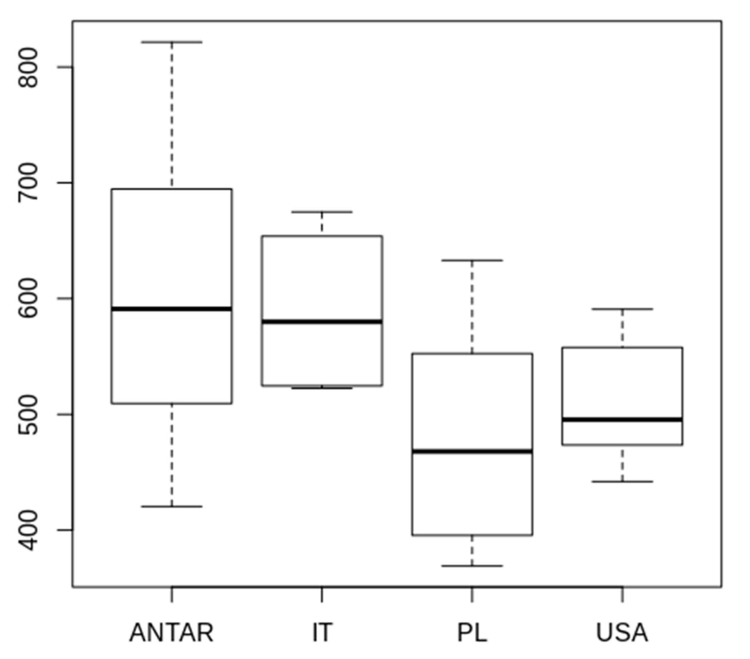
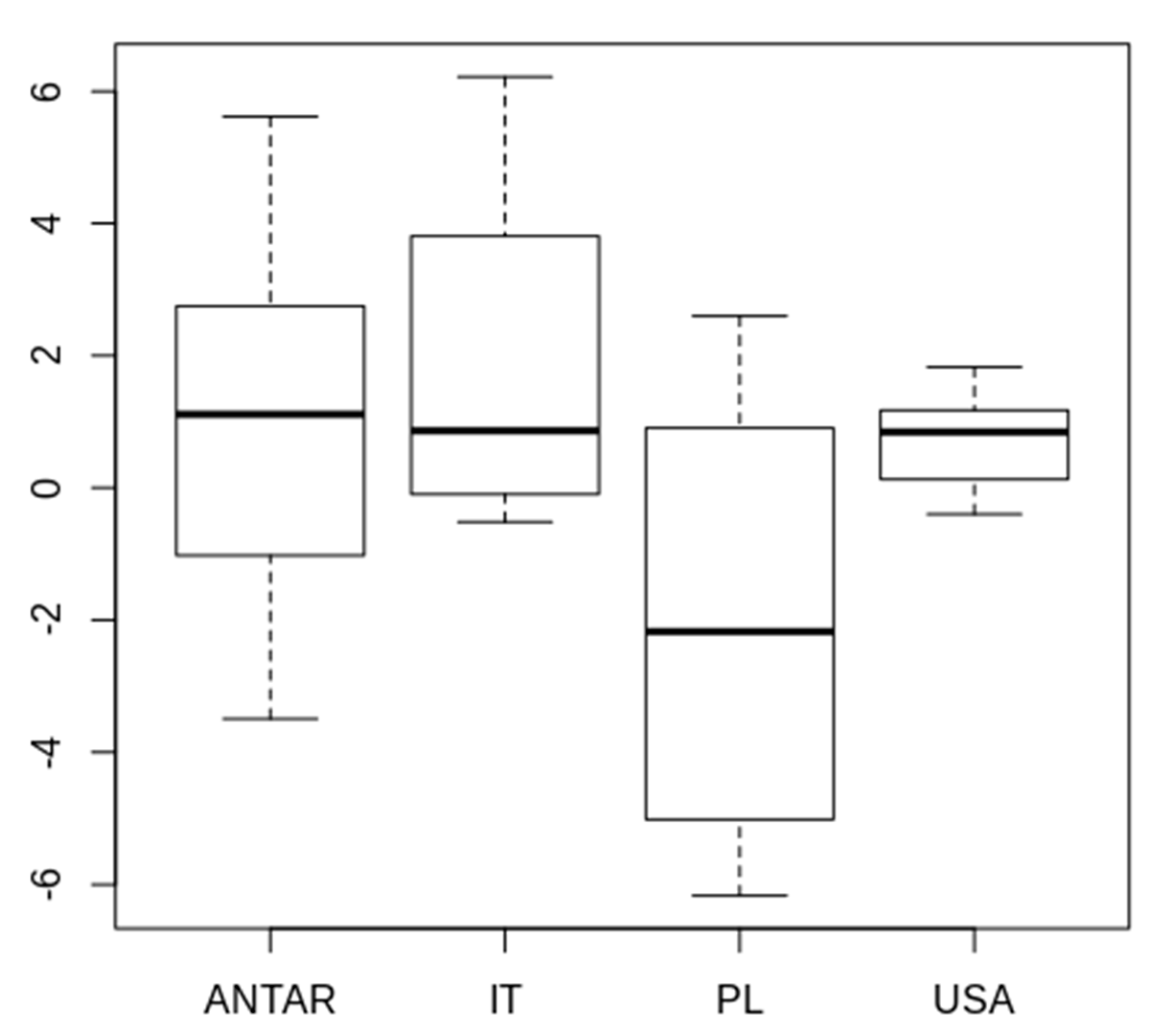
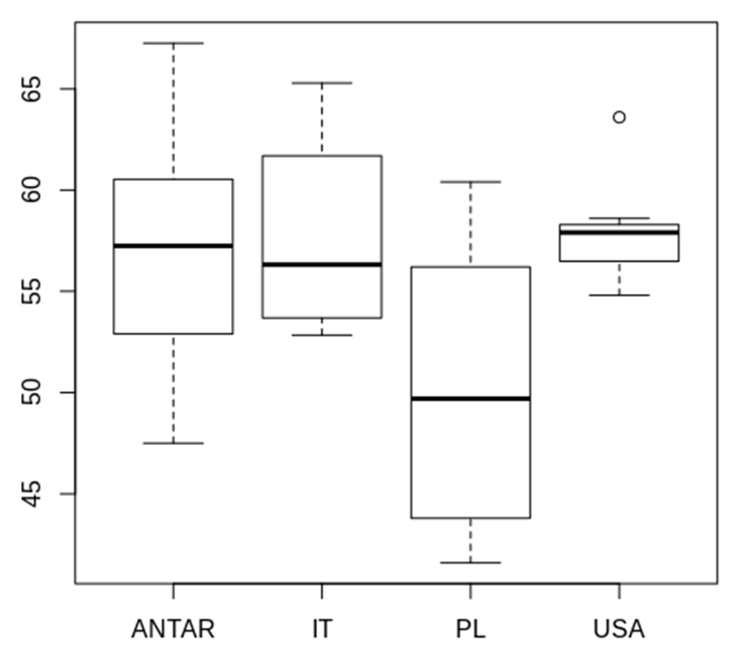
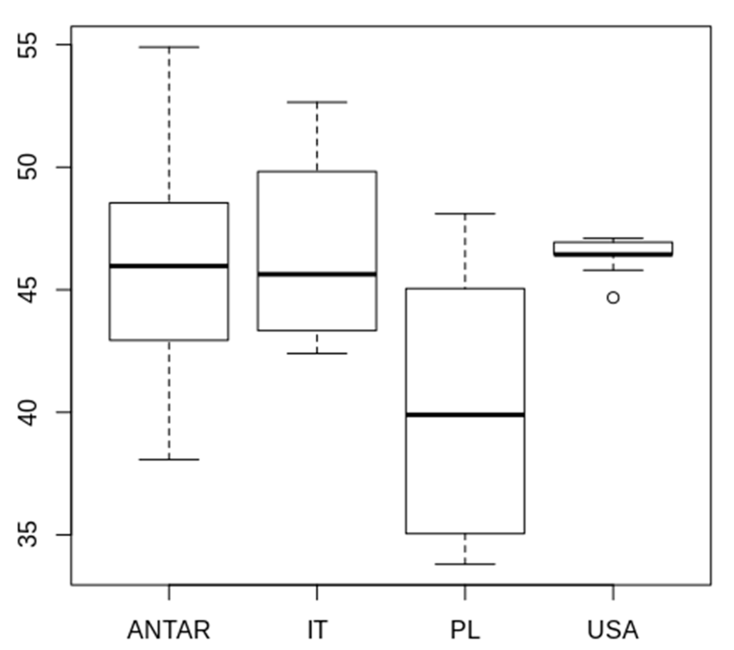
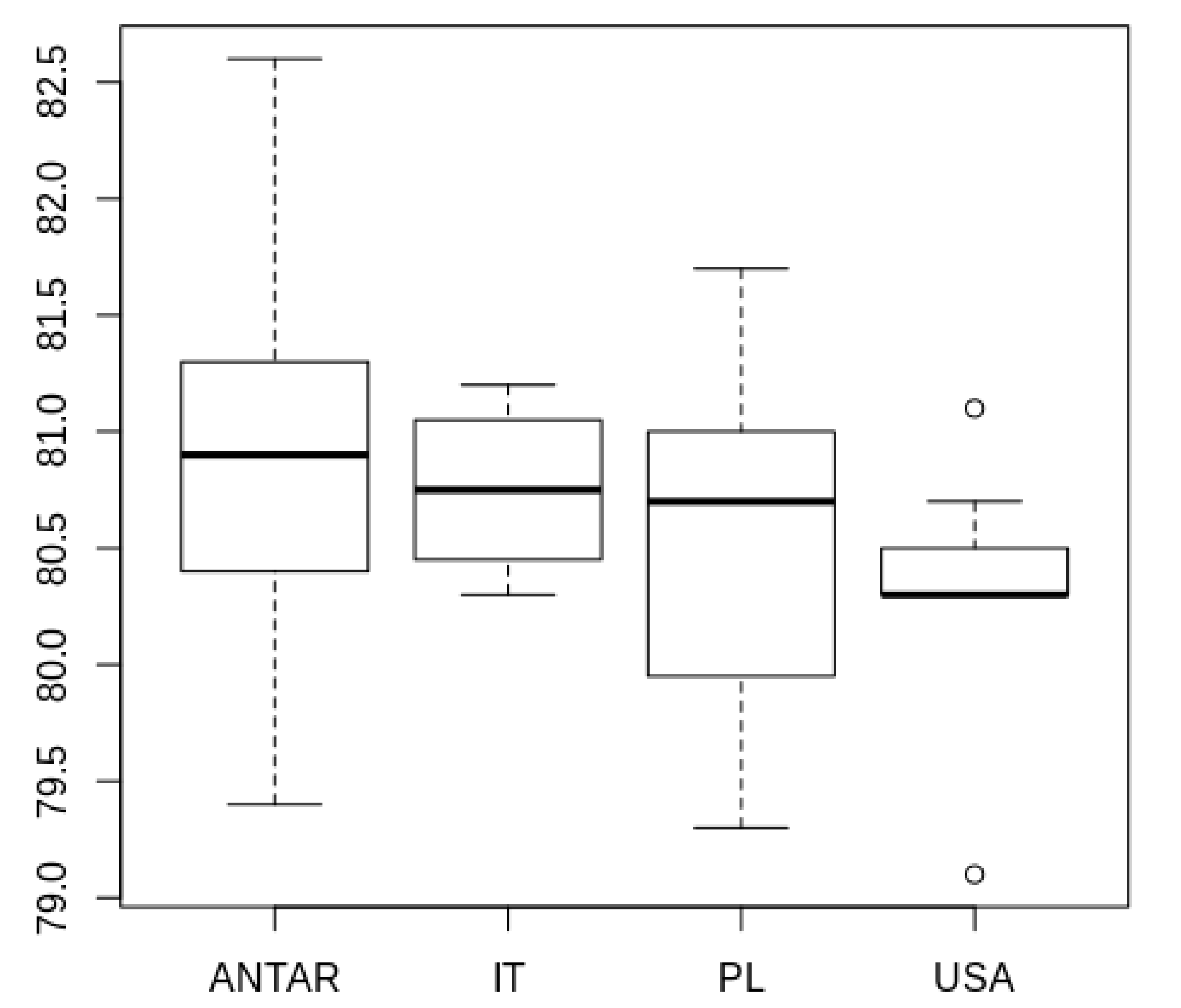
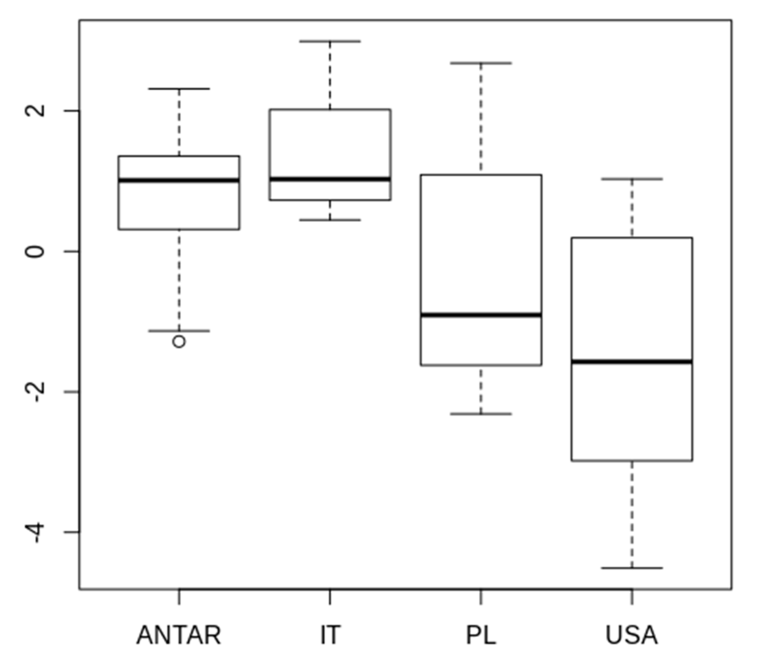
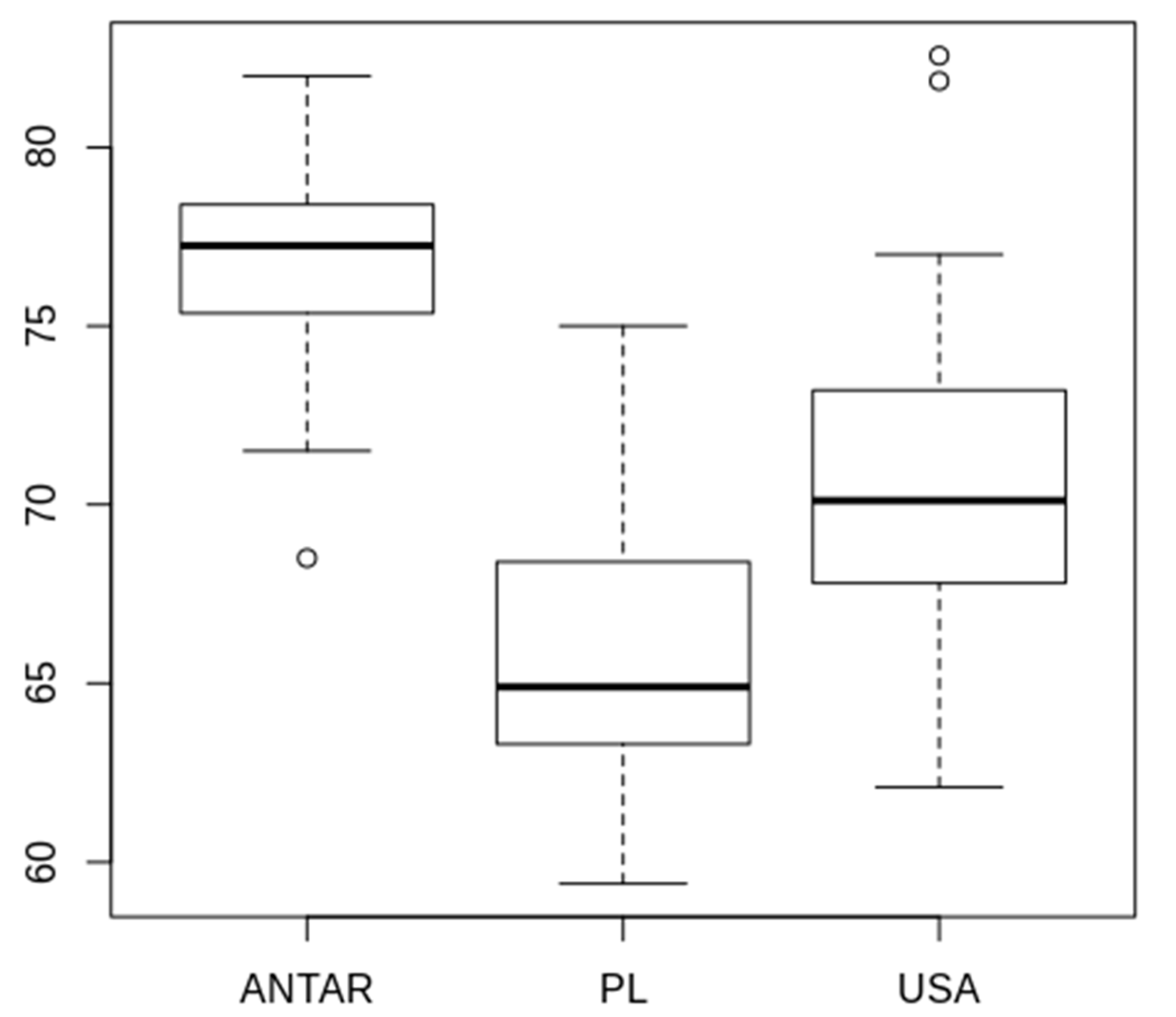
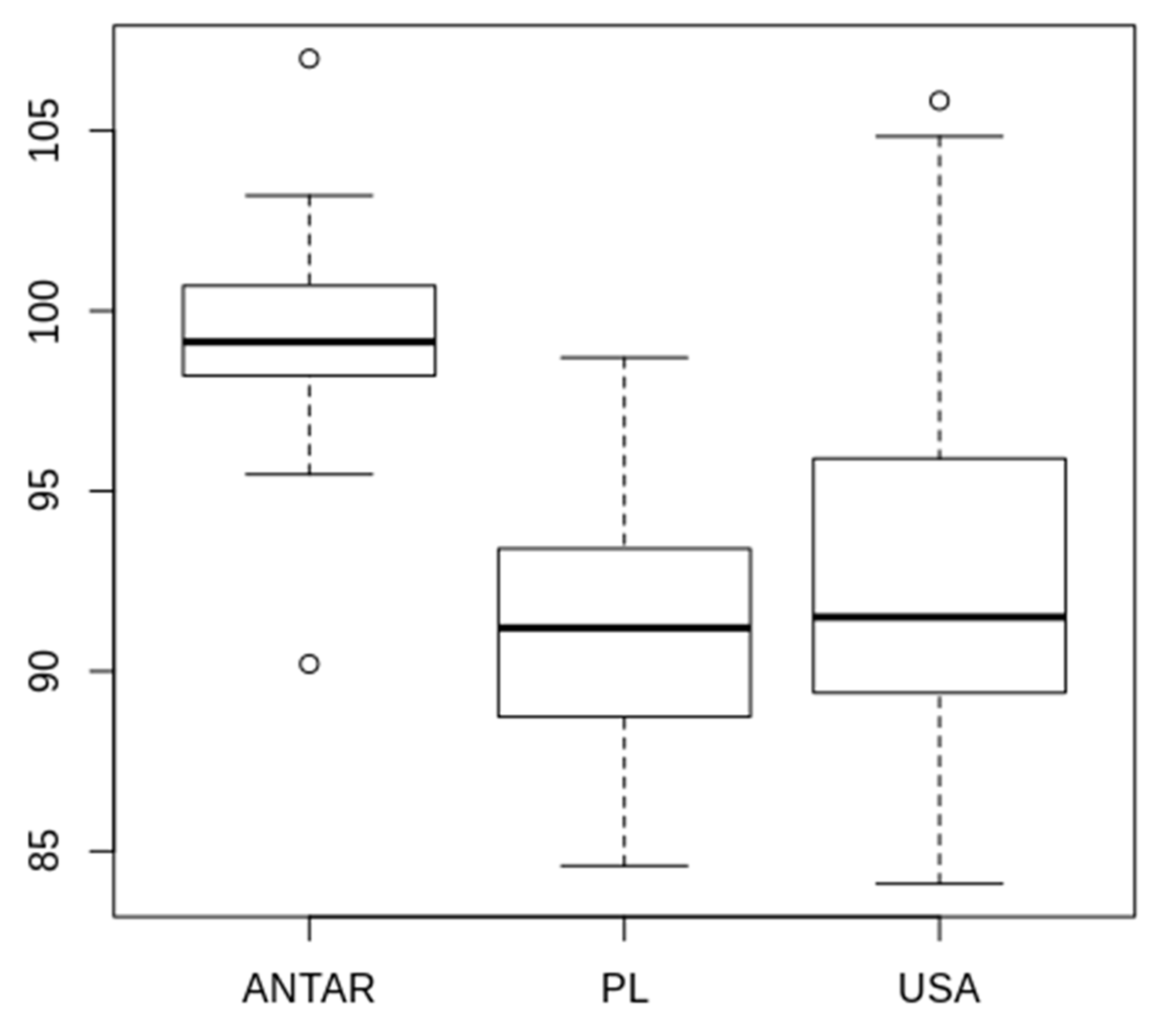
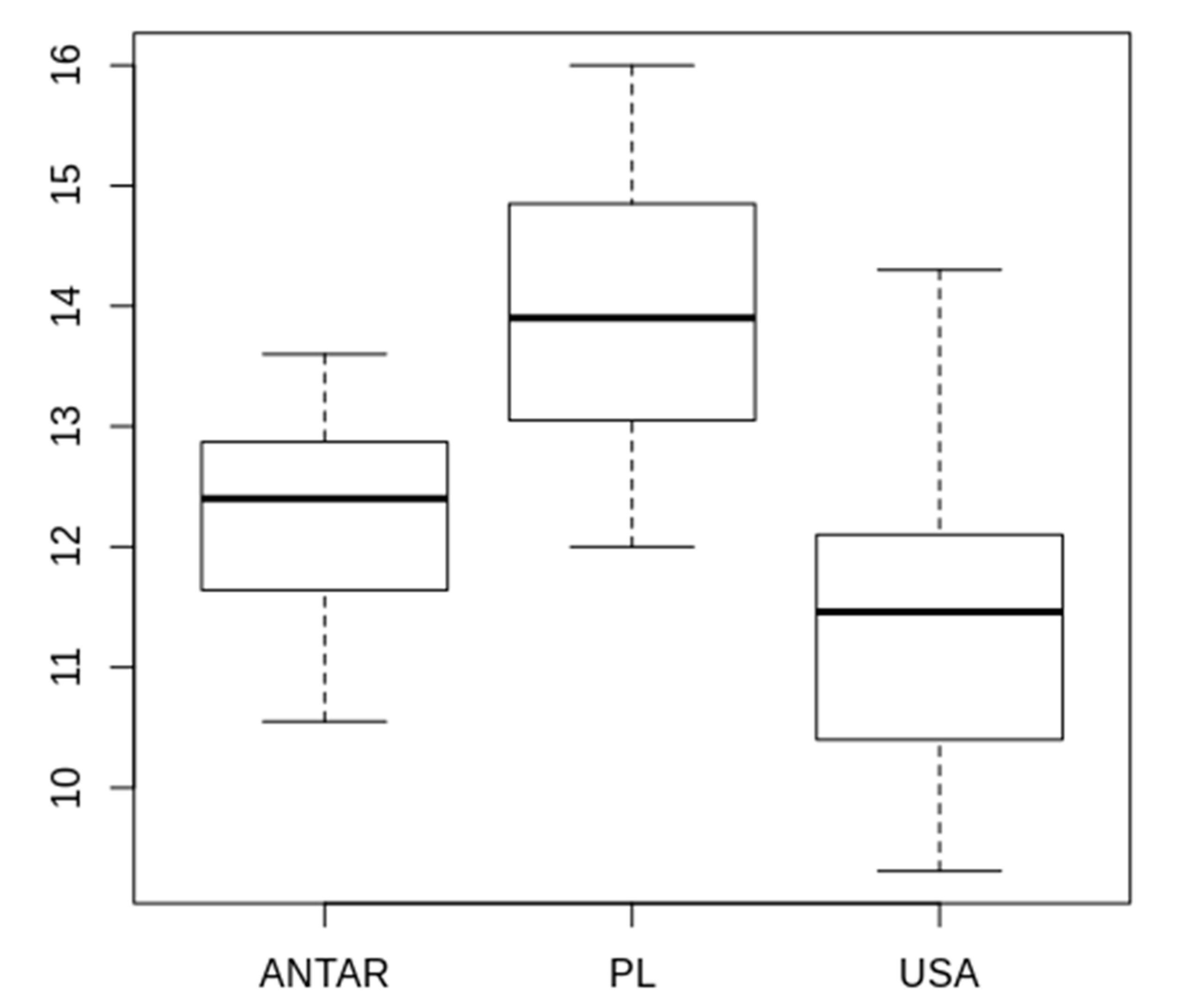
3.2. Morphometric Comparison of Different Populations of the Pam. fairbanksi

Significant differences in BL between the studied populations (Table 3, Table 4, Table 5, Table 6, Table 7, Table 8, Table 9 and Table 10 and Figure 4, Figure 5, Figure 6, Figure 7, Figure 8, Figure 9, Figure 10, Figure 11 and Figure 12) were found (df = 3, F = 4.184, p = 0.0115, n = 44), with specimens from Antarctic being significantly longer than the ones from Poland (p = 0.013, Figure 4). Similarly, the model with the first PCA component from the measurements of all the studied morphological characters as the dependent variable (df = 3, F = 4.721; p = 0.0065; n = 44) showed differences between Polish and Antarctic (p = 0.013) populations (Figure 5). ANOVA performed for BTL (df = 3, F = 5.741, p = 0.0023, n = 44) also showed differences between Polish and Antarctic (p = 0.072), as well as Polish and Alaskan (p = 0.0115) populations (Figure 6). Analysis for SSIP lengths (df = 3, F = 6.156, p = 0.0015, n = 44) showed the same tendency; SSIP in Polish populations was significantly smaller than in Antarctic (p = 0.0037) and Alaskan (p = 0.0126) (Figure 7).
The pt of BL also differed between the populations (df = 3, F = 5.371, p = 0.0034, n = 44), with specimens from Alaska having significantly lower pt of BL than those from Antarctic (p = 0.0029, Figure 8). Similar differences (p = 0.0021) between the two populations were found when first component of PCA for pt of all the measurements was analyzed (df = 3, F = 6.332, p = 0.0013, n = 44), with additional significant differences between Alaskan and Italian populations (p = 0.019, Figure 9). ANOVA performed for pt of SSIP showed no significant differences between the populations (df = 3, F = 1.196, p = 0.324, n = 44).
The ANOVA performed on EBD measurements of eggs (df = 2, F = 18.72, p < 0.0001, n = 49) showed significant differences between all the populations, where the eggs in Polish population were significantly smaller than these from Alaska (p = 0.0197) and Antarctica (p < 0.0001) and the eggs from Alaskan population clearly smaller than those from Antarctic (p = 0.0033) (Figure 10). Analysis on EFD values, however, showed no differences between measurements of Polish and Alaskan eggs (p = 1, n = 49, Figure 11), but eggs in Antarctic population were significantly bigger than these collected in Alaska (p = 0.0008) and Poland (p = 0.0002), the model was significant at p < 0.0001 (df = 2, F= 11.72). The differences between the studied populations were also visible in the size of PH (df = 2, F = 18.91, p < 0.0001, n = 44). Egg processes were longest in Polish population in comparison to both Antarctic (p = 0.0009) and Alaskan (p < 0.0001) populations. There was no significant difference between the length of egg processes in Alaskan and Antarctic populations (p = 0.1252, n = 49, Figure 12).

3.3. Genetic Comparison of Different Populations of the Pam. fairbanksi

All DNA barcode sequences of Pam. fairbanksi from Antarctic were obtained from DNA extracted from the eggs. In DNA extracted from adult individuals, signs of degradation were observed; however, newly designed degenerate primers to amplify COI marker allowed confirmation of molecular species identification. The COI sequences (GenBank accession numbers: MN964281-MN964282) were 639 bp long (using HCO2198 and LCO1490 primers and 392 bp long using designed ParCOIF and ParCOIR primers) and represented only one haplotype. No insertions, deletions, or stop codons were identified. The translation was successfully carried out with the invertebrate mitochondrial codon table and the –2th reading frame. The p-distances between COI haplotypes of all sequenced Pam. fairbanksi populations from Antarctica, Italy, Spain, USA (Alaska), and Poland ranged from 0.002% to 0.005% (an average distance of 0.003%). In case of COI polymorphism analyses, only one haplotype was observed in populations from Antarctica, USA (Alaska), and Poland, two haplotypes were found in population from Spain, and six haplotypes were observed in population from Italy (for details see Table 11).
The 18S rRNA sequences (GenBank accession numbers: MN960302-MN960304) were 1717 to 1725 bp long and one nucleotide substitution was found. Comparing with available in GenBank sequences of Pam. fairbanksi (from Italy and Alaska, alignment sequences were trimmed to 822 bp, see [28]) also showed one nucleotide substitution.
The 28S rRNA sequences (GenBank accession numbers: MN960306-MN960307) were 796 to 800 bp long and one nucleotide substitution was found. We obtained 28S rRNA sequences for Pam. fairbanksi for the very first time, so that no sequences of this species were available for comparison.

4. Discussion

4.1. Taxa

Most of species found in the present study are endemic to the Antarctic region. Barbaria pseudowendti, Das. improvisus, and Mes. blocki are known only from Enderby Land, East Antarctica [12]. Acutuncus antarcticus is a pan-Antarctic species, however, as it is a parthenogenetic species some populations are isolated and show quite large genetic distance which can suggest a process of speciation [5].
Hebesuncus mollispinus has quite recently been described as a species from Charcot Island, close to Antarctic Peninsula (West Antarctica) [15]. It was also reported from Alamode and Terra Firma Islands (close to Antarctic Peninsula), South Georgia (sub-Antarctica), and Argentinian Andes [15,66]. The present report is the first in East Antarctica. Such geographical range of this species suggests their wide distribution in the Antarctic region and also inhabiting glacier regions in South America.
The taxonomic status of Mil. quadrifidum is unclear, especially that their original description is very scanty [51] and lacks a detailed description of animal morphology. In the past it was synonymized with nominal Mil. tar. tardigradum Doyère [41], but recently Morek et al. [67] proposed it as a valid species being the only member of the genus Milnesium with four points on all secondary branches of claws. However, the same authors also suggested that this species needs a detailed redescription. Only recently, Kaczmarek et al. [68] described another species with four points on secondary branches of claws IV, i.e., Mil. wrightae Kaczmarek, Grobys, Kulpa, Bartylak, Kmita, Kepel, Kepel and Roszkowska [68]. Dastych [52] reported Mil. tar. tardigradum from King George Island and Enderby Land with many claw aberrations (i.e., different number of points on secondary branches of claws, even in the same animal). As the number of points on claws is rather constant (but may change during ontogenesis) and species-specific character [67,69,70], it should be considered that specimens of Mil. tar. tardigradum reported by Dastych [52] could possibly belong to a different species. In this situation it is impossible to determine a detailed morphology of Mil. quadrifidum or their distribution. The specimens found in the present study correspond to the original description of Mil. quadrifidum, but taking into consideration all the taxonomic problems addressed for this species, their presence in Antarctica should be confirmed after detailed redescription of this species from the type locality.
Paramacrobiotus fairbanksi was described from Alaska (USA) and later reported from Italy, Poland and Spain [25,26,27,28], and now we have provided the next record from Antarctica. This species perfectly fits the model taxon for the “everything is everywhere” hypothesis, i.e., is microscopic, easy to disperse, is able to cryptobiosis and it is parthenogenetic. However, before the final conclusion that Pam. fairbanksi is a true cosmopolitan species, its presence should also be confirmed on other continents.
Due to the lack of eggs, determination of the specimens of the genus Mesobiotus from two samples was not possible. It is of course highly probable that these specimens belong to Mes. blocki which was also found in the present study.

4.2. Morphometric Comparison of Different Populations of the Pam. fairbanksi

It is clearly visible in Table 3, Table 4, Table 5, Table 6, Table 7, Table 8, Table 9 and Table 10 and Figure 1, Figure 2, Figure 3, Figure 4, Figure 5, Figure 6, Figure 7, Figure 8 and Figure 9 and supported by the statistical analyses that some small morphometric differences are present between the populations of Pam. fairbanksi from different regions. However, measurements of all structures generally overlap which rather clearly allows the identification of this species even without genetic markers. This suggests that, based on the morphology, all specimens, even collected in different regions, should be correctly classified if a proper number of specimens is examined. Nonetheless, specimens from the Polish population are the smallest and lay the smallest eggs, equipped, however, with the longest egg processes. All observations and measurements of the Polish and Alaskan population of Pam. fairbanksi are based on cultured specimens, whereas in other cases specimens were extracted from the wild. In general, in both cultured populations, the length of the body is the smallest which could be caused by the well-known phenomenon when cultured populations of various animal species are dwarfing as a result of suboptimal conditions, high culture densities, and inbreeding. In addition, the measurements of other morphological structures of adults in Polish population are also the smallest, whereas, in contrast, in Alaska they are similar to those from Antarctic and Italy. Therefore, the observed dissimilarities could well be a sign of conserved differences in the morphology of the populations as a result of ongoing speciation.
It is also visible that the eggs of the Antarctic population are clearly bigger and have rather small egg processes. At present, we suggest that the harsh conditions in Antarctica favor laying larger eggs, while in cultures the eggs are smaller because of the lack of such selective pressure. In contrast, it is quite strange that the number of processes on the egg circumference in the Alaskan population is higher (10 to 20 processes, based on data from the original description) than in other populations (11 to 12 processes, based on the own observations). However, this could be an artefact caused by differences in the methodology used. Summarizing, at present we should consider this species as effectively morphologically defined with small interpopulation differences.

4.3. Genetic Comparison of Different Populations of the Pam. fairbanksi

Paramacrobiotus fairbanksi has a wide distribution and until now was observed in Italia, Spain [26], Poland [27], and Alaska [25]. From each locality, COI sequences of Pams fairbanksi are available. In the case of nucleotide markers, the 18S rRNA sequences have been obtained from Italia, Alaska, and presented in our study from Antarctica. In turn, the 28S rRNA sequences have been obtained only from our population from Antarctica. As examined in this study, Pam. fairbanksi population from Antarctica is the most similar to the population from Poland, with the same COI haplotype (Figure 13). Molecular analysis based on 18S rRNA sequences indicated no differences between our population from Antarctica and populations from Italy, whereas those based on COI sequences revealed low genetic distances, i.e., 0.002 to 0.003. The genetic distances, based on COI markers among Pam. fairbanksi from Antarctica and Alaska was 0.003, and one nucleotide substitution was indicated in 18S rRNA sequences. On the basis of this molecular data for populations from very distant regions, with physical barriers to gene flow, we showed the low evolutionary rate of Pam. fairbanksi.

Author Contributions

Conceptualization, Ł.K., M.M., B.G., M.R.; Methodology, Ł.K., M.M., B.G.; Software, Ł.K., M.M., B.G.; Validation, Ł.K., M.M., U.K., K.K., I.P., B.G., M.R.; Formal analysis, Ł.K.; Investigation, Ł.K., M.M., U.K., K.K., I.P., B.G., M.R.; Resources: Ł.K., M.M., I.P.; Data curation, Ł.K.; Writing—original draft preparation, Ł.K., M.M., B.G., M.R.; Writing—review and editing, Ł.K., M.M., U.K., K.K., I.P., B.G., M.R.; Visualization: B.G., M.R. Supervision, Ł.K.; Project administration, Ł.K.; Funding acquisition, Ł.K.; All authors have read and agreed to the published version of the manuscript.

Funding

The study was supported by the research grant of the National Science Centre, Poland, NCN 2016/21/B/NZ4/00131.

Acknowledgments

The isogenic strain of Pam. fairbanksi (Pam.fai_I_PL.018) was kindly provided by the Michalczyk Lab (Jagiellonian University, Kraków, Poland). The strain was established from a female isolated from a moss sample collected by Piotr Gąsiorek and has been maintained by Daniel Stec. We are also grateful to Robert Sobkowiak (Adam Mickiewicz University, Poznań, Poland) and Edyta Fiałkowska (Jagiellonian University, Kraków, Poland) for providing nematodes and rotifers as a food for the tardigrades.

Conflicts of Interest

The authors declare no conflict of interest.

References

  1. Kimble, J.M. Cryosols. Permafrost-Affected Soils; Springer: Berlin/Heidelberg, Germany, 2004; pp. 1–94. [Google Scholar]
  2. Velasco-Castrillón, A.; Gibson, J.A.E.; Stevens, M.I. A review of current Antarctic limno-terrestrial microfauna. Polar Biol. 2014, 37, 1517–1531. [Google Scholar]
  3. Richters, F. Vorläufiger Bericht über die antarktische Moosfauna. Verh. Dtsch. Zool. Ges. 1904, 14, 236–239. [Google Scholar]
  4. Pugh, P.J.A.; Convey, P. Surviving out in the cold: Antarctic endemic invertebrates and their refugia. J. Biogeogr. 2008, 35, 2176–2186. [Google Scholar] [CrossRef]
  5. Cesari, M.; McInnes, S.; Bertolani, R.; Rebecchi, L.; Guidetti, R. Genetic diversity and biogeography of the south polar water bear Acutuncus antarcticus (Eutardigrada: Hypsibiidae)—Evidence that it is a truly pan-Antarctic species. Invertebr. Syst. 2016, 30, 635–649. [Google Scholar] [CrossRef]
  6. Guidetti, R.; Bertolani, R. Tardigrade taxonomy: An updated check list of the taxa and a list of characters for their identification. Zootaxa 2005, 845, 1–46. [Google Scholar] [CrossRef]
  7. Degma, P.; Guidetti, R. Notes to the current checklist of Tardigrada. Zootaxa 2007, 1579, 41–53. [Google Scholar] [CrossRef]
  8. Vicente, F.; Bertolani, R. Considerations on the taxonomy of the Phylum Tardigrada. Zootaxa 2013, 3626, 245–248. [Google Scholar] [CrossRef]
  9. Degma, P.; Bertolani, R.; Guidetti, R. Actual Checklist of Tardigrada Species; Version 35: Edition: 31-07-2019; Università di Modena e Reggio Emilia: Modena, Italy, 2019. [Google Scholar] [CrossRef]
  10. Ramazzotti, G.; Maucci, W. Il Phylum Tardigrada. III edizione riveduta e aggiornata. Mem. Ist. Ital. Idrobiol. 1983, 41, 1–1012. [Google Scholar]
  11. Nelson, D.R.; Guidetti, R.; Rebecchi, L. Phylum Tardigrada. In Ecology and General Biology. Vol. 1. Thorp and Covich’s Freshwater Invertebrates; Throp, J.H., Rogers, D.C., Eds.; Academic Press: Cambridge, MA, USA, 2015; pp. 347–380. [Google Scholar] [CrossRef]
  12. Convey, P.; McInnes, S.J. Exceptional tardigrade-dominated ecosystems in Ellsworth Land, Antarctica. Ecology 2005, 86, 519–527. [Google Scholar] [CrossRef]
  13. Binda, M.G.; Pilato, G.; Lisi, O. Remarks on Macrobiotus furciger Murray, 1906 and description of three new species of the furciger group (Eutardigrada, Macrobiotidae). Zootaxa 2005, 1075, 55–68. [Google Scholar] [CrossRef]
  14. McInnes, S.J. Echiniscus corrugicaudatus (Heterotardigrada; Echiniscidae) a new species from Ellsworth Land, Antarctica. Polar Biol. 2010, 33, 59–70. [Google Scholar] [CrossRef]
  15. Pilato, G.; McInnes, S.J.; Lisi, O. Hebesuncus mollispinus (Eutardigrada, Hypsibiidae), a new species from maritime Antarctica. Zootaxa 2012, 3446, 60–68. [Google Scholar] [CrossRef]
  16. Guidetti, R.; Rebecchi, L.; Cesari, M.; McInnes, S.J. Mopsechiniscus franciscae, a new species of a rare genus of Tardigrada from continental Antarctica. Polar Biol. 2014, 37, 1221–1233. [Google Scholar] [CrossRef]
  17. Kaczmarek, Ł.; Janko, K.; Smykla, J.; Michalczyk, Ł. Soil tardigrades from the Antarctic Peninsula with a description of a new species and some remarks on the genus Ramajendas (Eutardigrada, Isohypsibiidae). Polar Rec. 2014, 50, 176–182. [Google Scholar] [CrossRef]
  18. Tsujimoto, M.; McInnes, S.J.; Convey, P.; Imura, S. Preliminary description of tardigrade species diversity and distribution pattern around coastal Syowa Station and inland Sør Rondane Mountains, Dronning Maud Land, East Antarctica. Polar Biol. 2014, 37, 1361–1367. [Google Scholar] [CrossRef] [Green Version]
  19. Vecchi, M.; Cesari, M.; Bertolani, R.; Jönsson, K.I.; Rebecchi, L.; Guidetti, R. Integrative systematic studies on tardigrades from Antarctica identify new genera and new species within Macrobiotoidea and Echiniscoidea. Invertebr. Syst. 2016, 30, 303–322. [Google Scholar] [CrossRef]
  20. Guidetti, R.; McInnes, S.J.; Cesari, M.; Rebecchi, L.; Rota-Stabelli, O. Evolutionary scenarios for the origin of an Antarctic tardigrade species based on molecular clock analyses and biogeographic data. Contrib. Zool. 2017, 86, 97–110. [Google Scholar] [CrossRef] [Green Version]
  21. Pilato, G.; Sabella, G.; D’Urso, V.; Lisi, O. Two new species of Eutardigrada from Victoria Land, Antarctica. Zootaxa 2017, 4317, 541–558. [Google Scholar] [CrossRef]
  22. Dastych, H. Redescription and revalidation of the sub-Antarctic tardigrade Hypsibius murrayi (Richters, 1907) based on the rediscovered type material (Tardigrada, Panarthropoda). Entomol. Heute 2018, 30, 95–115. [Google Scholar]
  23. Kaczmarek, Ł.; Parnikoza, I.; Gawlak, M.; Esefeld, J.; Peter, H.-U.; Kozeretska, I.; Roszkowska, M. Tardigrades from Larus dominicanus Lichtenstein, 1823 nests on the Argentine Islands (maritime Antarctic). Polar Biol. 2018, 41, 283–301. [Google Scholar] [CrossRef] [Green Version]
  24. Guidetti, R.; Massa, E.; Bertolani, R.; Rebecchi, L.; Cesari, M. Increasing knowledge of Antarctic biodiversity: New endemic taxa of tardigrades (Eutardigrada; Ramazzottiidae) and their evolutionary relationships. Syst. Biodivers. 2019, 17, 573–593. [Google Scholar] [CrossRef]
  25. Schill, R.O.; Forster, F.; Dandekar, T.; Wolf, N. Using compensatory base change analysis of internal transcribed spacer 2 secondary structures to identify three new species in Paramacrobiotus (Tardigrada). Org. Divers. Evol. 2010, 10, 287–296. [Google Scholar] [CrossRef]
  26. Guil, N.; Giribet, G. A comprehensive molecular phylogeny of tardigrades—Adding genes and taxa to a poorly resolved phylum-level phylogeny. Cladistics 2012, 28, 21–49. [Google Scholar] [CrossRef]
  27. Kosztyła, P.; Stec, D.; Morek, W.; Gąsiorek, P.; Zawierucha, Z.; Michno, K.; Małek, D.; Hlebowicz, K.; Ufir, K.; Laska, A.; et al. Experimental taxonomy confirms the environmental stability of morphometric traits in a taxonomically challenging group of microinvertebrates. Zool. J. Linn. Soc. 2016, 178, 765–775. [Google Scholar]
  28. Guidetti, R.; Cesari, M.; Bertolani, R.; Altiero, T.; Rebecchi, L. High diversity in species, reproductive modes and distribution within the Paramacrobiotus richtersi complex (Eutardigrada, Macrobiotidae). Zool. Lett. 2019, 5, 1. [Google Scholar] [CrossRef]
  29. Guidetti, R.; Gandolfi, A.; Rossi, V.; Bertolani, R. Phylogenetic analysis in Macrobiotidae (Eutardigrada, Parachela): A combined morphological and molecular approach. Zool. Scr. 2005, 34, 235–244. [Google Scholar] [CrossRef]
  30. Murray, J. Arctiscoida. Proc. Roy. Irish Acad. 1911, 31, 1–16. [Google Scholar]
  31. Beijerinck, M.W. De infusies en de ontdekking der bakterien. Jaarb. Kon Akad. Wetensch. 1913, 1–28. [Google Scholar]
  32. Baas Becking, L.G.M. Geobiologie of Inleiding Tot de Milieukunde; W.P. Van Stockum & Zoon N.V: Den Haag, The Netherlands, 1934; pp. 1–263. [Google Scholar]
  33. Cardillo, M.; Bromhan, L. Body size and risk of extinction in Australian mammals. Conserv. Biol. 2001, 15, 1435–1440. [Google Scholar] [CrossRef]
  34. Finlay, B.J. Global dispersal of free living microbial eukaryote species. Science 2002, 296, 1061–1063. [Google Scholar] [CrossRef] [Green Version]
  35. Foissner, W. Biogeography and dispersal of micro-organisms: A review emphasizing protists. Acta Protozool. 2006, 45, 111–136. [Google Scholar]
  36. Schön, I.; Martens, K.; Dijk, P. Lost Sex. The Evolutionary Biology of Parthenogenesis; Springer: Berlin/Heidelberg, Germany, 2009; pp. 1–615. [Google Scholar]
  37. Pilato, G.; Binda, M.G. Biogeography and limno-terrestrial tardigrades: Are they truly incompatible binomials? Zool. Anz. 2001, 240, 511–516. [Google Scholar] [CrossRef]
  38. Guil, N.; Sánchez-Moreno, S.; Machordom, A. Local biodiversity patterns in micrometazoans: Are tardigrades everywhere? Syst. Biodivers. 2009, 7, 259–268. [Google Scholar] [CrossRef]
  39. Faurby, S.; Jorgensen, A.; Kristensen, R.M.; Funch, P. Distribution and speciation in marine intertidal tardigrades: Testing the roles of climatic and geographical isolation. J. Biogeogr. 2012, 39, 1596–1607. [Google Scholar] [CrossRef]
  40. Gąsiorek, P.; Vončina, K.; Michalczyk, Ł. Echiniscus testudo (Doyère, 1840) in New Zealand: Anthropogenic dispersal or evidence for the ‘Everything is Everywhere’ hypothesis? N. Z. J. Zool. 2019, 46, 174–181. [Google Scholar]
  41. Doyère, P.L.N. Memoire sur les Tardigrades. Ann. Sci. Nat. Zool. Paris Ser. 2 1840, 14, 269–362. [Google Scholar]
  42. Stec, D.; Smolak, R.; Kaczmarek, Ł.; Michalczyk, Ł. An integrative description of Macrobiotus paulinae sp. nov. (Tardigrada: Eutardigrada: Macrobiotidae: Hufelandi group) from Kenya. Zootaxa 2015, 4052, 501–526. [Google Scholar] [CrossRef] [Green Version]
  43. Beasley, C.W. The Phylum Tardigrada, 3rd ed.; Ramazzotti, G., Maucci, W., Eds.; English Translation; Clark Beasley: Abilene, TX, USA, 1995; pp. 1–1014. [Google Scholar]
  44. Pilato, G.; Binda, M.G. Definition of families, subfamilies, genera and subgenera of the Eutardigrada, and keys to their identification. Zootaxa 2010, 2404, 1–52. [Google Scholar] [CrossRef] [Green Version]
  45. Kaczmarek, Ł.; Michalczyk, Ł. The Macrobiotus hufelandi group (Tardigrada) revisited. Zootaxa 2017, 4363, 101–123. [Google Scholar] [CrossRef]
  46. Kaczmarek, Ł.; Cytan, J.; Zawierucha, K.; Diduszko, D.; Michalczyk, Ł. Tardigrades from Peru (South America), with descriptions of three new species of Parachela. Zootaxa 2014, 3790, 357–379. [Google Scholar] [CrossRef] [Green Version]
  47. Pilato, G. Analisi di nuovi caratteri nello studio degli Eutardigradi. Animalia 1981, 8, 51–57. [Google Scholar]
  48. Michalczyk, Ł.; Kaczmarek, Ł. The Tardigrada Register: A comprehensive online data repository for tardigrade taxonomy. J. Limnol. 2013, 72, 175–181. [Google Scholar] [CrossRef]
  49. Bertolani, R.; Guidetti, R.; Marchioro, T.; Altiero, T.; Rebechci, L.; Cesari, M. Phylogeny of Eutardigrada: New molecular data and their morphological support lead to the identification of new evolutionary lineages. Mol. Phylogenetics Evol. 2014, 76, 110–126. [Google Scholar] [CrossRef] [PubMed]
  50. Perry, E.; Miller, W.R.; Kaczmarek, Ł. Recommended abbreviations for the names of genera of the phylum Tardigrada. Zootaxa 2019, 4608, 145–154. [Google Scholar] [CrossRef] [PubMed]
  51. Nederström, P. Die bis jetzt aus Finland bekannten Tardigraden. Acta Soc. Fauna Flora Fenn. 1919, 46, 1–25. [Google Scholar]
  52. Dastych, H. The Tardigrada from Antarctica with description of several new species. Acta Zool. Cracov. 1984, 27, 377–436. [Google Scholar]
  53. Pilato, G. The taxonomic value of the structures for the insertion of the stylet muscles in the Eutardigrada, and description of a new genus. Zootaxa 2013, 3721, 365–378. [Google Scholar] [CrossRef] [Green Version]
  54. Pilato, G.; Binda, M.G. Acutuncus, a new genus of Hypsibiidae (Eutardigrada). Entomol. Mitt. Zool. Mus. Hamburg 1997, 12, 159–162. [Google Scholar]
  55. Casquet, J.; Thebaud, C.; Gillespie, R.G. Chelex without boiling, a rapid and easy technique to obtain stable amplifiable DNA from small amounts of ethanol-stored spiders. Mol. Ecol. Resour. 2012, 12, 136–141. [Google Scholar] [CrossRef]
  56. Sands, C.J.; Mcinnes, S.J.; Marley, N.J.; Goodall–Copestake, W.P.; Convey, P.; Linse, K. Phylum Tardigrada: An “individual” approach. Cladistics 2008, 24, 861–871. [Google Scholar] [CrossRef]
  57. Mironov, S.V.; Dabert, J.; Dabert, M. A new feather mite species of the genus Proctophyllodes Robin, 1877 (Astigmata: Proctophyllodidae) from the long–tailed tit Aegithalos caudatus (Passeriformes: Aegithalidae): Morphological description with DNA barcode data. Zootaxa 2012, 3253, 54–61. [Google Scholar] [CrossRef] [Green Version]
  58. Folmer, O.; Black, M.; Hoeh, W.; Lutz, R.; Vrijenhoek, R. DNA primers for amplification of mitochondrial cytochrome c oxidase subunit I from diverse metazoan invertebrates. Mol. Mar. Biol. Biotechnol. 1994, 3, 294–299. [Google Scholar] [PubMed]
  59. Untergasser, A.; Cutcutache, I.; Koressaar, T.; Ye, J.; Faircloth, B.C.; Remm, M.; Rozen, S.G. 2012. Primer3—New capabilities and interfaces. Nucleic Acids Res. 2012, 40, e115. [Google Scholar] [CrossRef] [PubMed] [Green Version]
  60. Altschul, S.F.; Gish, W.; Miller, W.; Myers, E.W.; Lipman, D.J. Basic local alignment search tool. J. Mol. Biol. 1990, 215, 403–410. [Google Scholar] [CrossRef]
  61. Thompson, J.D.; Higgins, D.G.; Gibson, T.J. CLUSTAL W: Improving the sensitivity of progressive multiple sequence alignment through sequence weighting, positionspecific gap penalties and weight matrix choice. Nucleic Acids Res. 1994, 22, 4673–4680. [Google Scholar] [CrossRef] [PubMed] [Green Version]
  62. Hall, T.A. BioEdit: A user-friendly biological sequence alignment editor and analysis program for Windows 95/98/NT. Nucleic Acids Symp. 1999, 41, 95–98. [Google Scholar]
  63. Rice, P.; Longden, I.; Bleasby, A. EMBOSS: The European molecular biology open software suite. Trends Genet. 2000, 16, 276–277. [Google Scholar] [CrossRef]
  64. Goujon, M.; Mcwilliam, H.; Li, W.; Valentin, F.; Squizzato, S.; Paern, J.; Lopez, R. A new bioinformatics analysis tools framework at EMBL–EBI. Nucleic Acids Res. 2010, 38, 695–699. [Google Scholar] [CrossRef]
  65. Librado, P.; Rozas, J. DnaSP v5: A software for comprehensive analysis of DNA polymorphism data. Bioinformatics 2009, 25, 1451–1452. [Google Scholar] [CrossRef] [Green Version]
  66. Roszkowska, M.; Stec, D.; Ciobanu, D.A.; Kaczmarek, Ł. (2016) Tardigrades from Nahuel Huapi National Park (Argentina, South America) with descriptions of two new Macrobiotidae species. Zootaxa 2016, 4105, 243–260. [Google Scholar] [CrossRef]
  67. Morek, W.; Gąsiorek, P.; Stec, D.; Blagden, B.; Michalczyk, Ł. Experimental taxonomy exposes ontogenetic variability and elucidates the taxonomic value of claw configuration in Milnesium Doyère, 1840 (Tardigrada: Eutardigrada: Apochela). Contrib. Zool. 2016, 85, 173–200. [Google Scholar] [CrossRef] [Green Version]
  68. Kaczmarek, Ł.; Grobys, D.; Kulpa, A.; Bartylak, T.; Kmita, H.; Kepel, M.; Kepel, A.; Roszkowska, M. Two new species of the genus Milnesium Doyère, 1840 (Tardigrada, Apochela, Milnesiidae) from Madagascar. ZooKeys 2019, 884, 1–22. [Google Scholar] [CrossRef] [PubMed]
  69. Morek, W.; Stec, D.; Gąsiorek, P.; Surmacz, B.; Michalczyk, Ł. Milnesium tardigradum Doyère, 1840: The first integrative study of inter-population variability in a tardigrade species. J. Zool. Syst. Evol. Res. 2019, 57, 1–23. [Google Scholar] [CrossRef] [Green Version]
  70. Surmacz, B.; Morek, W.; Michalczyk, Ł. What if multiple claw configurations are present in a sample: A case study with the description of Milnesium pseudotardigradum sp. nov. with unique developmental variability. Zool. Stud. 2019, 58, 32. [Google Scholar]
Figure 1. Milnesium quadrifidum. (A) Dorso-ventral projection of the entire animal; (B) Pseudopores visible on the dorsal side of the body; (C) Claws II; (D) Claws IV. Scale bars in (μm). All PCM (Phase Contrast Microscopy).
Diversity 12 00108 g001
Figure 2. Paramacrobiotus fairbanksi (Polish population). (A) Dorso-ventral projection of the entire animal; (B) Claws I with smooth lunules; (C) Granulation on leg IV with visible smooth lunulae. Scale bars in (μm). All PCM.
Diversity 12 00108 g002
Figure 3. Paramacrobiotus fairbanksi (Polish population). (A,B) Egg chorion; (C) Egg process morphology; (D) The surface between egg processes with visible areoles. Scale bars in (μm). All PCM.
Diversity 12 00108 g003
Figure 4. Differences in the body length (in micrometres (μm)) between the studied populations of Paramacrobiotus fairbanksi Schill, Förster, Dandekar and Wolf [25] (Antar, Antarctic; IT, Italy; PL, Poland; USA, USA). Minimum, maximum, median, first quartile, and third quartile for each population are presented.
Diversity 12 00108 g004
Figure 5. Differences between the studied populations of Paramacrobiotus fairbanksi Schill, Förster, Dandekar and Wolf [25] expressed by the first component of PCA performed on the measurements of chosen morphological structures (Antar, Antarctic; IT, Italy; PL, Poland; USA, USA). Minimum, maximum, median, first quartile, and third quartile for each population are presented.
Diversity 12 00108 g005
Figure 6. Differences in the buccal tube length (in micrometres (μm)) between the studied populations of Paramacrobiotus fairbanksi Schill, Förster, Dandekar and Wolf [25] (Antar, Antarctic; IT, Italy; PL, Poland; USA, USA). Minimum, maximum, median, first quartile, and third quartile for each population are presented.
Diversity 12 00108 g006
Figure 7. Differences in the stylet supports insertion points (in micrometres (μm)) between the studied populations of Paramacrobiotus fairbanksi Schill, Förster, Dandekar and Wolf [25] (Antar, Antarctic; IT, Italy; PL, Poland; USA, USA). Minimum, maximum, median, first quartile, and third quartile for each population are presented.
Diversity 12 00108 g007
Figure 8. Differences in the pt of stylet supports insertion points ratio between the studied populations of Paramacrobiotus fairbanksi Schill, Förster, Dandekar and Wolf [25] (Antar, Antarctic; IT, Italy; PL, Poland; USA, USA). Minimum, maximum, median, first quartile, and third quartile for each population are presented.
Diversity 12 00108 g008
Figure 9. Differences between the studied populations of Paramacrobiotus fairbanksi Schill, Förster, Dandekar and Wolf [25] expressed by the first component of PCA performed on pt ratios of lengths of the measurements of chosen morphological structures (Antar, Antarctic; IT, Italy; PL, Poland; USA, USA). Minimum, maximum, median, first quartile, and third quartile for each population are presented.
Diversity 12 00108 g009
Figure 10. Differences in the egg bare diameter between the studied populations of Paramacrobiotus fairbanksi Schill, Förster, Dandekar and Wolf [25] (Antar, Antarctic; PL, Poland; USA, USA). Minimum, maximum, median, first quartile, and third quartile for each population are presented.
Diversity 12 00108 g010
Figure 11. Differences in the egg full diameter between the studied populations of Paramacrobiotus fairbanksi Schill, Förster, Dandekar and Wolf [25] (Antar, Antarctic; PL, Poland; USA, USA). Minimum, maximum, median, first quartile, and third quartile for each population are presented.
Diversity 12 00108 g011
Figure 12. Differences in the egg processes height between the studied populations of Paramacrobiotus fairbanksi Schill, Förster, Dandekar and Wolf [25] (Antar, Antarctic; PL, Poland; USA, USA). Minimum, maximum, median, first quartile, and third quartile for each population are presented.
Diversity 12 00108 g012
Figure 13. A World map with indicated haplotypes of Paramacrobiotus fairbanksi Schill, Förster, Dandekar and Wolf [25] found in different localities.
Diversity 12 00108 g013
Table 1. Samples and localities.
NoCoordinatesSample DescriptionExpedition
167°39′S, 46°09′EVechernia Mt Base, lichen Usnea sphacelata.9 BAE (2016–2017)
267°39′S, 46°09′EVechernia Mt Base, lichen Umbilicaria aprina.9 BAE (2016–2017)
369°24′S, 76°24′EProgress Base, lichen Umbilicaria decussata.9 BAE (2016–2017)
467°39′S, 46°09′EVechernia Mt Base, lichen Xanthoria elegans.9 BAE (2016–2017)
567°39′S, 46°09′EVechernia Mt Base, algae-bacterial mat from Nizhne Lake.7 BAE (2014–2015)
670°46′S, 11°49′EVechernia Mt Base, algae-bacterial mat from Nizhne Lake.5 BAE (2012–2013)
767°39′S, 46°09′EVechernia Mt Base, moss Ceratodon purpureus. 7 BAE (2014–2015)
869°24′S, 76°24′EProgress Base, malanized soil surface algae.9 BAE (2016–2017)
969°23′S, 76°23′EProgress Base, algae Parsiola crispa.9 BAE (2016–2017)
1069°22′S, 76°23′EProgress Base, sediment from bottom of the snow collector for fresh-water maker.9 BAE (2016–2017)
1167°39′S, 46°09′EProgress Base, soil with vegetation.9 BAE (2016–2017)
Table 2. Species list with their localities and remarks (exuvia not included; s/s + e, samples no/no specimens + eggs.
Taxons/s + eRemarks
Acu. antarcticus (Richters [3])6/1, 8/59, 9/205Pan-Antarctic species [5]
Bar. pseudowendti (Dastych [52])1/53, 2/3, 4/1Endemic species known only from Antarctica [52]
Das. improvisus (Dastych [52])2/9Species of the monotypic genus endemic for Antarctica This is a second report of this species [52]
Heb. mollispinus Pilato et al. [15]2/50, 4/4, 7/19Species known only from Antarctica and South America [15,66]
Mes. blocki (Dastych [52])7/25 + 68Endemic species known only from Antarctica [52]. This is second report of this species
Mesobiotus sp.3/4, 2/2Undefined species due to lack of eggs in the examined samples
Mil. quadrifidum Nederström [51]2/1Species with unclear taxonomic position [27]
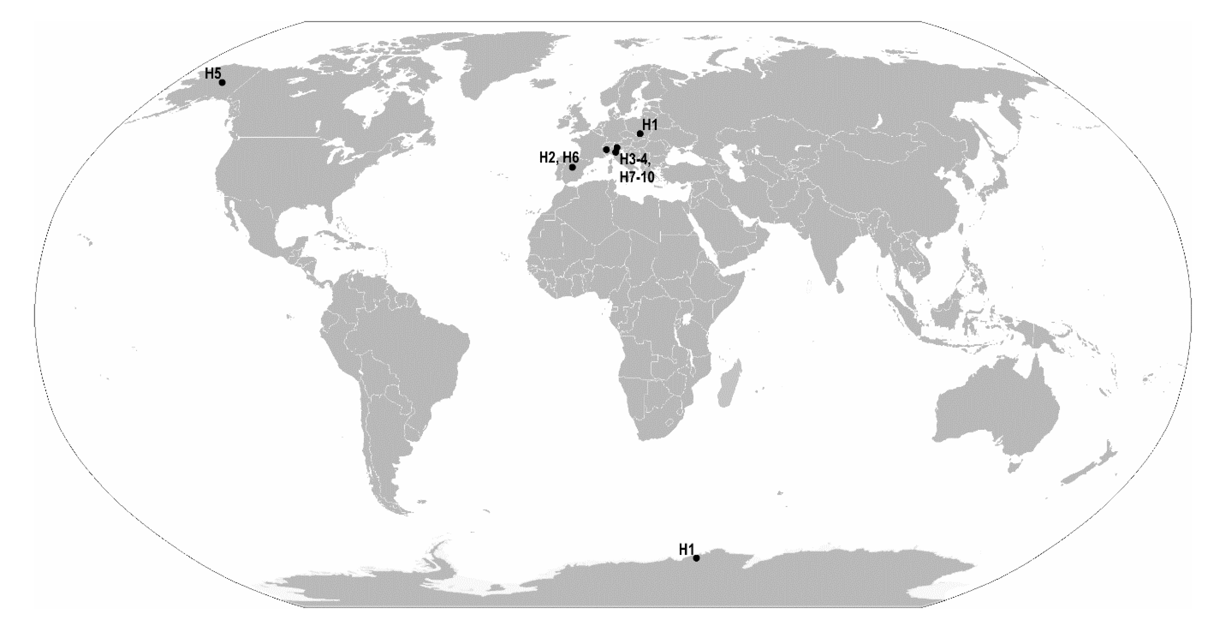
Pam. fairbanksi Schill et al. [25]7/25 + 59Probably cosmopolitan species up to now reported from Italy, Poland, Spain, USA (Alaska; type locality) [25,26,27,28,29]
Table 3. Measurements (in µm) and pt values of selected morphological structures of individuals of the Antarctic population of Paramacrobiotus fairbanksi Schill, Förster, Dandekar and Wolf [25] mounted in Hoyer’s medium (N, number of specimens/structures measured; RANGE refers to the smallest and the largest structure among all measured specimens; SD, standard deviation; pt, ratio of the length of a given structure to the length of the buccal tube expressed as a percentage).
CHARACTERNRANGEMEANSD
µmptµmptµmpt
Body length16420821 595112
Buccopharyngeal tube
Buccal tube length1647.567.3 57.15.4
Stylet support insertion point1638.154.979.482.646.180.94.40.8
Buccal tube external width1610.514.821.822.512.722.21.20.2
Buccal tube internal width167.711.316.018.19.716.91.00.5
Ventral lamina length1629.642.660.464.735.962.93.41.2
Placoid lengths
Macroplacoid 1167.812.116.418.410.017.41.30.7
Macroplacoid 2166.610.113.915.98.615.01.00.6
Macroplacoid 3169.214.019.421.111.720.41.30.5
Microplacoid164.16.98.210.95.18.90.70.6
Macroplacoid row1626.838.754.159.032.857.43.51.1
Placoid row1636.152.671.781.044.577.94.92.0
Claw 1 heights
External primary branch1512.317.625.726.915.026.31.60.5
External secondary branch159.813.920.021.511.820.61.20.4
Internal primary branch1511.916.424.125.514.224.91.40.5
Internal secondary branch159.813.319.321.011.620.31.10.4
Claw 2 heights
External primary branch1513.118.927.029.116.228.31.70.6
External secondary branch159.915.820.623.512.722.31.61.2
Internal primary branch1511.415.822.725.814.024.61.30.9
Internal secondary branch159.813.719.822.111.820.71.10.7
Claw 3 heights
External primary branch1513.318.627.429.116.128.41.60.5
External secondary branch1510.015.221.122.712.421.81.40.4
Internal primary branch1511.716.423.924.713.924.31.40.3
Internal secondary branch159.613.819.121.711.420.01.30.8
Claw 4 heights
Anterior primary branch1314.019.729.030.117.029.51.70.4
Anterior secondary branch1310.414.821.422.712.722.01.20.4
Posterior primary branch1215.220.429.731.017.730.31.50.3
Posterior secondary branch1211.115.421.622.913.022.21.20.3
Table 4. Measurements [in µm] of selected morphological structures of eggs of the Antarctic population of Paramacrobiotus fairbanksi Schill, Förster, Dandekar & Wolf [25] mounted in Hoyer’s medium (N–number of eggs/structures measured, RANGE refers to the smallest and the largest structure among all measured eggs; SD–standard deviation). * except of values of ratio which is presented as percentage and number of processes on the egg circumference which is as number.
CHARACTERNRANGEMEANSD
Egg bare diameter1668.582.076.53.5
Egg full diameter1690.2107.099.13.6
Process height4810.114.512.30.9
Process base width4814.619.816.81.4
* Process base/height ratio48130%146%137%5%
Inter-process distance484.88.16.10.7
* Number of processes on the egg circumference16121212.00.0
Table 5. Measurements (in µm) and pt values of selected morphological structures of individuals of the Polish (cultured) population of Paramacrobiotus fairbanksi Schill, Förster, Dandekar and Wolf [25] mounted in Hoyer’s medium (N, number of specimens/structures measured; RANGE refers to the smallest and the largest structure among all measured specimens; SD, standard deviation; pt, ratio of the length of a given structure to the length of the buccal tube expressed as a percentage).
CHARACTERNRANGEMEANSD
µmptµmptµmpt
Body length15369633 48396
Buccopharyngeal tube
Buccal tube length1541.660.4 50.26.7
Stylet support insertion point1533.848.179.381.740.480.55.30.7
Buccal tube external width158.313.118.422.110.420.61.50.8
Buccal tube internal width156.310.014.016.97.915.71.30.8
Ventral lamina length1526.439.061.665.431.963.64.31.3
Placoid lengths
Macroplacoid 1156.311.315.119.98.617.11.81.4
Macroplacoid 2155.49.412.916.57.514.81.41.1
Macroplacoid 3157.912.518.922.010.220.21.71.0
Microplacoid153.76.08.010.14.69.10.80.6
Macroplacoid row1521.435.351.462.328.356.24.92.8
Placoid row1528.948.069.581.437.975.36.63.8
Claw 1 heights
External primary branch1310.516.824.329.612.926.22.01.5
External secondary branch117.813.118.423.19.720.11.81.5
Internal primary branch1310.316.022.828.212.624.61.81.3
Internal secondary branch118.512.418.321.310.119.91.71.0
Claw 2 heights
External primary branch1211.517.825.131.413.927.52.21.7
External secondary branch118.813.620.823.811.022.11.81.0
Internal primary branch129.815.522.526.512.424.61.81.2
Internal secondary branch108.412.519.521.910.621.01.40.8
Claw 3 heights
External primary branch1311.417.625.331.013.827.61.81.7
External secondary branch128.514.319.025.210.921.81.91.5
Internal primary branch129.815.722.326.512.624.52.01.5
Internal secondary branch118.213.419.523.310.720.81.81.3
Claw 4 heights
Anterior primary branch1111.717.626.830.514.428.71.91.3
Anterior secondary branch109.013.420.523.510.921.81.81.0
Posterior primary branch1312.218.728.732.615.230.22.21.2
Posterior secondary branch129.315.220.726.811.623.21.91.5
Table 6. Measurements (in µm) of selected morphological structures of eggs of the Polish (cultured) population of Paramacrobiotus fairbanksi Schill, Förster, Dandekar and Wolf [25] mounted in Hoyer’s medium (N, number of eggs/structures measured; RANGE refers to the smallest and the largest structure among all measured eggs; SD, standard deviation). * except of values of ratio which is presented as percentage and number of processes on the egg circumference which is as number.
CHARACTERNRANGEMEANSD
Egg bare diameter1559.475.066.24.9
Egg full diameter1584.698.791.44.3
Process height4511.516.513.91.3
Process base width4514.319.216.41.1
* Process base/height ratio45100%136%118%11%
Inter-process distance453.88.15.50.8
* Number of processes on the egg circumference15111211.50.5
Table 7. Measurements (in µm) and pt values of selected morphological structures of individuals of the American (Alaskan) population of Paramacrobiotus fairbanksi Schill, Förster, Dandekar and Wolf [25] mounted in Hoyer’s medium (N, number of specimens/structures measured; RANGE refers to the smallest and the largest structure among all measured specimens; SD, standard deviation, pt, ratio of the length of a given structure to the length of the buccal tube expressed as a percentage). * measurement based partially on Schill et al. [25].
CHARACTERNRANGEMEANSDHolotype
µmptµmptµmptµmpt
*Body length9442591 51351474
Buccopharyngeal tube
*Buccal tube length954.863.6 57.92.556.4
Stylet support insertion point544.747.179.181.146.380.41.10.7??
*Buccal tube external width912.818.522.132.115.025.92.13.312.822.7
Buccal tube internal width510.812.218.721.511.219.40.61.2??
Ventral lamina length533.336.257.562.834.660.11.32.1??
Placoid lengths
*Macroplacoid 197.112.511.222.010.317.81.73.39.817.4
*Macroplacoid 297.610.413.117.79.015.61.01.68.214.5
Macroplacoid 3510.611.318.419.510.919.00.30.5??
Microplacoid54.24.77.38.14.47.60.20.4??
*Macroplacoid row927.235.248.260.131.955.22.75.127.248.2
Placoid row540.445.071.576.742.774.21.82.3??
Claw 1 heights
*External primary branch78.416.814.529.012.421.53.15.710.117.9
External secondary branch311.312.919.922.312.120.90.81.2??
Internal primary branch313.614.423.524.613.924.20.40.6??
Internal secondary branch311.012.319.521.011.620.10.70.8??
Claw 2 heights
*External primary branch88.617.015.729.713.322.93.96.710.318.3
External secondary branch412.514.221.924.613.423.20.81.3??
Internal primary branch315.115.325.926.115.226.00.10.1??
Internal secondary branch212.913.622.023.513.222.70.51.1??
Claw 3 heights
*External primary branch515.818.327.131.117.229.91.01.7??
External secondary branch512.414.221.424.513.022.60.71.2??
Internal primary branch313.215.323.326.114.024.31.21.6??
Internal secondary branch310.211.517.619.810.918.70.71.1??
Claw 4 heights
*Anterior primary branch99.619.516.633.415.026.03.56.310.518.6
Anterior secondary branch511.414.219.724.212.521.71.42.1??
Posterior primary branch515.317.926.930.716.528.71.11.7??
Posterior secondary branch512.113.121.522.612.722.10.40.5??
Table 8. Measurements (in µm) of selected morphological structures of eggs of the American (Alaskan) population of Paramacrobiotus fairbanksi Schill, Förster, Dandekar and Wolf [25] mounted in Hoyer’s medium (N, number of eggs/structures measured; RANGE refers to the smallest and the largest structure among all measured eggs; SD, standard deviation). * except of values of ratio which is presented as percentage and number of processes on the egg circumference which is as number; ** measurement based partially on Schill et al. [25].
CHARACTERNRANGEMEANSD
** Egg bare diameter1862.182.670.95.5
** Egg full diameter1884.1105.892.56.1
* Process height246.314.311.31.8
Process base width2414.122.618.02.4
*,** Process base/height ratio24120%325%164%43%
Inter-process distance93.411.56.12.9
*,** Number of processes on the egg circumference18102013.92.5
Table 9. Measurements (in µm) and pt values of selected morphological structures of individuals of the Italian population of Paramacrobiotus fairbanksi Schill, Förster, Dandekar and Wolf [25] mounted in Hoyer’s medium (N, number of specimens/structures measured; RANGE refers to the smallest and the largest structure among all measured specimens; SD, standard deviation, pt, ratio of the length of a given structure to the length of the buccal tube expressed as a percentage).
CHARACTERNRANGEMEANSD
µmptµmptµmpt
Body length4522675 58977
Buccopharyngeal tube
Buccal tube length452.865.3 57.75.5
Stylet support insertion point442.452.780.381.246.680.74.50.4
Buccal tube external width412.318.222.527.914.324.62.82.4
Buccal tube internal width49.615.517.623.811.820.22.72.6
Ventral lamina length433.743.061.765.937.264.34.41.9
Placoid lengths
Macroplacoid 149.114.217.321.811.119.22.21.9
Macroplacoid 248.612.714.819.49.816.91.92.1
Macroplacoid 3411.213.420.021.511.920.71.00.7
Microplacoid44.45.87.99.24.98.60.60.6
Macroplacoid row430.341.257.363.134.359.34.82.7
Placoid row438.553.872.682.444.176.26.74.5
Claw 1 heights
External primary branch314.916.425.127.315.626.30.81.1
External secondary branch310.411.818.121.811.219.60.81.9
Internal primary branch412.714.922.425.613.924.21.01.6
Internal secondary branch211.913.721.022.612.821.81.31.1
Claw 2 heights
External primary branch415.219.328.129.616.628.71.90.6
External secondary branch313.117.122.626.114.424.32.31.8
Internal primary branch413.016.724.726.414.625.41.70.8
Internal secondary branch111.811.821.721.711.821.7??
Claw 3 heights
External primary branch415.718.828.830.917.129.61.61.0
External secondary branch313.416.625.326.115.125.61.60.4
Internal primary branch413.115.924.225.814.324.81.40.7
Internal secondary branch0 ? ? ????
Claw 4 heights
Anterior primary branch216.316.928.031.016.629.50.42.1
Anterior secondary branch113.013.022.422.413.022.4??
Posterior primary branch315.317.426.729.516.628.11.21.4
Posterior secondary branch112.712.723.223.212.723.2??
Table 10. Measurements (in µm) of selected morphological structures of egg of the Italian population of Paramacrobiotus fairbanksi Schill, Förster, Dandekar and Wolf [25] mounted in Hoyer’s medium (N, number of eggs/structures measured; RANGE refers to the smallest and the largest structure among all measured eggs; SD, standard deviation). * except of values of ratio which is presented as percentage and number of processes on the egg circumference which is as number.
CHARACTERNRANGEMEANSD
Egg bare diameter167.067.067.0
Egg full diameter190.290.290.2
Process height311.017.313.33.5
Process base width315.820.818.72.6
* Process base/height ratio3112%180%145%34%
Inter-process distance34.05.04.40.5
* Number of processes on the egg circumference1111111.0
Table 11. Estimates of evolutionary divergence between COI haplotypes (H1-H10 *) based on p-distances (below the diagonal). Standard error estimates are shown above the diagonal and were obtained by a bootstrap procedure (500 replicates).
H1H2H3H4H5H6H7H8H9H10
H1 0.0030.0020.0020.0020.0020.0020.0020.0020.002
H20.005 0.0030.0020.0030.0030.0030.0030.0030.003
H30.0030.005 0.0020.0020.0020.0020.0020.0020.002
H40.0020.0030.002 0.0020.0020.0020.0020.0020.002
H50.0030.0050.0030.002 0.0020.0020.0020.0020.002
H60.0020.0050.0030.0020.003 0.0020.0020.0020.002
H70.0030.0050.0030.0020.0030.003 0.0020.0020.002
H80.0030.0050.0030.0020.0030.0030.003 0.0020.002
H90.0030.0050.0030.0020.0030.0030.0030.003 0.002
H100.0030.0050.0030.0020.0030.0030.0030.0030.003
* The list of COI haplotypes: H1, Antarctica (GenBank accession numbers: MN964281-MN964282, our study), Poland (GenBank accession number: KU513421, [27]); H2, Spain (GenBank accession number: FJ435809, [26]; H3, Italy (GenBank accession number: AY598778, [29]; H4, Italy (GenBank accession numbers: AY598779, MK041003, MK041007, MK041009, MK041011, [28,29]; H5, USA (Alaska) (GenBank accession number: EU244597, [25]; H6, Spain (GenBank accession number: FJ435808, [26]; H7, Italy (GenBank accession number: MK041004, [28]; H8, Italy (GenBank accession number: MK041005, [28]; H9, Italy (GenBank accession numbers: MK041006, MK041008, [28]; H10, Italy (GenBank accession number: MK041010, [28].

