Article

Microplastic Contamination Has Limited Effects on Coral Fertilisation and Larvae

1
College of Science & Engineering, James Cook University, Townsville, QLD 4810, Australia
2
AIMS@JCU, Division of Research & Innovation, James Cook University and Australian Institute of Marine Science, Townsville, QLD 4810, Australia
3
Australian Institute of Marine Science, Townsville, QLD 4810, Australia
4
Department of Biology, University of Victoria, Victoria, BC V8P 5C2, Canada
5
Centre for Environmental Sustainability and Remediation (EnSuRe), School of Science, RMIT University, GPO Box 2476, Melbourne, VIC 3001, Australia
*
Author to whom correspondence should be addressed.
Diversity 2019, 11(12), 228; https://doi.org/10.3390/d11120228
Submission received: 9 October 2019 / Revised: 22 November 2019 / Accepted: 22 November 2019 / Published: 28 November 2019

Abstract

:
Microplastics are ubiquitous throughout the world’s oceans and contaminate coral reef ecosystems. There is evidence of microplastic ingestion by corals and passive contact with coral tissues, causing adverse health effects that include energy expenditure for particle removal from the tissue surface, as well as reduced growth, tissue bleaching, and necrosis. Here, it was examined whether microplastic contamination impairs the success of gamete fertilisation, embryo development and larval settlement of the reef-building coral Acropora tenuis. Coral gametes and larvae were exposed to fifteen microplastic treatments using two types of plastic: (1) weathered polypropylene particles and (2) spherical polyethylene microbeads. The treatments ranged from five to 50 polypropylene pieces L−1 and 25 to 200 microbeads L−1. Fertilisation was only negatively affected by the largest weathered microplastics (2 mm2), but the effects were not dose dependent. Embryo development and larval settlement were not significantly impacted by either microplastic type. The study shows that moderate–high levels of marine microplastic contamination, specifically particles <2 mm2, will not substantially interfere with the success of critical early life coral processes.

1. Introduction

The amount of plastic pollution entering the marine environment is increasing [1] concurrent to plastic production (384 million Mt in 2017 alone [2,3]) and global microplastic contamination was recently estimated at between five and 51 × 1012 particles [4,5]. Microplastics, which refer to plastic pieces <5 mm in size [6], can form from the breakdown of larger plastic marine debris (i.e., secondary microplastics) or enter the marine environment via wastewater containing plastic fibres from clothing, and manufactured plastic particles used in personal care products and medicinal drugs (i.e., primary microplastics [7]). Plastic degrades slowly and wide-scale dispersal of marine microplastics by wind, tidal action, and water currents [8], has resulted in global documentation of contamination throughout the water column [4,9], in Arctic sea ice [10], surface waters of the Antarctic [11], in bottom sediments [12], at tropical coral reefs, including the Great Barrier Reef (GBR, [13,14,15]), and benthic organisms [16,17], including those found in the deep-sea [18]. This widespread contamination by microplastics suggests many marine organisms may experience a range of exposure scenarios, with implications for potential adverse effects on different life history stages and feeding behaviours.
While plastics of all sizes pose a physical threat to marine organisms through a range of interactions such as entanglement [19] and ingestion [20,21], they also pose a potential chemical threat due to their chemical additives (e.g., phthalates [22,23]). Microplastics in particular can act as a source and sink for pollutants such as heavy metals [24] or organic contaminants [25,26,27,28]. A range of physiological, behavioural, and ecotoxicological effects have been observed after exposure of microplastics to aquatic organisms (reviewed in [29]), including reduced feeding activity [27,30], growth inhibition [31,32,33], genotoxicity [34,35] and reduced reproductive fitness [32,36,37]. In contrast, a number of studies have also shown limited effects of microplastic exposure [38,39,40]. A recent review of research on the impacts of microplastics on aquatic organisms highlighted that the majority of field and laboratory research has focused on fish and small and large crustacea, and Annelida [29]. To date, fewer experiments have been conducted on Cnidaria [13,31,38,41,42,43], despite the documentation of microplastics in coral reef environments and organisms, including in north and central Australia, Mo’orea, and Hong Kong [13,14,15,44].
Scleractinian corals are the foundation species of coral reef ecosystems, providing structure, food, and shelter for a diverse array of associated organisms. How corals respond to and are impacted by microplastic contamination differs by species as corals exhibit a range of morphologies, feeding, and particle clearance strategies [31,41]. Ingestion of microplastics, an interaction that relies on chemosensory cues and the presence of a biofilm [42], has been observed by some coral species under laboratory conditions [13,31,38,41,42,45], with potential consequences on energetics, trophic transfer and chemical toxicity [42]. Several coral species (Acropora millepora, Acropora humilis, Porites cylindrica) exposed to chronic (4 week) polyethylene particles (37–163 µm) at a concentration of 4000 µg L−1 exhibited stress responses such as bleaching, while tissue necrosis was observed for Pocillopora spp. Porites lutea exhibited no signs of impaired health in the same experiment [41]. A microcosm study investigated the long-term (6 month) effects of 200 microplastic particles L−1 on corals of the genera Acropora, Pocillopora, Porites, and Heliopora. Species-specific effects on coral growth and photosynthetic performance, including elevated energy demands, were measured [31]. In addition, a two-day exposure of Montastrea cavernosa and Orbicella faveolata to 30 mg L−1 of microbeads resulted in no significant effects to coral calcification [38], however reduced calcification was observed in the cold-water coral, Lophelia pertusa, after chronic exposure to 500 µm low density polyethylene microbeads [45]. While studies such as these have identified ingestion, egestion, and signs of sub-lethal and lethal effects of microplastics on adult corals, there is limited information on the effects of microplastics on coral early life history processes, such as fertilization and larval settlement. Such processes are critical for continued reef growth and function, as well as reef restoration after detrimental stressors. Understanding the implications of contaminants on these processes is important since early pelagic stages of corals have limited ability to avoid water-borne contaminants and are often more sensitive to pollution and climate stressors than older coral stages [46,47,48,49].
Early life history stages of corals are considered particularly sensitive to toxicants and particulates in water due to their small size (<1 mm) and minimal defences [46,48,50]. For example, coral embryos lack a mucus membrane, making them particularly sensitive to physical encounters [51]. Physical contact with small (<63 µm) suspended particles such as sediments and coal can also impact essential early life history processes of corals such as fertilisation, embryo survivorship, and larval settlement [48,52,53,54]. Similarly, contact with microplastic could physically harm gametes or larvae, decreasing the success of coral fertilization and/or settlement, potentially impacting coral recruitment and the ongoing health and function of affected reefs. Thus, the present study investigated the effects of microplastic on three development processes, fertilization, embryo development, and larval settlement of the reef-building coral Acropora tenuis. Experimental exposures tested a range of microplastic sizes and concentrations, up to 200 particles L−1, which is a conservative estimate for future (2100) ocean surface water contamination levels [31,55].

2. Materials and Methods

2.1. Coral Spawning, Gamete Collection, and Fertilisation

The effects of microplastics on fertilization and larval settlement were tested using gametes and larvae from six Acropora tenuis colonies collected at Magnetic Island (19.198578° S, 146.791696° E) 3 days prior to the November 2015 full moon to coincide with the 2015 mass spawning event on the Great Barrier Reef (GBR). Acropora tenuis colonies were acclimated in outdoor 1000 L flow-through holding tanks located at the Australian Institute of Marine Science (AIMS) National Sea Simulator. All holding tanks were kept at ambient temperature (27 °C), 35.8 psu, and a natural light cycle until spawning occurred. Egg and sperm bundle collection and subsequent gamete separation was conducted in accordance with methods described in Negri and Heyward (2000) [56]. For fertilisation experiments, eggs from a single A. tenuis colony were combined with pooled sperm samples from five different A. tenuis colonies. Remaining gametes from the six colonies were fertilised and larvae were cultured in 500 L flow-through tanks, which were gently aerated after 36 h development [56]. Larvae were used for microplastic exposure experiments after 6 days of development. Water temperatures in the rearing tanks were kept consistent with reef temperatures (27–29 °C).

2.2. Microplastics Preparation and Treatments

For both gamete and larval experiments, 15 microplastic exposure treatments that differed in concentration and size were used ranging from 5 pieces L−1 to 200 pieces L−1 (Table 1). Weathered polypropylene collected from Cape Cleveland beach (Queensland, Australia) was cut with a scalpel under a dissecting microscope and measured with a ruler to obtain “large” (2 mm2), “medium” (1 mm2) and “small” (0.5 mm2) particles. Plastic pieces were rinsed in filtered sea water (0.22 µm) for 24 h prior to the experiment. Additionally, new 1 µm and 6 µm particle sized scientific grade polyethylene microbeads were tested. Microbeads were pipetted into a small vial and shaken vigorously in filtered water prior to use, diluted, and counted under a compound microscope to generate different concentrations. Weathered particles and microbeads of each size class were added to respective 200 mL jars containing 0.22 µm filtered sea water. Final concentrations of weathered particles were: 5, 15, and 50 pieces L−1 (i.e., 1, 3 and 10 pieces per jar), and concentrations of microbeads were: 25, 100, and 200 spheres L−1 (i.e., 5, 20 and 40 beads per jar). Each microplastic treatment was replicated 5 times. The tested concentrations were higher than those reported at reef environments, however, the upper limit of concentrations tested (200 L−1) is a conservative estimate for projected future (2100) ocean surface water contamination levels [31,55].

2.3. Effects of Microplastics on Fertilisation and Early Embryo Development

To assess the effects of microplastics on fertilisation success, 1 mL of sperm at a concentration of 2 × 10⁴ was transferred into each microplastic treatment (200 mL jar) for a 30 min pre-exposure to microplastics, after which eggs (1 mL) were combined with the sperm in each jar for fertilisation. Treatment jars were then placed sideways on rollers to generate water flow at ~0.3 revolutions s−1 and were left to fertilise for 2.5 h [54]. Two sets of controls (n = 5 each) were used, one with no plastics but continuous jar rolling (“control”) and one with no plastics left static on the table (“blank”) to control for possible effects of mechanical agitation [54]. Samples were then fixed (Z-fix, 4:1 FSW dilution) for later assessment. Fertilisation was assessed under a dissecting microscope, where fertilised eggs were characterized by at least one cleavage, and abnormal embryo development by irregular cleavage and cell shape and/or loss of cellular integrity.

2.4. Effects of Microplastics on Larval Settlement

This treatment tested the latent effect of 24 h exposure to microplastics in the water column on swimming larvae to undergo settlement and metamorphosis. Six-day old larvae were gently transferred into 200 mL microplastic treatment jars (n = 30 larvae per jar, n = 5 jars per treatment; Table 1). Jars were rolled continuously at ~0.3 revolutions s−1 for 24 h to maintain microplastics and larvae in suspension. Two sets of controls (n = 5 each) were used, one with no plastics but continuous jar rolling (“control”) and one with no plastics and left static on the table (“blank”). After 24 h, larvae (n = 150 per treatment) were gently transferred into 6-well plates (n = 10 larvae per well) and were induced to settle using small fragments (5 × 5 mm2) of live crustose coralline algae Porolithon onkodes [57]. Larvae were left for 24 h, after which settlement and metamorphosis was assessed. Settled larvae were identified as those that had undergone permanent attachment, flattening, and the development of an oral pore [57], while unsettled larvae continued to swim.

2.5. Statistical Analysis

All statistical tests were performed in R v. 3.5.2 (R Core Team 2018) and associated packages. Two-way analyses of variance (ANOVA) were performed to assess the effects of particle size and concentration of microplastics, as well as any interaction between the two, on both the fertilisation and settlement success of A. tenuis offspring. Fertilisation success was calculated as the ratio of fertilised eggs to total eggs, embryo abnormality was calculated as the ratio of abnormal fertilised eggs to fertilised eggs, and settlement success was calculated as the ratio of settled larvae to total larvae. To meet assumptions of normality, fertilisation success was arcsine-square root transformed, and both embryo abnormality and settlement success were square root transformed prior to hypothesis testing. Transformations were visually checked for normality by Q–Q plots and best models were chosen using Aikake Information Criterion corrected for small sample sizes (AICc). Multiple comparisons on the significant linear models were performed in emmeans [58] using Tukey’s test, and used to group differentially significant treatments. All data were visualised using ggplot2 [59].

3. Results

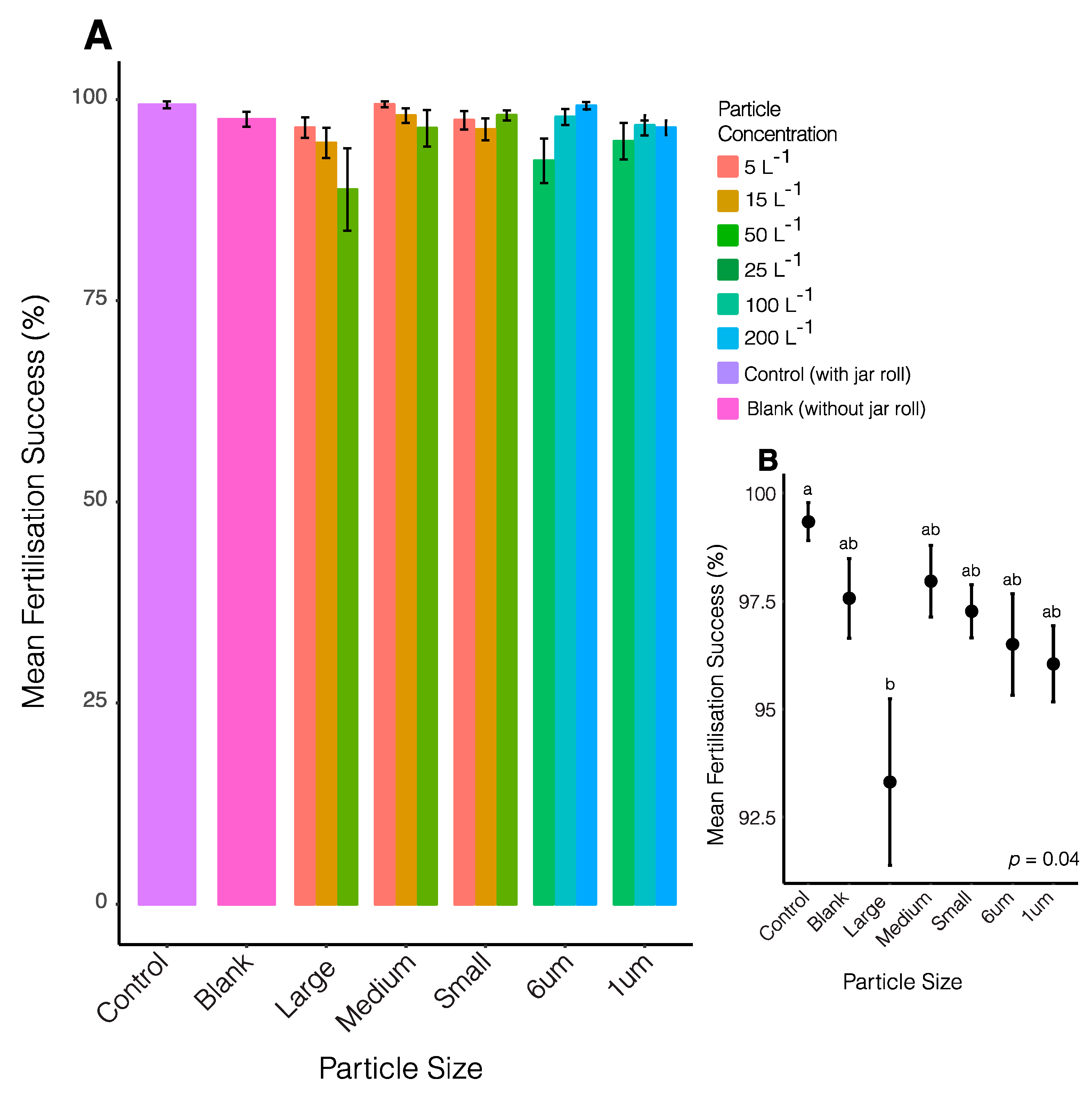
3.1. Fertilisation Success

There was a significant effect of particle size on fertilisation success (p = 0.03, Figure 1A, Table S1); however, there were no significant effects of particle concentration, nor was there a significant interaction between concentration and particle size. Despite the significant overall effect of particle size, further pairwise comparisons could find no significant differences among treatments (including particle concentration). In a comparison of particle size alone, large weathered plastic pieces (pooled 5 to 50 particles L−1) were shown to cause significantly lower fertilisation success (ANOVA: df = 6, F = 2.34, p = 0.04) than the control treatments. Large weathered plastic piece treatments had an average of 93.31 ± 0.02% fertilisation success in comparison to 99.33 ± 0.44% fertilisation success in controls, a 6.02% decrease (Figure 1B). Mean fertilisation success in all other treatments did not significantly differ from controls (within 3.5% on average).

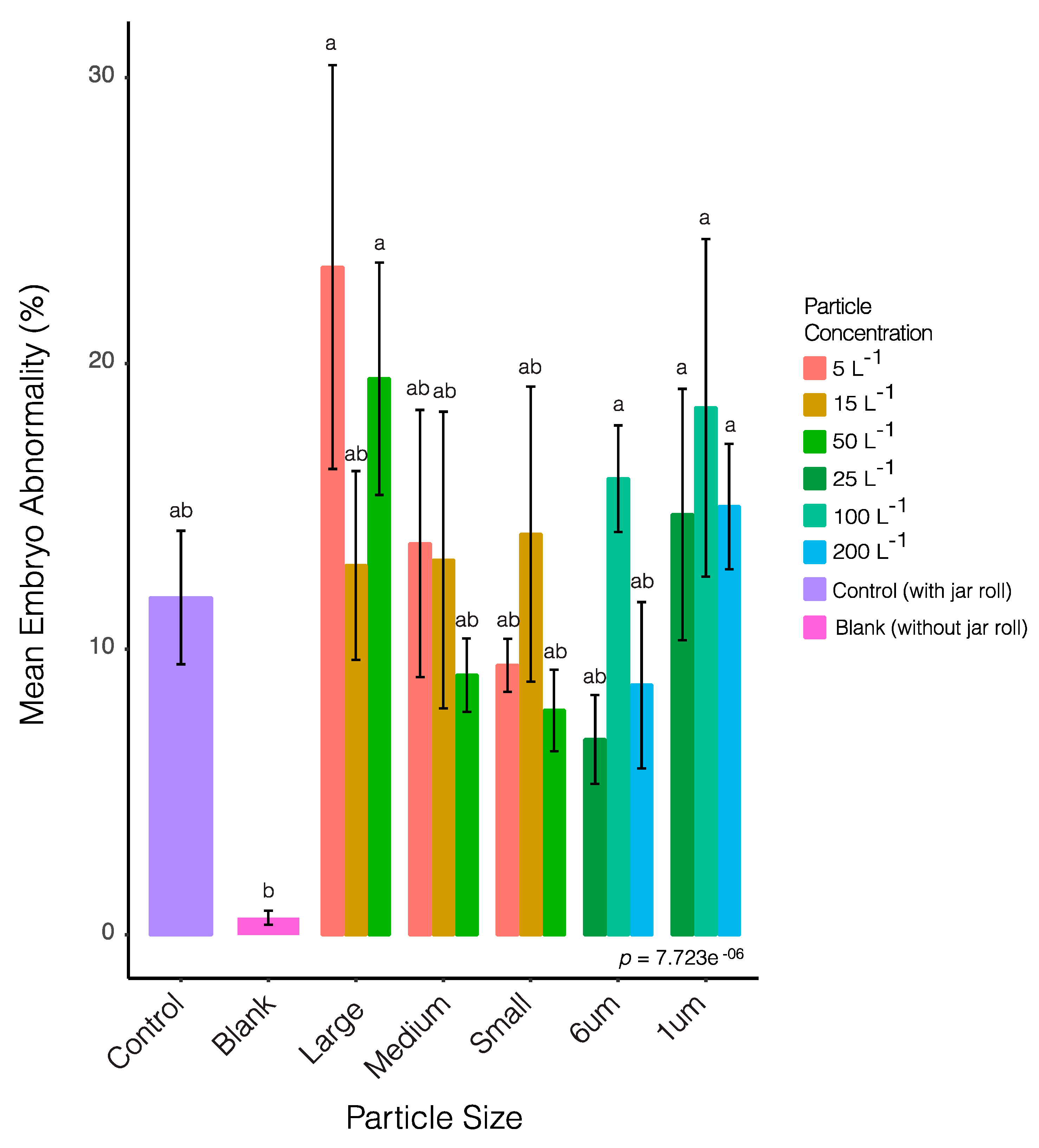
3.2. Embryo Abnormality

Abnormality in fertilised eggs (embryos) followed a similar pattern to fertilisation success; there was a significant effect of particle size, but no effect of particle concentration, nor a significant interaction between the two (Table S2). Further pairwise comparisons showed that embryo abnormality was significantly less in the blank treatment (adjusted p value = 4.66 × 10⁻⁶; Figure 2) than all other treatments, which exhibited on average only 0.60 ± 0.25% abnormality. The treatment with 5 pieces L−1 of large (2 mm2) weathered plastic contained the highest proportion of abnormal embryos (23.36 ± 7.08%), which was, on average, 11.6% higher than the mean abnormality measured in controls (11.81 ± 2.34%), however variability was high among replicates (Figure 2). Abnormality in all other microplastic treatments differed from controls by ≤8% on average.

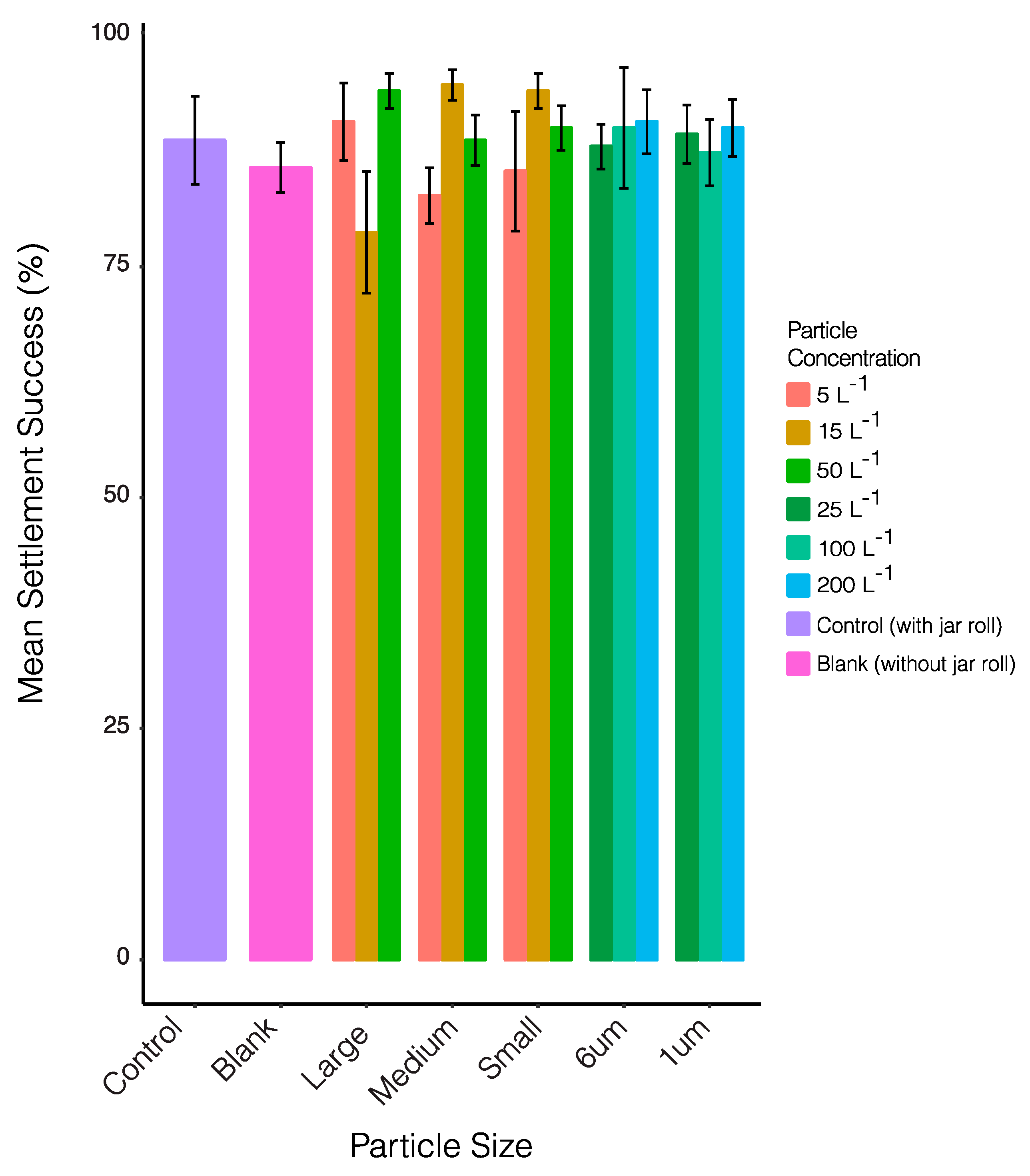
3.3. Settlement Success

All treatments showed settlement success within 9% of control levels. Although there was some variation among treatments (Figure 3), there were no significant effects of particle size or concentration on overall settlement success (p > 0.05; Table S3), despite some evidence of physical harm (Figure 4). The rolling of jars had no effect on the settlement success of the larvae (85.71 ± 2.72% blanks without rolling and 88.67 ± 4.77% controls with rolling).

4. Discussion

Globally, coral reefs are facing numerous stressors associated with anthropogenic activities such as increased sea surface temperatures and ocean acidification as a result of climate change [60,61,62,63], overfishing [64], and pollution [46,65,66,67,68,69]. Emerging contaminants, such as microplastics, represent an additional stressor potentially impacting these important tropical ecosystems and their associated organisms and function [68,69]. With increasing accounts of microplastic contamination at coral reefs [2,13,14,15,44], this experiment aimed to investigate the potential effects of two plastic types on three early life history stages of corals—fertilisation, early embryo development, and larval settlement. The experimental assays showed that short exposures to both types of microplastics exhibited minimal impacts on all three processes, even at relatively high particle concentrations. Further work is required to confirm the resistance of early life processes in corals to other plastic types such as microfibres, which accounted for 86% of all micro-debris measured at reef surface waters in the central Great Barrier Reef [15].
Only the large weathered plastic pieces (2 mm2) affected any of the early life stages and processes tested. Although statistically significant, the reduction in fertilisation success (by 6%, relative to controls) was not dose dependent and the fertilisation success in the presence of these large particles remained high at 93%. The measured impacts could have been due to physical contact between gametes and the plastic particles, as was observed between weathered plastic pieces and larvae. No early life history processes were inhibited by exposure to microbeads at any concentration or size. Investigations of the effects of microplastics on reproductive processes in other marine organisms highlight the variability of species sensitivities and tested exposure concentrations. For example, the oyster Crassostrea gigas exhibited reduced potential for reproduction when exposed to polystyrene microbeads (2 and 6 µm) for 2 months at a concentration of 0.023 mg L−1 [70]. Specifically, female oocyte numbers and sizes were reduced [70], while males exhibited a 23% reduction in sperm velocity, relative to controls. Such effects could lead to fertilization inhibition, reduced larvae survival and offspring growth [70]. Chronic (9 day) exposure of a marine copepod (Calanus helgolandicus) to 65 polystyrene microbeads mL−1 (i.e., 65,000 L−1) resulted in reduced egg size and hatching success [71]. Adult pearl oysters (Pinctada margaritifera) exposed to polystyrene microbeads (6 and 10 µm) for 2 months at concentrations of 0.25, 2.5, and 25 µg L−1 exhibited no significant difference in gonad development index or normal gametogenesis, relative to controls [72]. However, epithelial detachments and small holes were observed in gonadal tubules in certain treatments and a significantly higher number of oysters exhibited regression of gametogenesis. Due to differences in concentration unit reporting between studies it is difficult to directly compare results, however the highest concentration (200 particles L−1) tested in the present study was 325 times lower than the 65,000 microbeads L−1 that elicited reduced egg size and hatching success in C. helgolandicus [71].
The limited effect of microplastics on embryo development and larval settlement are consistent with other studies that have shown some tolerance of these early life stages of corals to other types of physical stressors (e.g., sediment and coal [52,73,74]). The effects on coral fertilisation by natural particulates is due to interactions with sperm that results in sperm sinking, sperm limitation at the surface near the buoyant eggs and ultimately reduced fertilisation rates [54]. The size and type of particle also has a strong effect on sperm limitation, with smaller particles, high in organic carbon having a far more pronounced effect (EC50 < 10 mg L−1) than larger, carbonate particles which only affect fertilisation at very high concentrations (EC50 > 800 mg L−1) [74]. Although small (1 and 6 µm), and seemingly able to associate with coral sperm (Figure 4A), the plastic beads were unlikely to have resulted in sperm sinking and limitation, hence fertilisation was not affected. It is not known how the large irregular plastic particles here (2 mm2) may have lowered fertilisation; but reduced interactions between eggs and sperm by physical blocking or attachment of some sperm are possible. Throughout the slow rolling exposures, coral eggs, embryos, and plastic particles were at the water surface, and some abnormalities including irregular cell division was observed. However, since there was a significant difference in abnormalities between control and blank treatments, we cannot decipher whether the observed abnormalities were caused by the presence of plastic particles or mechanical agitation (i.e., rolling). The coral larvae exposed to microplastics were ciliated and mobile and these characteristics, along with an ability to produce mucous, has been shown to protect larvae from exposures to very high concentrations of sediments [73]. Similar mechanisms are likely to have protected the swimming larvae in these tests to all microplastic types.
Microplastics have been demonstrated to act as vectors for pollution exposure to marine species [75] and these mechanisms and effects can extend to early life stages. For example, studies exposing the oyster Crassostrea gigas to microplastics have identified cellular impacts on spermatozoa, and a dose-dependent increase in reactive oxygen species production in spermatozoa after acute (up to 5 h) exposure to polystyrene nanoparticles coated with carboxylic groups [76]. A separate study found that both virgin (new) and weathered plastic pellets can act as vectors for pollution, with virgin pellets exhibiting more severe adverse effects than weathered pellets, with anomalous sea urchin (Lytechinus variegatus) embryonic development increasing by 58.1% and 66.5% in virgin pellet assays [77]. Toxicology was beyond the scope of the current study, however testing the influence of contamination and weathering of microplastics on early life stages of coral could be valuable.
There was little, or no impact of the plastics tested on the early life stages of A. tenuis but the comparative and ecological relevance of this finding is uncertain. It is difficult to make comparisons between studies and against environmental concentrations due to differences in life histories of the tested organisms, sizes, and concentrations of microplastics used, as well as variability in the reporting of units (e.g., weight/volume vs. number of plastics/volume) [78]. Examination of ecologically relevant concentrations (i.e., contamination levels measured in the field) is important to ensure impacts are not overestimated and sensationalized [79]. With respect to the concentration of particles in the water column, our tests reached as high as 50 and 200 pieces L−1 (large weathered and microbeads respectively), relatively high by published environmental levels [13,14,15], indicating microplastic particles of this type present little risk to the early life processes of the coral species A. tenuis.
Given the cumulative pressures faced by coral reefs and an increasing awareness of the ubiquity of microplastics in marine ecosystems, more work is clearly needed to assess the potential effects of a wider range of microplastic types, including fibres and highly weathered particles, on the early life stages of a wider range of coral-reef invertebrates. This will improve risk assessments for this emerging class of contaminant and inform priorities for the management of plastic pollution entering sensitive coral reef ecosystems.

Supplementary Materials

The following are available online at https://www.mdpi.com/1424-2818/11/12/228/s1, Table S1: Results of a linear model with arcsine square-root transformed fertilisation success as the response variable, against particle size (size class) and concentration as explanatory variables. “*” denotes significance at the α = 0.05 level; Table S2: Results of a linear model with square-root transformed embryo abnormality ratio as the response variable, against particle size (size class) and concentration as explanatory variables. “*” denotes significance at the α = 0.05 level; Table S3: Results of a linear model with square-root transformed settlement success as the response variable, against particle size (size class) and concentration as explanatory variables. No significance found.

Author Contributions

K.L.E.B. and N.M.H. conceived the ideas and designed the methodology; K.L.E.B. and A.P.N. collected the data; H.E.E. analysed and interpreted the data; K.L.E.B., N.M.H., A.P.N., H.E.E., and P.J.L. led the writing of the manuscript. All authors contributed critically to the drafts and gave final approval for publication.

Funding

This research was supported by AIMS@JCU Division of Research & Innovation at James Cook University (JCU) and the Australian Institute of Marine Science (AIMS) Townsville, Australia.

Acknowledgments

The Authors wish to acknowledge Holly Bennett, Michael Civiello, and staff at the National Sea Simulator (AIMS, Townsville) for their experimental support.

Conflicts of Interest

The authors declare no conflict of interest. The funders had no role in the design of the study; in the collection, analyses, or interpretation of data; in the writing of the manuscript, or in the decision to publish the results.

References

  1. Ostle, C.; Thompson, R.C.; Broughton, D.; Gregory, L.; Wootton, M.; Johns, D.G. The Rise in Ocean Plastics Evidenced from a 60-Year Time Series. Nat. Commun. 2019, 10, 1622. [Google Scholar] [CrossRef] [PubMed]
  2. Law, K.L. Plastics in the Marine Environment. Annu. Rev. Mar. Sci. 2017, 9, 205–229. [Google Scholar] [CrossRef] [PubMed]
  3. Geyer, R.; Jambeck, J.R.; Law, K.L. Production, Use, and Fate of All Plastics Ever Made. Sci. Adv. 2017, 3, e1700782. [Google Scholar] [CrossRef]
  4. Van Sebille, E.; Wilcox, C.; Lebreton, L.; Maximenko, N.; Hardesty, B.D.; van Franeker, J.A.; Eriksen, M.; Siegel, D.; Galgani, F.; Law, K.L. A Global Inventory of Small Floating Plastic Debris. Environ. Res. Lett. 2015, 10, 124006. [Google Scholar] [CrossRef]
  5. Eriksen, M.; Lebreton, L.C.M.; Carson, H.S.; Thiel, M.; Moore, C.J.; Borerro, J.C.; Galgani, F.; Ryan, P.G.; Reisser, J. Plastic Pollution in the World’s Oceans: More than 5 Trillion Plastic Pieces Weighing over 250,000 Tons Afloat at Sea. PLoS ONE 2014, 9, e111913. [Google Scholar] [CrossRef]
  6. Andrady, A.L. Microplastics in the Marine Environment. Mar. Pollut. Bull. 2011, 62, 1596–1605. [Google Scholar] [CrossRef]
  7. Zitko, V.; Hanlon, M. Another Source of Pollution by Plastics: Skin Cleaners with Plastic Scrubbers. Mar. Pollut. Bull. 1991, 22, 41–42. [Google Scholar] [CrossRef]
  8. Critchell, K.; Lambrechts, J. Modelling Accumulation of Marine Plastics in the Coastal Zone; What Are the Dominant Physical Processes? Estuar. Coast. Shelf Sci. 2016, 171, 111–122. [Google Scholar] [CrossRef]
  9. Thompson, R.C.; Olsen, Y.; Mitchell, R.P.; Davis, A.; Rowland, S.J.; John, A.W.G.; McGonigle, D.; Russell, A.E. Lost at Sea: Where Is All the Plastic? Science 2004, 304, 838. [Google Scholar] [CrossRef]
  10. Peeken, I.; Primpke, S.; Beyer, B.; Gütermann, J.; Katlein, C.; Krumpen, T.; Bergmann, M.; Hehemann, L.; Gerdts, G. Arctic Sea Ice Is an Important Temporal Sink and Means of Transport for Microplastic. Nat. Commun. 2018, 9, 1505. [Google Scholar] [CrossRef]
  11. Lacerda, A.L.d.F.; Rodrigues, L.d.S.; van Sebille, E.; Rodrigues, F.L.; Ribeiro, L.; Secchi, E.R.; Kessler, F.; Proietti, M.C. Plastics in Sea Surface Waters around the Antarctic Peninsula. Sci. Rep. 2019, 9, 3977. [Google Scholar] [CrossRef] [PubMed]
  12. Brandon, J.A.; Jones, W.; Ohman, M.D. Multidecadal Increase in Plastic Particles in Coastal Ocean Sediments. Sci. Adv. 2019, 5, eaax0587. [Google Scholar] [CrossRef] [PubMed]
  13. Hall, N.M.; Berry, K.L.E.; Rintoul, L.; Hoogenboom, M.O. Microplastic Ingestion by Scleractinian Corals. Mar. Biol. 2015, 162, 725–732. [Google Scholar] [CrossRef]
  14. Connors, E.J. Distribution and Biological Implications of Plastic Pollution on the Fringing Reef of Mo’orea, French Polynesia. PeerJ 2017, 5, e3733. [Google Scholar] [CrossRef] [PubMed]
  15. Jensen, L.H.; Motti, C.A.; Garm, A.L.; Tonin, H.; Kroon, F.J. Sources, Distribution and Fate of Microfibres on the Great Barrier Reef, Australia. Sci. Rep. 2019, 9, 9021. [Google Scholar] [CrossRef] [PubMed]
  16. Green, D.S. Effects of Microplastics on European Flat Oysters, Ostrea Edulis and Their Associated Benthic Communities. Environ. Pollut. 2016, 216, 95–103. [Google Scholar] [CrossRef]
  17. Watts, A.J.R.; Urbina, M.A.; Corr, S.; Lewis, C.; Galloway, T.S. Ingestion of Plastic Microfibers by the Crab Carcinus Maenas and Its Effect on Food Consumption and Energy Balance. Environ. Sci. Technol. 2015, 49, 14597–14604. [Google Scholar] [CrossRef]
  18. Taylor, M.L.; Gwinnett, C.; Robinson, L.F.; Woodall, L.C. Plastic Microfibre Ingestion by Deep-Sea Organisms. Sci. Rep. 2016, 6, 33997. [Google Scholar] [CrossRef]
  19. Hardesty, B.D.; Good, T.P.; Wilcox, C. Novel Methods, New Results and Science-Based Solutions to Tackle Marine Debris Impacts on Wildlife. Ocean Coast. Manag. 2015, 115, 4–9. [Google Scholar] [CrossRef]
  20. Kazour, M.; Jemaa, S.; Issa, C.; Khalaf, G.; Amara, R. Microplastics Pollution along the Lebanese Coast (Eastern Mediterranean Basin): Occurrence in Surface Water, Sediments and Biota Samples. Sci. Total Environ. 2019, 696, 133933. [Google Scholar] [CrossRef]
  21. Wright, S.L.; Thompson, R.C.; Galloway, T.S. The Physical Impacts of Microplastics on Marine Organisms: A Review. Environ. Pollut. 2013, 178, 483–492. [Google Scholar] [CrossRef] [PubMed]
  22. Lithner, D.; Larsson, Å.; Dave, G. Environmental and Health Hazard Ranking and Assessment of Plastic Polymers Based on Chemical Composition. Sci. Total Environ. 2011, 409, 3309–3324. [Google Scholar] [CrossRef] [PubMed]
  23. Groh, K.J.; Backhaus, T.; Carney-Almroth, B.; Geueke, B.; Inostroza, P.A.; Lennquist, A.; Leslie, H.A.; Maffini, M.; Slunge, D.; Trasande, L.; et al. Overview of Known Plastic Packaging-Associated Chemicals and Their Hazards. Sci. Total Environ. 2019, 651, 3253–3268. [Google Scholar] [CrossRef] [PubMed]
  24. Rochman, C.M.; Hentschel, B.T.; Teh, S.J. Long-Term Sorption of Metals Is Similar among Plastic Types: Implications for Plastic Debris in Aquatic Environments. PLoS ONE 2014, 9, e85433. [Google Scholar] [CrossRef] [PubMed]
  25. Teuten, E.L.; Rowland, S.J.; Galloway, T.S.; Thompson, R.C. Potential for Plastics to Transport Hydrophobic Contaminants. Environ. Sci. Technol. 2007, 41, 7759–7764. [Google Scholar] [CrossRef]
  26. Teuten, E.L.; Saquing, J.M.; Knappe, D.R.U.; Barlaz, M.A.; Jonsson, S.; Björn, A.; Rowland, S.J.; Thompson, R.C.; Galloway, T.S.; Yamashita, R.; et al. Transport and Release of Chemicals from Plastics to the Environment and to Wildlife. Philos. Trans. R. Soc. B Biol. Sci. 2009, 364, 2027–2045. [Google Scholar] [CrossRef]
  27. Bakir, A.; O’Connor, I.A.; Rowland, S.J.; Hendriks, A.J.; Thompson, R.C. Relative Importance of Microplastics as a Pathway for the Transfer of Hydrophobic Organic Chemicals to Marine Life. Environ. Pollut. 2016, 219, 56–65. [Google Scholar] [CrossRef]
  28. Koelmans, A.A.; Besseling, E.; Foekema, E.M. Leaching of Plastic Additives to Marine Organisms. Environ. Pollut. 2014, 187, 49–54. [Google Scholar] [CrossRef]
  29. De Sá, L.C.; Oliveira, M.; Ribeiro, F.; Rocha, T.L.; Futter, M.N. Studies of the Effects of Microplastics on Aquatic Organisms: What Do We Know and Where Should We Focus Our Efforts in the Future? Sci. Total Environ. 2018, 645, 1029–1039. [Google Scholar] [CrossRef]
  30. Weber, A.; Scherer, C.; Brennholt, N.; Reifferscheid, G.; Wagner, M. PET Microplastics Do Not Negatively Affect the Survival, Development, Metabolism and Feeding Activity of the Freshwater Invertebrate Gammarus Pulex. Environ. Pollut. 2018, 234, 181–189. [Google Scholar] [CrossRef]
  31. Reichert, J.; Arnold, A.L.; Hoogenboom, M.O.; Schubert, P.; Wilke, T. Impacts of Microplastics on Growth and Health of Hermatypic Corals Are Species-Specific. Environ. Pollut. 2019, 254, 113074. [Google Scholar] [CrossRef] [PubMed]
  32. Besseling, E.; Wang, B.; Lürling, M.; Koelmans, A.A. Nanoplastic Affects Growth of S. Obliquus and Reproduction of D. Magna. Environ. Sci. Technol. 2014, 48, 12336–12343. [Google Scholar] [CrossRef] [PubMed]
  33. Lo, H.K.A.; Chan, K.Y.K. Negative Effects of Microplastic Exposure on Growth and Development of Crepidula Onyx. Environ. Pollut. 2018, 233, 588–595. [Google Scholar] [CrossRef] [PubMed]
  34. Prokić, M.D.; Radovanović, T.B.; Gavrić, J.P.; Faggio, C. Ecotoxicological Effects of Microplastics: Examination of Biomarkers, Current State and Future Perspectives. TrAC Trends Anal. Chem. 2019, 111, 37–46. [Google Scholar] [CrossRef]
  35. Tallec, K.; Huvet, A.; Di Poi, C.; González-Fernández, C.; Lambert, C.; Petton, B.; Le Goïc, N.; Berchel, M.; Soudant, P.; Paul-Pont, I. Nanoplastics Impaired Oyster Free Living Stages, Gametes and Embryos. Environ. Pollut. 2018, 242, 1226–1235. [Google Scholar] [CrossRef] [Green Version]
  36. Aljaibachi, R.; Callaghan, A. Impact of Polystyrene Microplastics on Daphnia Magna Mortality and Reproduction in Relation to Food Availability. PeerJ 2018, 6, e4601. [Google Scholar] [CrossRef] [Green Version]
  37. Rist, S.; Baun, A.; Hartmann, N.B. Ingestion of Micro- and Nanoplastics in Daphnia Magna—Quantification of Body Burdens and Assessment of Feeding Rates and Reproduction. Environ. Pollut. 2017, 228, 398–407. [Google Scholar] [CrossRef] [Green Version]
  38. Hankins, C.; Duffy, A.; Drisco, K. Scleractinian Coral Microplastic Ingestion: Potential Calcification Effects, Size Limits, and Retention. Mar. Pollut. Bull. 2018, 135, 587–593. [Google Scholar] [CrossRef]
  39. Santana, M.F.M.; Moreira, F.T.; Pereira, C.D.S.; Abessa, D.M.S.; Turra, A. Continuous Exposure to Microplastics Does Not Cause Physiological Effects in the Cultivated Mussel Perna Perna. Arch. Environ. Contam. Toxicol. 2018, 74, 594–604. [Google Scholar] [CrossRef] [Green Version]
  40. Kroon, F.J.; Motti, C.E.; Jensen, L.H.; Berry, K.L.E. Classification of Marine Microdebris: A Review and Case Study on Fish from the Great Barrier Reef, Australia. Sci. Rep. 2018, 8, 16422. [Google Scholar] [CrossRef] [Green Version]
  41. Reichert, J.; Schellenberg, J.; Schubert, P.; Wilke, T. Responses of Reef Building Corals to Microplastic Exposure. Environ. Pollut. 2018, 237, 955–960. [Google Scholar] [CrossRef] [PubMed]
  42. Allen, A.S.; Seymour, A.C.; Rittschof, D. Chemoreception Drives Plastic Consumption in a Hard Coral. Mar. Pollut. Bull. 2017, 124, 198–205. [Google Scholar] [CrossRef] [PubMed]
  43. Murphy, F.; Quinn, B. The Effects of Microplastic on Freshwater Hydra Attenuata Feeding, Morphology & Reproduction. Environ. Pollut. 2018, 234, 487–494. [Google Scholar] [CrossRef] [PubMed]
  44. Ding, J.; Jiang, F.; Li, J.; Wang, Z.; Sun, C.; Wang, Z.; Fu, L.; Ding, N.X.; He, C. Microplastics in the Coral Reef Systems from Xisha Islands of South China Sea. Environ. Sci. Technol. 2019, 53, 8036–8046. [Google Scholar] [CrossRef] [PubMed]
  45. Chapron, L.; Peru, E.; Engler, A.; Ghiglione, J.F.; Meistertzheim, A.L.; Pruski, A.M.; Purser, A.; Vétion, G.; Galand, P.E.; Lartaud, F. Macro- and Microplastics Affect Cold-Water Corals Growth, Feeding and Behaviour. Sci. Rep. 2018, 8, 15299. [Google Scholar] [CrossRef] [PubMed] [Green Version]
  46. Fabricius, K.E. Effects of Terrestrial Runoff on the Ecology of Corals and Coral Reefs: Review and Synthesis. Mar. Pollut. Bull. 2005, 50, 125–146. [Google Scholar] [CrossRef]
  47. Humanes, A.; Noonan, S.H.C.; Willis, B.L.; Fabricius, K.E.; Negri, A.P. Cumulative Effects of Nutrient Enrichment and Elevated Temperature Compromise the Early Life History Stages of the Coral Acropora Tenuis. PLoS ONE 2016, 11, e0161616. [Google Scholar] [CrossRef]
  48. Jones, R.; Ricardo, G.F.; Negri, A.P. Effects of Sediments on the Reproductive Cycle of Corals. Mar. Pollut. Bull. 2015, 100, 13–33. [Google Scholar] [CrossRef]
  49. Jones, R.; Bessell-Browne, P.; Fisher, R.; Klonowski, W.; Slivkoff, M. Assessing the Impacts of Sediments from Dredging on Corals. Mar. Pollut. Bull. 2016, 102, 9–29. [Google Scholar] [CrossRef]
  50. Reichelt-Brushett, A.J.; Harrison, P.L. The Effect of Copper, Zinc and Cadmium on Fertilization Success of Gametes from Scleractinian Reef Corals. Mar. Pollut. Bull. 1999, 38, 6. [Google Scholar] [CrossRef]
  51. Heyward, A.J.; Negri, A.P. Turbulence, Cleavage, and the Naked Embryo: A Case for Coral Clones. Science 2012, 335, 1064. [Google Scholar] [CrossRef] [PubMed]
  52. Berry, K.L.E.; Hoogenboom, M.O.; Brinkman, D.L.; Burns, K.A.; Negri, A.P. Effects of Coal Contamination on Early Life History Processes of a Reef-Building Coral, Acropora Tenuis. Mar. Pollut. Bull. 2017, 114, 505–514. [Google Scholar] [CrossRef] [PubMed]
  53. Ricardo, G.F.; Jones, R.J.; Negri, A.P.; Stocker, R. That Sinking Feeling: Suspended Sediments Can Prevent the Ascent of Coral Egg Bundles. Sci. Rep. 2016, 6, 21567. [Google Scholar] [CrossRef] [PubMed]
  54. Ricardo, G.F.; Jones, R.J.; Clode, P.L.; Humanes, A.; Negri, A.P. Suspended Sediments Limit Coral Sperm Availability. Sci. Rep. 2016, 5, 18084. [Google Scholar] [CrossRef] [Green Version]
  55. Koelmans, A.A.; Kooi, M.; Law, K.L.; van Sebille, E. All Is Not Lost: Deriving a Top-down Mass Budget of Plastic at Sea. Environ. Res. Lett. 2017, 12, 114028. [Google Scholar] [CrossRef] [Green Version]
  56. Negri, A.P.; Heyward, A.J. Inhibition of Fertilization and Larval Metamorphosis of the Coral Acropora Millepora (Ehrenberg, 1834) by Petroleum Products. Mar. Pollut. Bull. 2000, 41, 420–427. [Google Scholar] [CrossRef]
  57. Heyward, A.J.; Negri, A.P. Natural Inducers for Coral Larval Metamorphosis. Coral Reefs 1999, 18, 273–279. [Google Scholar] [CrossRef]
  58. Lenth, R.; Singmann, H.; Love, J.; Buerkner, P.; Herve, M. Package “Emmeans”. Am. Stat. 2018, 34, 216–221. [Google Scholar]
  59. Hadley, W. Ggplot2; Springer Science+Business Media, LLC: New York, NY, USA, 2016. [Google Scholar]
  60. Hughes, T.P.; Kerry, J.T.; Álvarez-Noriega, M.; Álvarez-Romero, J.G.; Anderson, K.D.; Baird, A.H.; Babcock, R.C.; Beger, M.; Bellwood, D.R.; Berkelmans, R.; et al. Global Warming and Recurrent Mass Bleaching of Corals. Nature 2017, 543, 373–377. [Google Scholar] [CrossRef]
  61. Hoegh-Guldberg, O. Climate Change, Coral Bleaching and the Future of the World’s Coral Reefs. Mar. Freshw. Res. 1999, 50, 839. [Google Scholar] [CrossRef] [Green Version]
  62. Cinner, J.E.; McClanahan, T.R.; Graham, N.A.J.; Daw, T.M.; Maina, J.; Stead, S.M.; Wamukota, A.; Brown, K.; Bodin, Ö. Vulnerability of Coastal Communities to Key Impacts of Climate Change on Coral Reef Fisheries. Glob. Environ. Chang. 2012, 22, 12–20. [Google Scholar] [CrossRef]
  63. Pendleton, L.; Comte, A.; Langdon, C.; Ekstrom, J.A.; Cooley, S.R.; Suatoni, L.; Beck, M.W.; Brander, L.M.; Burke, L.; Cinner, J.E.; et al. Coral Reefs and People in a High-CO2 World: Where Can Science Make a Difference to People? PLoS ONE 2016, 11, e0164699. [Google Scholar] [CrossRef] [PubMed]
  64. Loh, T.-L.; McMurray, S.E.; Henkel, T.P.; Vicente, J.; Pawlik, J.R. Indirect Effects of Overfishing on Caribbean Reefs: Sponges Overgrow Reef-Building Corals. PeerJ 2015, 3, e901. [Google Scholar] [CrossRef] [PubMed] [Green Version]
  65. Brodie, J.; Wolanski, E.; Lewis, S.; Bainbridge, Z. An Assessment of Residence Times of Land-Sourced Contaminants in the Great Barrier Reef Lagoon and the Implications for Management and Reef Recovery. Mar. Pollut. Bull. 2012, 65, 267–279. [Google Scholar] [CrossRef] [PubMed]
  66. Waterhouse, J.; Brodie, J.; Lewis, S.; Mitchell, A. Quantifying the Sources of Pollutants in the Great Barrier Reef Catchments and the Relative Risk to Reef Ecosystems. Mar. Pollut. Bull. 2012, 65, 394–406. [Google Scholar] [CrossRef]
  67. Berry, K.L.E.; Seemann, J.; Dellwig, O.; Struck, U.; Wild, C.; Leinfelder, R.R. Sources and Spatial Distribution of Heavy Metals in Scleractinian Coral Tissues and Sediments from the Bocas Del Toro Archipelago, Panama. Environ. Monit. Assess. 2013, 185, 9089–9099. [Google Scholar] [CrossRef] [Green Version]
  68. Kroon, F.J.; Berry, K.L.E.; Brinkman, D.L.; Davis, A.; King, O.; Kookana, R.; Lewis, S.; Leusch, F.; Makarynskyy, O.; Melvin, S.; et al. Identification, Impacts, and Prioritisation of Emerging Contaminants Present in the GBR and Torres Strait Marine Environments. Tropical Water Quality Hub, National Environmental Science Programme, Technical Report, pg. 152. Available online: https://nesptropical.edu.au/wp-content/uploads/2016/05/NESP-TWQ-1.10-FINAL-REPORTa.pdf (accessed on 28 November 2019).
  69. Kroon, F.J.; Berry, K.L.E.; Brinkman, D.L.; Kookana, R.; Leusch, F.; Melvin, S.; Neale, P.; Negri, A.; Puotinen, M.; Tsang, J.J.; et al. Sources, Presence, and Potential Effects of Contaminants of Emerging Concern in the Marine Environments of the Great Barrier Reef and Torres Strait, Australia. Sci. Total Environ. 2019, in press. [Google Scholar] [CrossRef]
  70. Sussarellu, R.; Suquet, M.; Thomas, Y.; Lambert, C.; Fabioux, C.; Pernet, M.E.J.; Le Goïc, N.; Quillien, V.; Mingant, C.; Epelboin, Y.; et al. Oyster Reproduction Is Affected by Exposure to Polystyrene Microplastics. Proc. Natl. Acad. Sci. USA 2016, 113, 2430–2435. [Google Scholar] [CrossRef] [Green Version]
  71. Cole, M.; Lindeque, P.; Fileman, E.; Halsband, C.; Galloway, T.S. The Impact of Polystyrene Microplastics on Feeding, Function and Fecundity in the Marine Copepod Calanus Helgolandicus. Environ. Sci. Technol. 2015, 49, 1130–1137. [Google Scholar] [CrossRef]
  72. Gardon, T.; Reisser, C.; Soyez, C.; Quillien, V.; Le Moullac, G. Microplastics Affect Energy Balance and Gametogenesis in the Pearl Oyster Pinctada Margaritifera. Environ. Sci. Technol. 2018, 52, 5277–5286. [Google Scholar] [CrossRef] [Green Version]
  73. Ricardo, G.F.; Jones, R.J.; Clode, P.L.; Negri, A.P. Mucous Secretion and Cilia Beating Defend Developing Coral Larvae from Suspended Sediments. PLoS ONE 2016, 11, e0162743. [Google Scholar] [CrossRef] [PubMed]
  74. Ricardo, G.F.; Jones, R.J.; Clode, P.L.; Humanes, A.; Gioffre, N.; Negri, A.P. Sediment Characteristics Influence the Fertilisation Success of the Corals Acropora Tenuis and Acropora Millepora. Mar. Pollut. Bull. 2018, 135, 941–953. [Google Scholar] [CrossRef] [PubMed]
  75. Guo, X.; Wang, J. The Chemical Behaviors of Microplastics in Marine Environment: A Review. Mar. Pollut. Bull. 2019, 142, 1–14. [Google Scholar] [CrossRef] [PubMed]
  76. González-Fernández, C.; Tallec, K.; Le Goïc, N.; Lambert, C.; Soudant, P.; Huvet, A.; Suquet, M.; Berchel, M.; Paul-Pont, I. Cellular Responses of Pacific Oyster (Crassostrea Gigas) Gametes Exposed in Vitro to Polystyrene Nanoparticles. Chemosphere 2018, 208, 764–772. [Google Scholar] [CrossRef] [Green Version]
  77. Nobre, C.R.; Santana, M.F.M.; Maluf, A.; Cortez, F.S.; Cesar, A.; Pereira, C.D.S.; Turra, A. Assessment of Microplastic Toxicity to Embryonic Development of the Sea Urchin Lytechinus Variegatus (Echinodermata: Echinoidea). Mar. Pollut. Bull. 2015, 92, 99–104. [Google Scholar] [CrossRef]
  78. Miller, M.E.; Kroon, F.J.; Motti, C.A. Recovering Microplastics from Marine Samples: A Review of Current Practices. Mar. Pollut. Bull. 2017, 123, 6–18. [Google Scholar] [CrossRef]
  79. Lenz, R.; Enders, K.; Nielsen, T.G. Microplastic Exposure Studies Should Be Environmentally Realistic. Proc. Natl. Acad. Sci. USA 2016, 113, E4121–E4122. [Google Scholar] [CrossRef] [Green Version]
Figure 1. Mean (± SEM) fertilisation success in (A) all treatments of varying particle size and concentration and (B) among size class only. Large, medium, and small particle sizes refer to weathered plastics, while 6 µm and 1 µm particle sizes refer to microbeads. Similarly, weathered plastics were in concentrations of 5, 15 or 50 pieces L−1, whereas microbead treatments were in concentrations of 25, 100 or 200 beads L−1. Controls included plastic free jars that were rolled at ~0.3 revolutions s−1 to generate water flow, while blanks were not rolled and acted as experimental controls.
Diversity 11 00228 g001
Figure 2. Mean (± SEM) embryo abnormalities after 2.5 h exposures to microplastics among all treatments of varying particle size and concentration. Controls included plastic free jars that were rolled at ~0.3 revolutions s−1 to generate water flow, while blanks were not rolled and acted as experimental controls.
Diversity 11 00228 g002
Figure 3. Mean (± SEM) settlement success following exposure to microplastics for 24 h, among all treatments of varying particle size and concentration. “Control” refers to rolling jars, while “Blank” refers to non-rolling jars. Jars were rolled to create water movement and mimic surface water conditions. Controls included plastic free jars that were rolled at ~0.3 revolutions s−1 to generate water flow, while blanks were not rolled and acted as experimental controls.
Diversity 11 00228 g003
Figure 4. Early life history-microplastic interactions. (A) 1 µm microbeads (unknown concentration used for visual purposes only) appear to clump with sperm surrounding Acropora tenuis eggs. (B) physical contact between two larvae and a weathered plastic particle resulted in embedment of the plastic piece into the larvae. Scale bar = 0.5 mm.
Diversity 11 00228 g004
Table 1. Microplastic exposure treatments. Microplastic exposures were conducted in 200 mL of filtered sea water (0.22 µm).
No. of Plastic Pieces Per JarConcentration (No. Microplastic L−1)SizeShape
152, 1, 0.5 mm2Square
3152, 1, 0.5 mm2Square
10502, 1, 0.5 mm2Square
5251, 6 µmSphere
201001, 6 µmSphere
402001, 6 µmSphere

