
Lipid Alterations and Metabolism Disturbances in Selected Inflammatory Skin Diseases

Abstract
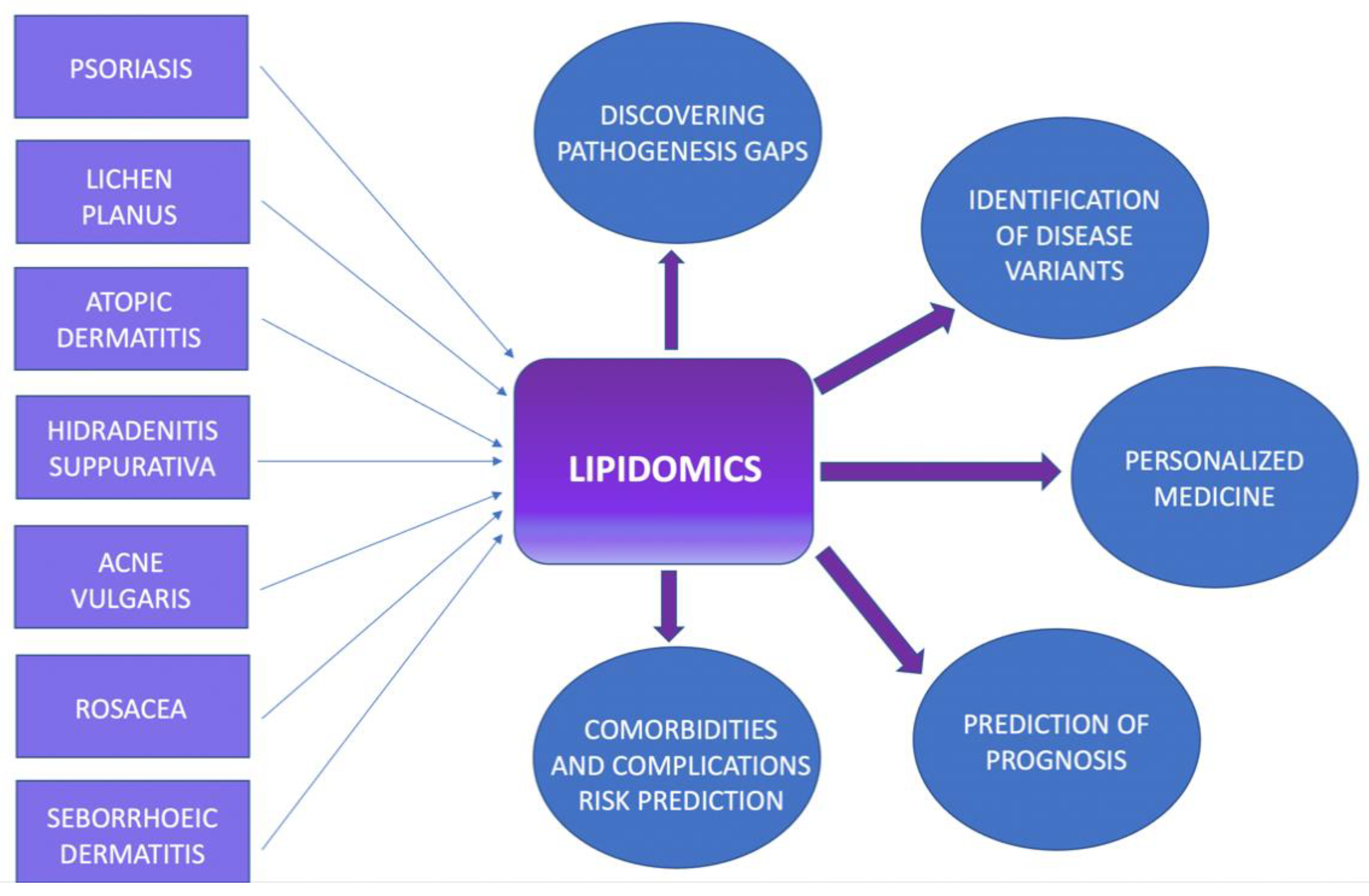
:1. Introduction
2. Psoriasis
3. Lichen Planus
4. Atopic Dermatitis
5. Hidradenitis Suppurativa
6. Seborrhoeic Dermatitis
7. Rosacea
8. Acne Vulgaris
9. Materials and Methods
10. Discussion and Conclusions
Author Contributions
Funding
Institutional Review Board Statement
Informed Consent Statement
Data Availability Statement
Conflicts of Interest
References
- Han, X.; Gross, R.W. The foundations and development of lipidomics. J. Lipid Res. 2022, 63, 100164. [Google Scholar] [CrossRef]
- Zeng, C.; Wen, B.; Hou, G.; Lei, L.; Mei, Z.; Jia, X.; Chen, X.; Zhu, W.; Li, J.; Kuang, Y.; et al. Lipidomics profiling reveals the role of glycerophospholipid metabolism in psoriasis. Gigascience 2017, 6, 1–11. [Google Scholar] [CrossRef] [Green Version]
- Fahy, E.; Subramaniam, S.; Brown, H.A.; Glass, C.K.; Merrill, A.H., Jr.; Murphy, R.C.; Raetz, C.R.; Russell, D.W.; Seyama, Y.; Shaw, W.; et al. A comprehensive classification system for lipids. J. Lipid Res. 2005, 46, 839–861. [Google Scholar] [CrossRef] [PubMed] [Green Version]
- Li, S.; Ganguli-Indra, G.; Indra, A.K. Lipidomic analysis of epidermal lipids: A tool to predict progression of inflammatory skin disease in humans. Expert Rev. Proteom. 2016, 13, 451–456. [Google Scholar] [CrossRef] [Green Version]
- Penno, C.A.; Jäger, P.; Laguerre, C.; Hasler, F.; Hofmann, A.; Gass, S.K.; Wettstein-Ling, B.; Schaefer, D.J.; Avrameas, A.; Raulf, F.; et al. Lipidomics profiling of hidradenitis suppurativa skin lesions reveals lipoxygenase pathway dysregulation and accumulation of proinflammatory leukotriene B4. J. Investig. Dermatol. 2020, 140, 2421–2432.e10. [Google Scholar] [CrossRef]
- Nowowiejska, J.; Baran, A.; Flisiak, I. Aberrations in lipid expression and metabolism in psoriasis. Int. J. Mol. Sci. 2021, 22, 6561. [Google Scholar] [CrossRef] [PubMed]
- The National Psoriais Foundation. NPF Psoriasis Statistics. Available online: https://www.psoriasis.org/psoriasis-statistics/ (accessed on 20 February 2023).
- Kamiya, K.; Kishimoto, M.; Sugai, J.; Komine, M.; Ohtsuki, M. Risk factors for the development of psoriasis. Int. J. Mol. Sci. 2019, 20, 4347. [Google Scholar] [CrossRef] [Green Version]
- Hirotsu, C.; Rydlewski, M.; Araújo, M.S.; Tufik, S.; Andersen, M.L. Sleep loss and cytokines levels in an experimental model of psoriasis. PLoS ONE 2012, 7, e51183. [Google Scholar] [CrossRef] [Green Version]
- Tokuyama, M.; Mabuchi, T. New treatment addressing the pathogenesis of psoriasis. Int. J. Mol. Sci. 2020, 21, 7488. [Google Scholar] [CrossRef]
- Bakshi, H.; Nagpal, M.; Singh, M.; Dhingra, G.A.; Aggarwal, G. Treatment of psoriasis: A comprehensive review of entire therapies. Curr. Drug Saf. 2020, 15, 82–104. [Google Scholar] [CrossRef] [PubMed]
- Wu, J.J.; Kavanaugh, A.; Lebwohl, M.G.; Gniadecki, R.; Merola, J.F. Psoriasis and metabolic syndrome: Implications for the management and treatment of psoriasis. J. Eur. Acad. Dermatol. Venereol. 2022, 36, 797–806. [Google Scholar] [CrossRef] [PubMed]
- Sommer, D.M.; Jenisch, S.; Suchan, M.; Christophers, E.; Weichenthal, M. Increased prevalence of the metabolic syndrome in patients with moderate to severe psoriasis. Arch. Dermatol. Res. 2006, 298, 321–328. [Google Scholar] [CrossRef]
- Ramezani, M.; Zavattaro, E.; Sadeghi, M. Evaluation of serum lipid, lipoprotein, and apolipoprotein levels in psoriatic patients: A systematic review and meta-analysis of case-control studies. Adv. Dermatol. Allergol. 2019, 36, 692–702. [Google Scholar] [CrossRef]
- Baran, A.; Kiluk, P.; Mysliwiec, H.; Flisiak, I. The role of lipids in psoriasis. Dermatol. Rev. 2017, 104, 619–635. [Google Scholar] [CrossRef] [Green Version]
- Myśliwiec, H.; Baran, A.; Harasim-Symbor, E.; Choromańska, B.; Myśliwiec, P.; Milewska, A.J.; Chabowski, A.; Flisiak, I. Increase in circulating sphingosine-1-phosphate and decrease in ceramide levels in psoriatic patients. Arch. Dermatol. Res. 2017, 309, 79–86. [Google Scholar] [CrossRef] [PubMed] [Green Version]
- Shih, C.M.; Chen, C.C.; Chu, C.K.; Wang, K.H.; Huang, C.Y.; Lee, A.W. The roles of lipoprotein in psoriasis. Int. J. Mol. Sci. 2020, 2, 859. [Google Scholar] [CrossRef] [Green Version]
- Hong, K.K.; Cho, H.R.; Ju, W.C.; Cho, Y.; Kim, N.I. A study on altered expression of serine palmitoyltransferase and ceramidase in psoriatic skin lesion. J. Korean Med. Sci. 2007, 22, 862–867. [Google Scholar] [CrossRef] [Green Version]
- Tawada, C.; Kanoh, H.; Nakamura, M.; Mizutani, Y.; Fujisawa, T.; Banno, Y.; Seishima, M. Interferon-γdecreases ceramides with long-chain fatty acids: Possible involvement in atopic dermatitis and psoriasis. J. Investig. Dermatol. 2014, 134, 712–718. [Google Scholar] [CrossRef] [Green Version]
- Elmets, C.A.; Leonardi, C.L.; Davis, D.M.; Gelfand, J.M.; Lichten, J.; Mehta, N.N.; Armstrong, A.W.; Connor, C.; Cordoro, K.M.; Elewski, B.E.; et al. Joint AAD-NPF guidelines of care for the management and treatment of psoriasis with awareness and attention to comorbidities. J. Am. Acad. Dermatol. 2019, 80, 1073–1113. [Google Scholar] [CrossRef] [Green Version]
- Okpala, I.C.; Akinboro, A.O.; Ezejoifor, I.O.; Onunu, A.N.; Okwara, B.U. Metabolic syndrome and dyslipidemia among nigerians with lichen planus: A cross-sectional study. Indian J. Dermatol. 2019, 64, 303–310. [Google Scholar] [PubMed]
- Özkur, E.; Ugurer, E.; Altunay, I.K. Dyslipidemia in Lichen Planus: A case-control study. Med. Bull. Sisli Etfal Hosp. 2020, 54, 62–66. [Google Scholar] [CrossRef] [PubMed]
- Tziotzios, C.; Lee, J.Y.W.; Brier, T.; Saito, R.; Hsu, C.K.; Bhargava, K.; Stefanato, C.M.; Fenton, D.A.; McGrath, J.A. Lichen planus and lichenoid dermatoses: Clinical overview and molecular basis. J. Am. Acad. Dermatol. 2018, 79, 789–804. [Google Scholar] [CrossRef] [PubMed]
- Husein-ElAhmed, H.; Gieler, U.; Steinhoff, M. Lichen planus: A comprehensive evidence-based analysis of medical treatment. J. Eur. Acad. Dermatol. Venereol. 2019, 33, 1847–1862. [Google Scholar] [CrossRef]
- Singla, R.; Ashwini, P.K.; Jayadev, B. Lichen planus and metabolic syndrome: Is there a relation? Indian Dermatol. Online J. 2019, 10, 555–559. [Google Scholar] [PubMed]
- Saleh, N.; Samir, N.; Megahed, H.; Farid, E. Homocysteine and other cardiovascular risk factors in patients with lichen planus. J. Eur. Acad. Dermatol. Venereol. 2013, 28, 1507–1513. [Google Scholar] [CrossRef] [PubMed]
- Nasiri, S.; Sadeghzadeh-Bazargan, A.; Robati, R.M.; Haghighatkhah, H.R.; Younespour, S. Subclinical atherosclerosis andcardiovascular markers in patients with lichen planus: A case–control study. Indian J. Dermatol. Venereol. Leprol. 2019, 85, 138–144. [Google Scholar]
- Daye, M.; Temiz, S.A.; Isık, B. The relationship between lichen planus and metabolic syndrome. J. Cosmet. Dermatol. 2021, 20, 2635–2639. [Google Scholar] [CrossRef]
- Dreiher, J.; Shapiro, J.; Cohen, A.D. Lichen planus and dyslipidaemia: A case-control study. Br. J. Dermatol. 2009, 161, 626–629. [Google Scholar] [CrossRef]
- Lai, Y.C.; Yew, Y.W.; Schwartz, R.A. Lichen planus and dyslipidemia: A systematic review and meta-analysis of observational studies. Int. J. Dermatol. 2016, 55, e295–e304. [Google Scholar] [CrossRef]
- Koseoglu, C.; Erdogan, M.; Ertem, A.G.; Koseoglu, G.; Akoglu, G.; Aktas, A.; Ozdemir, E.; Kurmus, O.; Durmaz, T.; Keles, T.; et al. Aortic elastic properties and myocardial performance index are impaired in patients with lichen planus. Med. Princ. Pract. 2016, 25, 247–253. [Google Scholar] [CrossRef]
- Aksu, F.; Karadag, A.S.; Caliskan, M.; Uzuncakmak, T.K.; Keles, N.; Ozlu, E.; Yilmaz, Y.; Akdeniz, N. Does Lichen Planus Cause Increased Carotid Intima-Media Thickness and Impaired Endothelial Function? Can. J. Cardiol. 2016, 32, 1246.e1–1246.e6. [Google Scholar] [CrossRef] [PubMed]
- Ilves, L.; Ottas, A.; Raam, L.; Zilmer, M.; Traks, T.; Jaks, V.; Kingo, K. Changes in Lipoprotein Particles in the Blood Serum of Patients with Lichen Planus. Metabolites 2023, 13, 91. [Google Scholar] [CrossRef] [PubMed]
- Wollenberg, A.; Barbarot, S.; Bieber, T.; Christen-Zaech, S.; Deleuran, M.; Fink-Wagner, A.; Gieler, U.; Girolomoni, G.; Lau, S.; Muraro, A.; et al. Consensus-based European guidelines for treatment of atopic eczema (atopic dermatitis) in adults and children: Part I. J. Eur. Acad. Dermatol. Venereol. 2018, 32, 657–682. [Google Scholar] [CrossRef] [PubMed] [Green Version]
- Janssens, M.; van Smeden, J.; Gooris, G.S.; Bras, W.; Portale, G.; Caspers, P.J.; Vreeken, R.J.; Hankemeier, T.; Kezic, S.; Wolterbeek, R.; et al. Increase in short-chain ceramides correlates with an altered lipid organization and decreased barrier function in atopic eczema patients. J. Lipid Res. 2012, 53, 2755–2766. [Google Scholar] [CrossRef] [PubMed] [Green Version]
- De Simoni, E.; Rizzetto, G.; Molinelli, E.; Lucarini, G.; Mattioli-Belmonte, M.; Capodaglio, I.; Ferretti, G.; Bacchetti, T.; Offidani, A.; Simonetti, O. Metabolic Comorbidities in Pediatric Atopic Dermatitis: A Narrative Review. Life 2022, 13, 2. [Google Scholar] [CrossRef]
- Brunner, P.M.; Guttman-Yassky, E. Racial differences in atopic dermatitis. Ann. Allergy Asthma Immunol. Off. Publ. Am. Coll. Allergy Asthma Immunol. 2019, 122, 449–455. [Google Scholar] [CrossRef] [Green Version]
- Ghosh, D.; Bernstein, J.A.; Khurana Hershey, G.K.; Rothenberg, M.E.; Mersha, T.B. Leveraging Multilayered “Omics” Data for Atopic Dermatitis: A Road Map to Precision Medicine. Front. Immunol. 2018, 9, 2727. [Google Scholar] [CrossRef] [Green Version]
- Shalom, G.; Dreiher, J.; Kridin, K.; Horev, A.; Khoury, R.; Battat, E.; Freud, T.; Comaneshter, D.; Cohen, A.D. Atopic Dermatitis and the Metabolic Syndrome: A Cross-sectional Study of 116,816 Patients. J. Eur. Acad. Dermatol. Venereol. 2019, 33, 1762–1767. [Google Scholar] [CrossRef]
- Tang, Z.; Shen, M.; Xiao, Y.; Liu, H.; Chen, X. Association Between Atopic Dermatitis, Asthma, and Serum Lipids: A UK Biobank Based Observational Study and Mendelian Randomization Analysis. Front. Med. 2022, 9, 810092. [Google Scholar] [CrossRef]
- Lee, J.H.; Jung, H.M.; Han, K.D.; Lee, S.H.; Lee, J.Y.; Park, Y.G.; Park, Y.M. Association Between Metabolic Syndrome and Atopic Dermatitis in Korean Adults. Acta Derm. Venereol. 2017, 97, 77–80. [Google Scholar] [CrossRef] [Green Version]
- Ali, Z.; Ulrik, C.S.; Agner, T.; Thomsen, S.F. Association between Atopic Dermatitis and the Metabolic Syndrome: A Systematic Review. Dermatology 2018, 234, 79–85. [Google Scholar] [CrossRef]
- Paller, A.; Jaworski, J.C.; Simpson, E.L.; Boguniewicz, M.; Russell, J.J.; Block, J.K.; Tofte, S.; Dunn, J.D.; Feldman, S.R.; Clark, A.R.; et al. Major Comorbidities of Atopic Dermatitis: Beyond Allergic Disorders. Am. J. Clin. Dermatol. 2018, 19, 821–838. [Google Scholar] [CrossRef]
- Kim, J.H.; Lee, S.W.; Yon, D.K.; Ha, E.K.; Jee, H.M.; Sung, M.; Sim, H.J.; Yoon, J.W.; Choi, S.H.; Shin, Y.H.; et al. Association of serum lipid parameters with the SCORAD index and onset of atopic dermatitis in children. Pediatr. Allergy Immunol. 2021, 32, 322–330. [Google Scholar] [CrossRef]
- Trieb, M.; Wolf, P.; Knuplez, E.; Weger, W.; Schuster, C.; Peinhaupt, M.; Holzer, M.; Trakaki, A.; Eichmann, T.; Lass, A.; et al. Abnormal composition and function of high-density lipoproteins in atopic dermatitis patients. Allergy 2019, 74, 398–402. [Google Scholar] [CrossRef] [PubMed] [Green Version]
- Hoji, A.; Kumar, R.; Gern, J.E.; Bendixsen, C.G.; Seroogy, C.M.; Cook-Mills, J.M. Cord blood sphingolipids are associated with atopic dermatitis and wheeze in the first year of life. J. Allergy Clin. Immunol. Glob. 2022, 1, 162–171. [Google Scholar] [CrossRef] [PubMed]
- Pavel, P.; Blunder, S.; Moosbrugger-Martinz, V.; Elias, P.M.; Dubrac, S. Atopic Dermatitis: The Fate of the Fat. Int. J. Mol. Sci. 2022, 23, 2121. [Google Scholar] [CrossRef] [PubMed]
- Ishikawa, J.; Narita, H.; Kondo, N.; Hotta, M.; Takagi, Y.; Masukawa, Y.; Kitahara, T.; Takema, Y.; Koyano, S.; Yamazaki, S.; et al. Changes in the ceramide profile of atopic dermatitis patients. J. Investig. Dermatol. 2010, 130, 2511–2514. [Google Scholar] [CrossRef] [Green Version]
- Agrawal, K.; Hassoun, L.A.; Foolad, N.; Pedersen, T.L.; Sivamani, R.K.; Newman, J.W. Sweat lipid mediator profiling: A noninvasive approach for cutaneous research. J. Lipid Res. 2017, 58, 188–195. [Google Scholar] [CrossRef] [Green Version]
- Emmert, H.; Baurecht, H.; Thielking, F.; Stölzl, D.; Rodriguez, E.; Harder, I.; Proksch, E.; Weidinger, S. Stratum corneum lipidomics analysis reveals altered ceramide profile in atopic dermatitis patients across body sites with correlated changes in skin microbiome. Exp. Dermatol. 2021, 30, 1398–1408. [Google Scholar] [CrossRef]
- Goldburg, S.R.; Strober, B.E.; Payette, M.J. Hidradenitis suppurativa: Epidemiology, clinical presentation, and pathogenesis. J. Am. Acad. Dermatol. 2020, 82, 1045–1058. [Google Scholar] [CrossRef]
- Mintoff, D.; Benhadou, F.; Pace, N.P.; Frew, J.W. Metabolic syndrome and hidradenitis suppurativa: Epidemiological, molecular, and therapeutic aspects. Int. J. Dermatol. 2022, 61, 1175–1186. [Google Scholar] [CrossRef]
- Miller, I.M.; Ellervik, C.; Vinding, G.R.; Zarchi, K.; Ibler, K.S.; Knudsen, K.M.; Jemec, G.B. Association of metabolic syndrome and hidradenitis suppurativa. JAMA Dermatol. 2014, 150, 1273–1280. [Google Scholar] [CrossRef] [PubMed] [Green Version]
- Kimball, A.B.; Sundaram, M.; Gauthier, G.; Guérin, A.; Pivneva, I.; Singh, R.; Ganguli, A. The Comorbidity Burden of Hidradenitis Suppurativa in the United States: A Claims Data Analysis. Dermatol. Ther. 2018, 8, 557–569. [Google Scholar] [CrossRef] [Green Version]
- González-Villanueva, I.; DeGracia, C.; Planells, M.; Poveda, I.; Álvarez, P.; Schneller-Pavalescu, L.; Betlloch, I.; Jemec, G.B.E.; Ramos, J.M.; Pascual, J.C. Hidradenitis Suppurativa is Associated with Non-alcoholic Fatty Liver Disease: A Cross-sectional Study. Acta Derm. Venereol. 2020, 100, adv00239. [Google Scholar] [CrossRef]
- Hernández, J.L.; Baldeón, C.; López-Sundh, A.E.; Ocejo-Vinyals, J.G.; Blanco, R.; González-López, M.A. Atherogenic index of plasma is associated with the severity of Hidradenitis Suppurativa: A case-control study. Lipids Health Dis. 2020, 19, 200. [Google Scholar] [CrossRef] [PubMed]
- Dauden, E.; Lazaro, P.; Aguilar, M.D.; Blasco, A.J.; Suarez, C.; Marin, I.; Queiro, R.; Bassas-Vila, J.; Martorell, A.; García-Campayo, J. Recommendations for the management of comorbidity in hidradenitis suppurativa. J. Eur. Acad. Dermatol. Venereol. 2018, 32, 129–144. [Google Scholar] [CrossRef]
- Adalsteinsson, J.A.; Kaushik, S.; Muzumdar, S.; Guttman, E.; Ungar, J. An Update on the Microbiology, Immunology and Genetics of Seborrheic Dermatitis. Exp. Dermatol. 2020, 29, 481–489. [Google Scholar] [CrossRef] [PubMed] [Green Version]
- Borda, L.J.; Perper, M.; Keri, J.E. Treatment of seborrheic dermatitis: A comprehensive review. J. Dermatol. Treat. 2019, 30, 158–169. [Google Scholar] [CrossRef] [PubMed]
- Akbaş, A.; Kılınç, F.; Şener, S.; Hayran, Y. Investigation of the relationship between seborrheic dermatitis and metabolic syndrome parameters. J. Cosmet. Dermatol. 2022, 21, 6079–6085. [Google Scholar] [CrossRef]
- Imamoglu, B.; Hayta, S.B.; Guner, R.; Akyol, M.; Ozcelik, S. Metabolic syndrome may be an important comorbidity in patients with seborrheic dermatitis. Arch. Med. Sci. Atheroscler. Dis. 2016, 1, e158–e161. [Google Scholar] [CrossRef]
- Gloor, M.; Wiegand, I.; Friederich, H.C. Uber Menge und Zusammensetzung der Hautoberflachenlipide beim sogenannten seborrhoischen Ekzem. Derm. Mschr. 1972, 158, 759–764. [Google Scholar]
- Passi, S.; Picardo, M.; Morrone, A.; De Luca, C.; Ippolito, F. Skin surface lipids in HIV sero-positive and HIV sero-negative patients affected with seborrheic dermatitis. J. Dermatol. Sci. 1991, 2, 84–91. [Google Scholar] [CrossRef] [PubMed]
- Pye, R.J.; Meyrick, G.; Burton, L.J. Skin surface lipids in seborrheic dermatitis. Br. J. Dermatol. 1987, 97, 12–13. [Google Scholar] [CrossRef]
- Suchonwanit, P.; Triyangkulsri, K.; Ploydaeng, M.; Leerunyakul, K. Assessing Biophysical and Physiological Profiles of Scalp Seborrheic Dermatitis in the Thai Population. BioMed Res. Int. 2019, 2019, 5128376. [Google Scholar] [CrossRef] [Green Version]
- van Zuuren, E.J.; Arents, B.W.M.; van der Linden, M.M.D.; Vermeulen, S.; Fedorowicz, Z.; Tan, J. Rosacea: New Concepts in Classification and Treatment. Am. J. Clin. Dermatol. 2021, 22, 457–465. [Google Scholar] [CrossRef] [PubMed]
- Akin Belli, A.; Ozbas Gok, S.; Akbaba, G.; Etgu, F.; Dogan, G. The relationship between rosacea and insulin resistance and metabolic syndrome. Eur. J. Dermatol. 2016, 26, 260–264. [Google Scholar] [CrossRef] [PubMed]
- Caf, N.; Özkök Akbulut, T.; Can, M.M.; Sarı, M.; Atsü, A.N.; Türkoğlu, Z. Evaluation of subclinical atherosclerosis in rosacea patients by flow-mediated dilatation method. J. Cosmet. Dermatol. 2023, 22, 1001–1010. [Google Scholar] [CrossRef]
- Li, S.; Cho, E.; Drucker, A.M.; Qureshi, A.A.; Li, W.Q. Obesity and risk for incident rosacea in US women. J. Am. Acad. Dermatol. 2017, 77, 1083–1087. [Google Scholar] [CrossRef]
- Aksoy, B.; Ekiz, Ö.; Unal, E.; Yavuz, G.O.; Gonul, M.; Cakmak, S.K.; Polat, M.; Bilgiç, Ö.; Selcuk, L.B.; Unal, I.; et al. Systemic comorbidities associated with rosacea: A multicentric retrospective observational study. Int. J. Dermatol. 2019, 58, 722–728. [Google Scholar] [CrossRef]
- Chen, Q.; Shi, X.; Tang, Y.; Wang, B.; Xie, H.F.; Shi, W.; Li, J. Association between rosacea and cardiometabolic disease: A systematic review and meta-analysis. J. Am. Acad. Dermatol. 2020, 83, 1331–1340. [Google Scholar] [CrossRef]
- Zhang, J.; Yan, Y.; Jiang, P.; Liu, Z.; Liu, Y.; Liu, Y.; Wang, X.; Li, M.; Xu, Y. Association between rosacea and cardiovascular disease: A systematic review and meta-analysis. J. Cosmet. Dermatol. 2021, 20, 2715–2722. [Google Scholar] [CrossRef]
- Hua, T.-C.; Chung, P.-I.; Chen, Y.-J.; Wu, L.-C.; Chen, Y.-D.; Hwang, C.-Y.; Chu, S.-Y.; Chen, C.-C.; Lee, D.-D.; Chang, Y.-T.; et al. Cardiovascular comorbidities in patients with rosacea: A nationwide case-control study from Taiwan. J. Am. Acad. Dermatol. 2015, 73, 249–254. [Google Scholar] [CrossRef]
- Li, Y.; Guo, L.; Hao, D.; Li, X.; Wang, Y.; Jiang, X. Association between Rosacea and Cardiovascular Diseases and Related Risk Factors: A Systematic Review and Meta-Analysis. BioMed Res. Int. 2020, 2020, 7015249. [Google Scholar] [CrossRef] [PubMed]
- Duman, N.; Ersoy Evans, S.; Atakan, N. Rosacea and cardiovascu- lar risk factors: A case control study. J. Eur. Acad. Dermatol. Venereol 2014, 28, 1165–1169. [Google Scholar] [CrossRef]
- Gürel, G.; Turan, Y. Noninvasive assessment of subclinical atherosclerosis in patients with rosacea. Ital. J. Dermatol. Venerol. 2021, 156, 51–56. [Google Scholar] [CrossRef] [PubMed]
- Son, J.H.; Chung, B.Y.; Jung, M.J.; Choi, Y.W.; Kim, H.O.; Park, C.W. The Risk of Rosacea According to Chronic Diseases and Medications: A 5-Year Retrospective, Multi-Institutional Case-Control Study. Ann. Dermatol. 2018, 30, 676–687. [Google Scholar] [CrossRef] [PubMed]
- Tsai, T.Y.; Chiang, Y.Y.; Huang, Y.C. Cardiovascular Risk and Comorbidities in Patients with Rosacea: A Systematic Review and Meta-analysis. Acta Derm. Venereol. 2020, 100, adv00300. [Google Scholar] [CrossRef] [PubMed]
- Belli, A.A.; Altun, I.; Altun, I. Thickness of carotid intima and epicardial fat in rosacea: A cross-sectional study. An. Bras. Dermatol. 2017, 92, 820–825. [Google Scholar] [CrossRef] [Green Version]
- Pye, R.J.; Meyrick, G.; Burton, J.L. Skin surface lipid composition in rosacea. Br. J. Dermatol. 1976, 94, 161–164. [Google Scholar] [CrossRef]
- Medgyesi, B.; Dajnoki, Z.; Béke, G.; Gáspár, K.; Szabó, I.L.; Janka, E.A.; Póliska, S.; Hendrik, Z.; Méhes, G.; Törőcsik, D.; et al. Rosacea Is Characterized by a Profoundly Diminished Skin Barrier. J. Investig. Dermatol. 2020, 140, 1938–1950.e5. [Google Scholar] [CrossRef] [Green Version]
- Haber, R.; El Gemayel, M. Comorbidities in rosacea: A systematic review and update. J. Am. Acad. Dermatol. 2018, 78, 786–792.e8. [Google Scholar] [CrossRef] [PubMed]
- Dosal, J.R.; Rodriguez, G.L.; Pezon, C.F.; Li, H.; Keri, J.E. Effect of tetracyclines on the development of vascular disease in veterans with acne or rosacea: A retrospective cohort study. J. Investig. Dermatol. 2014, 134, 2267–2269. [Google Scholar] [CrossRef] [Green Version]
- Okoro, O.E.; Adenle, A.; Ludovici, M.; Truglio, M.; Marini, F.; Camera, E. Lipidomics of facial sebum in the comparison between acne and non-acne adolescents with dark skin. Sci. Rep. 2021, 11, 16591. [Google Scholar] [CrossRef]
- Hazarika, N. Acne vulgaris: New evidence in pathogenesis and future modalities of treatment. J. Dermatol. Treat. 2021, 32, 277–285. [Google Scholar] [CrossRef] [PubMed]
- Jiang, H.; Li, C.Y.; Zhou, L.; Lu, B.; Lin, Y.; Huang, X.; Wei, B.; Wang, Q.; Wang, L.; Lu, J. Acne patients frequently associated with abnormal plasma lipid profile. J. Dermatol. 2015, 42, 296–299. [Google Scholar] [CrossRef] [Green Version]
- Yu, S.; Xiao, Z.; Ou Yang, X.; Wang, X.; Zhang, D.; Li, C. Untargeted metabolomics analysis of the plasma metabolic signature of moderate-to-severe acne. Clin. Chim. Acta 2022, 533, 79–84. [Google Scholar] [CrossRef]
- Chen, F.; Hu, X.; Dong, K. Consistency changes of potential lipid markers in acne patients of different ages and their role in acne pathogenesis. J. Cosmet. Dermatol. 2021, 20, 2031–2035. [Google Scholar] [CrossRef]
- Yang, M.; Zhou, M.; Wang, H.; He, C.; Yang, M.; Gao, Y.; Jia, Y. Lipidomics Reveals the Role of Glycoceramide and Phosphatidylethanolamine in Infantile Acne. J. Cosmet. Dermatol. 2021, 20, 947–954. [Google Scholar] [CrossRef] [PubMed]
- Zhou, M.; Yang, M.; Zheng, Y.; Dong, K.; Song, L.; He, C.; Liu, W.; Wang, Y.; Jia, Y. Skin surface lipidomics revealed the correlation between lipidomic profile and grade in adolescent acne. J. Cosmet. Dermatol. 2020, 19, 3349–3356. [Google Scholar] [CrossRef]
- Ottaviani, M.; Camera, E.; Picardo, M. Lipid mediators in acne. Mediat. Inflamm. 2010, 2010, 858176. [Google Scholar] [CrossRef] [Green Version]
- Pappas, A.; Johnsen, S.; Liu, J.C.; Eisinger, M. Sebum analysis of individuals with and without acne. Dermato-Endocrinol. 2009, 1, 157–161. [Google Scholar] [CrossRef] [PubMed] [Green Version]
- Camera, E.; Ludovici, M.; Tortorella, S.; Sinagra, J.L.; Capitanio, B.; Goracci, L.; Picardo, M. Use of lipidomics to investigate sebum dysfunction in juvenile acne. J. Lipid Res. 2016, 57, 1051–1058. [Google Scholar] [CrossRef] [PubMed] [Green Version]
- Melnik, B.C. Acne vulgaris: The metabolic syndrome of the pilo-sebaceous follicle. Clin. Dermatol. 2017, 36, 29–40. [Google Scholar] [CrossRef] [PubMed]


Disclaimer/Publisher’s Note: The statements, opinions and data contained in all publications are solely those of the individual author(s) and contributor(s) and not of MDPI and/or the editor(s). MDPI and/or the editor(s) disclaim responsibility for any injury to people or property resulting from any ideas, methods, instructions or products referred to in the content. |
© 2023 by the authors. Licensee MDPI, Basel, Switzerland. This article is an open access article distributed under the terms and conditions of the Creative Commons Attribution (CC BY) license (https://creativecommons.org/licenses/by/4.0/).
Share and Cite
Nowowiejska, J.; Baran, A.; Flisiak, I. Lipid Alterations and Metabolism Disturbances in Selected Inflammatory Skin Diseases. Int. J. Mol. Sci. 2023, 24, 7053. https://doi.org/10.3390/ijms24087053
Nowowiejska J, Baran A, Flisiak I. Lipid Alterations and Metabolism Disturbances in Selected Inflammatory Skin Diseases. International Journal of Molecular Sciences. 2023; 24(8):7053. https://doi.org/10.3390/ijms24087053
Chicago/Turabian StyleNowowiejska, Julia, Anna Baran, and Iwona Flisiak. 2023. "Lipid Alterations and Metabolism Disturbances in Selected Inflammatory Skin Diseases" International Journal of Molecular Sciences 24, no. 8: 7053. https://doi.org/10.3390/ijms24087053