
Characterization of IGF2R Molecular Expression in Canine Osteosarcoma as Part of a Novel Comparative Oncology Approach

Abstract
: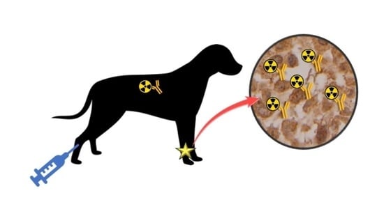
1. Introduction
2. Results
2.1. Clinical and Epidemiological Data
2.2. Histopathology
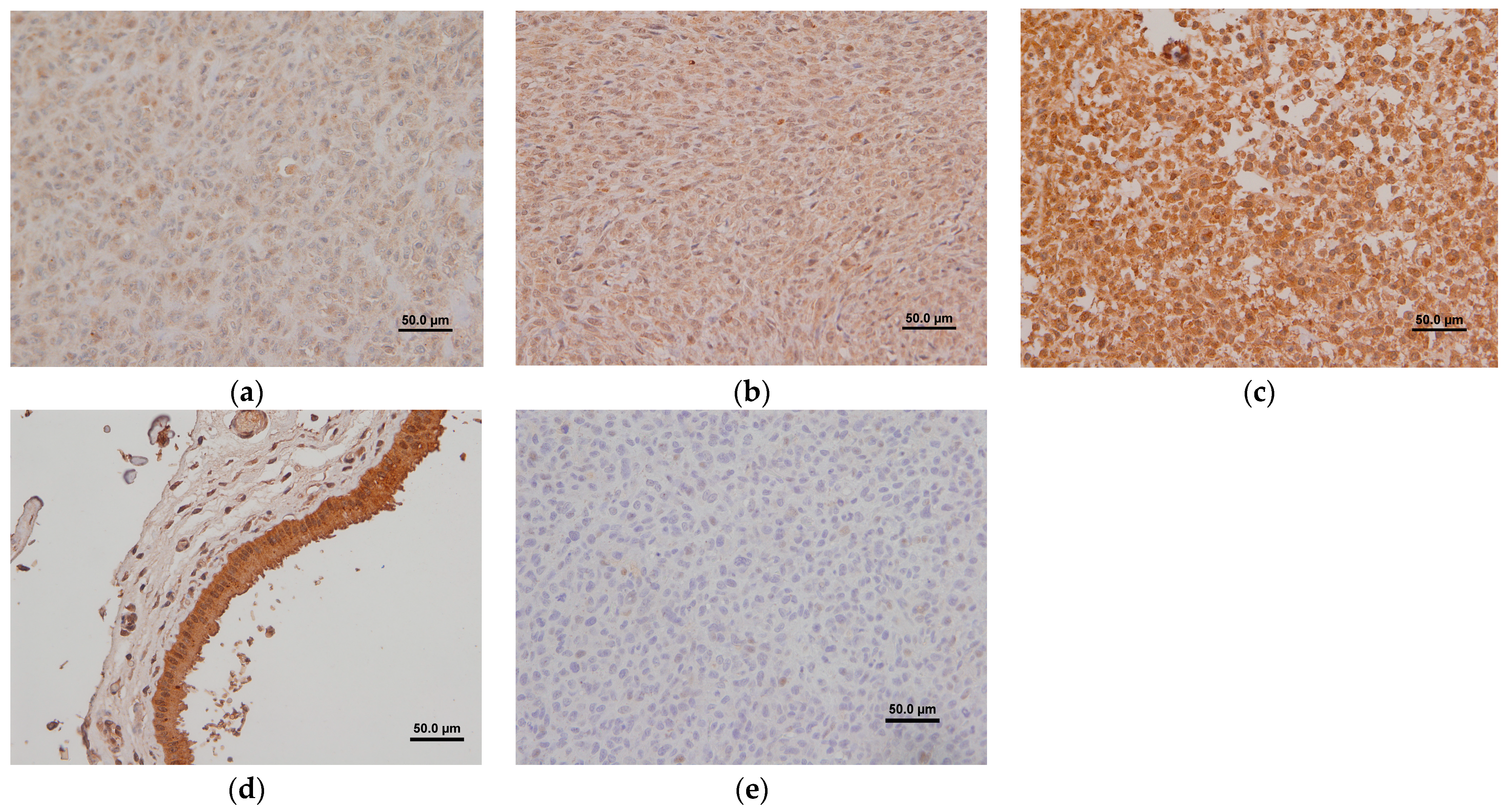
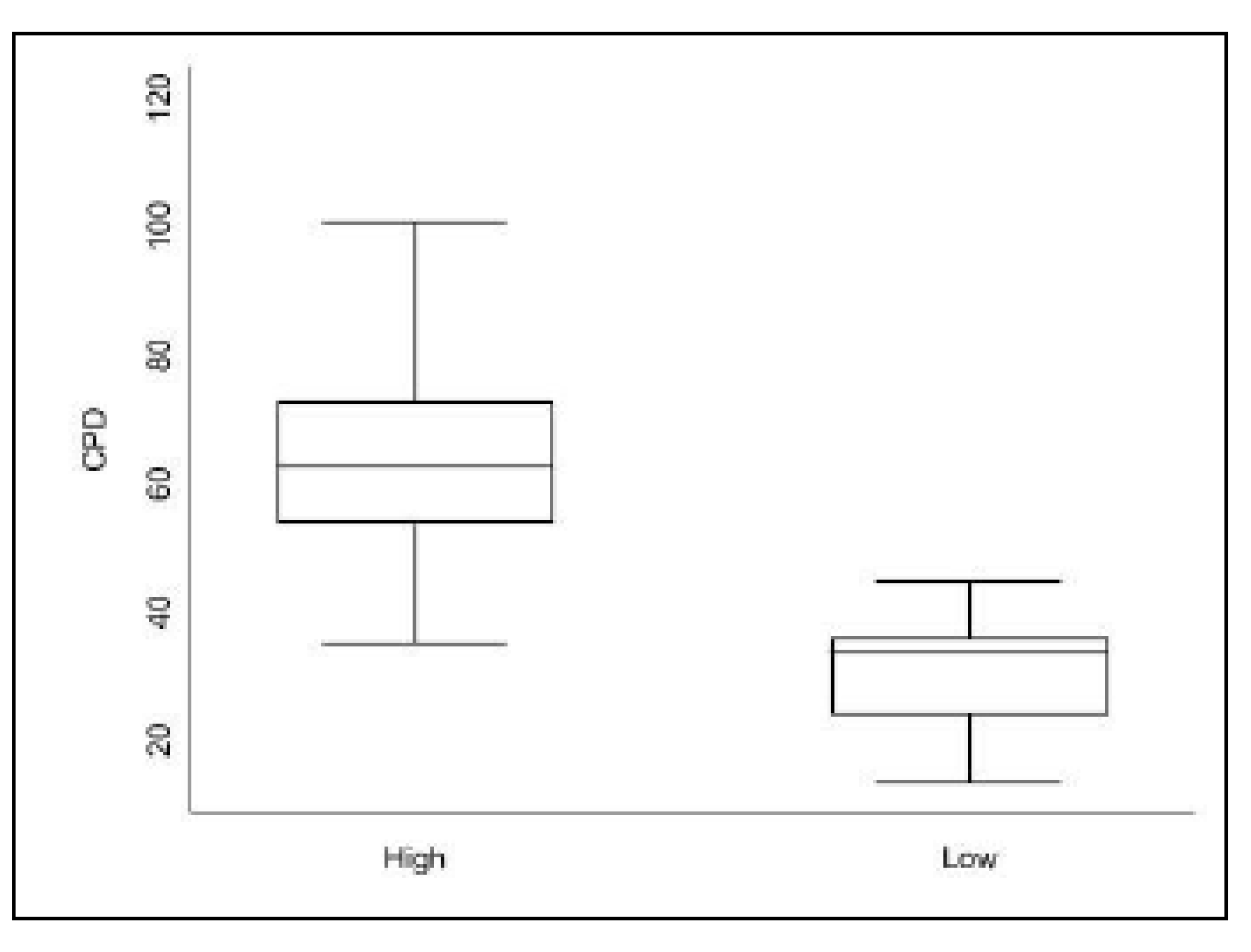
2.3. IGF2R Immunostaining Intensity in Canine OS
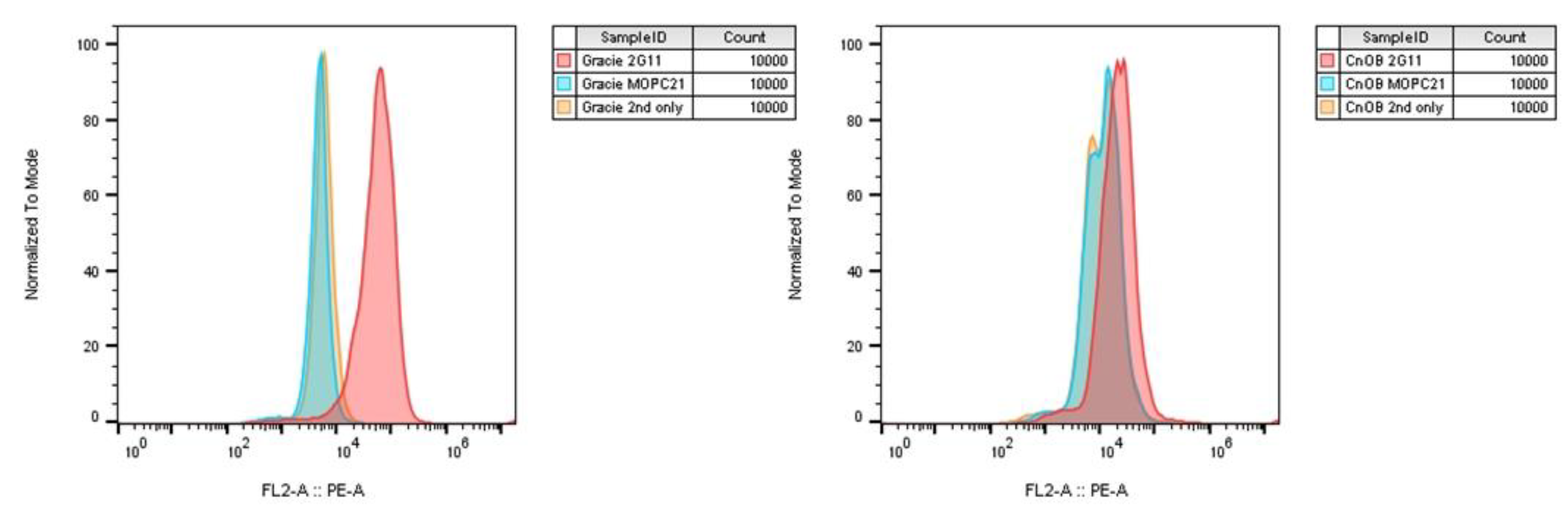
2.4. Flow Cytometry of Non-Neoplastic Canine Osteoblasts (CnOb) and Gracie (Canine OS) Cell Lines
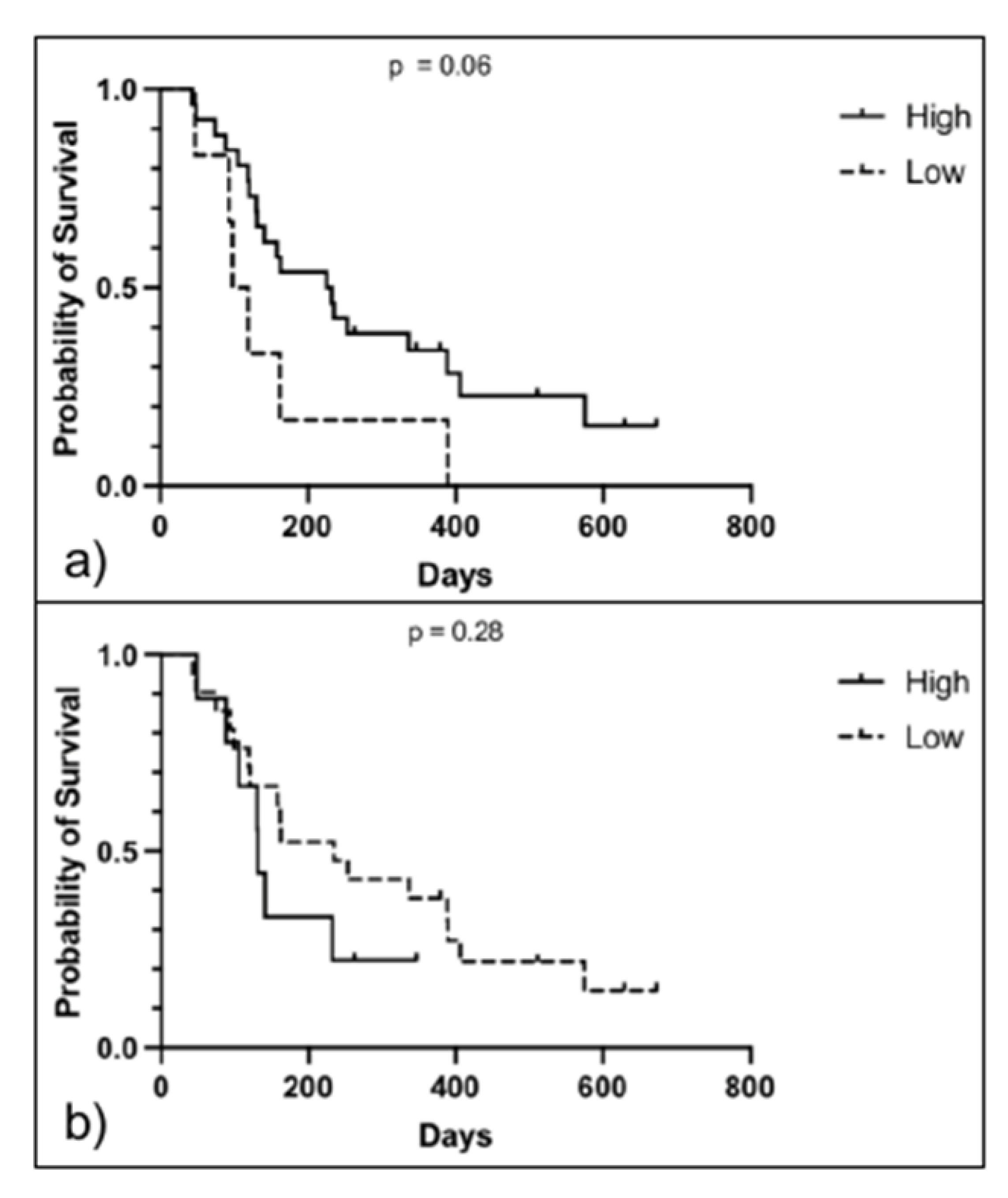
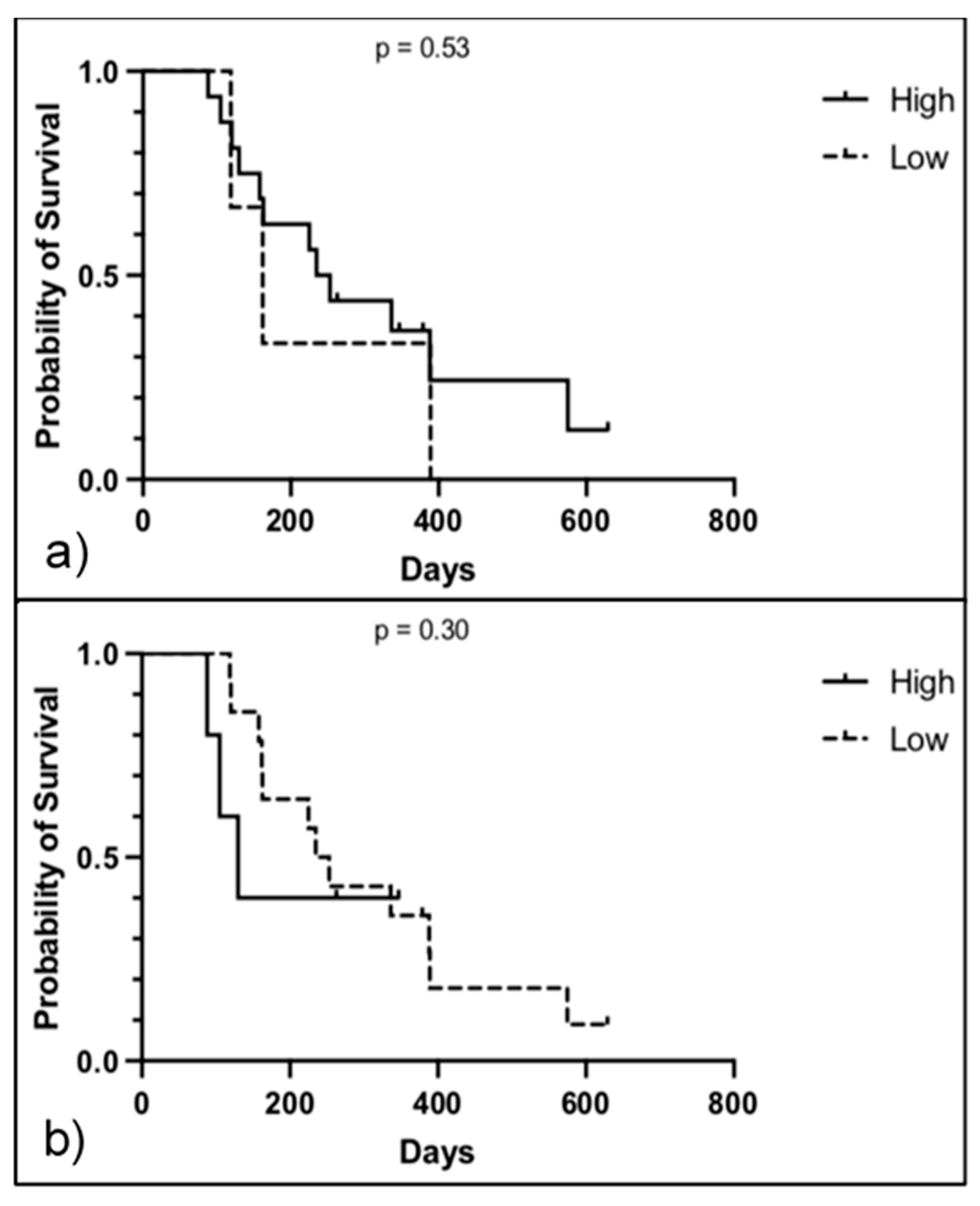
2.5. Survival Analysis Related to Immunostaining Intensity of IGF2R in Canine OS
3. Discussion
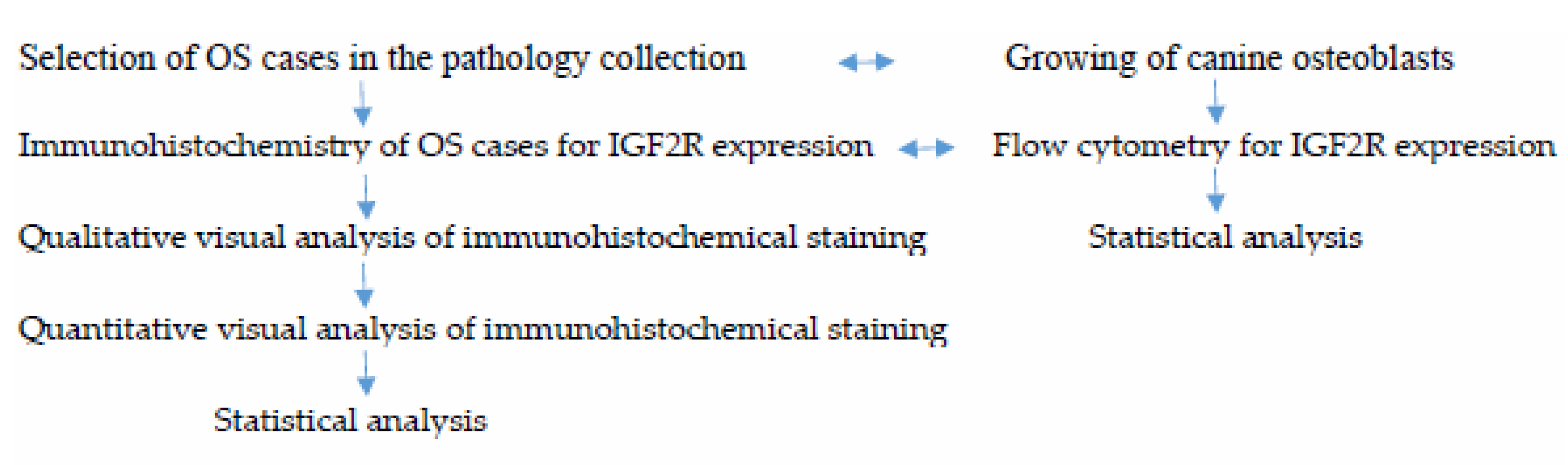
4. Materials and Methods
4.1. Case Selection
4.2. Immunohistochemistry
4.3. Qualitative Visual Analysis of Immunohistochemical Staining
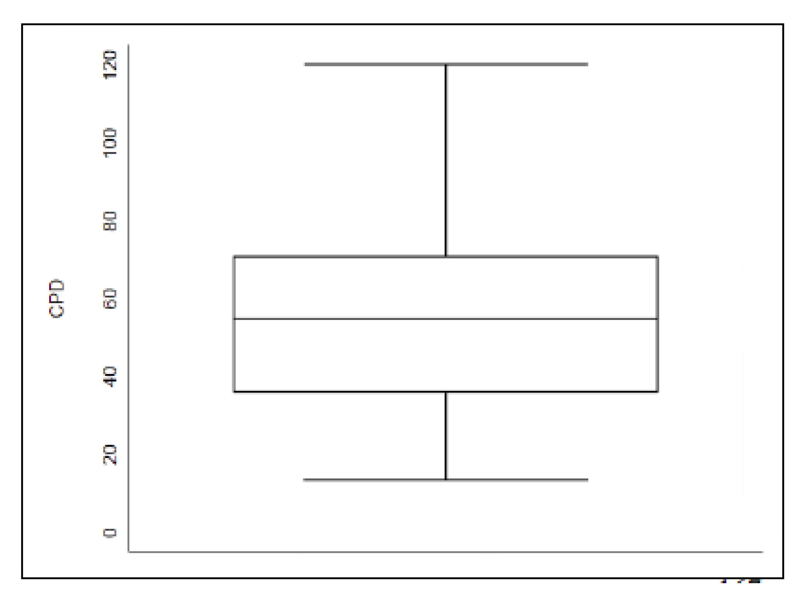
4.4. Quantitative Analysis of Immunohistochemical Staining
4.5. Cell Lines
4.6. Flow Cytometry
4.7. Statistical Analysis
5. Conclusions
Author Contributions
Funding
Institutional Review Board Statement
Informed Consent Statement
Data Availability Statement
Conflicts of Interest
References
- Ehrhart, N.P.; Neil, I.C.; Fan, T.M. Tumors of the skeletal system. In Withrow and MacEwen’s Small Animal Clinical Oncology, 6th ed.; Withrow, S.J., Vail, D.M., Eds.; Saunders: St. Louis, MI, USA, 2020; pp. 524–564. [Google Scholar]
- Liptak, J.M.; Dernell, W.S.; Ehrhart, N.P.; Withrow, S.J. Canine appendicular osteosarcoma: Diagnosis and palliative treatment. Compend. Contin. Educ. Vet. 2004, 26, 172–182. [Google Scholar]
- Mirabello, L.; Troisi, R.J.; Savage, S.A. Osteosarcoma incidence and survival rates from 1973 to 2004: Data from the surveillance, epidemiology, and end results program. Cancer 2009, 115, 1531–1543. [Google Scholar] [CrossRef] [PubMed] [Green Version]
- Spodnick, G.J.; Berg, J.; Rand, W.M.; Schelling, S.H.; Couto, G.; Harvey, H.J.; Henderson, R.A.; MacEwen, G.; Mauldin, N.; McCaw, D.L. Prognosis for dogs with appendicular osteosarcoma treated by amputation alone: 162 cases (1978–1988). J. Am. Vet. Med. Assoc. 1992, 200, 995–999. [Google Scholar] [PubMed]
- Bergman, P.J.; MacEwen, E.G.; Kurzman, I.D.; Henry, C.J.; Hammer, A.S.; Knapp, D.W.; Hale, A.; Kruth, S.A.; Klein, M.K.; Klausner, J.; et al. Amputation and Carboplatin for Treatment of Dogs with Osteosarcoma: 48 Cases (1991 to 1993). J. Vet. Intern. Med. 1996, 10, 76–81. [Google Scholar] [CrossRef] [PubMed]
- Phillips, B.; Powers, B.E.; Dernell, W.S.; Straw, R.C.; Khanna, C.; Hogge, G.S.; Vail, D.M. Use of single-agent carboplatin as adjuvant or neoadjuvant therapy in conjunction with amputation for appendicular osteosarcoma in dogs. J. Am. Anim. Hosp. Assoc. 2009, 45, 33–38. [Google Scholar] [CrossRef]
- Saam, D.E.; Liptak, J.M.; Stalker, M.J.; Chun, R. Predictors of outcome in dogs treated with adjuvant carboplatin for appendicular osteosarcoma: 65 cases (1996–2006). J. Am. Vet. Med. Assoc. 2011, 238, 195–206. [Google Scholar] [CrossRef] [Green Version]
- Farese, J.P.; Milner, R.; Thompson, M.S.; Lester, N.; Cooke, K.; Fox, L.; Hester, J.; Bova, F.J. Stereotactic radiosurgery for treatment of osteosarcomas involving the distal portions of the limbs in dogs. J. Am. Vet. Med. Assoc. 2004, 225, 1567–1572. [Google Scholar] [CrossRef]
- Covey, J.L.; Farese, J.P.; Bacon, N.; Schallberger, S.P.; Amsellem, P.; Cavanaugh, R.; Milner, R.J. Stereotactic Radiosurgery and Fracture Fixation in 6 Dogs with Appendicular Osteosarcoma. Vet. Surg. 2014, 43, 174–181. [Google Scholar] [CrossRef]
- Boston, S.E.; Vinayak, A.; Lu, X.; LaRue, S.; Bacon, N.; Bleedorn, J.A.; Souza, C.H.M.; Ehrhart, N.P. Outcome and complications in dogs with appendicular primary bone tumors treated with stereotactic radiotherapy and concurrent surgical stabilization. Vet. Surg. 2017, 46, 829–837. [Google Scholar] [CrossRef]
- Kim, C.; Matsuyama, A.; Mutsaers, A.J.; Woods, J.P. Retrospective evaluation of toceranib (Palladia) treatment for canine metastatic appendicular osteosarcoma. Can. Vet. J. 2017, 58, 1059–1064. [Google Scholar]
- Morello, E.; Martano, M.; Buracco, P. Biology, diagnosis and treatment of canine appendicular osteosarcoma: Similarities and differences with human osteosarcoma. Vet. J. 2011, 189, 268–277. [Google Scholar] [CrossRef] [PubMed]
- Rowell, J.L.; McCarthy, D.O.; Alvarez, C.E. Dog models of naturally occurring cancer. Trends Mol. Med. 2011, 17, 380–388. [Google Scholar] [CrossRef]
- Simpson, S.; Dunning, M.D.; de Brot, S.; Grau-Roma, L.; Mongan, N.P.; Rutland, C.S. Comparative review of human and canine osteosarcoma: Morphology, epidemiology, prognosis, treatment and genetics. Acta Vet. Scand. 2017, 59, 71. [Google Scholar] [CrossRef] [PubMed]
- Milenic, D.E.; Brady, E.D.; Brechbiel, M.W. Antibody-targeted radiation cancer therapy. Nat. Rev. Drug Discov. 2004, 3, 488–499. [Google Scholar] [CrossRef] [PubMed] [Green Version]
- Larson, S.M.; Carrasquillo, J.A.; Cheung, N.K.; Press, O.W. Radioimmunotherapy of human tumors. Nat. Rev. Cancer 2015, 15, 347–360. [Google Scholar] [CrossRef] [PubMed] [Green Version]
- Sartor, O.; Heinrich, D.; Mariados, N.; Méndez Vidal, M.J.; Keizman, D.; Thellenberg Karlsson, C.; Peer, A.; Procopio, G.; Frank, S.J.; Pulkkanen, K.; et al. Re-treatment with radium-223: 2-year follow-up from an international, open-label, phase ½ study in patients with castration-resistant prostate cancer and bone metastases. Prostate 2019, 79, 1683–1691. [Google Scholar] [CrossRef] [Green Version]
- Xofigo (radiumRa223dichloride) Injection for Intravenous Use [Package Insert]; Bayer HealthCare Pharmaceuticals Inc.: Wayne, NJ, USA, 2013; Available online: http://hcp.xofigo-us.com/index.php (accessed on 17 October 2018).
- Alex, N.E.; Christoph, B. Does 177Lu-labeled octreotate improve the rate of remission of endocrine gastroenteropancreatic tumors? Nat. Clin. Pract. Endocrinol. Metab. 2005, 1, 20–21. [Google Scholar]
- Zandee, W.T.; Feelders, R.A.; Duijzentkunst, D.A.S.; Hofland, J.; Metselaar, R.M.; Oldenburg, R.A.; van Linge, A.; Kam, B.L.R.; Teunissen, J.J.M.; Korpershoek, E.; et al. Treatment of inoperable or metastatic paragangliomas and pheochromocytomas with peptide receptor radionuclide therapy using 177Lu-DOTATATE. Eur. J. Endocrinol. 2019, 181, 45–53. [Google Scholar] [CrossRef]
- Sharkey, R.M.; Goldenberg, D.M. Perspectives on cancer therapy with radiolabeled monoclonal antibodies. J. Nucl. Med. 2005, 46 (Suppl. 1), 115S–127S. [Google Scholar]
- Geller, D.S.; Morris, J.; Revskaya, E.; Kahn, M.; Zhang, W.; Piperdi, S.; Park, A.; Koirala, P.; Guzik, H.; Hall, C.; et al. Targeted therapy of osteosarcoma with radiolabeled monoclonal antibody to an insulin-like growth factor-2 receptor (IGF2R). Nucl. Med. Biol. 2016, 43, 812–817. [Google Scholar] [CrossRef] [Green Version]
- Falls, J.G.; Pulford, D.J.; Wylie, A.A.; Jirtle, R.L. Genomic imprinting: Implications for human disease. Am. J. Pathol. 1999, 154, 635–647. [Google Scholar] [CrossRef] [PubMed] [Green Version]
- Toretsky, J.A.; Helman, L.J. Involvement of IGF-II in human cancer. J. Endocrinol. 1996, 149, 367–372. [Google Scholar] [CrossRef]
- Hassan, A.B.; Howell, J.A. Insulin-like growth factor II supply modifies growth of intestinal adenoma in Apc(Min/+) mice. Cancer Res. 2000, 60, 1070–1076. [Google Scholar] [PubMed]
- Foulstone, E.; Prince, S.; Zaccheo, O.; Burns, J.L.; Harper, J.; Jacobs, C.; Church, D.; Hassan, A.B. Insulin-like growth factor ligands, receptors, and binding proteins in cancer. J. Pathol. 2005, 205, 145–153. [Google Scholar] [CrossRef] [PubMed]
- Hassan, S.E.; Ba, M.B.; Kim, M.Y.; Lin, J.; Bs, S.P.; Gorlick, R.; Geller, D.S. Cell surface receptor expression patterns in osteosarcoma. Cancer 2012, 118, 740–749. [Google Scholar] [CrossRef] [PubMed]
- Savage, S.A.; Woodson, K.; Walk, E.; Modi, W.; Liao, J.; Douglass, C.; Hoover, R.N.; Chanock, S.J.; National Osteosarcoma Etiology Study Group. National Osteosarcoma Etiology Study Group. Analysis of genes critical for growth regulation identifies Insulin- like Growth Factor 2 Receptor variations with possible functional significance as risk factors for osteosarcoma. Cancer Epidemiol. Biomark. Prev. 2007, 16, 1667–1674. [Google Scholar] [CrossRef] [Green Version]
- Brown, J.; Delaine, C.; Zaccheo, O.J.; Siebold, C.; Gilbert, R.; Van Boxel, G.; Denley, A.; Wallace, J.C.; Hassan, A.; Forbes, B.; et al. Structure and functional analysis of the IGF-II/IGF2R interaction. EMBO J. 2008, 27, 265–276. [Google Scholar] [CrossRef] [Green Version]
- Burland, O.S.; Skretting, A.; Solheim, O.P.; Aas, M. Targeted radiotherapy of osteosarcoma using 153SM-Edtmp: A new promising approach. Acta Oncol. 1996, 35, 381–384. [Google Scholar] [CrossRef]
- Popwell, S.J.; Schulz, M.D.; Wagener, K.B.; Batich, C.D.; Milner R., J.; Lagmay, J.; Bolch, W.E. Synthesis of polymeric phosphonates for selective delivery of radionuclides to osteosarcoma. Cancer Biother. Radiopharm. 2014, 29, 273–282. [Google Scholar] [CrossRef]
- Westrom, S.; Bondsdorff, T.B.; Abbas, N.; Bruland, O.S.; Jonasdottir, T.J.; Maelandsmo, G.M.; Larsen, R.H. Evaluation of CD146 as target for radioimmunotherapy against osteosarcoma. PLoS ONE 2016, 11, e0165382. [Google Scholar] [CrossRef] [Green Version]
- Li, H.K.; Hasegawa, S.; Nakajima, J.I.; Morokoshi, Y.; Miengishi, K.; Nagatsu, K. Targeted cancer cell ablation in mice by an α particle emitting astatine-211-labelled antibody again major histocompatibility complex class I chain-related protein A and B. Biochem. Biophys. Res. Commun. 2018, 506, 1078–1084. [Google Scholar] [CrossRef]
- Karkare, S.; Allen, K.J.H.; Jiao, R.; Malo, M.E.; Dawicki, W.; Helal, M.; Godson, D.L.; Dickinson, R.; MacDonald-Dickinson, V.; Yang, R.; et al. Detection and targeting insulin growth factor receptor type 2 (IGF2R) in osteosarcoma PDX in mouse models and in canine osteosarcoma tumors. Sci. Rep. 2019, 9, 11476. [Google Scholar] [CrossRef] [PubMed] [Green Version]
- Boerman, I.; Selvarajah, G.T.; Nielen, M.; Kirpensteijn, J. Prognostic factors in canine appendicular osteosarcoma—A meta-analysis. BMC Vet. Res. 2012, 8, 56. [Google Scholar] [CrossRef] [Green Version]
- Garzotto, C.K.; Berg, J.; Hoffmann, W.E.; Rand, W.M. Prognostic significance of serum alkaline phosphatase activity in canine appendicular osteosarcoma. J. Vet. Intern. Med. 2000, 14, 587–592. [Google Scholar] [CrossRef]
- Ren, H.-Y.; Sun, L.-L.; Li, H.-Y.; Ye, Z.-M. Prognostic Significance of Serum Alkaline Phosphatase Level in Osteosarcoma: A Meta-Analysis of Published Data. BioMed Res. Int. 2015, 2015, 160835. [Google Scholar] [CrossRef] [PubMed] [Green Version]
- Griffin, L.R.; Thamm, D.H.; Brody, A.; Selmic, L.E. Prognostic value of fluorine18 flourodeoxyglucose positron emission tomography/computed tomography in dogs with appendicular osteosarcoma. J. Vet. Int. Med. 2019, 33, 820–826. [Google Scholar] [CrossRef] [Green Version]
- Al-Khan, A.A.; Nimmo, J.S.; Tayebi, M.; Ryan, S.D.; Simcock, J.O.; Tarzi, R.; Kuntz, C.A.; Saad, E.S.; Day, M.J.; Richardson, S.J.; et al. Parathyroid hormone receptor 1 (PTHR1) is a prognostic indicator in canine osteosarcoma. Sci. Rep. 2020, 10, 1564. [Google Scholar] [CrossRef] [Green Version]
- Maniscalco, L.; Iussich, S.; Morello, E.; Martano, M.; Gattino, F.; Miretti, S.; Biolatti, B.; Accornero, P.; Martignani, E.; Sánchez-Céspedes, R.; et al. Increased expression of insulin-like growth factor-1 receptor is correlated with worse survival in canine appendicular osteosarcoma. Vet. J. 2015, 205, 272–280. [Google Scholar] [CrossRef]
- Schott, C.R.; Tatiersky, L.J.; Foster, R.A.; Wood, G.A. Histologic Grade Does Not Predict Outcome in Dogs with Appendicular Osteosarcoma Receiving the Standard of Care. Vet. Pathol. 2018, 55, 202–211. [Google Scholar] [CrossRef] [Green Version]
- Al-Khan, A.; Nimmo, J.; Day, M.; Tayebi, M.; Ryan, S.; Kuntz, C.; Simcock, J.; Tarzi, R.; Saad, E.; Richardson, S.; et al. Fibroblastic Subtype has a Favourable Prognosis in Appendicular Osteosarcoma of Dogs. J. Comp. Pathol. 2020, 176, 133–144. [Google Scholar] [CrossRef]
- Reubi, J.C.; Schaer, J.C.; Markwalder, R.; Waser, B.; Horisberger, U.; Laissue, J. Distribution of somatostatin receptors in normal and neoplastic human tissues: Recent advances and potential relevance. Yale J. Biol. Med. 1997, 70, 471–479. [Google Scholar]
- Sachpekidis, C.; Jackson, D.B.; Soldatos, T.G. Radioimmunotherapy in Non-Hodgkin’s Lymphoma: Retrospective Adverse Event Profiling of Zevalin and Bexxar. Pharmaceuticals 2019, 12, 141. [Google Scholar] [CrossRef] [Green Version]
- Skandalis, S.S.; Labropoulou, V.T.; Ravazoula, P.; Likaki-Karatza, E.; Dobra, K.; Kalofonos, H.P.; Karamanos, N.K.; Theocharis, A.D. Versican but not decorin accumulation is related to malignancy in mammographically detected high density and malignant appearing microcalcifications in non-palpable breast carcinomas. BMC Cancer 2011, 11, 314. [Google Scholar] [CrossRef] [Green Version]
- Damasceno, K.A.; Ferreira, E.; Estrela-Lima, A.; Gamba Cde, O.; Miranda, F.F.; Alves, M.R.; Rocha, R.M.; de Barros, A.L.; Cassali, G.D. HER-2 and EGFR mRNA Expression and Its Relationship with Versican in Malignant Matrix-Producing Tumors of the Canine Mammary Gland. PLoS ONE 2016, 11, e0160419. [Google Scholar] [CrossRef]


| Targeting Molecule | Radionuclide | Advantages | Disadvantages | Reference |
|---|---|---|---|---|
| EDTMP | 153Sm | Ease of synthesis and administration | Lack of targeting soft tissue metastasis | [30] |
| Polymeric Phosphonates | 153Sm | Ease of synthesis and administration | Non-specific targeting of non-osseous tumors based on enhanced permeability and retention (the EPR effect) | [31] |
| Antibody to CD146 | 177Lu | Antigen-specific delivery of the radionuclide to the tumor | Bone marrow as a possible dose-limiting organ | [32] |
| Antibody to human IGF2R | 188Re | Antigen-specific delivery of the radionuclide to the tumor | Antibody binds only to human IGF2R making it difficult to assess toxicity in animal models | [22] |
| Antibody to major histocompatibility complex class I chain-related protein A and B | 211At | Antigen-specific delivery of the radionuclide to the tumor | Availability of 211At is limited to only a few places around the world | [33] |
| Antibody to human, murine, and canine IGF2R | 177Lu | Antigen-specific delivery of the radionuclide to the tumor; possible to use for toxicity evaluation in rodents and dogs | The antibody is murine which precludes its repeated administrations to humans | [34] |
| # | Breed | Sex | Age | Location |
|---|---|---|---|---|
| 1 | Labrador Retriever Cross | FS | 10 | Distal radius |
| 2 | German Shepherd Cross | FS | 9 | Proximal humerus |
| 3 | Boxer Cross | MC | 10 | Femoral head |
| 4 | Boxer | FS | 9.5 | Distal humerus |
| 5 | Rottweiler | MC | 10 | Proximal tibia |
| 6 | Rottweiler | MC | 9 | Distal tibia |
| 7 | Labrador Retriever Cross | FS | 7 | Distal femur |
| 8 | German Shepherd | MC | 9 | Distal radius |
| 9 | Rottweiler | FS | 10 | Proximal femur |
| 10 | Great Dane | FS | 7 | Distal radius |
| 11 | Rottweiler Cross | FS | 9 | Proximal humerus |
| 12 | Rottweiler | FS | 9 | Distal radius |
| 13 | Bull Mastiff | MC | 9 | Proximal humerus |
| 14 | Boxer Cross | FS | 6 | Scapula |
| 15 | Doberman Cross | MC | 7 | Proximal humerus |
| 16 | Border Collie Cross | MC | 9 | Distal radius |
| 17 | Rottweiler | MC | 8 | Mid humerus |
| 18 | Rottweiler | FS | 9 | Distal femur |
| 19 | Pitbull | FS | 8 | Proximal humerus |
| 20 | Goldendoodle | FS | 8.5 | Proximal humerus |
| 21 | Giant Schnauzer Cross | FS | 6 | Distal tibia |
| 22 | Rottweiler Cross | MC | 9 | Distal femur |
| 23 | Shetland Sheepdog | M | 12 | Proximal humerus |
| 24 | Labrador Retriever | FS | 7.5 | Proximal humerus |
| 25 | Golden Retriever | MC | 8 | Proximal humerus |
| 26 | Labrador Retriever Cross | FS | 11 | Proximal humerus |
| 27 | Belgian Malinois Cross | MC | 7 | Distal tibia |
| 28 | Greyhound | MC | 7.5 | Distal radius |
| 29 | Rottweiler | M | 8 | Proximal femur |
| 30 | Rottweiler Cross | FS | 10 | Distal femur |
| 31 | Labrador Retriever | FS | 10 | Distal humerus |
| 32 | German Shepherd | FS | 11 | Proximal humerus |
| 33 | Malamute | MC | 8 | Distal radius |
| 34 | Rottweiler | FS | 6.5 | Proximal humerus |
| OS Subtype | Primary Treatment | ST (Days) | Status | Visual Intensity Score | Corrected Pixel Density | Corrected Pixel Density Score | |
|---|---|---|---|---|---|---|---|
| 1 | Ob OSA | Amp | 43 | dead | High | 69.91 | High |
| 2 | Ob OSA | Amp + Carboplatin (6) | 235 | dead | High | 54.97 | High |
| 3 | Ob OSA | Amp | NA | LFU | Low | 44.62 | Low |
| 4 | Ob OSA | Amp | 47 | dead | Low | 33.67 | Low |
| 5 | Ob OSA | Amp + Carboplatin (2) | 163 | dead | High | 54.92 | Low |
| 6 | Ob OSA | Amp | 131 | dead | High | 71.03 | High |
| 7 | Ob OSA | Amp + Carboplatin (3) | 575 | dead | High | 58.18 | High |
| 8 | Ob OSA | Amp + Carboplatin (6) | 388 | dead | High | 35.13 | Low |
| 9 | Ob OSA | Amp + Carboplatin (2) | 105 | dead | High | 100.89 | High |
| 10 | Ob OSA | Amp + palladia | 93 | dead | Low | 40.14 | Low |
| 11 | Ob OSA | SRT + Carboplatin (6) | 379 | dead | High | 59.55 | High |
| 12 | Ob OSA | Amp + Carboplatin (6) | 253 | dead | High | 42.57 | Low |
| 13 | Ob OSA | Palliative | 74 | dead | High | 50.96 | Low |
| 14 | Ch OSA | Amp | 48 | dead | High | 120.24 | High |
| 15 | Ob OSA | Amp + Carboplatin (4) | 119 | dead | Low | 23.92 | Low |
| 16 | Ob OSA | Amp | 232 | dead | High | 72.73 | High |
| 17 | Ob OSA | Amp | 406 | dead | Low | 35.94 | Low |
| 18 | Ob OSA | Amp | 141 | dead | High | 79.78 | High |
| 19 | Ob OSA | Amp | 672 | dead | High | 21.65 | Low |
| 20 | Fb OSA | Amp + Carboplatin (6) | 162 | dead | Low | 24.55 | Low |
| 21 | Ob OSA | Amp + Carboplatin (2) | 88 | dead | High | 93.68 | High |
| 22 | Ch OSA | Amp + Carboplatin (6) | 336 | dead | High | 62.52 | High |
| 23 | Ob OSA | Amp + Carboplatin (3) | 347 | alive | High | 71.82 | High |
| 24 | Ob OSA | Amp + CHOP | 98 | dead | Low | 17.55 | Low |
| 25 | Ob OSA | Amp + Carboplatin (6) | 389 | dead | Low | 34.34 | Low |
| 26 | Ob OSA | Amp + Carboplatin (3) | 130 | dead | High | 112.58 | High |
| 27 | Ob OSA | Amp + Carboplatin (6) | 629 | alive | High | 41.28 | Low |
| 28 | Ch OSA | BPN + palliative RT + Carbo (6) | 511 | alive | High | 64.92 | High |
| 29 | Ob OSA | Euthanasia | 0 | dead | Low | 13.40 | Low |
| 30 | Ob OSA | Euthanasia | 0 | dead | High | 65.77 | High |
| 31 | Ob OSA | Amp + Carboplatin (6) | 120 | dead | High | 57.58 | High |
| 32 | Ob OSA | Amp + Carboplatin (1) | 158 | dead | High | 53.74 | Low |
| 33 | Ob OSA | Amp + Carboplatin (1) | 225 | dead | High | 52.99 | Low |
| 34 | Ob OSA | Amp + Carboplatin (4) | 263 | alive | High | 72.69 | High |
Disclaimer/Publisher’s Note: The statements, opinions and data contained in all publications are solely those of the individual author(s) and contributor(s) and not of MDPI and/or the editor(s). MDPI and/or the editor(s) disclaim responsibility for any injury to people or property resulting from any ideas, methods, instructions or products referred to in the content. |
© 2023 by the authors. Licensee MDPI, Basel, Switzerland. This article is an open access article distributed under the terms and conditions of the Creative Commons Attribution (CC BY) license (https://creativecommons.org/licenses/by/4.0/).
Share and Cite
Boisclair, C.; Dickinson, R.; Giri, S.; Dadachova, E.; MacDonald-Dickinson, V. Characterization of IGF2R Molecular Expression in Canine Osteosarcoma as Part of a Novel Comparative Oncology Approach. Int. J. Mol. Sci. 2023, 24, 1867. https://doi.org/10.3390/ijms24031867
Boisclair C, Dickinson R, Giri S, Dadachova E, MacDonald-Dickinson V. Characterization of IGF2R Molecular Expression in Canine Osteosarcoma as Part of a Novel Comparative Oncology Approach. International Journal of Molecular Sciences. 2023; 24(3):1867. https://doi.org/10.3390/ijms24031867
Chicago/Turabian StyleBoisclair, Charles, Ryan Dickinson, Sabeena Giri, Ekaterina Dadachova, and Valerie MacDonald-Dickinson. 2023. "Characterization of IGF2R Molecular Expression in Canine Osteosarcoma as Part of a Novel Comparative Oncology Approach" International Journal of Molecular Sciences 24, no. 3: 1867. https://doi.org/10.3390/ijms24031867