Review

The Impact of Neurotransmitters on the Neurobiology of Neurodegenerative Diseases

Department of Drug Discovery and Biomedical Sciences, College of Pharmacy, University of South Carolina, 715 Sumter Street, Columbia, SC 29208, USA
*
Author to whom correspondence should be addressed.
Int. J. Mol. Sci. 2023, 24(20), 15340; https://doi.org/10.3390/ijms242015340
Submission received: 26 September 2023 / Revised: 16 October 2023 / Accepted: 17 October 2023 / Published: 19 October 2023
(This article belongs to the Collection Feature Papers in Molecular Neurobiology)

Abstract

:
Neurodegenerative diseases affect millions of people worldwide. Neurodegenerative diseases result from progressive damage to nerve cells in the brain or peripheral nervous system connections that are essential for cognition, coordination, strength, sensation, and mobility. Dysfunction of these brain and nerve functions is associated with Alzheimer’s disease, Parkinson’s disease, Huntington’s disease, Amyotrophic lateral sclerosis, and motor neuron disease. In addition to these, 50% of people living with HIV develop a spectrum of cognitive, motor, and/or mood problems collectively referred to as HIV-Associated Neurocognitive Disorders (HAND) despite the widespread use of a combination of antiretroviral therapies. Neuroinflammation and neurotransmitter systems have a pathological correlation and play a critical role in developing neurodegenerative diseases. Each of these diseases has a unique pattern of dysregulation of the neurotransmitter system, which has been attributed to different forms of cell-specific neuronal loss. In this review, we will focus on a discussion of the regulation of dopaminergic and cholinergic systems, which are more commonly disturbed in neurodegenerative disorders. Additionally, we will provide evidence for the hypothesis that disturbances in neurotransmission contribute to the neuronal loss observed in neurodegenerative disorders. Further, we will highlight the critical role of dopamine as a mediator of neuronal injury and loss in the context of NeuroHIV. This review will highlight the need to further investigate neurotransmission systems for their role in the etiology of neurodegenerative disorders.

1. Introduction

The prevalence of neurodegenerative disorders (NDs) is increasing in the United States. It is projected that 13.8 million people will be affected by Alzheimer’s disease (AD) by 2050 [1]. Additionally, the incidence of Parkinson’s disease (PD) has increased from 1990 to 2019 [2]. FDA-approved medications for alleviating the symptoms of AD and PD involve the rebalance of neurotransmitter (NT) systems. For instance, in AD patients, acetylcholine esterase inhibitors are used to increase acetyl choline (ACh) available for signaling [3]. Moreover, levodopa (L-DOPA), along with monoamine oxidase (MAO) inhibitors and dopamine (DA) agonists, have been used clinically as major medications for the treatment of symptoms associated with PD [4]. The current generation of medication in development for NDs is being designed to target underlying disease pathogenesis and pathology rather than alleviating disease-induced symptoms. For the treatment of AD, Aducanumab was approved for medical use in the United States by FDA in 2021. Aducanumab is a medication used in the management and treatment of AD, which is an amyloid-β (Aβ) -directed monoclonal antibody [5]. However, the use of medications targeting Aβ is controversial, as this class of drugs has not been highly efficacious at rescuing cognitive decline in AD patients. Regardless, the development of drugs that target the underlying etiology of NDs, which function to prevent neuronal injury, is of great clinical interest.
While the etiology of different NDs varies, a unifying feature of NDs is the activation of downstream pathways that activate cellular death, including but not limited to apoptosis and pyroptosis. Activation of these pathways is brought about by neurotoxic molecules which promote inflammation and/or mitochondrial dysfunction. Importantly, dysregulation of NTs induces such neurotoxic molecules. For example, methamphetamine (meth) dysregulates DA homeostasis via interaction with the DA transporter (DAT), leading to autooxidation of DA and the production of neurotoxic reactive oxygen species (ROS). The meth-induced production of ROS from dysregulation of DA metabolism has been directly linked as the cause of meth-induced neurotoxicity [6].
We propose there are two paths underlying NT-mediated neurotoxicity. First, the direct path involves neurotoxic NT metabolites, which can directly influence cell stress response pathways. As described above, neurotoxicity and cell death induced by meth represents the direct pathway for NT-mediated neurotoxicity. This phenomenon has been observed in the context of PD, where inappropriately formed DA metabolites in the brain were found to be directly responsible for mediating alpha-synuclein (αSyn) induced cell—death [7]. Second, the indirect path involves the overactivation of NT receptors by dysregulated NT systems. Specifically, the inappropriate activation of G-protein coupled receptors (GPCRs) can increase intracellular Ca2+ levels. Increased Ca2+ can then perturb mitochondrial function and activate mitochondrial-induced apoptosis [8]. In this review, we will discuss the dopaminergic and cholinergic neurotransmission, two NT systems commonly dysregulated in NDs. Further, we will discuss evidence for both pyroptosis and mitochondrial-mediated apoptosis in NDs. We will also review the possible mechanisms by which dysregulated NT systems observed in NDs can activate downstream pathways to bring about neuronal death. Lastly, we will highlight the role of DA as a critical mediator of both synaptodendritic injuries as well as neuronal loss observed in HIV-positive persons. With this review, we aim to shed light on how the dysregulation of NT systems impacts the neurobiology of NDs.

2. Dopaminergic Neurotransmission

2.1. Regulation of Dopamine Synthesis

DA is a neurotransmitter that is involved in several pathways in the brain, including the nigrostriatal, mesolimbic, and mesocortical projections. The production of DA, its vesicular localization and release, and its extracellular persistence are regulated by the activity of tyrosine hydroxylase (TH), the vesicular monoamine transporter (VMAT-2), and the DAT [9]. DA is produced from the aromatic amino acid tyrosine, after which it is converted by the rate-limiting enzyme TH into L-3,4-dihydroxyphenylalanine (L-DOPA). L-dopa is then converted into DA via the enzyme aromatic amino acid decarboxylase (AADC) [10]. Once in dopaminergic terminals, DA molecules are sequestered and packaged into secretory vesicles via transport proteins [11]. The signal termination and degradation mechanisms for DA transmission include enzymatic processing by monoamine oxidase (MAO-B), which is located inside of the presynaptic terminal, and catechol-o-methyltransferase (COMT), which is located inside the synaptic cleft [9,12]. When DA is released into the synapse, it can bind to both presynaptic and postsynaptic DA receptors, after which it is transported back into the presynaptic terminal by the DAT. Once in the cytosol, DA will either be repackaged into synaptic vesicles or become degraded [13].

2.2. Dopamine Transporter Regulation

DA receptors can be divided into either D1- or D2-like receptor families. The D1 type family includes G protein-coupled receptors D1 and D5, which are excitatory and expressed post-synaptically. D1-like receptors are coupled to the stimulatory G-protein Gs and can induce excitatory transmission by upregulating adenylate cyclase activity and subsequent production of cyclic AMP (cAMP). Conversely, the D2-like family includes D2, D3, and D4 receptors, which are expressed in both presynaptic and postsynaptic terminals. D2-like receptors are coupled to the G protein Gi and induce inhibitory transmission by reducing cAMP production via inhibition of adenylate cyclase activity [14]. DA signaling is primarily terminated by the reuptake of DA by DAT into the cytosol and then by VMAT-2, which transports DA from the cytosol into synaptic vesicles [9]. DAT is a member of the Na+/Cl coupled neurotransmitter solute carrier 6 (SLC6) family and is highly conserved among humans and other mammals [9,12]. The DAT consists of 12 transmembrane helices and large N and C terminal tails located intracellularly, which contain sites for post-translational modifications [15,16]. The kinetics of DA transport through DAT follow the Michaelis-Menten model [17]. DAT transports DA into the cytosol by a channel-like mechanism involving the transition between inward-facing and outward-facing confirmations [12,15,18]. This alternation of conformations is accomplished by the symport of two sodium ions and one chloride ion into the cytosol along their concentration gradients, which are established by the Na+/K+-ATPase [19]. First, DAT occupies the “outward facing” conformation wherein substrates bind to their uptake sites on the extracellular side of the transporter. Once the substrate binds the DAT, a conformational change occurs wherein DAT occupies the “inward facing” conformation, and the translocates into the neuron.
Dopaminergic neurotransmission is altered by DAT-binding pharmacological agents which bind near the DA uptake site, including cocaine and meth [12,17]. Specifically, cocaine competitively inhibits the DA uptake site on DAT and induces DAT conformational transition at the outward-facing stage, leading to increased extracellular DA levels. Conversely, meth-like drugs act as a DAT substrate and stabilize the inward-facing conformation of DAT, thus causing the efflux of DA through DAT from the intracellular to the extracellular space. While these drugs interact with DAT near the DA uptake site, modulation of DAT function at allosteric binding sites also alters DA transport dynamics. Currently, the development of allosteric modulators for the DAT is being investigated for their therapeutic potential for substance use disorder and comorbid HIV-1 infection [20,21].
Post-translational modification of DAT by phosphorylation is important for modifying DAT function [9,22]. Specifically, the four threonine (Thr43, 46, 48, 62) and eight serine (Ser 2, 4, 7,13, 14, 44, 45, 64) residues on the N terminus of DAT can undergo phosphorylation by protein kinase C (PKC), protein kinase A (PKA), protein kinase G, and Ca2+ calmodulin protein kinase II [9,23]. PKC-mediated phosphorylation of DAT regulates DAT endocytosis, transport, and efflux. Specifically, PKC can phosphorylate serine residues on the intracellular N-terminus tail [15,22,24]. PKC-mediated phosphorylation of DAT changes DAT function by stimulating clathrin and dynamin-mediated endocytosis to remove DAT from the surface of the plasma membrane [25,26]. Another kinase that may modify DAT function is the extracellular signal-regulated Kinase (ERK). ERK phosphorylates the proline residue next to Thr53, thus altering DAT structure and increasing DAT’s DA transport capacity [15].
Other post-translational biochemical modifications to DAT can also regulate the protein’s function, including glycosylation, palmitoylation, and ubiquitination. Glycosylation of asparagine residues located on the extracellular loop of DAT helps to stabilize DAT localization to the plasma membrane and increases DA uptake via DAT. Further, glycosylation of DAT is increased with age and has been shown to play an important role in the susceptibility of substance use disorders [9]. Palmitoylation of C-terminus residues to add saturated fatty acids to DAT functions to reduce PKC-mediated endocytosis of DAT. Specifically, palmitoyl acyltransferases known as DHHC enzymes (2, 3, 8, 15 and 17) covalently attach a palmitoyl group via a thioester bond to the Cys580 reside of DAT on the C-terminus. This, in turn, stabilizes DAT expression and uptake kinetics [27]. Lastly, ubiquitination of the lysine residues on the N terminus by ubiquitin E3 ligases Nedd4-2 and Parkin regulates the recycling and degradation of DAT [22].
In addition to biochemical modifications, DAT function is also regulated through protein–protein interactions, such a D2 receptors (D2R) and sigma receptors. D2R expressed in presynaptic terminals can interact with the N-terminus of DAT via its intracellular loop. D2R interaction with DAT has been shown to enhance DA uptake and facilitate recruitment of DAT to the plasma membrane [28], and further, has been attributed to DA-induced neurotoxicity [29]. Sigma receptors expressed in dopaminergic regions have been shown to affect DA synthesis, release, and reuptake in the substantia nigra (SNr) and the ventral tegmental area (VTA) [30,31]. There are two established sigma receptor types, σ1R and σ2R. The σ1R functions to regulate Ca2+ homeostasis; however, σ1R, which is generally localized near the plasma membrane, has been shown to regulate several different transmembrane protein activities through direct protein–protein interaction. Regarding dopaminergic proteins, σ1R has been shown to directly modulate D1 and D2 receptors, as well as DAT. The impact of σ1R interaction with DAT on DA transport remains unclear, but evidence does suggest that σ1R activation may increase DA uptake through DAT [31]. Considering the importance of σ1R in mediating the effects of DAT inhibitors such as meth on DA release, characterizing the σ1R/DAT interaction is of significant interest [32].

2.3. Vesicular Monoamine Transporter Regulation

Vesicular monoamine transporters, including VMAT-1 and VMAT-2, belong to the SLC18 family of vesicular amine transporters. VMAT-1 is primarily expressed in neuroendocrine cells, whereas VMAT-2 is expressed in the central peripheral and enteric nervous systems and is localized in presynaptic terminals [9,33]. VMAT-2 plays a critical role in sorting, storing, and releasing NTs in order to protect neurons by counteracting intracellular toxicity [34,35]. Generally, VMAT-2 inhibitors are classified as being either “reserpine-” (irreversible) or “tetrabenazine” (TBZ)-like [36,37]. VMAT-2 is critical for mediating the psychoactive effects of amphetamines. Amphetamines both inhibit DA uptake through VMAT-2 at reserpine sites and release DA from vesicles into the cytosol, which is then reverse transported through DAT [38]. The loading of NTs into vesicles by VMAT-2 occurs against their concentration gradient. Therefore, VMAT-2 functions as an H+-antiporter, releasing two protons from the vesicle for every monoamine translocated into the vesicle. This process is accomplished using proton gradients, which are established using H+- ATPases located on the lumen of vesicles [35]. Amphetamine-induced release of DA from vesicles is a result of disrupting this proton gradient.
VMAT-2 localization and function are regulated by post-translational modifications, including glycosylation and phosphorylation. VMAT-2 is synthesized in the ER and then glycosylated in the Golgi apparatus. Colocalization of the N-linked glycosylation loop (located between TM1 and TM2) and the C-termini tail of VMAT-2 is necessary for the localization of VMAT-2 to large dense core vesicles [39]. Further, phosphorylation of serine residues near the C-terminus of VMAT-2 by serine/threonine-directed kinase CKII is also thought to contribute to VMAT-2 localization to large dense core vesicles, as evidenced in PC12 cells [40]. Lastly, phosphorylation is also suspected to regulate VMAT-2 function. Phosphorylation of N-terminal residues Ser15 and Ser18 by PKC was found to be necessary for meth-stimulated efflux of 5-HT [41]. Thus, the N-terminal and C-terminal domains of VMAT-2 located in the cytoplasm are important for regulating VMAT-2 localization as well as function.

3. Cholinergic Neurotransmission

3.1. Regulation of Acetylcholine Transmission

Cholinergic neurotransmission is essential for cognitive function, synaptic plasticity, and memory [42,43]. Acetylcholine (ACh) is released by cholinergic neurons, which are primarily located in the subcortical regions with projections to the cortical areas [44,45]. Choline acetyltransferase (ChaT) is responsible for ACh synthesis and is required for cholinergic neurotransmission in the central and peripheral nervous system [46]. ChaT is produced in the soma and then localized mainly in the axon terminal, where it catalyzes the synthesis of ACh from choline and acetyl-CoA [44,47]. Like DA release, the exocytotic release of ACh is dependent on the SNARE protein complex [48]. After exocytotic release, ACh interacts with cholinergic receptors to relay information in the CNS and PNS and mediate synaptic transmission at neuromuscular junctions [49]. Acetylcholinesterase (AChE), primarily found at postsynaptic neuromuscular junctions, metabolizes ACh in the extracellular space into acetate and choline, thus terminating the ACh signal [50]. Inhibition of AChE causes an accumulation of ACh in the synaptic cleft and continuous activation of the cholinergic receptors. Therefore, AChE inhibitors are used in various pharmacological treatments, including the treatment of AD, which is characterized by deficits in ACh levels [44]. However, inappropriate inhibition of AChE has negative consequences, as increased ACh can lead to overstimulation of cholinergic receptors and have harmful effects [51]. Finally, released choline in the synaptic cleft is transported back into synaptic terminals by the choline transporter (CHT). The CHT is a sodium-coupled transporter containing 13 transmembrane domains and belongs to the SLC5A7 family of glucose transporters. Regulation of choline uptake is primarily accomplished through the trafficking of CHT to the plasma membrane. CHT recycling occurs via clathrin-mediated endocytosis, wherein endosomes either reenter the synaptic vesicle cycle or mature into lysosomes, upon which CHT is then degraded [52].

3.2. Acetylcholine Signal Transduction

There are two types of cholinergic receptors: nicotinic and muscarinic receptors [51]. Nicotinic acetylcholine receptors (nAChRs) are found in the central and peripheral nervous system and neuromuscular junction [51,53]. nAChRs are formed by the assembly of five transmembrane subunits. Here, we mainly discuss neuronal nAChRs, which are assembled either as homo-pentamers of α7, α8, and α9 or hetero-pentamers of α2–α6 in combination with β2–β4 or α9 with α10 subunits [54,55].
Activation of nAChRs initiates the opening of an ion pore channel, allowing sodium and calcium influx and potassium efflux across the cell membrane, which in turn regulates neuronal membrane potential and excitability [42]. The nAChRs-mediated ion channel activation initiates a tertiary conformational transition of nAChRs, which includes distinct resting, open, and desensitized states. These conformational state transitions are dynamic and depend on nAChRs subtype and agonist concentration [56]. The most predominant subtypes of nAChRs expressed in the human brain are the heteromeric α4β2 and the homomeric α7 [57,58]. The α4β2 subtype is important for mediating nicotinic effects on DA in the mesolimbic system, whereas the α7 subtype is involved in glutamatergic and dopaminergic release in the CNS. Both α4β2 and α7 subtypes contribute to the pathogenesis of a range of neurological disorders, including AD, schizophrenia, PD, and depression [59,60,61].
The muscarinic receptor family includes five different metabotropic G-coupled protein receptors involved in the parasympathetic nervous system in the brain, which can activate a multitude of signaling pathways important for the modulation of neuronal excitability, synaptic plasticity, and feedback regulation of ACh release [44,51]. Receptors M1, M3, and M5 are coupled with a Gq subunit, leading to IP3-mediated Ca2+ release through phospholipase C activation, while M2 and M4 receptors are coupled with a Gi subunit and function to inhibit adenylate cyclase and prevent the formation of cAMP [14]. For example, M2 can inhibit beta-adrenergic stimulated relaxation, and further, M3 plays a role in the control of the contraction of airway smooth muscle [62]. In the CNS, muscarinic receptors are located both pre- and post-synaptically on neurons. Expression patterns of muscarinic receptor subtypes vary across the CNS. Due to the role of ACh in cognition and substance use disorders, drugs that modulate the function of muscarinic receptors are of great interest and have been reviewed extensively [63].

3.3. Vesicular Acetylcholine Transporter Regulation

At the presynaptic terminal, synthesized ACh is packaged into synaptic vesicles for storage by the vesicular acetylcholine transporter (VAChT). VAChT is the third member of the SLC18 gene group, SLC18A3. The structure of VAChT includes 12 transmembrane domains, which are bundled into two groups (TM1–6 and TM7–12), with the N- and C-termini extending into the cytoplasm [64]. VAChT functions to transport ACh against its concentration gradient by coupling ACh transport to the efflux of two protons [65]. These protons are transported with their concentration gradient, which is established by an H+-ATpase. ACh release is triggered by Ca2+ influx through specific presynaptic Ca2+ channels. VAChT trafficking is regulated by a di-leucine motif on its C-terminus, which helps to localize VAChT to synaptic vesicles. One well-established drug that targets VAChT function is vesicamol, a cell-permeable non-competitive inhibitor that interacts with the C-terminus of VAChT to prevent repackaging of ACh into vesicles. In the context of NDs, decreased VAChT mRNA levels have been observed in patients with AD compared to healthy controls [66]. Decreased VAChT availability reduces the amount of ACh available for release, thus negatively impacting memory. Henceforth, changes in VAChT expression may underlie observed deficits in memory associated with AD.

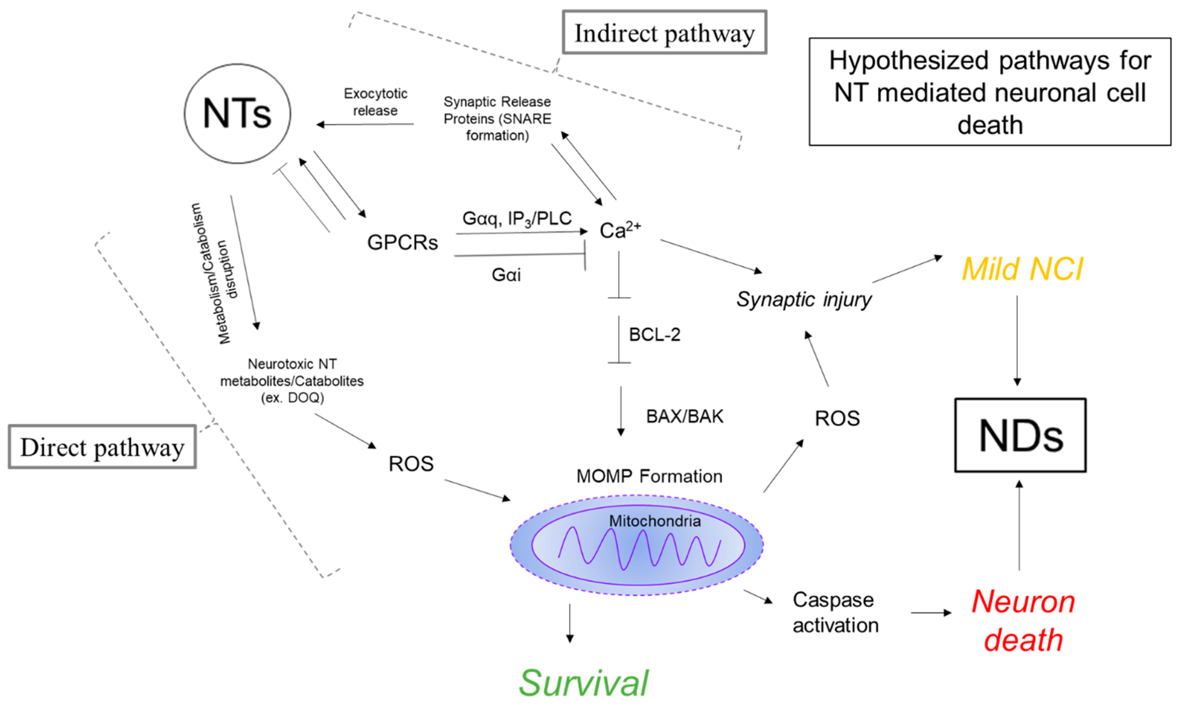
4. Neurotransmitter Hypothesis of Neurodegeneration

4.1. Evidence for Pyroptosis in Neurodegenerative Diseases

One major form of cell death evidenced in NDs is the inflammatory-mediated process called pyroptosis [67]. Pyroptosis is caused by the activation of inflammatory caspase-1/4/5 or 11) and is distinct from traditional apoptosis, as pyroptosis leads to the release of inflammatory cytokines [68]. Specifically, the cytosolic pattern recognition receptor (PRR) protein NLRP3-mediated inflammasome has been implicated as a mediator for NDs [69,70]. In the context of PD, the release of the inflammatory cytokine IL-1β is promoted by the misfolded αSyn protein, a key hallmark of PD. IL-1β primes NLRP3-inflammasomes for activation by αSyn, thus producing pyroptosis [71]. The canonical proteins thought to mediate AD, Aβ, and phosphorylated Tau have also been shown to initiate NLRP3- and NLRP1-mediated neuronal pyroptosis [72,73]. Recent evidence identified cleaved Gasdermin D (GSDMD), a marker for pyroptosis, in microglia and astrocytes near local neuronal loss sites and Aβ plaques in post-mortem brain tissue of AD patients [74]. Lastly, Huntington’s disease (HD) is characterized by a repeat expansion of CAG (>35) in the IT15 gene, which encodes for a protein associated with axonal transport [75]. Several forms of neuronal cell death, including necroptosis, ferroptosis, and apoptosis, have been implicated in HD. Recent evidence also suggests the role of pyroptosis in HD, as evidenced by reduced disease progression in an HD mouse model treated with the NLRP3 inhibitor MCC950 [76,77]. While multiple forms of neuronal cell death are involved in the neurodegeneration of NDs, activation of NLRP3 inflammasomes is of great interest in the pathophysiology of neurodegeneration.

4.2. Dysregulation of Neurotransmission in Neurodegenerative Disorders

AD is a neurodegenerative disorder characterized by memory loss and behavioral and psychological symptoms. An imbalance of different NTs, including glutamate (Glu), γ-Aminobutyric acid (GABA), ACh, DA, and 5-HT, is well evidenced in AD. For example, cholinergic neuronal loss and decreased molecules responsible for ACh metabolism and neurotransmission, such as acetylcholine esterase, are observed in AD [78]. Furthermore, monoaminergic systems are reduced in patients with AD, which may be due to increased MAO activity observed in at-risk persons carrying the ε4 allele of apolipoprotein E [79]. Specifically, disturbances in the dopaminergic system have been evidenced in both AD and PD. Moreover, patients with AD have reduced D1R and D2R expression [80] and selective neuronal loss in the VTA [78]. Regarding the major neurotransmitters in the CNS, Glu, and GABA, Aβ peptides can impact glutamatergic (excitatory) neurotransmission through several routes, including inhibition of Glu reuptake, enhanced Glu release, and dysregulation of Glu/glutamine synthesis [81]. Circulating levels of GABA, which are responsible for the inhibition of synaptic firing, are reduced in the CSF of AD patients. A feedback loop between GABAA receptor activation and an increase in neurofibrillary tangles (NFTs) as a result of increased tau phosphorylation has been postulated as a contributor to AD pathology [82].
In PD, neuronal loss is restricted to dopaminergic neurons, primarily in the SNr [83]. Specifically, neurotoxic dopaminergic metabolites (primarily aminochrome and 5,6-indolequinone) are hypothesized to mediate dopaminergic loss due to the adducts they form with αSyn [84]. Indeed, DA metabolites and catabolites are a major source of oxidative stress observed in PD and are elevated in clinical CSF samples from PD patients compared to healthy controls [85]. Together, these present a possible role for dysregulation of DA homeostasis in the pathology of PD. Further, the serotonergic system is implicated in the difficulty of treating PD. While it is controversial whether PD pathology directly impacts 5-HT systems, it has been reported that the serotonergic system plays a vital role in L-DOPA-induced dyskinesias (LID) by promoting the non-physiological release of DA [86]. Understanding how serotonergic pathways influence dopaminergic release and motor function is of critical interest for improving therapeutic interventions for PD.

4.3. Evidence for Neurotransmission as a Mediator of Neurodegeneration

4.3.1. Calcium Function in the Neuron

Due to its electrochemical nature and ability to interact with complex binding sites, Ca2+ plays a variety of roles in the body, serving as a versatile carrier of information in many signaling networks [87]. Here, we will mainly focus on Ca2+ as it functions in neurons and in the context of NDs. In neurons, Ca2+ regulates the exocytotic release of NTs from vesicles. Voltage-gated channels (Cav) expressed on the plasma membrane of axon terminals are activated in response to membrane depolarization. Upon activation, these channels allow for the influx of Ca2+ ions into cytosolic space. Various Cav channels have been identified and are characterized based on the threshold of depolarization required for their activation. In synapses, there are two primary Cavs responsible for Ca2+ influx, Cav 2.1 and Cav 2.2, and are differentiated by their specific α1 subunit composition (Cav 2.1 is α1A, Cav 2.2 is α1B) [88]. Vesicles that are “primed” at the plasma membrane in the active zone undergo fusion in response to Ca2+ binding. Specifically, Ca2+ causes synaptotagmin-1 to be released from the SNARE (soluble N -ethylmaleimide sensitive factor attachment protein receptor) complex. The synaptotagmin-1 interacts with the plasma membrane and cooperates with the SNARE complex to trigger vesicle fusion [89]. The SNARE complex forms within milliseconds due to “nanodomains” of localized increases in Ca2+, which occur after Cav activation. These local increases in Ca2+ are coupled to the active zone such that NT release may occur on a sub-second timescale. After the tripartite complex is formed, fusion occurs, at which point NTs are released via exocytosis into the synaptic cleft, completing the transmission of the signal [90].
In both neuron and non-neuronal cells, Ca2+ acts as a secondary messenger to carry information and mediate changes at the level of transcription. Although the role of Ca2+ in many different modulatory functions in cells has been reviewed previously [91], we will focus on the role of Ca2+ within the context of the GPCR (Gαq) activation pathway. Upon binding to a ligand, a guanosine diphosphate (GDP) bound to the α subunit of the GPCR is replaced with GTP, upon which the Gαq-GTP subunit dissociates from the GPCR complex to interact with downstream effector molecules [92]. The Gαq is inactivated by intrinsic hydrolysis of GTP to GDP, upon which the inactive Gαq-GDP complex binds to the GPCR again to await reactivation. The canonical pathway in which the Gαq-GTP effects changes in intracellular activity is through activation of phospholipase C (PLC) enzymatic hydrolysis of phospholipid phosphatidylinositol bisphosphate (PIP2) to Inositol-1,4,5-triphosphate (IP3) and diacylglycerol (DAG) [93]. DAG activates protein kinase C (PKC), which in turn can activate protein kinase D (PKD) through phosphorylation of PKD residues Ser744 and Ser748. PKD activation is associated with many different cellular functions, including the promotion of antiapoptotic pathways [94]. On the other hand, IP3 functions to mobilize the release of intracellular stores of Ca2+ from the ER through the activation of IP3 receptors (IP3Rs). Intracellular stores of Ca2+ in the ER are maintained in the millimolar range, whereas cytosolic levels of Ca2+ generally do not exceed 100 nM. Two major types of channels are expressed on the ER, which are Ca2+ permeable: ryanodine receptors (RyR) and IP3 receptors (IP3R). When activated, these channels increase cytosolic Ca2+ concentrations. The function and structure of RyRs and IP3Rs have been reviewed elsewhere [95]. To return the cytosol back to its low-level Ca2+ state, Ca2+ ions are transported back into the ER for storage by sarcoendoplasmic reticulum calcium ATPase (SERCA) pumps. Additionally, negative feedback is provided by the elevated Ca2+, which can subsequently regulate this effect by inhibiting the opening of the RyR and IP3R channels. Further, Ca2+, along with DAG, can activate PKC-mediated phosphorylation of PLC. This, in turn, reduces PLC activity, thus providing an additional level of negative regulatory feedback for intracellular Ca2+ release from the ER.

4.3.2. Mitochondrial Function in the Neuron

Mitochondria are double-walled organelles that hold the electron transport chain and play host to the final steps of anaerobic respiration. It is estimated that a single neuron may contain millions of mitochondria [96]. This is necessary, as the brain relies primarily on mitochondria for ATP production as its energy source. In neurons, mitochondria are important for tight regulation of intracellular Ca2+ levels. Mitochondria interact with the ER at specific sites called mitochondria-associated membranes (MAMs). It is at these sites where Ca2+ (released from the ER via IP3Rs) can be transported into the mitochondria via the mitochondrial calcium uniporter (MCU) [97]. The coupling of Ca2+ release from the ER to uptake into the mitochondria at MAMs functions to provide Ca2+ for calcium-dependent enzymes functioning in the Krebs Cycle during anaerobic respiration. While Ca2+ transport to the mitochondria is necessary for meeting bioenergetic demands, excessive Ca2+ in the mitochondria is not conducive to cell survival [98]. When mitochondrial Ca2+ levels are excessive, mitochondrial permeability transition pores open (mPTPs) [99]. The formation of mPTPs causes depolarization of the mitochondrial membrane and swelling of the mitochondria, thus releasing pro-apoptotic proteins. Specifically, depolarization of the outer mitochondria membrane releases apoptotic peptidase activating factor 1 (APAF1), which activates caspase 9, to then activate downstream “executioner caspases” 3 and 7. Additionally, Ca2+ regulates mitochondrial depolarization by regulating the activation of the pro-apoptotic family of B cell lymphoma (Bcl-2) proteins, which contains both pro- (Bax) and anti- (Bcl-2) apoptotic proteins (Figure 1) [100].

4.3.3. Neurotransmitter Mediated Dysregulation of Ca2+

Various neurotransmission systems are disrupted in NDs. Independent from this, mitochondrial function is highly dependent on homeostasis Ca2+, and disruption of mitochondrial function (with or without Ca2+ disturbances) can mediate mitochondrial-induced apoptotic pathways. Therefore, understanding how NTs disrupt intracellular Ca2+ to then (indirectly) initiate mitochondrial-mediated apoptosis is of great interest.
Extracellular levels of NTs and intracellular levels of Ca2+ regulate one another. As described previously, local influxes of Ca2+ via Cav channels are required for the exocytotic release of NTs into the synaptic cleft for neurotransmission. Conversely, NTs can regulate intracellular Ca2+ levels through activation of GPCRs, which are expressed pre- or post-synaptically, as well as near the soma. GPCRs regulate NT release through modulation of Ca2+, primarily through inhibition of voltage-gated calcium channels by Gβγ subunits [101]. However, more importantly, NTs can activate GPCRs coupled to Gαq/s can increase intracellular Ca2+ levels [14] by generating IP3 (Gαq path) to release intracellular stores of Ca2+ [102]. There are many GPCRs coupled to Gαq that are involved in the transmission of signals across synapses. Some examples include metabotropic Glu receptors (mGluRs) 1 and 5, heteromers D1R-D2R and D2R-D5R, mAChR-1, 5-HT-R 2C, α-1 adrenergic receptors, and heteromeric GABAB(1,2) receptors [103,104,105,106,107,108]. Considering each NT system contains at least one Gαq coupled receptor in its toolbox of receptors used to transduce its signal, there are many possible pathways by which NTs can dysregulate intracellular Ca2+ levels through overactivation of GPCRs. Considering the role of GPCRs in regulating internal Ca2+ levels and the importance of Ca2+ in promoting mitochondrial-induced apoptosis, overactivation of GPCRs by disturbances in NT levels is a major concern for the pathophysiology of NDs [109].
In AD, increased intracellular Ca2+ is regulated by several factors, including GPCRs such as mGluRs and mAChRs [110,111]. Indeed, increased ROS and reduced mitochondrial function and size are observed in AD [112]. In the context of PD, neurotoxic elevated cytosolic DA is regulated in the SNr by Ca2+ steady-state levels [7]. In the SNr, mitochondria must work to meet a high bioenergetic demand due to the SNr’s extensive axonal arbor. This is accomplished in part due to the coupling of Cav1.3 channel-mediated Ca2+ release to mitochondrial oxidative phosphorylation. However, while this feature helps SNr neurons to meet energy demands, it puts these neurons at higher risk for mitochondrial stress and ROS [113]. Independent of NTs, αSyn aggregation, the hallmark of PD pathology, increases with Ca2+ levels. IP3 kinase B (ITPKB), which inactivates IP3, a secondary messenger that increases the release of Ca2+ from ER stores, was found to negatively regulate αSyn aggregation. Considering how NTs and Ca2+ levels regulate one another, disruption of NTs to increase cytosolic Ca2+ may contribute to αSyn aggregation. Understanding how disturbances in NTs may translate to changes in GPCR activation is needed to understand how NTs may contribute to the neurobiology of AD and PD.

5. Neurodegeneration and HIV-1 Associated Neurocognitive Disorders

5.1. NeuroHIV Background

HIV-1-associated neurocognitive disorders collectively referred to as “HAND” include asymptomatic neurocognitive impairment (ANI), HIV-associated mild neurocognitive disorder (MND), and HIV-associated dementia (HAD) [114]. The most severe form of HAND, HAD, includes Parkinsonian-like symptoms such as bradykinesia, rigidity, and hypophonia [115]. In addition to the other NDs discussed in this review, NLRP3 inflammasomes have been evidenced in the neurobiology of NeuroHIV. The HIV-1 protein transactivator of transcription (Tat) induces expression of microglial NLRP3 and IL1β, which results in synaptodendritic injury to neurons [116]. Another HIV-1 protein tied to HAND progression, glycoprotein 120 (gp120), also has been shown to promote neuroinflammation and death through microglial NLRP3 and IL1β mediated pyroptosis [117]. NLRP3-mediated inflammasome-induced pyroptosis is a major pathway toward neuronal loss, which should be characterized further in the context of NeuroHIV.

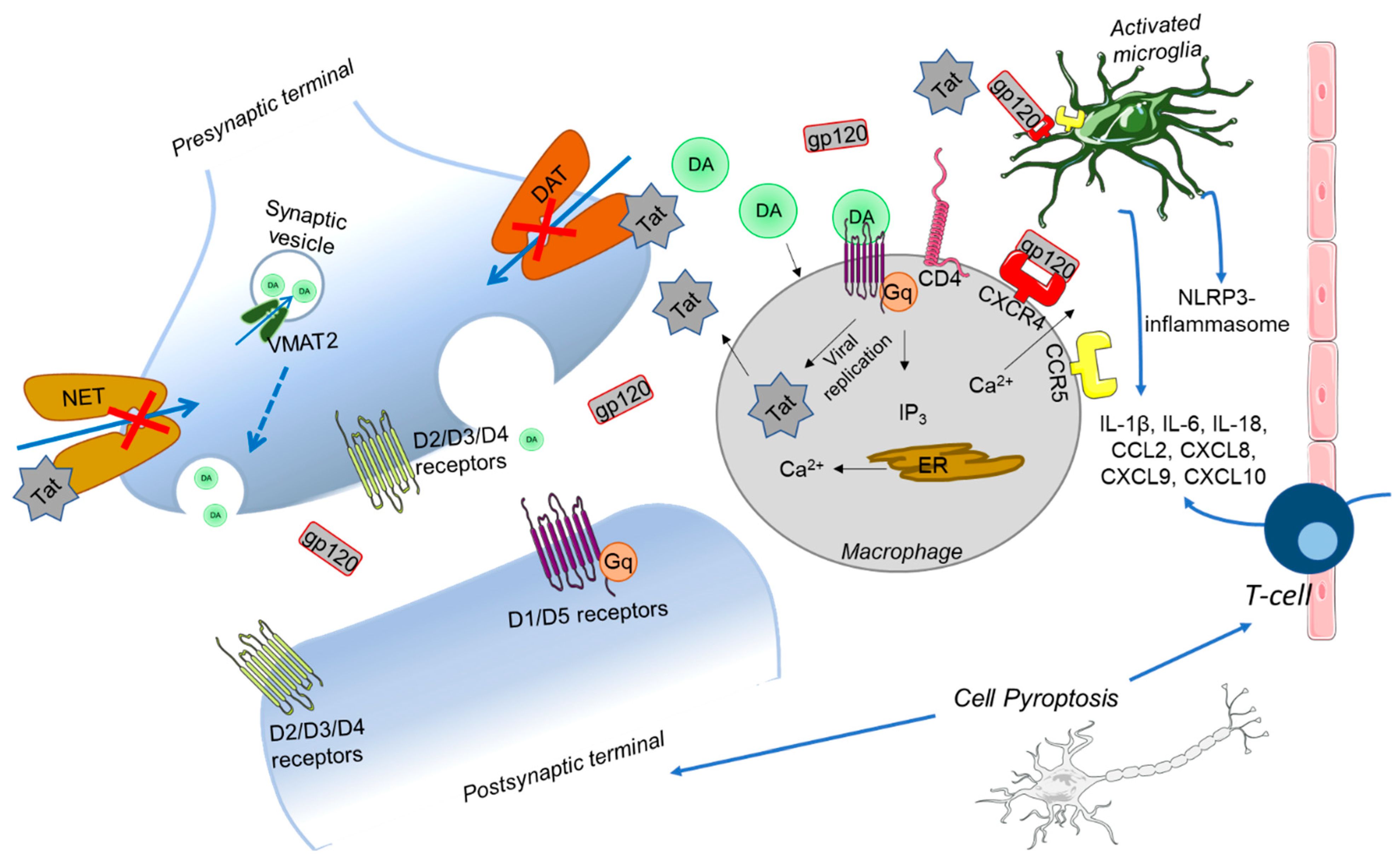
5.2. Disturbances in Neurotransmitter Systems in HAND

Dopaminergic neurotransmission has been implicated as a mediator of HAND pathology. Brain volume measured in patients with HAD post-mortem showed a reduced volume of basal ganglia, which correlated with the severity of HAD [118]. Further, clinical reports of DA in the CSF of HIV-1 infected persons show increased DA levels in early disease [119,120] and decreased DA in late disease stages [121,122,123], even when HIV-1 viremia is suppressed under combined antiretroviral therapy (cART) treatment [124,125]. Combined computational modeling and mutagenesis approaches identified the HIV-1 protein Tat as a negative allosteric modulator of the DA transporter (DAT) [126,127], as well as the NE transporter (NET) [128,129,130]. Acute expression of physiologically relevant levels of Tat [131,132,133,134] increases phasic-like vesicular release of DA, an effect that can be reversed by a novel allosteric modulator of DAT in inducible transgenic mice [20,135]. In addition to Tat, gp120 has also been shown to disturb monoamine systems through inhibition of DA uptake through DAT in various cell models [136,137,138]. Unsurprisingly, clinical reports using PET scan imaging found decreased expression of DAT in patients with HAD, independent of comorbid substance use status [139]. Indeed, disturbances in DA function are an important feature in NeuroHIV, which persists even in the post-cART era.

5.3. Evidence for Mitochondria and Ca2+ Disturbances in HAND

It has long been evidenced that Tat and gp120 are neurotoxic proteins, inducing neuronal apoptosis [140,141,142] preferentially in dopaminergic neurons [136,143,144]. Tat and gp120 have long been shown to disturb Ca2+ homeostasis by initiating IP3-mediated Ca2+ release from ER stores [145,146]. Further, Tat has been shown to initiate mitochondrial-induced apoptosis through Ca2+-mediated mitochondrial generation of ROS and caspase activation in hippocampal neurons [147]. Therefore, Tat-induced disturbances in Ca2+ homeostasis, which induce mitochondrial-mediated neurotoxicity, in addition to NLRP3 inflammasome-mediated pyroptosis, may also be a major contributor to the neurobiology of HAND [148].
While downstream effects of Tat on Ca2+ disturbances are well evidenced at this point, mechanisms by which these two key neurotoxic HIV-1 proteins initiate these effects are still coming to light. Tat-induced increases in intracellular Ca2+ levels have been attributed to the overactivation of L-type Ca2+ channels [149], as well as the potentiation of N-methyl-D-aspartate receptor (NMDAR) mediated Ca2+ influx [150]. In cortical neurons, Tat induces Ca2+ release from ER stores through a ryanodine receptor (RyR) dependent mechanism [151]. Although these reports do well to show that Tat does indeed disrupt Ca2+ through multiple Ca2+ regulatory mechanisms, it is not known whether Tat directly interacts with these channels to alter their function. Tat-induced disturbances of Ca2+ have been considered as the underlying mechanism for Tat-induced alterations in NT systems [152], including not only DA systems but GABA [153] and ACh as well [154]. It would not be surprising if Ca2+ dysregulation contributed to the Tat-induced disruption of NT systems, considering the critical role of Ca2+ in exocytotic NT release. However, direct interaction between Tat and molecular players has, to our knowledge, only been confirmed with monoamine transporters DAT and NET [130,155]. Therefore, the inverse relationship, wherein Tat-induced disturbances in Ca2+ homeostasis are downstream effects of Tat-induced disturbances in NT systems, we argue, is a possibility. NTs can dysregulate Ca2+ homeostasis by signaling through GPCRs and downstream signaling cascades triggered by activation of specific GPCR subtypes. Tat-induced effects on ACh release in cortical synaptosomes were shown to involve the mGluR1. These receptors, upon activation, provided regulatory feedback to increase Ca2+ release from the ER, increase Ca2+ influx from NMDARs, and finally increase noradrenaline release [156]. Additionally, Tat alters the expression and activity of DA receptors, another family of GPCRs. Expression of various DA receptors (D1, D2, D4, and D5) is reduced in the NAc of iTat-tg mice [157]. Further, Tat increases the excitability of D1 medium spiny neurons in the NAc by triggering IP3-mediated release of Ca2+ to depolarize the neuronal membrane [158]. Independent of Tat, extracellular DA can increase IP3-mediated Ca2+ release in macrophages via D5 receptors coupled to Gq proteins [159]. DA has also been shown to increase the release of inflammatory cytokines from human macrophages [160]. Considering the ability of Tat to increase extracellular DA release in the striatum [135], the aforementioned studies present two different mechanisms by which Tat-induced perturbances in extracellular DA may contribute to dopaminergic-specific neuronal damage observed in HAND. Specifically, Tat-induced disruption of DA neurotransmission may initiate neuronal loss not only due to mitochondrial-associated apoptosis via GPCR-mediated release of internal Ca2+ stores but also due to DA-induced neuroinflammation and downstream activation of pyroptosis in response to cytokine release from macrophages. Thus, preventing the effects of proteins such as Tat from disturbing NT systems is of great interest in mitigating the neuronal loss and damage underlying the neurobiology of HAND (Figure 2).

5.4. Mechanisms for Synaptic Injury

During the era of cART, the incidence of HAND has remained steady; however, fewer persons with HIV-1 are developing HAD and are instead experiencing either ANI or MND [161]. Neurocognitive impairments that precede HAD correlate with neuronal damage, and mechanisms by which HIV-1 induces synaptodendritic injury have been extensively reviewed [162]. Unsurprisingly, the molecular factors associated with neuronal loss, such as Ca2+ and mitochondrial instability, are also associated with changes in HIV-1 protein-induced changes in synaptic and dendritic morphology. HIV-1 protein Tat was found to increase cytosolic Ca2+ through NMDARs, which is largely responsible for Tat-induced dendrite swelling in mouse striatal neurons [163]. Additionally, gp120 induced cytoskeletal abnormalities, including rod-like complexes composed of cofilin and actin in hippocampal neurons, which was dependent on ROS formation by NADPH-oxidase 2 (NOX2) [164]. Key molecular factors thought to mediate neuronal loss, such as Ca2+ and ROS generation, are also shared in the pathways associated with synaptic injury, which are likely to underlie the neurocognitive impairment observed in the milder forms of HAND. Thus, targeting the upstream effects of these neurotoxic HIV-1 proteins may mitigate not only neuronal death but synaptodendritic injury as well.

6. Crosstalk between Inflammation and Mitochondrial Induced Cell Death

In this review, we have primarily covered two major pathways involved in the pathophysiology of neurodegeneration in NDs: NLRP3 inflammasome-mediated pyroptosis and Ca2+-mediated mitochondrial-induced apoptosis. These two pathways are not mutually exclusive, and multiple facets of crosstalk are present. For example, Ca2+ influx can directly or indirectly (via ER stress and ROS) activate NLRP3 inflammasomes. Additionally, inositol-requiring transmembrane kinase/endoribonuclease 1α (IRE1α), an ER stress sensor, regulates the release of inflammatory cytokines which can activate the NLRP3 inflammasome while also further producing ROS, which induces mitochondrial damage [165]. Crosstalk between ER stress and NLRP3 inflammasomes has been implicated in non-neuronal forms of cell death, such as cardiovascular disease [166] and renal ischemia [167]. In astroglia cells, ethanol-induced NLRP3 inflammasome activation was mediated by mitochondrial ROS generation. Interestingly, this study showed that ~73% of cell death was attributed to caspase-1-mediated pyroptosis, and the other ~25% was attributed to caspase-3-mediated apoptosis [168]. Both pyroptosis and apoptosis have been implicated as forms of cell death featured in the NDs discussed in this review. Therefore, therapeutic strategies for NDs should be developed to target upstream of the activation of these forms of cell death (such as disturbances in NT systems) to ensure that all forms of neuronal loss (and injury) can be prevented.

7. Conclusions

Different types of cell death, including (but not limited to) pyroptosis and apoptosis, are a featured hallmark in NDs. The machinery necessary for these forms of cell death is ubiquitously present in all cell types, which begs the question—why do some cells die and not others? More specifically, how is cell-specific loss/injury brought about in various NDs? We propose that the unique pattern of neurodegeneration evidenced in various NDs such as AD, PD, and HAD may be partially explained by their disruption of specific NT systems. In the context of NeuroHIV, extensive studies demonstrate a clear link between perturbation of monoaminergic transmission by exposure of the CNS to HIV-1 viral proteins and risk for development of HAND. Importantly, abused substances directly target NT transmission and exacerbate NT-mediated development of neurodegenerative diseases. Further work is needed to determine whether the viral protein-induced dysregulation of NT systems is sufficient to induce neuronal damage through the activation of downstream pathways, such as mitochondrial-mediated apoptosis or the release of inflammatory cytokines from macrophages to induce pyroptosis. Currently, there are no promising therapeutic strategies for neurodegenerative disease. Considering the progressive nature of neurodegenerative diseases, it is probably unsurprising that aging is a risk factor for NDs, and as populations live longer, NDs are becoming more prevalent [169]. It should be considered that the neuronal damage and/or loss induced by disturbances in NTs may be due to a smaller magnitude of mitochondrial stress (or inflammation) sustained over a long period of time rather than a large immediate effect as one might see for instance, with abused substances such as meth or cocaine [170]. Based on our current understanding of the neuropathology of NDs, establishing an early intervention strategy would be beneficial to prevent disturbances in NT transmission. For example, the effectiveness of an early therapeutic intervention for HAND to preserve neurocognitive functions in HIV-infected individuals may ultimately depend on a therapeutic strategy that combines compound(s) that specifically attenuate Tat binding site(s) in DAT with antiretroviral therapy without affecting the normal function of DAT [20,135]. In summary, this review sheds light on the impact of disturbances of NT systems on the development of various NDs.

Funding

This work was supported by grants from the National Institutes of Health National Institute on Drug Abuse to Jun Zhu (DA035714, DA047924, DA057866) and Sarah E. Davis (DA057163).

Conflicts of Interest

The authors declare no conflict of interest.

References

  1. Zhang, X.X.; Tian, Y.; Wang, Z.T.; Ma, Y.H.; Tan, L.; Yu, J.T. The epidemiology of alzheimer’s disease modifiable risk factors and prevention. J. Prev. Alzheimers Dis. 2021, 8, 313–321. [Google Scholar] [CrossRef] [PubMed]
  2. Ou, Z.; Pan, J.; Tang, S.; Duan, D.; Yu, D.; Nong, H.; Wang, Z. Global trends in the incidence, prevalence, and years lived with disability of parkinson’s disease in 204 countries/territories from 1990 to 2019. Front. Public Health 2021, 9, 776847. [Google Scholar] [CrossRef] [PubMed]
  3. Breijyeh, Z.; Karaman, R. Comprehensive review on alzheimer’s disease: Causes and treatment. Molecules 2020, 25, 5789. [Google Scholar] [CrossRef] [PubMed]
  4. Jankovic, J.; Tan, E.K. Parkinson’s disease: Etiopathogenesis and treatment. J. Neurol. Neurosurg. Psychiatr. 2020, 91, 795–808. [Google Scholar] [CrossRef]
  5. Beshir, S.A.; Aadithsoorya, A.M.; Parveen, A.; Goh, S.S.L.; Hussain, N.; Menon, V.B. Aducanumab therapy to treat alzheimer’s disease: A narrative review. Int. J. Alzheimers Dis. 2022, 2022, 9343514. [Google Scholar] [CrossRef]
  6. Meiser, J.; Weindl, D.; Hiller, K. Complexity of dopamine metabolism. Cell Commun. Signal. 2013, 11, 34. [Google Scholar] [CrossRef]
  7. Larsen, K.E.; Fon, E.A.; Hastings, T.G.; Edwards, R.H.; Sulzer, D. Methamphetamine-induced degeneration of dopaminergic neurons involves autophagy and upregulation of dopamine synthesis. J. Neurosci. 2002, 22, 8951–8960. [Google Scholar] [CrossRef]
  8. Mosharov, E.V.; Larsen, K.E.; Kanter, E.; Phillips, K.A.; Wilson, K.; Schmitz, Y.; Krantz, D.E.; Kobayashi, K.; Edwards, R.H.; Sulzer, D. Interplay between cytosolic dopamine, calcium, and alpha-synuclein causes selective death of substantia nigra neurons. Neuron 2009, 62, 218–229. [Google Scholar] [CrossRef]
  9. Hajnóczky, G.; Csordás, G.; Das, S.; Garcia-Perez, C.; Saotome, M.; Sinha Roy, S.; Yi, M. Mitochondrial calcium signalling and cell death: Approaches for assessing the role of mitochondrial Ca2+ uptake in apoptosis. Cell Calcium 2006, 40, 553–560. [Google Scholar] [CrossRef]
  10. German, C.L.; Baladi, M.G.; McFadden, L.M.; Hanson, G.R.; Fleckenstein, A.E. Regulation of the dopamine and vesicular monoamine transporters: Pharmacological targets and implications for disease. Pharmacol. Rev. 2015, 67, 1005–1024. [Google Scholar] [CrossRef]
  11. Liu, Y.; Edwards, R.H. The role of vesicular transport proteins in synaptic transmission and neural degeneration. Annu. Rev. Neurosci. 1997, 20, 125–156. [Google Scholar] [CrossRef] [PubMed]
  12. Mulvihill, K.G. Presynaptic regulation of dopamine release: Role of the DAT and VMAT2 transporters. Neurochem. Int. 2019, 122, 94–105. [Google Scholar] [CrossRef]
  13. Nirenberg, M.J.; Vaughan, R.A.; Uhl, G.R.; Kuhar, M.J.; Pickel, V.M. The dopamine transporter is localized to dendritic and axonal plasma membranes of nigrostriatal dopaminergic neurons. J. Neurosci. 1996, 16, 436–447. [Google Scholar] [CrossRef]
  14. Predescu, D.-V.; Crețoiu, S.M.; Crețoiu, D.; Pavelescu, L.A.; Suciu, N.; Radu, B.M.; Voinea, S.-C. G Protein-Coupled Receptors (GPCRs)-Mediated Calcium Signaling in Ovarian Cancer: Focus on GPCRs activated by Neurotransmitters and Inflammation-Associated Molecules. Int. J. Mol. Sci. 2019, 20, 5568. [Google Scholar] [CrossRef]
  15. Vaughan, R.A.; Foster, J.D. Mechanisms of dopamine transporter regulation in normal and disease states. Trends Pharmacol. Sci. 2013, 34, 489–496. [Google Scholar] [CrossRef]
  16. Guptaroy, B.; Fraser, R.; Desai, A.; Zhang, M.; Gnegy, M.E. Site-directed mutations near transmembrane domain 1 alter conformation and function of norepinephrine and dopamine transporters. Mol. Pharmacol. 2011, 79, 520–532. [Google Scholar] [CrossRef]
  17. Jones, S.R.; Joseph, J.D.; Barak, L.S.; Caron, M.G.; Wightman, R.M. Dopamine neuronal transport kinetics and effects of amphetamine. J. Neurochem. 1999, 73, 2406–2414. [Google Scholar] [CrossRef]
  18. Carvelli, L.; McDonald, P.W.; Blakely, R.D.; DeFelice, L.J. Dopamine transporters depolarize neurons by a channel mechanism. Proc. Natl. Acad. Sci. USA 2004, 101, 16046–16051. [Google Scholar] [CrossRef]
  19. Shan, J.; Javitch, J.A.; Shi, L.; Weinstein, H. The substrate-driven transition to an inward-facing conformation in the functional mechanism of the dopamine transporter. PLoS ONE 2011, 6, e16350. [Google Scholar] [CrossRef]
  20. Zhu, J.; Quizon, P.M.; Wang, Y.; Adeniran, C.A.; Strauss, M.J.; Jiménez-Torres, A.C.; Patel, P.; Cirino, T.J.; Eans, S.O.; Hammond, H.R.; et al. SRI-32743, a novel allosteric modulator, attenuates HIV-1 Tat protein-induced inhibition of the dopamine transporter and alleviates the potentiation of cocaine reward in HIV-1 Tat transgenic mice. Neuropharmacology 2022, 220, 109239. [Google Scholar] [CrossRef]
  21. Cao, J.; Slack, R.D.; Bakare, O.M.; Burzynski, C.; Rais, R.; Slusher, B.S.; Kopajtic, T.; Bonifazi, A.; Ellenberger, M.P.; Yano, H.; et al. Novel and High Affinity 2-[(Diphenylmethyl)sulfinyl]acetamide (Modafinil) Analogues as Atypical Dopamine Transporter Inhibitors. J. Med. Chem. 2016, 59, 10676–10691. [Google Scholar] [CrossRef] [PubMed]
  22. Adkins, E.M.; Samuvel, D.J.; Fog, J.U.; Eriksen, J.; Jayanthi, L.D.; Vaegter, C.B.; Ramamoorthy, S.; Gether, U. Membrane mobility and microdomain association of the dopamine transporter studied with fluorescence correlation spectroscopy and fluorescence recovery after photobleaching. Biochemistry 2007, 46, 10484–10497. [Google Scholar] [CrossRef] [PubMed]
  23. Gorentla, B.K.; Moritz, A.E.; Foster, J.D.; Vaughan, R.A. Proline-directed phosphorylation of the dopamine transporter N-terminal domain. Biochemistry 2009, 48, 1067–1076. [Google Scholar] [CrossRef]
  24. Foster, J.D.; Pananusorn, B.; Vaughan, R.A. Dopamine transporters are phosphorylated on N-terminal serines in rat striatum. J. Biol. Chem. 2002, 277, 25178–25186. [Google Scholar] [CrossRef] [PubMed]
  25. Cervinski, M.A.; Foster, J.D.; Vaughan, R.A. Psychoactive substrates stimulate dopamine transporter phosphorylation and down-regulation by cocaine-sensitive and protein kinase C-dependent mechanisms. J. Biol. Chem. 2005, 280, 40442–40449. [Google Scholar] [CrossRef] [PubMed]
  26. Loder, M.K.; Melikian, H.E. The dopamine transporter constitutively internalizes and recycles in a protein kinase C-regulated manner in stably transfected PC12 cell lines. J. Biol. Chem. 2003, 278, 22168–22174. [Google Scholar] [CrossRef] [PubMed]
  27. Bolland, D.E.; Moritz, A.E.; Stanislowski, D.J.; Vaughan, R.A.; Foster, J.D. Palmitoylation by multiple DHHC enzymes enhances dopamine transporter function and stability. ACS Chem. Neurosci. 2019, 10, 2707–2717. [Google Scholar] [CrossRef]
  28. Lee, F.J.S.; Pei, L.; Moszczynska, A.; Vukusic, B.; Fletcher, P.J.; Liu, F. Dopamine transporter cell surface localization facilitated by a direct interaction with the dopamine D2 receptor. EMBO J. 2007, 26, 2127–2136. [Google Scholar] [CrossRef]
  29. Su, P.; Liu, F. A peptide disrupting the D2R-DAT interaction protects against dopamine neurotoxicity. Exp. Neurol. 2017, 295, 176–183. [Google Scholar] [CrossRef]
  30. Nguyen, E.C.; McCracken, K.A.; Liu, Y.; Pouw, B.; Matsumoto, R.R. Involvement of sigma (sigma) receptors in the acute actions of methamphetamine: Receptor binding and behavioral studies. Neuropharmacology 2005, 49, 638–645. [Google Scholar] [CrossRef]
  31. Sambo, D.O.; Lebowitz, J.J.; Khoshbouei, H. The sigma-1 receptor as a regulator of dopamine neurotransmission: A potential therapeutic target for methamphetamine addiction. Pharmacol. Ther. 2018, 186, 152–167. [Google Scholar] [CrossRef] [PubMed]
  32. Hedges, D.M.; Obray, J.D.; Yorgason, J.T.; Jang, E.Y.; Weerasekara, V.K.; Uys, J.D.; Bellinger, F.P.; Steffensen, S.C. Methamphetamine Induces Dopamine Release in the Nucleus Accumbens Through a Sigma Receptor-Mediated Pathway. Neuropsychopharmacology 2018, 43, 1405–1414. [Google Scholar] [CrossRef] [PubMed]
  33. Tarakad, A.; Jimenez-Shahed, J. VMAT2 inhibitors in neuropsychiatric disorders. CNS Drugs 2018, 32, 1131–1144. [Google Scholar] [CrossRef] [PubMed]
  34. Lawal, H.O.; Krantz, D.E. SLC18: Vesicular neurotransmitter transporters for monoamines and acetylcholine. Mol. Aspects Med. 2013, 34, 360–372. [Google Scholar] [CrossRef] [PubMed]
  35. Lohr, K.M.; Bernstein, A.I.; Stout, K.A.; Dunn, A.R.; Lazo, C.R.; Alter, S.P.; Wang, M.; Li, Y.; Fan, X.; Hess, E.J.; et al. Increased vesicular monoamine transporter enhances dopamine release and opposes Parkinson disease-related neurodegeneration in vivo. Proc. Natl. Acad. Sci. USA 2014, 111, 9977–9982. [Google Scholar] [CrossRef]
  36. Lee, N.-R.; Zheng, G.; Leggas, M.; Janganati, V.; Nickell, J.R.; Crooks, P.A.; Bardo, M.T.; Dwoskin, L.P. GZ-11608, a Vesicular Monoamine Transporter-2 Inhibitor, Decreases the Neurochemical and Behavioral Effects of Methamphetamine. J. Pharmacol. Exp. Ther. 2019, 371, 526–543. [Google Scholar] [CrossRef]
  37. Yehuda, S.; Rabinovitz, S.; Mostofsky, D.I. Treatment with a polyunsaturated fatty acid prevents deleterious effects of Ro4-1284. Eur. J. Pharmacol. 1999, 365, 27–34. [Google Scholar] [CrossRef]
  38. Nickell, J.R.; Siripurapu, K.B.; Vartak, A.; Crooks, P.A.; Dwoskin, L.P. The vesicular monoamine transporter-2: An important pharmacological target for the discovery of novel therapeutics to treat methamphetamine abuse. Adv. Pharmacol. 2014, 69, 71–106. [Google Scholar] [CrossRef]
  39. Yao, J.; Hersh, L.B. The vesicular monoamine transporter 2 contains trafficking signals in both its N-glycosylation and C-terminal domains. J. Neurochem. 2007, 100, 1387–1396. [Google Scholar] [CrossRef]
  40. Fei, H.; Grygoruk, A.; Brooks, E.S.; Chen, A.; Krantz, D.E. Trafficking of vesicular neurotransmitter transporters. Traffic 2008, 9, 1425–1436. [Google Scholar] [CrossRef]
  41. Torres, B.; Ruoho, A.E. N-terminus regulation of VMAT2 mediates methamphetamine-stimulated efflux. Neuroscience 2014, 259, 194–202. [Google Scholar] [CrossRef] [PubMed]
  42. Bye, L.J.; Finol-Urdaneta, R.K.; Tae, H.-S.; Adams, D.J. Nicotinic acetylcholine receptors: Key targets for attenuating neurodegenerative diseases. Int. J. Biochem. Cell Biol. 2023, 157, 106387. [Google Scholar] [CrossRef] [PubMed]
  43. Warburton, E.C.; Koder, T.; Cho, K.; Massey, P.V.; Duguid, G.; Barker, G.R.I.; Aggleton, J.P.; Bashir, Z.I.; Brown, M.W. Cholinergic neurotransmission is essential for perirhinal cortical plasticity and recognition memory. Neuron 2003, 38, 987–996. [Google Scholar] [CrossRef] [PubMed]
  44. Sam, C.; Bordoni, B. Physiology, Acetylcholine. In StatPearls; StatPearls Publishing: Treasure Island, FL, USA, 2023. [Google Scholar]
  45. Waxenbaum, J.A.; Reddy, V.; Varacallo, M. Anatomy, autonomic nervous system. In StatPearls; StatPearls Publishing: Treasure Island, FL, USA, 2023. [Google Scholar]
  46. Prado, M.A.M.; Reis, R.A.M.; Prado, V.F.; de Mello, M.C.; Gomez, M.V.; de Mello, F.G. Regulation of acetylcholine synthesis and storage. Neurochem. Int. 2002, 41, 291–299. [Google Scholar] [CrossRef]
  47. Dey, S.; Ray, K. Cholinergic activity is essential for maintaining the anterograde transport of Choline Acetyltransferase in Drosophila. Sci. Rep. 2018, 8, 8028. [Google Scholar] [CrossRef]
  48. Han, J.; Pluhackova, K.; Böckmann, R.A. The multifaceted role of SNARE proteins in membrane fusion. Front. Physiol. 2017, 8, 5. [Google Scholar] [CrossRef]
  49. Rodríguez Cruz, P.M.; Cossins, J.; Beeson, D.; Vincent, A. The neuromuscular junction in health and disease: Molecular mechanisms governing synaptic formation and homeostasis. Front. Mol. Neurosci. 2020, 13, 610964. [Google Scholar] [CrossRef]
  50. Trang, A.; Khandhar, P.B. Physiology, Acetylcholinesterase. In StatPearls; StatPearls Publishing: Treasure Island, FL, USA, 2023. [Google Scholar]
  51. Carlson, A.B.; Kraus, G.P. Physiology, Cholinergic Receptors. In StatPearls; StatPearls Publishing: Treasure Island, FL, USA, 2023. [Google Scholar]
  52. Ojiakor, O.A.; Rylett, R.J. Modulation of sodium-coupled choline transporter CHT function in health and disease. Neurochem. Int. 2020, 140, 104810. [Google Scholar] [CrossRef]
  53. Papke, R.L. Merging old and new perspectives on nicotinic acetylcholine receptors. Biochem. Pharmacol. 2014, 89, 1–11. [Google Scholar] [CrossRef]
  54. Ho, T.N.T.; Abraham, N.; Lewis, R.J. Synthesis of full-length homodimer αD-VxXXB that targets human α7 nicotinic acetylcholine receptors. RSC Med. Chem. 2022, 13, 1410–1419. [Google Scholar] [CrossRef]
  55. Wittenberg, R.E.; Wolfman, S.L.; De Biasi, M.; Dani, J.A. Nicotinic acetylcholine receptors and nicotine addiction: A brief introduction. Neuropharmacology 2020, 177, 108256. [Google Scholar] [CrossRef] [PubMed]
  56. Albuquerque, E.X.; Pereira, E.F.R.; Alkondon, M.; Rogers, S.W. Mammalian nicotinic acetylcholine receptors: From structure to function. Physiol. Rev. 2009, 89, 73–120. [Google Scholar] [CrossRef] [PubMed]
  57. Millar, N.S.; Harkness, P.C. Assembly and trafficking of nicotinic acetylcholine receptors (Review). Mol. Membr. Biol. 2008, 25, 279–292. [Google Scholar] [CrossRef] [PubMed]
  58. Colombo, S.F.; Mazzo, F.; Pistillo, F.; Gotti, C. Biogenesis, trafficking and up-regulation of nicotinic ACh receptors. Biochem. Pharmacol. 2013, 86, 1063–1073. [Google Scholar] [CrossRef] [PubMed]
  59. Freedman, R.; Hall, M.; Adler, L.E.; Leonard, S. Evidence in postmortem brain tissue for decreased numbers of hippocampal nicotinic receptors in schizophrenia. Biol. Psychiatry 1995, 38, 22–33. [Google Scholar] [CrossRef]
  60. Wang, H.Y.; Lee, D.H.; D’Andrea, M.R.; Peterson, P.A.; Shank, R.P.; Reitz, A.B. beta-Amyloid(1-42) binds to alpha7 nicotinic acetylcholine receptor with high affinity. Implications for Alzheimer’s disease pathology. J. Biol. Chem. 2000, 275, 5626–5632. [Google Scholar] [CrossRef]
  61. D’Andrea, M.R.; Nagele, R.G. Targeting the alpha 7 nicotinic acetylcholine receptor to reduce amyloid accumulation in Alzheimer’s disease pyramidal neurons. Curr. Pharm. Des. 2006, 12, 677–684. [Google Scholar] [CrossRef]
  62. Kudlak, M.; Tadi, P. Physiology, Muscarinic Receptor. In StatPearls; StatPearls Publishing: Treasure Island, FL, USA, 2023. [Google Scholar]
  63. Moran, S.P.; Maksymetz, J.; Conn, P.J. Targeting muscarinic acetylcholine receptors for the treatment of psychiatric and neurological disorders. Trends Pharmacol. Sci. 2019, 40, 1006–1020. [Google Scholar] [CrossRef]
  64. Prado, V.F.; Roy, A.; Kolisnyk, B.; Gros, R.; Prado, M.A.M. Regulation of cholinergic activity by the vesicular acetylcholine transporter. Biochem. J. 2013, 450, 265–274. [Google Scholar] [CrossRef]
  65. Nguyen, M.L.; Cox, G.D.; Parsons, S.M. Kinetic parameters for the vesicular acetylcholine transporter: Two protons are exchanged for one acetylcholine. Biochemistry 1998, 37, 13400–13410. [Google Scholar] [CrossRef]
  66. Chen, K.H.; Reese, E.A.; Kim, H.-W.; Rapoport, S.I.; Rao, J.S. Disturbed neurotransmitter transporter expression in Alzheimer’s disease brain. J. Alzheimers Dis. 2011, 26, 755–766. [Google Scholar] [CrossRef] [PubMed]
  67. Fleming, A.; Bourdenx, M.; Fujimaki, M.; Karabiyik, C.; Krause, G.J.; Lopez, A.; Martín-Segura, A.; Puri, C.; Scrivo, A.; Skidmore, J.; et al. The different autophagy degradation pathways and neurodegeneration. Neuron 2022, 110, 935–966. [Google Scholar] [CrossRef] [PubMed]
  68. Song, H.; Yang, B.; Li, Y.; Qian, A.; Kang, Y.; Shan, X. Focus on the mechanisms and functions of pyroptosis, inflammasomes, and inflammatory caspases in infectious diseases. Oxid. Med. Cell Longev. 2022, 2022, 2501279. [Google Scholar] [CrossRef] [PubMed]
  69. Blevins, H.M.; Xu, Y.; Biby, S.; Zhang, S. The NLRP3 inflammasome pathway: A review of mechanisms and inhibitors for the treatment of inflammatory diseases. Front. Aging Neurosci. 2022, 14, 879021. [Google Scholar] [CrossRef] [PubMed]
  70. He, Y.; Hara, H.; Núñez, G. Mechanism and regulation of NLRP3 inflammasome activation. Trends Biochem. Sci. 2016, 41, 1012–1021. [Google Scholar] [CrossRef]
  71. Wang, S.; Yuan, Y.-H.; Chen, N.-H.; Wang, H.-B. The mechanisms of NLRP3 inflammasome/pyroptosis activation and their role in Parkinson’s disease. Int. Immunopharmacol. 2019, 67, 458–464. [Google Scholar] [CrossRef]
  72. Mangalmurti, A.; Lukens, J.R. How neurons die in Alzheimer’s disease: Implications for neuroinflammation. Curr. Opin. Neurobiol. 2022, 75, 102575. [Google Scholar] [CrossRef]
  73. Milner, M.T.; Maddugoda, M.; Götz, J.; Burgener, S.S.; Schroder, K. The NLRP3 inflammasome triggers sterile neuroinflammation and Alzheimer’s disease. Curr. Opin. Immunol. 2021, 68, 116–124. [Google Scholar] [CrossRef]
  74. Moonen, S.; Koper, M.J.; Van Schoor, E.; Schaeverbeke, J.M.; Vandenberghe, R.; von Arnim, C.A.F.; Tousseyn, T.; De Strooper, B.; Thal, D.R. Pyroptosis in Alzheimer’s disease: Cell type-specific activation in microglia, astrocytes and neurons. Acta Neuropathol. 2023, 145, 175–195. [Google Scholar] [CrossRef]
  75. Vitet, H.; Brandt, V.; Saudou, F. Traffic signaling: New functions of huntingtin and axonal transport in neurological disease. Curr. Opin. Neurobiol. 2020, 63, 122–130. [Google Scholar] [CrossRef]
  76. Paldino, E.; Fusco, F.R. Emerging role of NLRP3 inflammasome/pyroptosis in huntington’s disease. Int. J. Mol. Sci. 2022, 23, 8363. [Google Scholar] [CrossRef]
  77. Chen, K.-P.; Hua, K.-F.; Tsai, F.-T.; Lin, T.-Y.; Cheng, C.-Y.; Yang, D.-I.; Hsu, H.-T.; Ju, T.-C. A selective inhibitor of the NLRP3 inflammasome as a potential therapeutic approach for neuroprotection in a transgenic mouse model of Huntington’s disease. J. Neuroinflammation 2022, 19, 56. [Google Scholar] [CrossRef]
  78. Wong, K.Y.; Roy, J.; Fung, M.L.; Heng, B.C.; Zhang, C.; Lim, L.W. Relationships between Mitochondrial Dysfunction and Neurotransmission Failure in Alzheimer’s Disease. Aging Dis. 2020, 11, 1291–1316. [Google Scholar] [CrossRef] [PubMed]
  79. Quartey, M.O.; Nyarko, J.N.K.; Pennington, P.R.; Heistad, R.M.; Klassen, P.C.; Baker, G.B.; Mousseau, D.D. Alzheimer Disease and Selected Risk Factors Disrupt a Co-regulation of Monoamine Oxidase-A/B in the Hippocampus, but Not in the Cortex. Front. Neurosci. 2018, 12, 419. [Google Scholar] [CrossRef] [PubMed]
  80. Rabbito, A.; Dulewicz, M.; Kulczyńska-Przybik, A.; Mroczko, B. Biochemical markers in alzheimer’s disease. Int. J. Mol. Sci. 2020, 21, 1989. [Google Scholar] [CrossRef] [PubMed]
  81. Bukke, V.N.; Archana, M.; Villani, R.; Romano, A.D.; Wawrzyniak, A.; Balawender, K.; Orkisz, S.; Beggiato, S.; Serviddio, G.; Cassano, T. The dual role of glutamatergic neurotransmission in alzheimer’s disease: From pathophysiology to pharmacotherapy. Int. J. Mol. Sci. 2020, 21, 7452. [Google Scholar] [CrossRef]
  82. Li, Y.; Sun, H.; Chen, Z.; Xu, H.; Bu, G.; Zheng, H. Implications of gabaergic neurotransmission in alzheimer’s disease. Front. Aging Neurosci. 2016, 8, 31. [Google Scholar] [CrossRef]
  83. Klein, M.O.; Battagello, D.S.; Cardoso, A.R.; Hauser, D.N.; Bittencourt, J.C.; Correa, R.G. Dopamine: Functions, Signaling, and Association with Neurological Diseases. Cell Mol. Neurobiol. 2019, 39, 31–59. [Google Scholar] [CrossRef]
  84. Latif, S.; Jahangeer, M.; Maknoon Razia, D.; Ashiq, M.; Ghaffar, A.; Akram, M.; El Allam, A.; Bouyahya, A.; Garipova, L.; Ali Shariati, M.; et al. Dopamine in Parkinson’s disease. Clin. Chim. Acta 2021, 522, 114–126. [Google Scholar] [CrossRef]
  85. Masato, A.; Plotegher, N.; Boassa, D.; Bubacco, L. Impaired dopamine metabolism in Parkinson’s disease pathogenesis. Mol. Neurodegener. 2019, 14, 35. [Google Scholar] [CrossRef]
  86. Muñoz, A.; Lopez-Lopez, A.; Labandeira, C.M.; Labandeira-Garcia, J.L. Interactions Between the Serotonergic and Other Neurotransmitter Systems in the Basal Ganglia: Role in Parkinson’s Disease and Adverse Effects of L-DOPA. Front. Neuroanat. 2020, 14, 26. [Google Scholar] [CrossRef] [PubMed]
  87. Carafoli, E.; Krebs, J. Why calcium? how calcium became the best communicator. J. Biol. Chem. 2016, 291, 20849–20857. [Google Scholar] [CrossRef] [PubMed]
  88. Dolphin, A.C.; Lee, A. Presynaptic calcium channels: Specialized control of synaptic neurotransmitter release. Nat. Rev. Neurosci. 2020, 21, 213–229. [Google Scholar] [CrossRef] [PubMed]
  89. Zhou, Q.; Zhou, P.; Wang, A.L.; Wu, D.; Zhao, M.; Südhof, T.C.; Brunger, A.T. The primed SNARE-complexin-synaptotagmin complex for neuronal exocytosis. Nature 2017, 548, 420–425. [Google Scholar] [CrossRef]
  90. Verhage, M.; Sørensen, J.B. Vesicle docking in regulated exocytosis. Traffic 2008, 9, 1414–1424. [Google Scholar] [CrossRef]
  91. Brini, M.; Calì, T.; Ottolini, D.; Carafoli, E. Intracellular calcium homeostasis and signaling. Met. Ions Life Sci. 2013, 12, 119–168. [Google Scholar] [CrossRef]
  92. Zhang, L.; Shi, G. Gq-Coupled Receptors in Autoimmunity. J. Immunol. Res. 2016, 2016, 3969023. [Google Scholar] [CrossRef]
  93. Mizuno, N.; Itoh, H. Functions and regulatory mechanisms of Gq-signaling pathways. Neurosignals 2009, 17, 42–54. [Google Scholar] [CrossRef]
  94. Wang, Q.J. PKD at the crossroads of DAG and PKC signaling. Trends Pharmacol. Sci. 2006, 27, 317–323. [Google Scholar] [CrossRef]
  95. Woll, K.A.; Van Petegem, F. Calcium-release channels: Structure and function of IP3 receptors and ryanodine receptors. Physiol. Rev. 2022, 102, 209–268. [Google Scholar] [CrossRef]
  96. Misgeld, T.; Schwarz, T.L. Mitostasis in neurons: Maintaining mitochondria in an extended cellular architecture. Neuron 2017, 96, 651–666. [Google Scholar] [CrossRef] [PubMed]
  97. Patergnani, S.; Suski, J.M.; Agnoletto, C.; Bononi, A.; Bonora, M.; De Marchi, E.; Giorgi, C.; Marchi, S.; Missiroli, S.; Poletti, F.; et al. Calcium signaling around Mitochondria Associated Membranes (MAMs). Cell Commun. Signal 2011, 9, 19. [Google Scholar] [CrossRef] [PubMed]
  98. Pinton, P.; Giorgi, C.; Siviero, R.; Zecchini, E.; Rizzuto, R. Calcium and apoptosis: ER-mitochondria Ca2+ transfer in the control of apoptosis. Oncogene 2008, 27, 6407–6418. [Google Scholar] [CrossRef] [PubMed]
  99. Kim, J.-S.; He, L.; Lemasters, J.J. Mitochondrial permeability transition: A common pathway to necrosis and apoptosis. Biochem. Biophys. Res. Commun. 2003, 304, 463–470. [Google Scholar] [CrossRef]
  100. Gogvadze, V.; Orrenius, S.; Zhivotovsky, B. Multiple pathways of cytochrome c release from mitochondria in apoptosis. Biochim. Biophys. Acta 2006, 1757, 639–647. [Google Scholar] [CrossRef]
  101. Betke, K.M.; Wells, C.A.; Hamm, H.E. GPCR mediated regulation of synaptic transmission. Prog. Neurobiol. 2012, 96, 304–321. [Google Scholar] [CrossRef]
  102. Spangler, S.M.; Bruchas, M.R. Optogenetic approaches for dissecting neuromodulation and GPCR signaling in neural circuits. Curr. Opin. Pharmacol. 2017, 32, 56–70. [Google Scholar] [CrossRef]
  103. Borroto-Escuela, D.O.; Fuxe, K. Oligomeric Receptor Complexes and Their Allosteric Receptor-Receptor Interactions in the Plasma Membrane Represent a New Biological Principle for Integration of Signals in the CNS. Front. Mol. Neurosci. 2019, 12, 230. [Google Scholar] [CrossRef]
  104. Akam, E.C.; Challiss, R.A.; Nahorski, S.R. G(q/11) and G(i/o) activation profiles in CHO cells expressing human muscarinic acetylcholine receptors: Dependence on agonist as well as receptor-subtype. Br. J. Pharmacol. 2001, 132, 950–958. [Google Scholar] [CrossRef]
  105. Hasbi, A.; O’Dowd, B.F.; George, S.R. Heteromerization of dopamine D2 receptors with dopamine D1 or D5 receptors generates intracellular calcium signaling by different mechanisms. Curr. Opin. Pharmacol. 2010, 10, 93–99. [Google Scholar] [CrossRef]
  106. Spoida, K.; Masseck, O.A.; Deneris, E.S.; Herlitze, S. Gq/5-HT2c receptor signals activate a local GABAergic inhibitory feedback circuit to modulate serotonergic firing and anxiety in mice. Proc. Natl. Acad. Sci. USA 2014, 111, 6479–6484. [Google Scholar] [CrossRef] [PubMed]
  107. McElligott, Z.A.; Klug, J.R.; Nobis, W.P.; Patel, S.; Grueter, B.A.; Kash, T.L.; Winder, D.G. Distinct forms of Gq-receptor-dependent plasticity of excitatory transmission in the BNST are differentially affected by stress. Proc. Natl. Acad. Sci. USA 2010, 107, 2271–2276. [Google Scholar] [CrossRef] [PubMed]
  108. Bettler, B.; Kaupmann, K.; Mosbacher, J.; Gassmann, M. Molecular structure and physiological functions of GABA(B) receptors. Physiol. Rev. 2004, 84, 835–867. [Google Scholar] [CrossRef] [PubMed]
  109. Boczek, T.; Mackiewicz, J.; Sobolczyk, M.; Wawrzyniak, J.; Lisek, M.; Ferenc, B.; Guo, F.; Zylinska, L. The Role of G Protein-Coupled Receptors (GPCRs) and Calcium Signaling in Schizophrenia. Focus on GPCRs Activated by Neurotransmitters and Chemokines. Cells 2021, 10, 1228. [Google Scholar] [CrossRef] [PubMed]
  110. Foster, T.C. Calcium homeostasis and modulation of synaptic plasticity in the aged brain. Aging Cell 2007, 6, 319–325. [Google Scholar] [CrossRef] [PubMed]
  111. Sushma; Mondal, A.C. Role of GPCR signaling and calcium dysregulation in Alzheimer’s disease. Mol. Cell Neurosci. 2019, 101, 103414. [Google Scholar] [CrossRef]
  112. Celsi, F.; Pizzo, P.; Brini, M.; Leo, S.; Fotino, C.; Pinton, P.; Rizzuto, R. Mitochondria, calcium and cell death: A deadly triad in neurodegeneration. Biochim. Biophys. Acta 2009, 1787, 335–344. [Google Scholar] [CrossRef]
  113. Zampese, E.; Surmeier, D.J. Calcium, bioenergetics, and parkinson’s disease. Cells 2020, 9, 2045. [Google Scholar] [CrossRef]
  114. Antinori, A.; Arendt, G.; Becker, J.T.; Brew, B.J.; Byrd, D.A.; Cherner, M.; Clifford, D.B.; Cinque, P.; Epstein, L.G.; Goodkin, K.; et al. Updated research nosology for HIV-associated neurocognitive disorders. Neurology 2007, 69, 1789–1799. [Google Scholar] [CrossRef]
  115. Berger, J.R.; Arendt, G. HIV dementia: The role of the basal ganglia and dopaminergic systems. J. Psychopharmacol. 2000, 14, 214–221. [Google Scholar] [CrossRef]
  116. Kannan, M.; Singh, S.; Chemparathy, D.T.; Oladapo, A.A.; Gawande, D.Y.; Dravid, S.M.; Buch, S.; Sil, S. HIV-1 Tat induced microglial EVs leads to neuronal synaptodendritic injury: Microglia-neuron cross-talk in NeuroHIV. Extracell. Vesicles Circ. Nucl. Acids 2022, 3, 133–149. [Google Scholar] [CrossRef] [PubMed]
  117. He, X.; Yang, W.; Zeng, Z.; Wei, Y.; Gao, J.; Zhang, B.; Li, L.; Liu, L.; Wan, Y.; Zeng, Q.; et al. NLRP3-dependent pyroptosis is required for HIV-1 gp120-induced neuropathology. Cell Mol. Immunol. 2020, 17, 283–299. [Google Scholar] [CrossRef] [PubMed]
  118. Aylward, E.H.; Henderer, J.D.; McArthur, J.C.; Brettschneider, P.D.; Harris, G.J.; Barta, P.E.; Pearlson, G.D. Reduced basal ganglia volume in HIV-1-associated dementia: Results from quantitative neuroimaging. Neurology 1993, 43, 2099–2104. [Google Scholar] [CrossRef] [PubMed]
  119. Scheller, C.; Arendt, G.; Nolting, T.; Antke, C.; Sopper, S.; Maschke, M.; Obermann, M.; Angerer, A.; Husstedt, I.W.; Meisner, F.; et al. Increased dopaminergic neurotransmission in therapy-naïve asymptomatic HIV patients is not associated with adaptive changes at the dopaminergic synapses. J. Neural Transm. 2010, 117, 699–705. [Google Scholar] [CrossRef]
  120. Horn, A.; Scheller, C.; du Plessis, S.; Arendt, G.; Nolting, T.; Joska, J.; Sopper, S.; Maschke, M.; Obermann, M.; Husstedt, I.W.; et al. Increases in CSF dopamine in HIV patients are due to the dopamine transporter 10/10-repeat allele which is more frequent in HIV-infected individuals. J. Neural Transm. 2013, 120, 1411–1419. [Google Scholar] [CrossRef]
  121. Berger, J.R.; Kumar, M.; Kumar, A.; Fernandez, J.B.; Levin, B. Cerebrospinal fluid dopamine in HIV-1 infection. AIDS 1994, 8, 67–71. [Google Scholar] [CrossRef]
  122. Sardar, A.M.; Czudek, C.; Reynolds, G.P. Dopamine deficits in the brain: The neurochemical basis of parkinsonian symptoms in AIDS. Neuroreport 1996, 7, 910–912. [Google Scholar] [CrossRef]
  123. Larsson, M.; Hagberg, L.; Forsman, A.; Norkrans, G. Cerebrospinal fluid catecholamine metabolites in HIV-infected patients. J. Neurosci. Res. 1991, 28, 406–409. [Google Scholar] [CrossRef]
  124. Kumar, A.M.; Fernandez, J.B.; Singer, E.J.; Commins, D.; Waldrop-Valverde, D.; Ownby, R.L.; Kumar, M. Human immunodeficiency virus type 1 in the central nervous system leads to decreased dopamine in different regions of postmortem human brains. J. Neurovirol. 2009, 15, 257–274. [Google Scholar] [CrossRef]
  125. Kumar, A.M.; Ownby, R.L.; Waldrop-Valverde, D.; Fernandez, B.; Kumar, M. Human immunodeficiency virus infection in the CNS and decreased dopamine availability: Relationship with neuropsychological performance. J. Neurovirol. 2011, 17, 26–40. [Google Scholar] [CrossRef]
  126. Zhu, J.; Mactutus, C.F.; Wallace, D.R.; Booze, R.M. HIV-1 Tat protein-induced rapid and reversible decrease in [3H]dopamine uptake: Dissociation of [3H]dopamine uptake and [3H]2beta-carbomethoxy-3-beta-(4-fluorophenyl)tropane (WIN 35,428) binding in rat striatal synaptosomes. J. Pharmacol. Exp. Ther. 2009, 329, 1071–1083. [Google Scholar] [CrossRef] [PubMed]
  127. Zhu, J.; Ananthan, S.; Zhan, C.-G. The role of human dopamine transporter in NeuroAIDS. Pharmacol. Ther. 2018, 183, 78–89. [Google Scholar] [CrossRef] [PubMed]
  128. Strauss, M.J.; Porter, K.D.; Quizon, P.M.; Davis, S.E.; Lin, S.; Yuan, Y.; Martinez-Muniz, G.A.; Sun, W.-L.; Zhan, C.-G.; Zhu, J. Mutations of tyrosine 467 in the human norepinephrine transporter attenuate HIV-1 Tat-induced inhibition of dopamine transport while retaining physiological function. PLoS ONE 2022, 17, e0275182. [Google Scholar] [CrossRef] [PubMed]
  129. Strauss, M.; O’Donovan, B.; Ma, Y.; Xiao, Z.; Lin, S.; Bardo, M.T.; Ortinski, P.I.; McLaughlin, J.P.; Zhu, J. [3H]Dopamine Uptake through the Dopamine and Norepinephrine Transporters is Decreased in the Prefrontal Cortex of Transgenic Mice Expressing HIV-1 Transactivator of Transcription Protein. J. Pharmacol. Exp. Ther. 2020, 374, 241–251. [Google Scholar] [CrossRef] [PubMed]
  130. Adeniran, C.; Yuan, Y.; Davis, S.E.; Lin, C.; Xu, J.; Zhu, J.; Zhan, C.-G. Binding Mode of Human Norepinephrine Transporter Interacting with HIV-1 Tat. ACS Chem. Neurosci. 2021, 12, 1519–1527. [Google Scholar] [CrossRef]
  131. Kim, B.O.; Liu, Y.; Ruan, Y.; Xu, Z.C.; Schantz, L.; He, J.J. Neuropathologies in transgenic mice expressing human immunodeficiency virus type 1 Tat protein under the regulation of the astrocyte-specific glial fibrillary acidic protein promoter and doxycycline. Am. J. Pathol. 2003, 162, 1693–1707. [Google Scholar] [CrossRef]
  132. Henderson, L.J.; Johnson, T.P.; Smith, B.R.; Reoma, L.B.; Santamaria, U.A.; Bachani, M.; Demarino, C.; Barclay, R.A.; Snow, J.; Sacktor, N.; et al. Presence of Tat and transactivation response element in spinal fluid despite antiretroviral therapy. AIDS 2019, 33 (Suppl. 2), S145–S157. [Google Scholar] [CrossRef]
  133. Gerena, Y.; Menéndez-Delmestre, R.; Delgado-Nieves, A.; Vélez, J.; Méndez-Álvarez, J.; Sierra-Pagan, J.E.; Skolasky, R.L.; Henderson, L.; Nath, A.; Wojna, V. Release of soluble insulin receptor from neurons by cerebrospinal fluid from patients with neurocognitive dysfunction and HIV infection. Front. Neurol. 2019, 10, 285. [Google Scholar] [CrossRef]
  134. Johnson, T.P.; Patel, K.; Johnson, K.R.; Maric, D.; Calabresi, P.A.; Hasbun, R.; Nath, A. Induction of IL-17 and nonclassical T-cell activation by HIV-Tat protein. Proc. Natl. Acad. Sci. USA 2013, 110, 13588–13593. [Google Scholar] [CrossRef]
  135. Davis, S.E.; Ferris, M.J.; Ananthan, S.; Augelli-Szafran, C.E.; Zhu, J. Novel Allosteric Modulator Southern Research Institute-32743 Reverses HIV-1 Transactivator of Transcription-Induced Increase in Dopamine Release in the Caudate Putamen of Inducible Transactivator of Transcription Transgenic Mice. J. Pharmacol. Exp. Ther. 2023, 384, 306–314. [Google Scholar] [CrossRef]
  136. Hu, S.; Sheng, W.S.; Lokensgard, J.R.; Peterson, P.K.; Rock, R.B. Preferential sensitivity of human dopaminergic neurons to gp120-induced oxidative damage. J. Neurovirol. 2009, 15, 401–410. [Google Scholar] [CrossRef] [PubMed]
  137. Bennett, B.A.; Rusyniak, D.E.; Hollingsworth, C.K. HIV-1 gp120-induced neurotoxicity to midbrain dopamine cultures. Brain Res. 1995, 705, 168–176. [Google Scholar] [CrossRef] [PubMed]
  138. Wallace, D.R.; Dodson, S.; Nath, A.; Booze, R.M. Estrogen attenuates gp120- and tat1-72-induced oxidative stress and prevents loss of dopamine transporter function. Synapse 2006, 59, 51–60. [Google Scholar] [CrossRef] [PubMed]
  139. Wang, G.-J.; Chang, L.; Volkow, N.D.; Telang, F.; Logan, J.; Ernst, T.; Fowler, J.S. Decreased brain dopaminergic transporters in HIV-associated dementia patients. Brain 2004, 127, 2452–2458. [Google Scholar] [CrossRef]
  140. Silverstein, P.S.; Shah, A.; Weemhoff, J.; Kumar, S.; Singh, D.P.; Kumar, A. HIV-1 gp120 and drugs of abuse: Interactions in the central nervous system. Curr. HIV Res. 2012, 10, 369–383. [Google Scholar] [CrossRef]
  141. Rappaport, J.; Joseph, J.; Croul, S.; Alexander, G.; Del Valle, L.; Amini, S.; Khalili, K. Molecular pathway involved in HIV-1-induced CNS pathology: Role of viral regulatory protein, Tat. J. Leukoc. Biol. 1999, 65, 458–465. [Google Scholar] [CrossRef]
  142. Fitting, S.; Booze, R.M.; Mactutus, C.F. HIV-1 proteins, Tat and gp120, target the developing dopamine system. Curr. HIV Res. 2015, 13, 21–42. [Google Scholar] [CrossRef]
  143. Bansal, A.K.; Mactutus, C.F.; Nath, A.; Maragos, W.; Hauser, K.F.; Booze, R.M. Neurotoxicity of HIV-1 proteins gp120 and Tat in the rat striatum. Brain Res. 2000, 879, 42–49. [Google Scholar] [CrossRef]
  144. Agrawal, L.; Louboutin, J.-P.; Marusich, E.; Reyes, B.A.S.; Van Bockstaele, E.J.; Strayer, D.S. Dopaminergic neurotoxicity of HIV-1 gp120: Reactive oxygen species as signaling intermediates. Brain Res. 2010, 1306, 116–130. [Google Scholar] [CrossRef]
  145. Haughey, N.J.; Mattson, M.P. Calcium dysregulation and neuronal apoptosis by the HIV-1 proteins Tat and gp120. J. Acquir. Immune Defic. Syndr. 2002, 31 (Suppl. 2), S55–S61. [Google Scholar] [CrossRef]
  146. Haughey, N.J.; Holden, C.P.; Nath, A.; Geiger, J.D. Involvement of inositol 1,4,5-trisphosphate-regulated stores of intracellular calcium in calcium dysregulation and neuron cell death caused by HIV-1 protein tat. J. Neurochem. 1999, 73, 1363–1374. [Google Scholar] [CrossRef] [PubMed]
  147. Kruman, I.I.; Nath, A.; Mattson, M.P. HIV-1 protein Tat induces apoptosis of hippocampal neurons by a mechanism involving caspase activation, calcium overload, and oxidative stress. Exp. Neurol. 1998, 154, 276–288. [Google Scholar] [CrossRef] [PubMed]
  148. Andhavarapu, S.; Katuri, A.; Bryant, J.; Patel, V.; Gupta, U.; Asemu, G.; Makar, T.K. Intersecting roles of ER stress, mitochondrial dysfunction, autophagy, and calcium homeostasis in HIV-associated neurocognitive disorder. J. NeuroVirology 2020, 26, 664–675. [Google Scholar] [CrossRef]
  149. Strijbos, P.J.; Zamani, M.R.; Rothwell, N.J.; Arbuthnott, G.; Harkiss, G. Neurotoxic mechanisms of transactivating protein Tat of Maedi-Visna virus. Neurosci. Lett. 1995, 197, 215–218. [Google Scholar] [CrossRef] [PubMed]
  150. Haughey, N.J.; Nath, A.; Mattson, M.P.; Slevin, J.T.; Geiger, J.D. HIV-1 Tat through phosphorylation of NMDA receptors potentiates glutamate excitotoxicity. J. Neurochem. 2001, 78, 457–467. [Google Scholar] [CrossRef]
  151. Norman, J.P.; Perry, S.W.; Reynolds, H.M.; Kiebala, M.; De Mesy Bentley, K.L.; Trejo, M.; Volsky, D.J.; Maggirwar, S.B.; Dewhurst, S.; Masliah, E.; et al. HIV-1 Tat activates neuronal ryanodine receptors with rapid induction of the unfolded protein response and mitochondrial hyperpolarization. PLoS ONE 2008, 3, e3731. [Google Scholar] [CrossRef]
  152. Hu, X.-T. HIV-1 Tat-Mediated Calcium Dysregulation and Neuronal Dysfunction in Vulnerable Brain Regions. Curr. Drug Targets 2016, 17, 4–14. [Google Scholar] [CrossRef]
  153. Musante, V.; Summa, M.; Neri, E.; Puliti, A.; Godowicz, T.T.; Severi, P.; Battaglia, G.; Raiteri, M.; Pittaluga, A. The HIV-1 viral protein Tat increases glutamate and decreases GABA exocytosis from human and mouse neocortical nerve endings. Cereb. Cortex 2010, 20, 1974–1984. [Google Scholar] [CrossRef]
  154. Feligioni, M.; Raiteri, L.; Pattarini, R.; Grilli, M.; Bruzzone, S.; Cavazzani, P.; Raiteri, M.; Pittaluga, A. The human immunodeficiency virus-1 protein Tat and its discrete fragments evoke selective release of acetylcholine from human and rat cerebrocortical terminals through species-specific mechanisms. J. Neurosci. 2003, 23, 6810–6818. [Google Scholar] [CrossRef]
  155. Yuan, Y.; Huang, X.; Midde, N.M.; Quizon, P.M.; Sun, W.-L.; Zhu, J.; Zhan, C.-G. Molecular mechanism of HIV-1 Tat interacting with human dopamine transporter. ACS Chem. Neurosci. 2015, 6, 658–665. [Google Scholar] [CrossRef]
  156. Neri, E.; Musante, V.; Pittaluga, A. Effects of the HIV-1 viral protein TAT on central neurotransmission: Role of group I metabotropic glutamate receptors. In Neuroinflammation in Neuronal Death and Repair; Elsevier: Amsterdam, The Netherlands, 2007; Volume 82, pp. 339–356. [Google Scholar]
  157. Kesby, J.P.; Najera, J.A.; Romoli, B.; Fang, Y.; Basova, L.; Birmingham, A.; Marcondes, M.C.G.; Dulcis, D.; Semenova, S. HIV-1 TAT protein enhances sensitization to methamphetamine by affecting dopaminergic function. Brain Behav. Immun. 2017, 65, 210–221. [Google Scholar] [CrossRef]
  158. Brailoiu, G.C.; Deliu, E.; Barr, J.L.; Console-Bram, L.M.; Ciuciu, A.M.; Abood, M.E.; Unterwald, E.M.; Brailoiu, E. HIV Tat excites D1 receptor-like expressing neurons from rat nucleus accumbens. Drug Alcohol. Depend. 2017, 178, 7–14. [Google Scholar] [CrossRef]
  159. Nickoloff-Bybel, E.A.; Mackie, P.; Runner, K.; Matt, S.M.; Khoshbouei, H.; Gaskill, P.J. Dopamine increases HIV entry into macrophages by increasing calcium release via an alternative signaling pathway. Brain Behav. Immun. 2019, 82, 239–252. [Google Scholar] [CrossRef]
  160. Nolan, R.A.; Muir, R.; Runner, K.; Haddad, E.K.; Gaskill, P.J. Role of Macrophage Dopamine Receptors in Mediating Cytokine Production: Implications for Neuroinflammation in the Context of HIV-Associated Neurocognitive Disorders. J. Neuroimmune Pharmacol. 2019, 14, 134–156. [Google Scholar] [CrossRef] [PubMed]
  161. Saylor, D.; Dickens, A.M.; Sacktor, N.; Haughey, N.; Slusher, B.; Pletnikov, M.; Mankowski, J.L.; Brown, A.; Volsky, D.J.; McArthur, J.C. HIV-associated neurocognitive disorder-pathogenesis and prospects for treatment. Nat. Rev. Neurol. 2016, 12, 309. [Google Scholar] [CrossRef] [PubMed]
  162. Irollo, E.; Luchetta, J.; Ho, C.; Nash, B.; Meucci, O. Mechanisms of neuronal dysfunction in HIV-associated neurocognitive disorders. Cell Mol. Life Sci. 2021, 78, 4283–4303. [Google Scholar] [CrossRef] [PubMed]
  163. Fitting, S.; Knapp, P.E.; Zou, S.; Marks, W.D.; Bowers, M.S.; Akbarali, H.I.; Hauser, K.F. Interactive HIV-1 Tat and morphine-induced synaptodendritic injury is triggered through focal disruptions in Na⁺ influx, mitochondrial instability, and Ca²⁺ overload. J. Neurosci. 2014, 34, 12850–12864. [Google Scholar] [CrossRef] [PubMed]
  164. Smith, L.K.; Babcock, I.W.; Minamide, L.S.; Shaw, A.E.; Bamburg, J.R.; Kuhn, T.B. Direct interaction of HIV gp120 with neuronal CXCR4 and CCR5 receptors induces cofilin-actin rod pathology via a cellular prion protein- and NOX-dependent mechanism. PLoS ONE 2021, 16, e0248309. [Google Scholar] [CrossRef]
  165. Li, W.; Cao, T.; Luo, C.; Cai, J.; Zhou, X.; Xiao, X.; Liu, S. Crosstalk between ER stress, NLRP3 inflammasome, and inflammation. Appl. Microbiol. Biotechnol. 2020, 104, 6129–6140. [Google Scholar] [CrossRef]
  166. Chen, X.; Guo, X.; Ge, Q.; Zhao, Y.; Mu, H.; Zhang, J. ER stress activates the NLRP3 inflammasome: A novel mechanism of atherosclerosis. Oxid. Med. Cell Longev. 2019, 2019, 3462530. [Google Scholar] [CrossRef]
  167. Ni, H.; Ou, Z.; Wang, Y.; Liu, Y.; Sun, K.; Zhang, J.; Zhang, J.; Deng, W.; Zeng, W.; Xia, R.; et al. XBP1 modulates endoplasmic reticulum and mitochondria crosstalk via regulating NLRP3 in renal ischemia/reperfusion injury. Cell Death Discov. 2023, 9, 69. [Google Scholar] [CrossRef] [PubMed]
  168. Alfonso-Loeches, S.; Ureña-Peralta, J.R.; Morillo-Bargues, M.J.; Oliver-De La Cruz, J.; Guerri, C. Role of mitochondria ROS generation in ethanol-induced NLRP3 inflammasome activation and cell death in astroglial cells. Front. Cell Neurosci. 2014, 8, 216. [Google Scholar] [CrossRef] [PubMed]
  169. Hebert, L.E.; Weuve, J.; Scherr, P.A.; Evans, D.A. Alzheimer disease in the United States (2010–2050) estimated using the 2010 census. Neurology 2013, 80, 1778–1783. [Google Scholar] [CrossRef] [PubMed]
  170. Franceschi, C.; Campisi, J. Chronic inflammation (inflammaging) and its potential contribution to age-associated diseases. J. Gerontol. A Biol. Sci. Med. Sci. 2014, 69 (Suppl. S1), S4–S9. [Google Scholar] [CrossRef]
Figure 1. Two pathways are proposed for NT-mediated neuronal cell death and damage. The direct pathway involves neurotoxic metabolites or catabolites of NTs, which generate ROS, stimulating mitochondrial response. The indirect pathway involves the overactivation of G-protein coupled receptors to increase Ca2+ levels past physiologically normal levels, resulting in cellular stress responses and subsequent activation of mitochondrial-mediated apoptosis. Both pathways may facilitate synaptic injury through increased ROS formation. Molecular factors that contribute to the dysregulation of NT systems may mediate mild Neurocognitive Impairment (NCI) or neuronal cell death through the outlined pathways.
Ijms 24 15340 g001
Figure 2. Hypothesized pathway for DA mediated HAND neurobiology. HIV-1 enters the CNS by infecting macrophages, which subsequently translocate across the blood–brain barrier. Once in the CNS, viral replication takes place in infected macrophages in the basal ganglia. HIV proteins such as Tat and gp120 are then released from infected cells into the extracellular space. Extracellular Tat can directly block DA and NE transporter activity (red “X”), subsequently increasing extracellular DA levels (dashed arrow). DA may then increase viral replication in macrophages and promote the release of inflammatory cytokines from macrophages, presenting a pathway by which the NT DA mediates neurotoxicity in subcortical brain regions containing dopaminergic neurons. Additionally, extracellular gp120 interacts with CXCR5 and CCR5 receptors in microglia and macrophages, which also contributes to neurotoxicity through activation of the NLRP3-inflammasome.
Ijms 24 15340 g002
Disclaimer/Publisher’s Note: The statements, opinions and data contained in all publications are solely those of the individual author(s) and contributor(s) and not of MDPI and/or the editor(s). MDPI and/or the editor(s) disclaim responsibility for any injury to people or property resulting from any ideas, methods, instructions or products referred to in the content.

