Article

Transcription Factor TaMYB30 Activates Wheat Wax Biosynthesis

College of Life Sciences, Qingdao University, Qingdao 266071, China
*
Author to whom correspondence should be addressed.
These authors contributed equally to this work.
Int. J. Mol. Sci. 2023, 24(12), 10235; https://doi.org/10.3390/ijms241210235
Submission received: 4 June 2023 / Revised: 11 June 2023 / Accepted: 14 June 2023 / Published: 16 June 2023
(This article belongs to the Special Issue Molecular Mechanisms of Plant Abiotic Stress Tolerance)

Abstract

:
The waxy cuticle covers a plant’s aerial surface and contributes to environmental adaptation in land plants. Although past decades have seen great advances in understanding wax biosynthesis in model plants, the mechanisms underlying wax biosynthesis in crop plants such as bread wheat remain to be elucidated. In this study, wheat MYB transcription factor TaMYB30 was identified as a transcriptional activator positively regulating wheat wax biosynthesis. The knockdown of TaMYB30 expression using virus-induced gene silencing led to attenuated wax accumulation, increased water loss rates, and enhanced chlorophyll leaching. Furthermore, TaKCS1 and TaECR were isolated as essential components of wax biosynthetic machinery in bread wheat. In addition, silencing TaKCS1 and TaECR resulted in compromised wax biosynthesis and potentiated cuticle permeability. Importantly, we showed that TaMYB30 could directly bind to the promoter regions of TaKCS1 and TaECR genes by recognizing the MBS and Motif 1 cis-elements, and activate their expressions. These results collectively demonstrated that TaMYB30 positively regulates wheat wax biosynthesis presumably via the transcriptional activation of TaKCS1 and TaECR.

1. Introduction

The lipophilic cuticle covers the above-ground portions of plants and represents a true interphase between plant aerial organs and surrounding environments [1,2,3]. The cuticle is mainly composed of cutin matrices filled and covered with wax mixtures [1,2,3]. Through preventing organ fusion and promoting root formation, the cuticle tightly regulates plant development [4,5,6]. In addition, the waxy cuticle greatly contributes to the plant’s adaption to environmental challenges associated with terrestrialization such as water deficit (drought), high salinity, extreme temperatures (cold and heat), ultraviolet (UV) radiation, and attacks from pathogens and pests (P&Ps) [7,8,9,10,11]. Increasing studies suggested that cuticle-associated traits have been selected and/or manipulated for genetic improvement in important crop plants such as hexaploid bread wheat (Triticum aestivum) [12,13].
Plant cuticular wax mixtures consist of very long-chain (VLC, >C20) fatty acids and their derivatives such as alcohols, alkanes, aldehydes, ketones, and esters [1,2,3]. As summarized by prior reviews, cuticle wax biosynthesis mainly occurs in the endoplasmic reticulum (ER) of plant epidermal cells [1,2,3]. Briefly, C16 and C18 fatty acids trafficked from the plastid are first esterified with coenzyme A (CoA) by long-chain acyl-coenzyme A synthases (LACS) to form acyl-CoAs, and these C16 and C18 acyl-CoAs undergo aliphatic chain elongation under the action of fatty acid elongase (FAE) enzyme complexes comprising ketoacyl-CoA synthases (KCS), hydroxyacyl-CoA dehydratases (HCD), ketoacyl-CoA reductases (KCR), and enoyl-CoA reductases (ECR), as well as ECERIFERUM2-LIKE (CER2-LIKE) proteins, to generate VLC acyl-CoAs [14,15,16,17,18,19,20]. VLC acyl-CoAs could either enter the alkane-forming pathway to generate VLC alkanes, secondary alcohols, and ketones or be converted to VLC primary alcohols by an alternative alcohol-forming pathway. VLC primary alcohols could be esterified with VLC acyl-CoAs to form wax esters [21,22,23,24,25,26]. These wax components, the VLC alcohols, alkanes, aldehydes, ketones, and esters, are then transported through the Golgi and trans-Golgi network (TGN)-trafficking pathways to the plasma membrane (PM), and then deposited to the cuticle via (PM) localized ATP-binding cassette transporter subfamily G (ABCG) subfamily half transporters and the lipid transfer proteins (LTPs) [27,28,29,30].
Accumulating evidence suggests that cuticular wax biosynthesis is tightly regulated at transcriptional levels, which has been overviewed recently [31]. The SHINE (SHN) clade of the AP2 domain transcription factors AtSHN1, AtSHN2, and AtSHN3 were first identified as transcriptional activators of cuticle lipid biosynthesis in the dicot model plant Arabidopsis thaliana [32,33,34]. Orthologs of AtSHN1, AtSHN2, and AtSHN3 have been characterized as key regulators of wax biosynthesis in other plant species such as Hordeum vulgare, T. aestivum, and Physcomitrium patens [35,36,37,38,39]. In addition, myeloblastosis (MYB)-type transcription factors AtMYB16, AtMYB30, AtMYB41, AtMYB94, AtMYB96, and AtMYB106 are revealed to become widely involved in the transcriptional regulation of wax biosynthesis in A. thaliana [1,2,3,40,41,42]. For instance, leaf cuticular wax components and the expressions of wax biosynthesis genes were altered in the Arabidopsis MYB30 knockout mutant and overexpressing lines [43]. Similarly, the knockdown of MdMYB30, an apple (Malus domestica) ortholog of the AtMYB30 gene, attenuated wax deposition in apples [44]. In addition, wax biosynthesis is demonstrated to be regulated by MYB transcription factors in bread wheat. For instance, wheat MYB transcription factor TaMYB96/TaEPBM1 is recently identified as a wheat ortholog of the AtMYB96 and demonstrated to activate wax biosynthesis genes and potentiate wheat wax biosynthesis [45,46].
In this study, three closely homologous MYB genes TaMYB30-5A, TaMYB30-5B, and TaMYB30-5D were demonstrated to positively regulate wheat wax biosynthesis. The silencing of all endogenous TaMYB30 genes resulted in attenuated wax accumulation and potentiated cuticle permeability in bread wheat. Wheat ortholog genes of KCS1 and ECR (TaKCS1-4A, TaKCS1-4B, TaKCS1-4D, TaECR-3A, TaECR-3B, and TaECR-3D) were found to be essential for the wheat wax biosynthesis. Knockdown expression levels of all endogenous TaKCS1 or TaECR genes led to reduced wax accumulation and enhanced cuticle permeability in bread wheat. In addition, TaMYB30 proteins were found to be transcriptional activators and could directly activate the expressions of wax biosynthesis genes TaKCS1 and TaECR. These results strongly suggest that the transcriptional activator TaMYB30 positively regulates wheat wax biosynthesis, possibly via the transcriptional activation of wax biosynthesis genes TaEDS1 and TaSRAD1.

2. Results

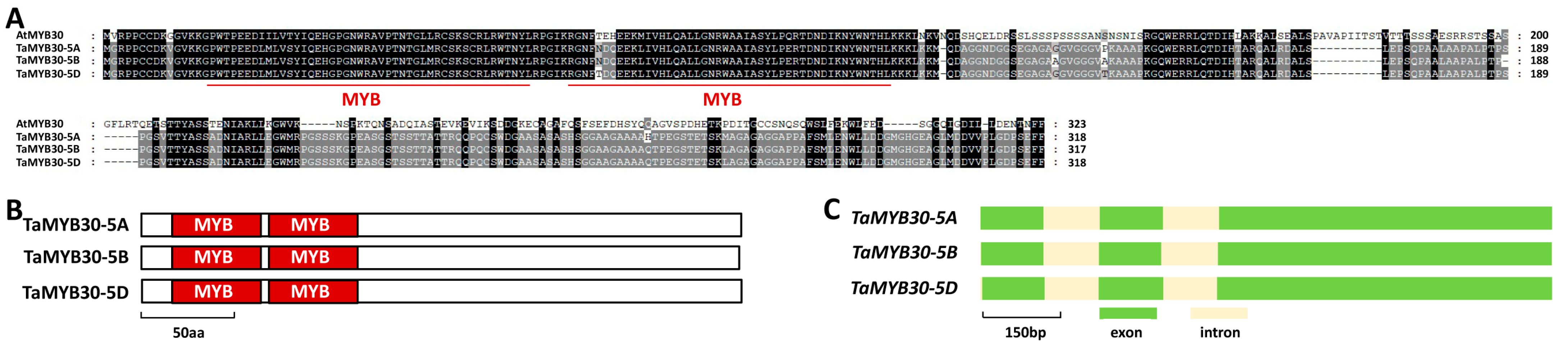
2.1. Identification of Wheat TaMYB30 Based on Homology with Arabidopsis AtMYB30

The MYB transcription factor AtMYB30 was demonstrated to be a key regulator of wax biosynthesis in the dicot model plant Arabidopsis [43]. In this study, we are interested in characterizing the function of the wheat homolog of AtMYB30 in the wax biosynthesis of bread wheat. To this end, we first searched the reference genome of the hexaploid bread wheat by using the amino acid sequence of Arabidopsis AtMYB30 (At3g28910) as a query and obtained TaMYB30, the most closed homologs of AtMYB30, in bread wheat. Three highly homologous sequences of TaMYB30 genes separately located on chromosomes 5A, 5B, and 5D were obtained from the genome sequence of the hexaploid wheat and designated as TaMYB30-5A (TraesCS5A02G227400), TaMYB30-5B (TraesCS5B02G226100), and TaMYB30-5D (TraesCS5D02G234800), respectively. As shown in Figure 1A, these predicted TaMYB30-5A, TaMYB30-5B, and TaMYB30-5D proteins shared about 45% identities with Arabidopsis AtMYB30. The TaMYB30-5A, TaMYB30-5B, and TaMYB30-5D proteins all contain two conserved MYB motifs at their N-termini (Figure 1B). The coding regions of these allelic TaMYB30 genomic sequences all contained three exons and two introns (Figure 1C).

2.2. TaMYB30 Positively Regulates Wheat Wax Biosynthesis

To study the function of TaMYB30 in wheat wax biosynthesis, we performed barley stripe mosaic virus (BSMV)-induced gene silencing (BSMV-VIGS) to silence all endogenous TaMYB30 genes in the leaves of the wheat cultivar Yannong 999. BSMV-infected wheat plants were employed as the negative controls. As revealed by the quantitative reverse-transcription polymerase chain reaction (qRT-PCR) assay, the TaMYB30 expression level was significantly reduced in the BSMV-TaMYB30as-infected wheat leaves (Figure 2A). We then conducted the gas chromatography-mass spectrometry (GC-MS) assay to measure cuticular wax constituents released from BSMV-γ and BSMV-TaMYB30as plants. As shown in Figure 2B, the amount of total cuticular wax was reduced from 12.1 μg cm−2 in the wheat leaves infected with BSMV-γ to 2.29 μg cm−2 in the BSMV-TaMYB30as-infected wheat plants (Figure 2B). Further wax component analyses revealed that the most abundant wax constituent, C28-alcohol, decreased from 8.05 μg cm−2 in the BSMV-γ wheat leaves to 1.05 μg cm−2 in the BSMV-TaMYB30as plants (Figure 2C). Likewise, other VLC fatty acids, alcohols, aldehydes, alkanes, and esters all showed an obvious reduction in the BSMV-TaMYB30as-infected wheat plants compared with BSMV-γ control plants (Figure 2C). These results indicated that the silencing of TaMYB30 significantly attenuated wax biosynthesis in wheat leaves and suggested that TaMYB30 positively regulates wheat wax biosynthesis.
Changes in wax deposition usually resulted in altered cuticle permeability. Compared with the BSMV-γ controls, the leaves of the BSMV-TaMYB30as-infected wheat plants displayed higher water loss rates with a loss of approximately 70% of the total water after 12 h (Figure 2D). Consistently, the chlorophyll leaching assay demonstrated that the BSMV-TaMYB30as-infected wheat plants released chlorophyll faster than the BSMV-γ-infected wheat plants (Figure 2E). Collectively, these results suggested that the silencing of TaMYB30 resulted in attenuated wax biosynthesis and enhanced surface barrier properties in wheat leaves.

2.3. Homology-Based Identification of TaKCS1 and TaECR in Bread Wheat

Previous studies revealed that the Arabidopsis MYB transcription factor AtMYB30 could regulate the expressions of wax biosynthesis genes AtKCS1 and AtECR [43]. We were interested in testing the potential regulation of TaMYB30 on these wax biosynthesis genes in bread wheat. To this end, we searched the reference genome of the hexaploid bread wheat by using the amino acid sequence of Arabidopsis AtKCS1 (At1g01120) and AtECR (At3g55360) as a query and obtained TaKCS1 and TaECR1, the most closed homologs of AtKCS1 and AtECR1, in bread wheat. Three highly homologous sequences of TaKCS1 genes separately located on chromosomes 4A, 4B, and 4D were obtained from the genome sequence of the hexaploid wheat and designated as TaKCS1-4A (TraesCS4A02G068400), TaKCS1-4B (TraesCS4B02G225500), and TaKCS1-4D (TraesCS4D02G226100), respectively. Similarly, three highly homologous sequences of TaECR genes separately located on chromosomes 3A, 3B, and 3D were obtained from the genome sequence of the hexaploid wheat and designated as TaECR-3A, TaECR-3B, and TaECR-3D, respectively [33].
As shown in Figure 3A, these predicted TaKCS1-4A, TaKCS1-4B, and TaKCS1-4D proteins shared about 43% identities with Arabidopsis AtKCS1. In addition, the TaKCS1-4A, TaKCS1-4B, and TaKCS1-4D proteins all contain a central FAE1/Type III polyketide synthase-like protein (FAE1_CUT1_RppA) domain, and the C-terminal 3-Oxoacyl-[acyl-carrier-protein (ACP)] synthase III C terminal (ACP_syn_III_C) domain (Figure 3B). The coding regions of these allelic TaKCS1 genomic sequences all contained one exon and zero introns (Figure 3C). The predicted TaECR-3A, TaECR-3B, and TaECR-3D proteins shared about 81% identities with Arabidopsis AtECR (Figure 3D). In addition, the TaECR-3A, TaECR-3B, and TaECR-3D proteins all contain a C-terminal 3-oxo-5-alpha-steroid 4-dehydrogenase (3-oxo-5_α-steroid 4-DH_C) domain (Figure 3E). The coding regions of these allelic TaECR genomic sequences all contained four exons and three introns (Figure 3F).

2.4. TaKCS1 and TaECR Positively Contribute to Wheat Wax Biosynthesis

To study the function of TaKCS1 and TaECR in wheat wax biosynthesis, we employed the BSMV-VIGS to silence all endogenous TaKCS1 or TaECR genes in the leaves of the wheat cultivar Yannong 999. As shown in Figure 4A, the expression levels of TaKCS1 or TaECR genes are significantly reduced in the BSMV-TaKCS1as- or BSMV-TaECRas-infected wheat plants (Figure 4A). We then measured cuticular wax constituents released from BSMV-γ, BSMV-TaKCS1as, or BSMV-TaECRas plants by GC-MS assay. As shown in Figure 4B, the amount of total cuticular wax decreased from 11.68 μg cm−2 in the BSMV-γ leaves to 2.89 μg cm−2 and 2.51 μg cm−2 in the wheat leaves infected with BSMV-TaKCS1as and BSMV-TaECRas, respectively (Figure 4B). Wax component analyses revealed that the amounts of VLC fatty acids, alcohols, aldehydes, alkanes, and esters were reduced significantly in the BSMV-TaKCS1as and BSMV-TaECRas leaves compared with the BSMV-γ controls (Figure 4C). These results collectively showed that the knockdown of TaKCS1 and TaECR expression levels remarkably attenuated wax biosynthesis in wheat leaves and confirmed that TaKCS1 and TaECR positively contribute to wheat wax biosynthesis.
Thereafter, we analyzed the cuticle permeability in the TaKCS1- or TaECR-silenced wheat plants by measuring the water loss rate and chlorophyll extracted. As shown in Figure 4D, the leaves of the BSMV-TaKCS1as- or BSMV-TaECRas-infected wheat plants displayed higher water loss rates than the BSMV-γ controls. Consistent with this, the chlorophyll leaching assay showed that the wheat leaves with silenced TaKCS1 or TaECR genes released chlorophyll faster than the BSMV-γ controls (Figure 4E). Overall, these results suggested that the knockdown of the TaKCS1 and TaECR expression levels led to compromised attenuated wax biosynthesis and enhanced surface barrier properties in wheat leaves.

2.5. TaMYB30 Activates Expression of TaKCS1 and TaECR

To determine the potential regulation of TaMYB30 on the expression of TaKCS1 and TaECR in bread wheat, we employed BSMV-VIGS to silence all endogenous TaMYB30 genes in the leaves of wheat cultivar Yannong 999. Thereafter, expression levels of TaKCS1 or TaECR genes were analyzed by qRT-PCR assay. As shown in Figure 5A, the silencing of TaMYB30 genes resulted in above 70% decreases in the expression levels of TaKCS1 and TaECR, suggesting that the MYB transcription factor TaMYB30 positively regulates the expressions of TaKCS1 and TaECR.
To quantify the transcriptional activity of TaMYB30 proteins, we performed the Arabidopsis leaf protoplast transfection assay, in which a luciferase (LUC) reporter was co-transfected with effector constructs expressing TaMYB30-5A, TaMYB30-5B, or TaMYB30-5D protein, and a control plasmid, to analyze the reporter luciferase activity (LucA). The Gal4 DNA-binding domain (DBD) was used to determine the basal LUC activity. As shown in Figure 5B, the LucA ratio has increased from 1 for the DBD control to above 2.2 under the expression of DBD-TaMYB30-5A, DBD-TaMYB30-5B, or DBD-TaMYB30-5D, indicating that TaMYB30 proteins exhibit transcriptional activator activity.
It has been demonstrated that wheat MYB transcription factor TaMYB96/TaEPBM1, a homolog of TaMYB30, could directly bind to the cis-elements MBS and Motif 1 [45,46]. Since MBS and Motif 1 cis-elements were found in the promoter regions of TaKCS1 and TaECR genes, we examined whether TaMYB30 proteins could directly bind to the TaKCS1 and TaECR promoters and activate their expressions. In the Arabidopsis leaf protoplast transfection assay, LUC reporters containing promoter regions of TaKCS1-4A, TaKCS1-4B, TaKCS1-4D, TaECR-3A, TaECR-3B, or TaECR-3D genes were co-transfected with effector constructs overexpressing TaMYB30-5A, TaMYB30-5B, or TaMYB30-5D proteins, and a control plasmid. Wild-type promoter regions of TaKCS1-4A, TaKCS1-4B, TaKCS1-4D, TaECR-3A, TaECR-3B, or TaECR-3D genes harboring the cis-elements MBS and Motif 1, and TaKCS1-4A, TaKCS1-4B, TaKCS1-4D, TaECR-3A, TaECR-3B, or TaECR-3D promoter mutants containing mutated MBS and Motif 1 cis-elements, were employed. As shown in Figure 5C,D, the LucA ratio obtained from LUC reporters containing wild-type TaKCS1 and TaECR promoters increased to above 2.07 in the presence of over-accumulated TaMYB30 proteins, compared with 1 for the Gal4 DNA-binding domain (DBD) proteins and with less than 0.3 for the empty vector (EV) control. In contrast, the LucA ratio obtained from mutant TaKCS1 and TaECR promoters was not significantly affected by over-accumulated TaMYB30 proteins (Figure 5C,D). This result implies that the transcriptional activators TaMYB30-5A, TaMYB30-5B, and TaMYB30-5D could directly bind to the promoter regions of TaKCS1 and TaECR genes by recognizing the MBS and Motif 1 cis-elements, and activate their expression in plant cells.

3. Discussion

3.1. TaMYB30 Is a Positive Regulator of Wax Biosynthesis in Bread Wheat

In this study, TaMYB30 is characterized as a positive regulator of wheat wax biosynthesis. The knockdown of TaMYB30 expression using BSMV-VIGS resulted in attenuated wax biosynthesis and enhanced cuticle permeability. Increasing evidence revealed that wheat wax biosynthesis is governed by transcription factors and their interactors. For instance, the knockout or knockdown expression of the transcription factor gene TaSHN1/WIN1 attenuated wheat wax biosynthesis, whereas the transgenic overexpression of the TaSHN1/WIN1 gene resulted in enhanced wax accumulation in bread wheat [38,39,47]. The mediator component CYCLIN-DEPENDENT KINASE8 (TaCDK8) was demonstrated to interact and phosphorylate the transcription factor TaSHN1/WIN1 [38,48,49,50]. The silencing of TaCDK8 by BSMV-VIGS assay resulted in attenuated wax accumulation in wheat leaves, indicating that TaCDK8 positively regulates wheat wax biosynthesis [38]. Interestingly, AtCDK8 interacts with AtSHN1 to potentiate wax biosynthesis in the dicot model plant A. thaliana, implying that the SHN1-CDK8 interacting module regulates wax biosynthesis in dicot and monocot plants [51]. The wheat R2R3-type MYB transcription factor TaMYB96/TaEPBM1 was revealed to trigger cuticular wax biosynthesis [45,46]. The TaGCN5-TaADA2 histone acetyltransferase module was shown to associate with TaMYB96/TaEPBM1 and mediate histone acetylation at the promoter regions of TaMYB96/TaEPBM1 target genes [45]. The knockdown of TaGCN5 or TaADA2 expression levels led to reduced wax accumulation, indicating that the TaGCN5-TaADA2 histone acetyltransferase module interacts with TaMYB96/TaEPBM1 to trigger wax biosynthesis [45]. Similarly, the CHD3-type chromatin remodeling factor TaCHR729 could interact with the basic helix-loop-helix (bHLH) transcription factor TaKPAB1 and mediate the deposition of the permissive epigenetic mark H3K4me3 at TaKCS6 promoters [52,53]. These studies demonstrated that transcription factors interact with other regulators such as mediator components and epigenetic components to fine-tune wheat wax biosynthesis. Therefore, it is intriguing to identify TaMYB30-interacting partners and characterize their potential roles in the regulation of wheat wax biosynthesis in future research.

3.2. Wheat TaKCS1 and TaECR Are Essential Components of Wax Biosynthetic Machinery

TaKCS1 is identified as an important component of wax biosynthetic machinery in bread wheat. In A. thaliana, AtKCS1 encodes a 3-ketoacyl-CoA synthase involved in wax biosynthesis [54]. The T-DNA-tagged kcs1-1 mutant displayed a decrease of up to 80% in the levels of VLC wax alcohols and aldehydes, but much smaller effects on the major Arabidopsis wax components the C29 alkanes and on the total wax load, suggesting the potential redundancy in the KCS activities involved in Arabidopsis wax synthesis [54]. In contrast, the barley eceriferum-zh (cer-zh) mutant displays an obvious glossy and wax-deficient phenotype [55]. CER-ZH encodes the β-ketoacyl-CoA synthase HvKCS1, a barley ortholog of AtKCS1. In this study, the silencing of TaKCS1 by BSMV-VIGS led to compromised wax biosynthesis, increased water loss rates, and enhanced chlorophyll leaching [55]. These results indicate the potential functional conservation of KCS1 in monocot and dicot plants, and that the distinct contribution of KCS1 to total wax load in Arabidopsis, barley, and wheat might result from different wax compositions among these plant species.
As an obligate biotrophic fungal pathogen, Blumeria graminis forma specialis tritici (B.g. tritici) has evolved sophisticated mechanisms to infect the host wheat plants. Increasing evidence suggests that wax signals from wheat epidermis could be harnessed by the host-adapted B.g. tritici fungus to promote pre-penetration infection processes [38,45,52]. For instance, the silencing of wheat wax biosynthesis genes TaWIN1, TaKCS6 and TaECR resulted in the attenuated conidial germination of B.g. tritici. In this study, the knockdown of TaMYB30 or TaKCS1 expressions led to reduced cuticular wax biosynthesis. It is therefore intriguing to examine whether and how TaMYB30 and TaKCS1 affect pre-penetration events such as conidial germination in the infection of B.g. tritici in future studies.
The Enoyl-CoA Reductase (ECR) gene encodes the enoyl-CoA reductase that catalyzes the biosynthesis of VLC acyl-CoAs in the fatty acid elongase complex [56]. Recent phylogenetic analysis showed that ECR genes are highly conserved among land plants. The knockout of ECR genes could significantly attenuate wax biosynthesis in Arabidopsis, whereas the ectopic overexpression of CsECR cloned from the “Newhall” navel orange (Citrus sinensis [L.] Osbeck cv. Newhall) increased cuticular wax accumulation in the tomato (Solanum lycopersicum) and enhanced its drought resistance [57,58]. In this and previous studies, the silencing of TaECR by BSMV-VIGS resulted in reduced wax biosynthesis and enhanced cuticle permeability, as well as the attenuated germination of B.g. tritici conidia [45]. These data suggested that ECR plays a key role in the wax biosynthesis among dicot and monocot plants, and has potential values in crop breeding for improving stress resilience and disease resistance.

3.3. TaMYB30 Activates Expression of TaKCS1 and TaECR1 to Positively Regulate Wheat Wax

Three closely homologous TaMYB30-5A, TaMYB30-5B, and TaMYB30-5D proteins were identified as transcriptional activators in this study. Importantly, TaMYB30-5A, TaMYB30-5B, or TaMYB30-5D could directly bind to the promoter regions of TaKCS1 and TaECR genes by recognizing the MBS and Motif 1 cis-elements, and stimulate their expression in plant cells. These data collectively suggested that TaMYB30 positively regulates wheat wax biosynthesis presumably via the transcriptional activation of TaKCS1 and TaECR (Figure 5E). It was previously reported that the expressions of Arabidopsis wax biosynthesis genes such as AtKCS1, AtECR, etc. were reduced in the AtMYB30-knockout mutant but enhanced in the AtMYB30-overexpressing lines, indicating that the up-regulation of wax biosynthesis genes by MYB30 and its orthologs is conserved among dicots and monocots [43].
It was previously demonstrated that another R2R3-type MYB transcription factor TaMYB96/TaEPBM1 could activate the expression of TaECR genes and positively regulate wheat wax biosynthesis [45]. In this study, the transcriptional activator TaMYB30 was shown to directly recognize the promoter regions of TaECR-3A, TaECR-3B, and TaECR-3D genes, and to activate their expression, suggesting that a single wax biosynthesis gene might be targeted by different MYB transcription factors. Accumulating studies revealed that plant wax biosynthesis is tightly regulated at the transcriptional level and affected by developmental stages and environmental conditions. It is intriguing to examine the interplays among different MYB transcription factors in the regulation of wax biosynthesis in response to developmental and environmental cues in future research.

4. Materials and Methods

4.1. Plant Materials

The plant seedlings of bread wheat cultivar Yannong 999 were grown in a growth chamber under a 16-h/8-h, 20 °C/18 °C day/night cycle with 70% relative humidity. A. thaliana Col-0 used in this study was grown in the greenhouse under a 16-h/8-h light period at 23 ± 1 °C with 70% relative humidity.

4.2. Quantitative Real-Time PCR (qRT-PCR)

The cuticular wax composition analysis was performed as described [38]. Briefly, total RNA was extracted from the wheat leaves using the EasyPure Plant RNA kit (Transgenbiotech, Beijing, China) and was used to synthesize the cDNA template using the TransScript one-step gDNA removal and cDNA synthesis supermix (Transgenbiotech, Beijing, China). The Real-time PCR assay was performed using the ABI real-time PCR system with the qPCR Master Mix (Invitrogen, Carlsbad, CA, USA). The expression of TaGADPH was set as the internal control and the expression levels of TaGAPDH, TaMYB30, TaKCS1, and TaECR were analyzed using the primer pairs 5′-TTAGACTTGCGAAGCCAGCA-3′/5′-AAATGCCCTTGAGGTTTCCC-3′, 5′-TGCTCGTCTCCTACATCCA-3′/5′-TATCGCCGCCCAACGGTTG-3′, 5′-CTACTCCTTCGTCCGCCTC-3′/5′-GTGATCTTGGTCTGGAACG-3′, and 5′-TGAAGGTCTCCGTCGTGTCC-3′/5′-CAGAAGAAGAGCGTGCTGTAG-3′, respectively.

4.3. BSMV-Mediated Gene Silencing

Antisense fragments of TaMYB30, TaKCS1, and TaECR were cloned into the pCa-γbLIC vector to create the BSMV-TaMYB30as, BSMV-TaKCS1as, and BSMV-TaECRas constructs using the primer pairs 5′-AAGGAAGTTTATAGAAGAACTCACTGGGGTC-3′/5′-AACCACCACCACCGTGCCAGCAGCCGCAGTGCTC-3′, 5′-AAGGAAGTTTATGTG CACGCAGAAGTGCTCG-3′/5′-AACCACCACCACCGTGGGACGAGTGCTTCAAGT GC-3′, and 5′-AAGGAAGTTTGGAGGAAGAATCACCCATCG-3′/5′-AACCACCACC ACCGT TCAACATTGTGACATGCGCAAAC-3′, respectively. The BSMV-mediated gene silencing in wheat leaves was performed as described by Kong and Chang [38].

4.4. Water Loss Rate and Chlorophyll Leaching Tests

The water loss rate tests were performed as described previously [59,60]. Briefly, the leaves (n = 5) with BSMV virus symptoms about 2 weeks post-BSMV-infection were dipped in ultrapure water under dark conditions for 1 h to maintain the stomatal closure, and leaf weights were then measured per 1 h for 12 h. The water loss rate tests were performed as described [59,61]. Wheat plants (n = 5) with BSMV virus symptoms about 2 weeks post-BSMV-infection were kept in the dark and immersed in ultrapure water for 1 h. Then leaves were detached and dipped in 80% ethanol, and the amount of chlorophyll extracted was measured at 647 and 664 nm with a spectrophotometer (Cary 60 UV-Vis, Agilent Technologies, Santa Clara, CA, USA) every 60 min for 12 h.

4.5. Wax Composition Analysis

The cuticular wax composition analysis was performed as described [38]. The leaves (n = 5) with BSMV virus symptoms about 2 weeks post-BSMV-infection were dipped into chloroform (Merck, Rahway, NJ, USA). The extracts dried under N2 were derivatized at 70 °C for 30 min through reaction with bis-N,O- trimethylsilyl trifluoroacetamide and analyzed with a capillary GC (5890 Series II, Agilent Technologies) and a flame ionization detector (6890 N, Agilent Technologies) with a mass spectrometer (MSD 5973, Agilent Technologies) as previously described [38]. Quantification was based on FID peak areas relative to the internal standard n-Tetracosane (Merck) and the dry, delipidated tissue weights.

4.6. Arabidopsis Protoplast Transactivation Assay

The isolation and transformation of the Arabidopsis protoplast were conducted as described previously by Zhi et al. [62]. The vector pRT-DBD and pRT were employed to generate construct expression proteins with and without DBD fusion, respectively. The effector constructs, including pRT, pRT-BD, and derivatives, were mixed with the reporter gene and co-delivered into the Arabidopsis protoplasts by transfection. At 48 h after transfection, the LUC activity was analyzed using a Promega dual-luciferase reporter assay system (Promega, Madison, WI, USA, E1910) following the manufacturer’s manual.

Author Contributions

L.L. and C.C. planned and designed the research; L.L. and X.W. performed most of the experiments with help from X.W.; L.L., H.L. and C.C. analyzed data and wrote the manuscript with contribution from X.W. All authors have read and agreed to the published version of the manuscript.

Funding

This work was supported by the Natural Science Foundation of Shandong Province (ZR2022MC008, ZR2017BC109), the Qingdao Science and Technology Bureau Fund (17-1-1-50-jch), and the Qingdao University Fund (DC1900005385).

Institutional Review Board Statement

Not applicable.

Informed Consent Statement

Not applicable.

Data Availability Statement

Data presented here are available on request from correspondence.

Acknowledgments

We thank Michael Moustakas for the kind invitation to contribute this work to special issue ‘Molecular Mechanisms of Plant Abiotic Stress Tolerance’. We are also grateful to anonymous reviewers for their very helpful comments on this manuscript.

Conflicts of Interest

The authors declare no conflict of interest.

References

  1. Yeats, T.H.; Rose, J.K. The formation and function of plant cuticles. Plant Physiol. 2013, 163, 5–20. [Google Scholar] [CrossRef] [PubMed] [Green Version]
  2. Martin, L.B.; Rose, J.K. There’s more than one way to skin a fruit: Formation and functions of fruit cuticles. J. Exp. Bot. 2014, 65, 4639–4651. [Google Scholar] [CrossRef] [Green Version]
  3. Lee, S.B.; Suh, M.C. Advances in the understanding of cuticular waxes in Arabidopsis thaliana and crop species. Plant Cell Rep. 2015, 34, 557–572. [Google Scholar] [CrossRef] [PubMed]
  4. Kurdyukov, S.; Faust, A.; Nawrath, C.; Bär, S.; Voisin, D.; Efremova, N.; Franke, R.; Schreiber, L.; Saedler, H.; Métraux, J.P.; et al. The epidermis-specific extracellular BODYGUARD controls cuticle development and morphogenesis in Arabidopsis. Plant Cell 2006, 18, 321–339. [Google Scholar] [CrossRef] [Green Version]
  5. Ingram, G.; Nawrath, C. The roles of the cuticle in plant development: Organ adhesions and beyond. J. Exp. Bot. 2017, 68, 5307–5321. [Google Scholar] [CrossRef] [PubMed] [Green Version]
  6. Weng, H.; Molina, I.; Shockey, J.; Browse, J. Organ fusion and defective cuticle function in a lacs1 lacs2 double mutant of Arabidopsis. Planta 2010, 231, 1089–1100. [Google Scholar] [CrossRef]
  7. Wang, X.; Chang, C. Exploring and exploiting cuticle biosynthesis for abiotic and biotic stress tolerance in wheat and barley. Front. Plant Sci. 2022, 13, 1064390. [Google Scholar] [CrossRef]
  8. Kong, L.; Liu, Y.; Zhi, P.; Wang, X.; Xu, B.; Gong, Z.; Chang, C. Origins and evolution of cuticle biosynthetic machinery in land plants. Plant Physiol. 2020, 184, 1998–2010. [Google Scholar] [CrossRef]
  9. Li, H.; Chang, C. Evolutionary insight of plant cuticle biosynthesis in bryophytes. Plant Signal. Behav. 2021, 16, 1943921. [Google Scholar] [CrossRef]
  10. Liu, L.; Wang, X.; Chang, C. Toward a smart skin: Harnessing cuticle biosynthesis for crop adaptation to drought, salinity, temperature, and ultraviolet stress. Front. Plant Sci. 2022, 13, 961829. [Google Scholar] [CrossRef]
  11. Wang, X.; Kong, L.; Zhi, P.; Chang, C. Update on cuticular wax biosynthesis and its roles in plant disease resistance. Int. J. Mol. Sci. 2020, 21, 5514. [Google Scholar] [CrossRef] [PubMed]
  12. Liu, X.; Feakins, S.J.; Ma, X.F.; Anderson, J.D.; Vidal, E.; Blancaflor, E.B. Crop breeding has increased the productivity and leaf wax n-alkane concentration in a series of five winter wheat cultivars developed over the last 60 years. J. Plant Physiol. 2019, 243, 153056. [Google Scholar] [CrossRef] [PubMed]
  13. Petit, J.; Bres, C.; Mauxion, J.P.; Bakan, B.; Rothan, C. Breeding for cuticle-associated traits in crop species: Traits, targets, and strategies. J. Exp. Bot. 2017, 68, 5369–5387. [Google Scholar] [CrossRef] [PubMed]
  14. Bach, L.; Michaelson, L.V.; Haslam, R.; Bellec, Y.; Gissot, L.; Marion, J.; Da Costa, M.; Boutin, J.P.; Miquel, M.; Tellier, F.; et al. The very-long-chain hydroxy fatty acyl-CoA dehydratase PASTICCINO2 is essential and limiting for plant development. Proc. Natl. Acad. Sci. USA 2008, 105, 14727–14731. [Google Scholar] [CrossRef] [PubMed] [Green Version]
  15. Beaudoin, F.; Wu, X.; Li, F.; Haslam, R.P.; Markham, J.E.; Zheng, H.; Napier, J.A.; Kunst, L. Functional characterization of the Arabidopsis β-ketoacyl-coenzyme A reductase candidates of the fatty acid elongase. Plant Physiol. 2009, 150, 1174–1191. [Google Scholar] [CrossRef] [PubMed] [Green Version]
  16. Kim, J.; Jung, J.H.; Lee, S.B.; Go, Y.S.; Kim, H.J.; Cahoon, R.; Markham, J.E.; Cahoon, E.B.; Suh, M.C. Arabidopsis 3-ketoacyl-coenzyme A synthase 9 is involved in the synthesis of tetracosanoic acids as precursors of cuticular waxes, suberins, sphingolipids, and phospholipids. Plant Physiol. 2013, 162, 567–580. [Google Scholar] [CrossRef] [Green Version]
  17. Lee, S.B.; Jung, S.J.; Go, Y.S.; Kim, H.U.; Kim, J.K.; Cho, H.J.; Park, O.K.; Suh, M.C. Two Arabidopsis 3-ketoacyl CoA synthase genes, KCS20 and KCS2/DAISY, are functionally redundant in cuticular wax and root suberin biosynthesis, but differentially controlled by osmotic stress. Plant J. 2009, 60, 462–475. [Google Scholar] [CrossRef]
  18. Huang, H.; Yang, X.; Zheng, M.; Chen, Z.; Yang, Z.; Wu, P.; Jenks, M.A.; Wang, G.; Feng, T.; Liu, L.; et al. An ancestral role for 3-KETOACYL-COA SYNTHASE3 as a negative regulator of plant cuticular wax synthesis. Plant Cell 2023, 35, 2251–2270. [Google Scholar] [CrossRef]
  19. Haslam, T.M.; Haslam, R.; Thoraval, D.; Pascal, S.; Delude, C.; Domergue, F.; Fernández, A.M.; Beaudoin, F.; Napier, J.A.; Kunst, L.; et al. ECERIFERUM2-LIKE proteins have unique biochemical and physiological functions in very-long-chain fatty acid elongation. Plant Physiol. 2015, 167, 682–692. [Google Scholar] [CrossRef] [Green Version]
  20. Haslam, T.; Gerelle, W.; Graham, S.; Kunst, L. The unique role of the ECERIFERUM2-LIKE clade of the BAHD acyltransferase superfamily in cuticular wax metabolism. Plants 2017, 6, 23. [Google Scholar] [CrossRef] [Green Version]
  21. Bourdenx, B.; Bernard, A.; Domergue, F.; Pascal, S.; Léger, A.; Roby, D.; Pervent, M.; Vile, D.; Haslam, R.P.; Napier, J.A.; et al. Overexpression of Arabidopsis ECERIFERUM1 promotes wax very-long-chain alkane biosynthesis and influences plant response to biotic and abiotic stresses. Plant Physiol. 2011, 156, 29–45. [Google Scholar] [CrossRef] [Green Version]
  22. Bernard, A.; Domergue, F.; Pascal, S.; Jetter, R.; Renne, C.; Faure, J.D.; Haslam, R.P.; Napier, J.A.; Lessire, R.; Joubes, J. Reconstitution of plant alkane biosynthesis in yeast demonstrates that Arabidopsis ECERIFERUM1 and ECERIFERUM3 are core components of a very-long-chain alkane synthesis complex. Plant Cell 2012, 24, 3106–3118. [Google Scholar] [CrossRef] [PubMed] [Green Version]
  23. Pascal, S.; Bernard, A.; Deslous, P.; Gronnier, J.; Fournier-Goss, A.; Domergue, F.; Rowland, O.; Joubès, J. Arabidopsis CER1-LIKE1 functions in a cuticular very-long-chain alkane-forming complex. Plant Physiol. 2019, 179, 415–432. [Google Scholar] [CrossRef] [Green Version]
  24. Rowland, O.; Zheng, H.; Hepworth, S.R.; Lam, P.; Jetter, R.; Kunst, L. CER4 encodes an alcohol-forming fatty acyl-coenzyme A reductase involved in cuticular wax production in Arabidopsis. Plant Physiol. 2006, 142, 866–877. [Google Scholar] [CrossRef] [PubMed] [Green Version]
  25. Yang, X.; Zhao, H.; Kosma, D.K.; Tomasi, P.; Dyer, J.M.; Li, R.; Liu, X.; Wang, Z.; Parsons, E.P.; Jenks, M.A.; et al. The acyl desaturase CER17 is involved in producing wax unsaturated primary alcohols and cutin monomers. Plant Physiol. 2017, 173, 1109–1124. [Google Scholar] [CrossRef] [PubMed] [Green Version]
  26. Li, F.; Wu, X.; Lam, P.; Bird, D.; Zheng, H.; Samuels, L.; Jetter, R.; Kunst, L. Identification of the wax ester synthase/acyl-coenzyme A: Diacylglycerol acyltransferase WSD1 required for stem wax ester biosynthesis in Arabidopsis. Plant Physiol. 2008, 148, 97–107. [Google Scholar] [CrossRef] [Green Version]
  27. Pighin, J.A.; Zheng, H.; Balakshin, L.J.; Goodman, I.P.; Western, T.L.; Jetter, R.; Kunst, L.; Samuels, A.L. Plant cuticular lipid export requires an ABC transporter. Science 2004, 306, 702–704. [Google Scholar] [CrossRef] [PubMed] [Green Version]
  28. Debono, A.; Yeats, T.H.; Rose, J.K.; Bird, D.; Jetter, R.; Kunst, L.; Samuels, L. Arabidopsis LTPG is a glycosylphosphatidylinositol-anchored lipid transfer protein required for export of lipids to the plant surface. Plant Cell 2009, 21, 1230–1238. [Google Scholar] [CrossRef] [Green Version]
  29. Ichino, T.; Yazaki, K. Modes of secretion of plant lipophilic metabolites via ABCG transporter-dependent transport and vesicle-mediated trafficking. Curr. Opin. Plant Biol. 2022, 66, 102184. [Google Scholar] [CrossRef]
  30. Nguyen, V.; Lee, S.B.; Suh, M.C.; An, G.; Jung, K.H. OsABCG9 is an important ABC transporter of cuticular wax deposition in rice. Front. Plant Sci. 2018, 9, 960. [Google Scholar] [CrossRef]
  31. Lee, S.B.; Suh, M.C. Regulatory mechanisms underlying cuticular wax biosynthesis. J. Exp. Bot. 2022, 73, 2799–2816. [Google Scholar] [CrossRef] [PubMed]
  32. Broun, P.; Poindexter, P.; Osborne, E.; Jiang, C.Z.; Riechmann, J.L. WIN1, a transcriptional activator of epidermal wax accumulation in Arabidopsis. Proc. Natl. Acad. Sci. USA 2004, 101, 4706–4711. [Google Scholar] [CrossRef] [Green Version]
  33. Aharoni, A.; Dixit, S.; Jetter, R.; Thoenes, E.; van Arkel, G.; Pereira, A. The SHINE clade of AP2 domain transcription factors activates wax biosynthesis, alters cuticle properties, and confers drought tolerance when overexpressed in Arabidopsis. Plant Cell 2004, 16, 2463–2480. [Google Scholar] [CrossRef] [Green Version]
  34. Kannangara, R.; Branigan, C.; Liu, Y.; Penfield, T.; Rao, V.; Mouille, G.; Höfte, H.; Pauly, M.; Riechmann, J.L.; Broun, P. The transcription factor WIN1/SHN1 regulates cutin biosynthesis in Arabidopsis thaliana. Plant Cell 2007, 19, 1278–1294. [Google Scholar] [CrossRef] [Green Version]
  35. Kumar, A.; Yogendra, K.N.; Karre, S.; Kushalappa, A.C.; Dion, Y.; Choo, T.M. WAX INDUCER1 (HvWIN1) transcription factor regulates free fatty acid biosynthetic genes to reinforce cuticle to resist Fusarium head blight in barley spikelets. J. Exp. Bot. 2016, 67, 4127–4139. [Google Scholar] [CrossRef] [Green Version]
  36. Djemal, R.; Khoudi, H. The barley SHN1-type transcription factor HvSHN1 imparts heat, drought and salt tolerances in transgenic tobacco. Plant Physiol. Biochem. 2021, 164, 44–53. [Google Scholar] [CrossRef]
  37. Kim, R.J.; Lee, S.B.; Pandey, G.; Suh, M.C. Functional conservation of an AP2/ERF transcription factor in cuticle formation suggests an important role in the terrestrialization of early land plants. J. Exp. Bot. 2022, 73, 7450–7466. [Google Scholar] [CrossRef]
  38. Kong, L.; Chang, C. Suppression of wheat TaCDK8/TaWIN1 interaction negatively affects germination of Blumeria graminis f.sp. tritici by interfering with very-long-chain aldehyde biosynthesis. Plant Mol. Biol 2018, 96, 165–178. [Google Scholar] [CrossRef]
  39. McAllister, T.; Campoli, C.; Eskan, M.; Liu, L.; McKim, S.M. A gene encoding a SHINE1/WAX INDUCER1 transcription factor controls cuticular wax in barley. Agronomy 2022, 12, 1088. [Google Scholar] [CrossRef]
  40. Lee, S.B.; Kim, H.U.; Suh, M.C. MYB94 and MYB96 additively activate cuticular wax biosynthesis in Arabidopsis. Plant Cell Physiol. 2016, 57, 2300–2311. [Google Scholar] [CrossRef] [PubMed] [Green Version]
  41. Lee, S.B.; Suh, M.C. Cuticular wax biosynthesis is up-regulated by the MYB94 transcription factor in Arabidopsis. Plant Cell Physiol. 2015, 56, 48–60. [Google Scholar] [CrossRef] [PubMed] [Green Version]
  42. Seo, P.J.; Lee, S.B.; Suh, M.C.; Park, M.J.; Go, Y.S.; Park, C.M. The MYB96 transcription factor regulates cuticular wax biosynthesis under drought conditions in Arabidopsis. Plant Cell 2011, 23, 1138–1152. [Google Scholar] [CrossRef] [PubMed] [Green Version]
  43. Raffaele, S.; Vailleau, F.; Léger, A.; Joubès, J.; Miersch, O.; Huard, C.; Blée, E.; Mongrand, S.; Domergue, F.; Roby, D.A. MYB transcription factor regulates very-long-chain fatty acid biosynthesis for activation of the hypersensitive cell death response in Arabidopsis. Plant Cell 2008, 20, 752–767. [Google Scholar] [CrossRef] [Green Version]
  44. Zhang, Y.L.; Zhang, C.L.; Wang, G.L.; Wang, Y.X.; Qi, C.H.; Zhao, Q.; You, C.X.; Li, Y.Y.; Hao, Y.J. The R2R3 MYB transcription factor MdMYB30 modulates plant resistance against pathogens by regulating cuticular wax biosynthesis. BMC Plant Biol. 2019, 19, 362. [Google Scholar] [CrossRef] [PubMed] [Green Version]
  45. Kong, L.; Zhi, P.; Liu, J.; Li, H.; Zhang, X.; Xu, J.; Zhou, J.; Wang, X.; Chang, C. Epigenetic activation of Enoyl-CoA Reductase by an acetyltransferase complex triggers wheat wax biosynthesis. Plant Physiol. 2020, 183, 1250–1267. [Google Scholar] [CrossRef]
  46. He, J.; Li, C.; Hu, N.; Zhu, Y.; He, Z.; Sun, Y.; Wang, Z.; Wang, Y. ECERIFERUM1-6A is required for the synthesis of cuticular wax alkanes and promotes drought tolerance in wheat. Plant Physiol. 2022, 190, 1640–1657. [Google Scholar] [CrossRef]
  47. Bi, H.; Shi, J.; Kovalchuk, N.; Luang, S.; Bazanova, N.; Chirkova, L.; Zhang, D.; Shavrukov, Y.; Stepanenko, A.; Tricker, P.; et al. Overexpression of the TaSHN1 transcription factor in bread wheat leads to leaf surface modifications, improved drought tolerance, and no yield penalty under controlled growth conditions. Plant Cell Environ. 2018, 41, 2549–2566. [Google Scholar] [CrossRef]
  48. Mao, X.; Weake, V.M.; Chapple, C. Mediator function in plant metabolism revealed by large-scale biology. J. Exp. Bot. 2019, 70, 5995–6003. [Google Scholar] [CrossRef]
  49. Agrawal, R.; Jiří, F.; Thakur, J.K. The kinase module of the Mediator complex: An important signalling processor for the development and survival of plants. J. Exp. Bot. 2021, 72, 224–240. [Google Scholar] [CrossRef]
  50. Chen, J.; Yang, S.; Fan, B.; Zhu, C.; Chen, Z. The Mediator complex: A central coordinator of plant adaptive responses to environmental stresses. Int. J. Mol. Sci. 2022, 23, 6170. [Google Scholar] [CrossRef]
  51. Zhu, Y.; Schluttenhoffer, C.M.; Wang, P.; Fu, F.; Thimmapuram, J.; Zhu, J.K.; Lee, S.Y.; Yun, D.J.; Mengiste, T. CYCLIN-DEPENDENT KINASE8 differentially regulates plant immunity to fungal pathogens through kinase-dependent and -independent functions in Arabidopsis. Plant Cell 2014, 26, 4149–4170. [Google Scholar] [CrossRef] [Green Version]
  52. Wang, X.; Zhi, P.; Fan, Q.; Zhang, M.; Chang, C. Wheat CHD3 protein TaCHR729 regulates the cuticular wax biosynthesis required for stimulating germination of Blumeria graminis f.sp. tritici. J. Exp. Bot. 2019, 70, 701–713. [Google Scholar] [CrossRef] [Green Version]
  53. Zhi, P.; Chang, C. Exploiting epigenetic variations for crop disease resistance improvement. Front. Plant Sci. 2021, 12, 692328. [Google Scholar] [CrossRef] [PubMed]
  54. Todd, J.; Post-Beittenmiller, D.; Jaworski, J.G. KCS1 encodes a fatty acid elongase 3-ketoacyl-CoA synthase affecting wax biosynthesis in Arabidopsis thaliana. Plant J. 1999, 17, 119–130. [Google Scholar] [CrossRef]
  55. Li, C.; Haslam, T.M.; Krüger, A.; Schneider, L.M.; Mishina, K.; Samuels, L.; Yang, H.; Kunst, L.; Schaffrath, U.; Nawrath, C.; et al. The β-ketoacyl-CoA synthase HvKCS1, encoded by Cer-zh, plays a key role in synthesis of barley leaf wax and germination of barley powdery mildew. Plant Cell Physiol. 2018, 59, 811–827. [Google Scholar] [CrossRef] [Green Version]
  56. Kohlwein, S.D.; Eder, S.; Oh, C.S.; Martin, C.E.; Gable, K.; Bacikova, D.; Dunn, T. Tsc13p is required for fatty acid elongation and localizes to a novel structure at the nuclear-vacuolar interface in Saccharomyces cerevisiae. Mol. Cell Biol. 2001, 21, 109–125. [Google Scholar] [CrossRef] [PubMed] [Green Version]
  57. Zheng, H.; Rowland, O.; Kunst, L. Disruptions of the Arabidopsis Enoyl-CoA reductase gene reveal an essential role for very-long-chain fatty acid synthesis in cell expansion during plant morphogenesis. Plant Cell 2005, 17, 1467–1481. [Google Scholar] [CrossRef] [Green Version]
  58. Liu, D.; Guo, W.; Guo, X.; Yang, L.; Hu, W.; Kuang, L.; Huang, Y.; Xie, J.; Liu, Y. Ectopic overexpression of CsECR from navel orange increases cuticular wax accumulation in tomato and enhances its tolerance to drought stress. Front. Plant Sci. 2022, 13, 924552. [Google Scholar] [CrossRef]
  59. Li, T.; Sun, Y.; Liu, T.; Wu, H.; An, P.; Shui, Z.; Wang, J.; Zhu, Y.; Li, C.; Wang, Y.; et al. TaCER1-1A is involved in cuticular wax alkane biosynthesis in hexaploid wheat and responds to plant abiotic stresses. Plant Cell Environ. 2019, 42, 3077–3091. [Google Scholar] [CrossRef] [PubMed]
  60. Kosma, D.K.; Bourdenx, B.; Bernard, A.; Parsons, E.P.; Lü, S.Y.; Joubès, J.; Jenks, M.A. The impact of water deficiency on leaf cuticle lipids of Arabidopsis. Plant Physiol. 2009, 151, 1918–1929. [Google Scholar] [CrossRef] [Green Version]
  61. Lolle, S.J.; Hsu, W.; Pruitt, R.E. Genetic analysis of organ fusion in Arabidopsis thaliana. Genetics 1998, 149, 607–619. [Google Scholar] [CrossRef] [PubMed]
  62. Zhi, P.; Kong, L.; Liu, J.; Zhang, X.; Wang, X.; Li, H.; Sun, M.; Li, Y.; Chang, C. Histone deacetylase TaHDT701 functions in TaHDA6-TaHOS15 complex to regulate wheat defense responses to Blumeria graminis f.sp. tritici. Int. J. Mol. Sci. 2020, 21, 2640. [Google Scholar] [CrossRef] [PubMed] [Green Version]
Figure 1. Homology-based identification of TaMYB30 in bread wheat. (A) Protein sequence comparison of wheat TaMYB30 and Arabidopsis AtMYB30. (B) Domain structure of wheat TaMYB30 proteins. (C) Gene architectures of wheat TaMYB30 genes.
Ijms 24 10235 g001
Figure 2. Functional analyses of wheat TaMYB30 in wheat wax biosynthesis. (A) qRT-PCR analysis of TaMYB30 expression levels in the wheat leaves infected with BSMV-γ or BSMV-TaMYB30as. (B) Total cuticular wax amounts in the wheat leaves infected with BSMV-γ or BSMV-TaMYB30as. (C) Amounts of major wax components in the BSMV-γ and BSMV-TaMYB30as wheat leaves. FA, fatty acid; ALC, alcohol; ALD, aldehyde; ALK, alkane; ALKE, alkyl ester; N. I., not identified compound. (D) Water loss rates and (E) chlorophyll extraction levels were measured using leaves infected with BSMV-γ or BSMV-TaMYB30as. For (AE), three independent biological replicates were statistically analyzed for each treatment (t-test; ** p < 0.01).
Ijms 24 10235 g002
Figure 3. Identification of wheat TaKCS1 and TaECR based on homology with Arabidopsis AtKCS1 and AtECR. (A) Protein sequence comparison of wheat TaKCS1 and Arabidopsis AtKCS1. (B) Domain structure of wheat TaKCS1 proteins. (C) Gene architectures of wheat TaKCS1 genes. (D) Protein sequence comparison of wheat TaECR and Arabidopsis AtECR. (E) Domain structure of wheat TaECR proteins. (F) Gene architectures of wheat TaECR genes.
Ijms 24 10235 g003
Figure 4. Functional analyses of wheat TaKCS1 and TaECR in wheat wax biosynthesis. (A) qRT-PCR analysis of TaKCS1 and TaECR expression levels in the wheat leaves infected with BSMV-γ, BSMV-TaKCS1as, or BSMV-TaECRas. (B) Total cuticular wax amounts in the wheat leaves infected with BSMV-γ, BSMV-TaKCS1as, or BSMV-TaECRas. (C) Amounts of major wax components in the BSMV-γ, BSMV-TaKCS1as, and BSMV-TaECRas wheat leaves. FA, fatty acid; ALC, alcohol; ALD, aldehyde; ALK, alkane; ALKE, alkyl ester; N. I., not identified compound. (D) Water loss rates and (E) chlorophyll extraction levels were measured using leaves infected with BSMV-γ, BSMV-TaKCS1as, or BSMV-TaECRas. For (AE), three independent biological replicates were statistically analyzed for each treatment (t-test; ** p < 0.01).
Ijms 24 10235 g004
Figure 5. Analysis of the transcriptional activation of TaKCS1 and TaECR genes by TaMYB30. (A) qRT-PCR analysis of TaKCS1 and TaECR expression levels in TaMYB30-silenced wheat leaves. (B) Transactivation activity analysis of TaMYB30 in Arabidopsis cells. The LUC reporter used in this assay contains 5× Gal4 UAS. Indicated effectors were co-expressed with the LUC reporter in the Arabidopsis protoplast by transfection. The LUC activity was then examined using a dual-luciferase reporter assay system. (C) Schematic depiction of the LUCIFERASE (LUC) reporter containing a wild-type or mutant (labeled as pro and mpro) promoter fragments of TaKCS1 and TaECR genes. (D) Transactivation of TaKCS1 and TaECR by TaMYB30 in Arabidopsis protoplast. LUC activity was normalized to that obtained from protoplasts expressing DBD alone. For (A,B,D), three independent biological replicates were statistically analyzed for each treatment (t-test; ** p < 0.01). (E) A proposed model of the action of TaMYB30 in the regulation of wheat wax biosynthesis. Transcription activator TaMYB30 directly binds to the promoter regions of TaKCS1 and TaECR genes by recognizing the MBS and Motif 1 cis-elements, and activates their expressions, which leads to the potentiated wax biosynthesis.
Ijms 24 10235 g005
Disclaimer/Publisher’s Note: The statements, opinions and data contained in all publications are solely those of the individual author(s) and contributor(s) and not of MDPI and/or the editor(s). MDPI and/or the editor(s) disclaim responsibility for any injury to people or property resulting from any ideas, methods, instructions or products referred to in the content.

