Article

Identification and Expression Analysis of Dsx and Its Positive Transcriptional Regulation of IAG in Black Tiger Shrimp (Penaeus monodon)

1
Key Laboratory of South China Sea Fishery Resources Exploitation & Utilization, Ministry of Agriculture and Rural Affairs, South China Sea Fisheries Research Institute, Chinese Academy of Fishery Sciences, Guangzhou 510300, China
2
Shenzhen Base of South China Sea Fisheries Research Institute, Chinese Academy of Fishery Sciences, Shenzhen 518108, China
3
College of Fisheries and Life Science, Shanghai Ocean University, Shanghai 201306, China
4
Key Laboratory of Efficient Utilization and Processing of Marine Fishery Resources of Hainan Province, Sanya Tropical Fisheries Research Institute, Sanya 572018, China
*
Author to whom correspondence should be addressed.
Int. J. Mol. Sci. 2022, 23(20), 12701; https://doi.org/10.3390/ijms232012701
Submission received: 2 October 2022 / Revised: 17 October 2022 / Accepted: 19 October 2022 / Published: 21 October 2022
(This article belongs to the Section Molecular Genetics and Genomics)

Abstract

:
Doublesex (Dsx) is a polymorphic transcription factor of the DMRTs family, which is involved in male sex trait development and controls sexual dimorphism at different developmental stages in arthropods. However, the transcriptional regulation of the Dsx gene is largely unknown in decapods. In this study, we reported the cDNA sequence of PmDsx in Penaeus monodon, which encodes a 257 amino acid polypeptide. It shared many similarities with Dsx homologs and has a close relationship in the phylogeny of different species. We demonstrated that the expression of the male sex differentiation gene Dsx was predominantly expressed in the P. monodon testis, and that PmDsx dsRNA injection significantly decreased the expression of the insulin-like androgenic gland hormone (IAG) and male sex-determining gene while increasing the expression of the female sex-determining gene. We also identified a 5′-flanking region of PmIAG that had two potential cis-regulatory elements (CREs) for the PmDsx transcription. Further, the dual-luciferase reporter analysis and truncated mutagenesis revealed that PmDsx overexpression significantly promoted the transcriptional activity of the PmIAG promoter via a specific CRE. These results suggest that PmDsx is engaged in male reproductive development and positively regulates the transcription of the PmIAG by specifically binding upstream of the promoter of the PmIAG. It provides a theoretical basis for exploring the sexual regulation pathway and evolutionary dynamics of Dmrt family genes in P. monodon.

1. Introduction

Insulin-like androgenic gland hormone (IAG), secreted by the androgenic glands (AG) [1], is hypothesized to play a role in male sexual differentiation and maintenance of sexual characteristics [2]. Since IAG was first identified in a decapod (the red-clawed crayfish Cherax quadricarinatus) in 2007, it has also been found in many crustacean species such as Macrobrachium rosenbergii [3]; Macrobrachium nipponensis [4,5]; Penaeus monodon [6]; Fenneropenaeus chinensis [7], and Portunus pelagicus [8]. The mechanism by which IAG mediates male sexual differentiation and gonad development has been demonstrated in crustaceans [9,10]. For example, silencing of IAG expression with siRNA [11] and dsRNA [12] in M. rosenbergii causes functional sex reversal and stagnation of testicular spermatogenesis. Meanwhile, EsIAG knockdown may produce partial “neo-female” in the males of the Eriocheir sinensis [13]. In addition, it has been shown that IAG is engaged in the initiation and maintenance of key reproductive hormones that regulate the formation of sexually dimorphic traits. For instance, in Scylla paramamosain, crustacean female sex hormone (CFSH) positively regulates female germinal pore formation and inhibits the expression of Sp-IAG in males by controlling STAT (Signal Transducer and Activator of Transcription), suggesting that IAG is involved in the transcriptional mechanisms of sex regulation [14].
Doublesex (Dsx) is a polymorphic transcription factor that was first identified in Drosophila melanogaster and shown to control sexual dimorphism at different developmental stages [15]. In different organisms, sex-specific Dsx has distinct and conserved effects on sexual characteristics, such as yolk protein (Yp) transcription [16], male courtship songs [17], male-specific sexual combs, and female-specific fertilization sacs [18]. In insects such as D. melanogaster, the Dsx gene has sex-specific splicing in males and females, generating male (Dsx M) and female-specific (Dsx F) Dsx isoforms [19]. Dsx F and Dsx M have opposing effects and compete with each other to regulate specific genes [20]. Dsx plays an integral role in the sexual differentiation and development of different insect species. In previous studies, a transgenic TALENs system was used to target the female-specific isoform (Bmdsx F) and the male-specific isoform (Bmdsx M) of the Bmdsx gene in Bombyx mori, resulting in female/male-specific sterility [18,21].
There is no splicing of Dsx genes and no splice variant forms in nematodes, echinoderms, vertebrates, and arachnids. Interestingly, in the crustacean Daphnia magna, two types of Dsx (Dsx1 and Dsx2) were found to be involved in male trait development. Knockdown of DapmaDsx1 in male embryos results in the production of female traits including ovarian maturation, while ectopic expression of DapmaDsx1 in female embryos leads to the development of a male-like phenotype. However, silencing and overexpression of Dsx2 did not cause any significant phenotypic changes [22]. Moreover, as a crustacean, only one Dsx gene has been identified in the M. rosenbergii (MrDsx) and has been implicated in its potential role in the regulation of molt, growth, and sexual differentiation. Furthermore, crustaceans Dsx/Dmrt was reported to play the role in the regulation of the IAG gene expression by binding the promoter region to affect development and maintain male sexual characteristics [23,24,25].
The black tiger shrimp (Penaeus monodon) (Crustacea: Decapoda: Prawns), is an economically significant crustacean species that is widely farmed along the southern coast of China. P. monodon has significant sexual dimorphism whereby females outgrow males. Though there have been many efforts to reveal the process of sexual dimorphism [26,27,28], the expression and function of critical genes that participate in sex determination and differentiation remain in deep research. In this study, we identified and characterized sex-determining homologs of the Dsx gene (PmDsx) from P. monodon and investigated its temporal and spatial sexual dimorphism expression profiles. Furthermore, we report that PmDsx binds directly to the cis-regulatory element (CRE) within the PmIAG promoter in vitro; it confirmed its positive regulatory role in activating the male-biased gene to participate in the process of sex determination and differentiation of P. monodon in vivo.

2. Results

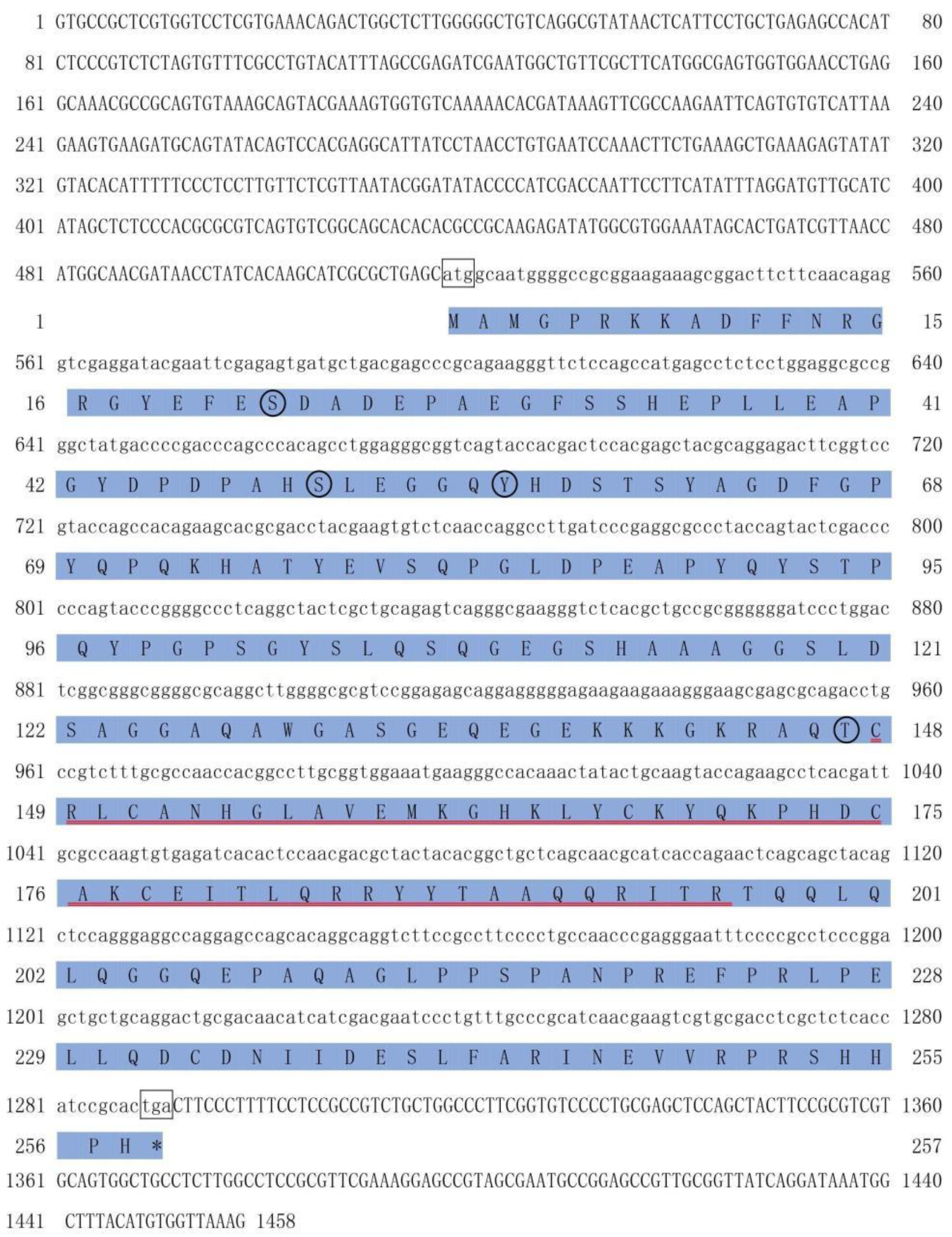
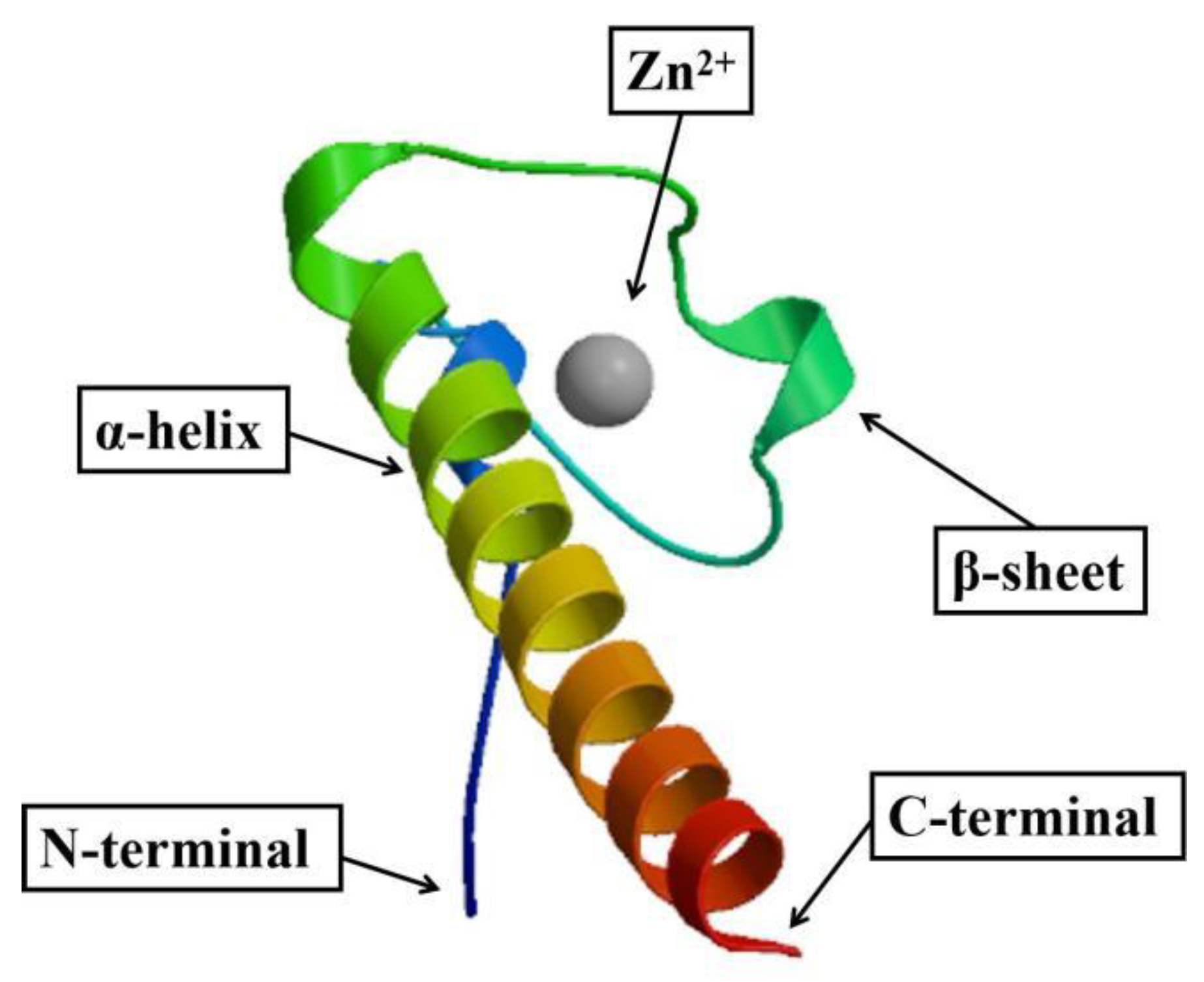
2.1. cDNA Cloning and Sequence Analysis of PmDsx

The PmDsx mRNA sequence is 1458 bp in length (GenBank: MT521744.1) and the Open Reading Frame (ORF) is from positions 518 to 1291 bp (774 bp). This nucleotide sequence translated to a peptide sequence of 257 amino acids with a calculated molecular weight of 28.214 kDa. No signal peptide and glycosylation site but four phosphorylation sites were detected in this amino acid sequence, including Tyrosine (Y56), Threonine (T147), Serine (S22), and Serine (S50). ExPASy tool revealed that PmDsx protein possessed a conserved DM domain between amino acids 148 and 196 (Figure 1). The three-dimensional model obtained showed that the DM domain was composed of several zinc finger structures and contained an α-helix and two anti-parallel β-sheets. There was a pair of cysteine residues at the N-terminal and a pair of histidine residues at the C-terminal of the zinc fingers, which had formed a cavity to accommodate exactly one Zn2+ (Figure 2).

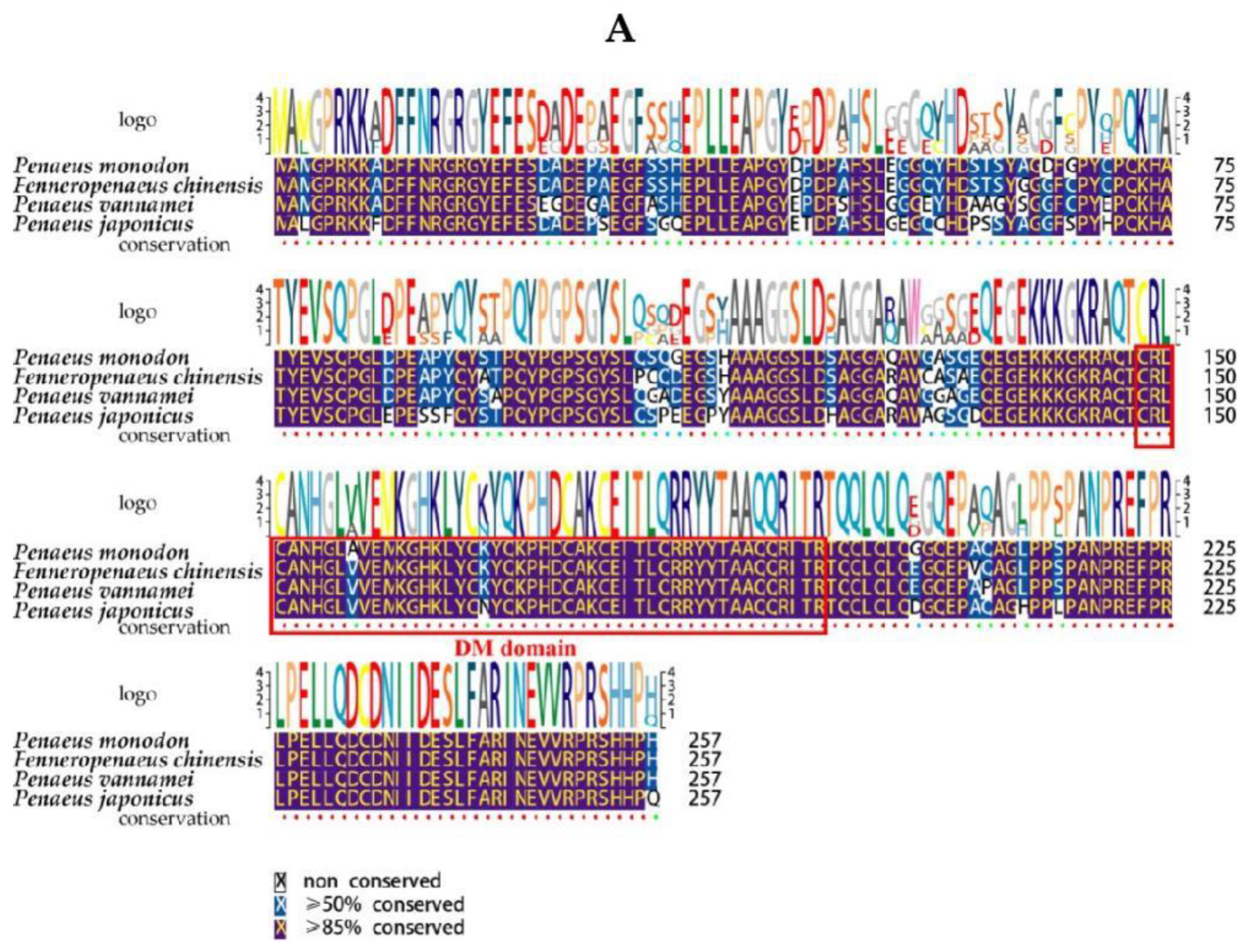
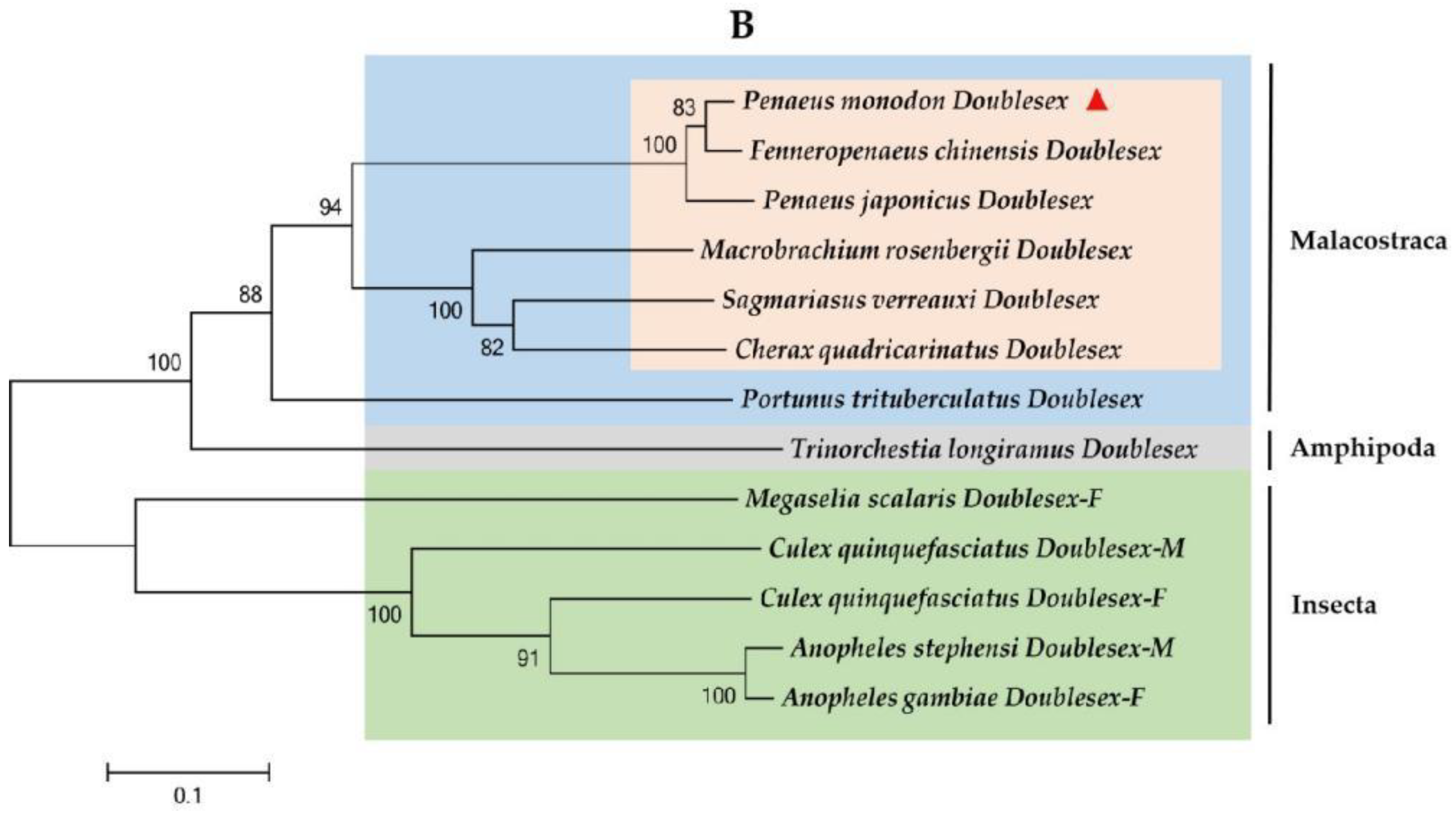
2.2. Multiple Sequence Alignment and Phylogenetic Analysis of PmDsx

Basic Local Alignment Search Tool (BLAST) analysis revealed that the complete amino acid sequence of PmDsx shared high sequence identity with Dsx sequences from other Decapoda; the results showed that the predicted protein sequence had the highest homology with F. chinensis (94.55% similarity), followed by Penaeus japonicus (87.11% similarity), M. rosenbergii (67.50% similarity), C. quadricarinatus (45.97% similarity), and Sagmariasus verreauxi (37.87% similarity), which all contain a highly conserved DM structural domain (Figure 3A). To assess the evolutionary relationship of PmDsx with Dsx of other species, phylogenetic trees of Dsx protein sequences of several species were generated using the NJ method. The results showed that the evolutionary tree of Dsx was divided into two main clusters: crustaceans and insects. Among the crustaceans, P. monodon clustered into a small branch with F. chinensis and P. japonicus, and belongs to a large branch with C. quadricarinatus, S. verreauxi, M. rosenbergii, and Portunus trituberculatus. The insects branch included C. quinquefasciatus, Anopheles Stephens, and Megaselia scalaris (Figure 3B).

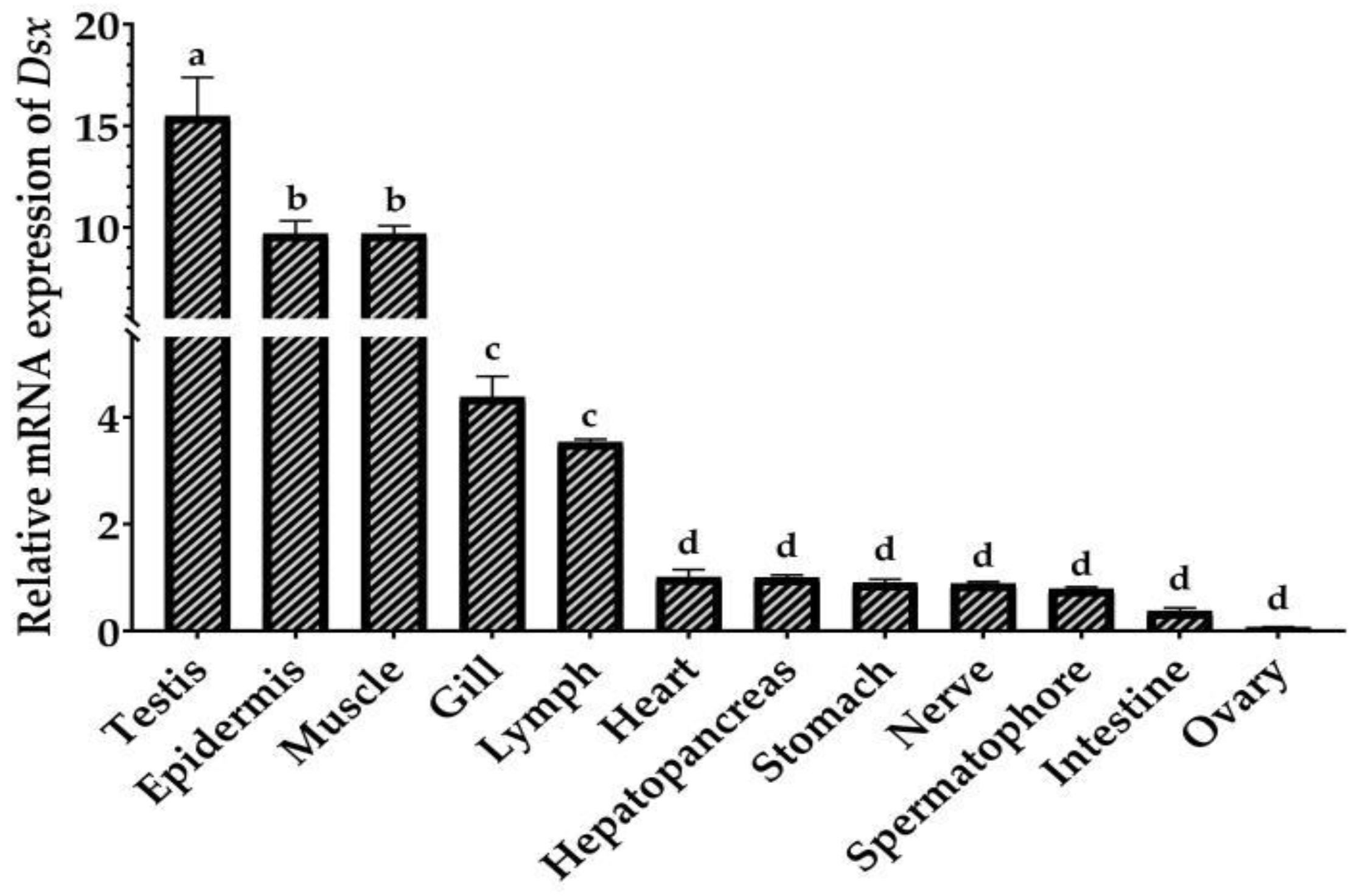
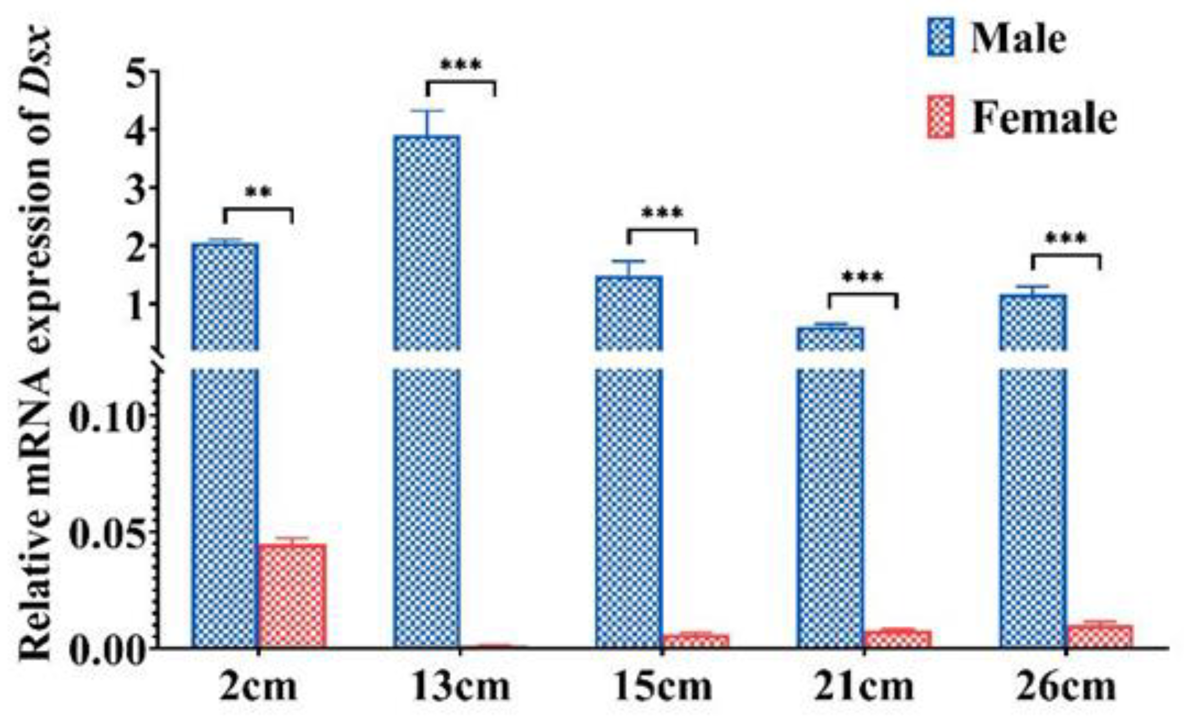
2.3. Tissue Distribution and Sexually Dimorphic Expression of PmDsx

Based on EF-1ɑ as a control, qRT-PCR was used to detect the spatial expression pattern of PmDsx in 12 tissues (Figure 4). The results showed that PmDsx was expressed in all tissues, with the highest expression level in testis, followed by the epidermis, muscle, gill, and lymph, low expression in the heart, hepatopancreas, stomach, nerve, and lowest expression in spermatophore, intestine, and ovary. (p < 0.05). Moreover, we examined the expression of PmDsx in individuals at different developmental stages. We used the whole shrimp with 2 cm body length because their gonads did not grow yet, but we can distinguish their sex by qPCR method of our lab, and then used the testis of male shrimp and ovary of female shrimp with 13 or 26 cm body length for qPCR analysis [26]. The results showed that the expression of PmDsx in the reproductive system of juvenile shrimp (2cm), young shrimp (13~15cm), and mature shrimp (21~26cm) showed significant sex dimorphism. The expression in male individuals and reproductive systems was significantly higher than that in female individuals and reproductive systems and decreased with the maturation of the testis. Similarly, the expression of Dsx was higher in female larvae and decreased significantly with the development of the ovary (Figure 5).

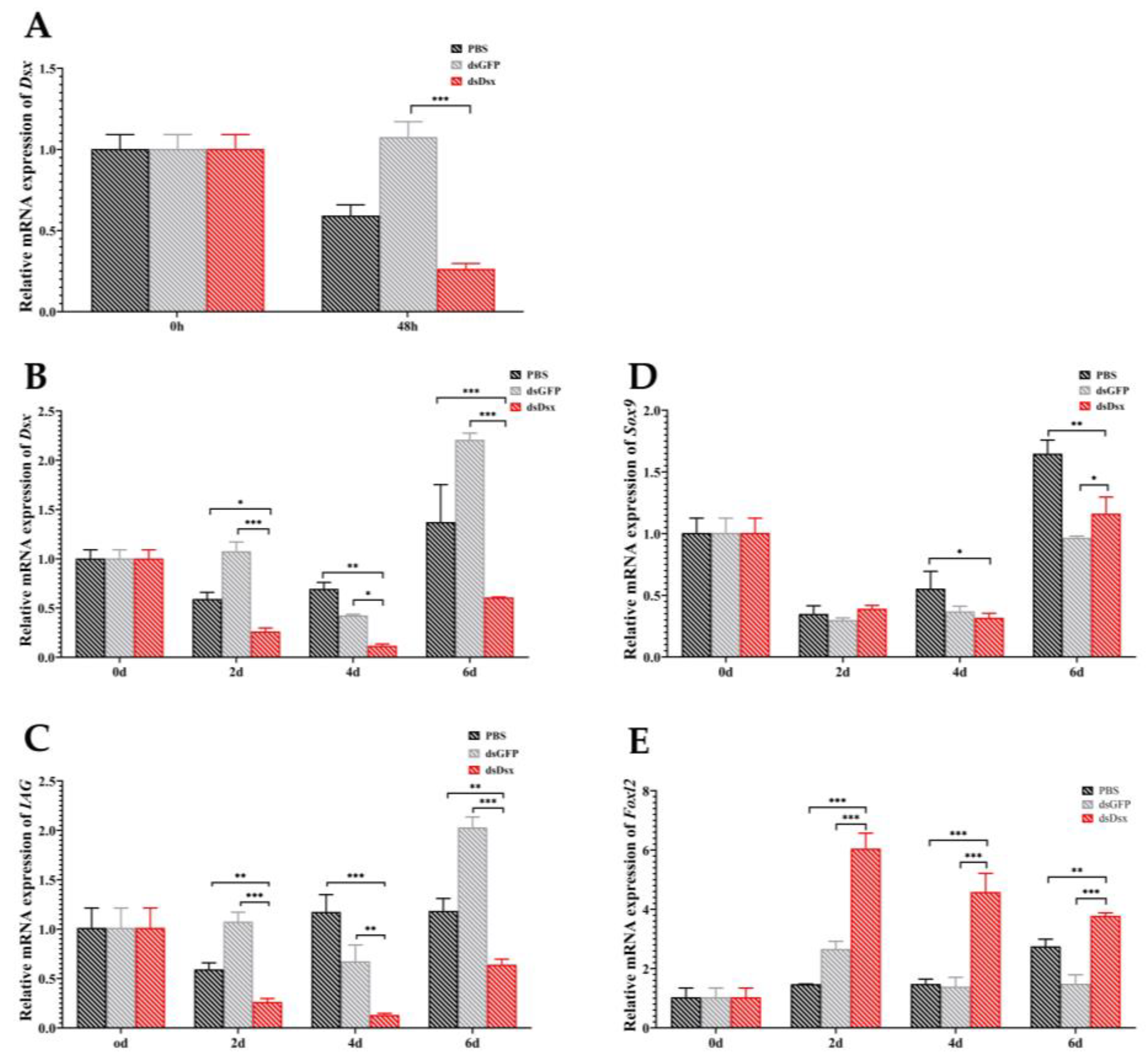
2.4. RNA Interference of PmDsx Reduces the Transcript Level of Sex-Related Genes

The interference efficiency and target gene expression were detected by qRT-PCR. The results showed that PmDsx expression was significantly decreased at 48 h, indicating that RNA interference was effective. In addition, we explored the effect of PmDsx knockout on the following sex-related genes: IAG, SRY-related HMG-box gene 9 (Sox9), and Forkhead transcription factor L2 (Foxl2). The results are shown in the figure when PmDsx was silenced, the expression of PmDsx in dsDsx group was significantly down-regulated compared with PBS group and dsGFP group at 2, 4, and 6 days, and the dsDsx group at 6 days showed a rebound in PmDsx expression compared to the dsDsx group at 2, 4 days. Moreover, the expression trends of PmIAG genes were basically the same as of PmDsx. The expression of PmSox9 expression in the dsDsx group was lower than in the dsGFP group at 4 days and lower than in the PBS group at 4, and 6 days. In 2, and 4 days, the expression of PmSox9 was slightly decreased. On the contrary, the expression of PmFoxl2 was significantly increased compared with PBS group and dsGFP group at 2, 4, and 6 days, also PmDsx expression was remarkably upregulated in the dsDsx group at 2, 4, and 6 days compared to 0 days (Figure 6).

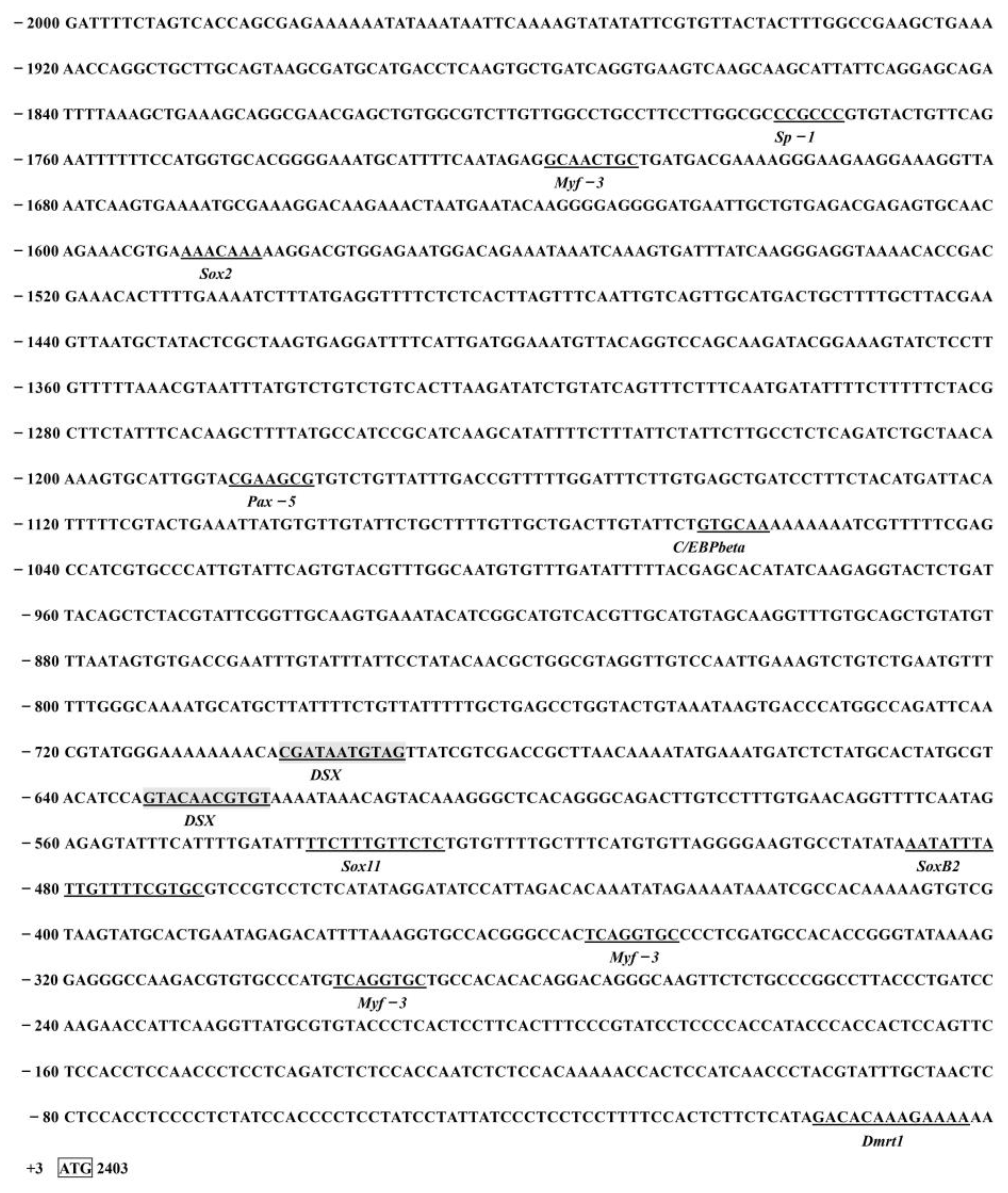
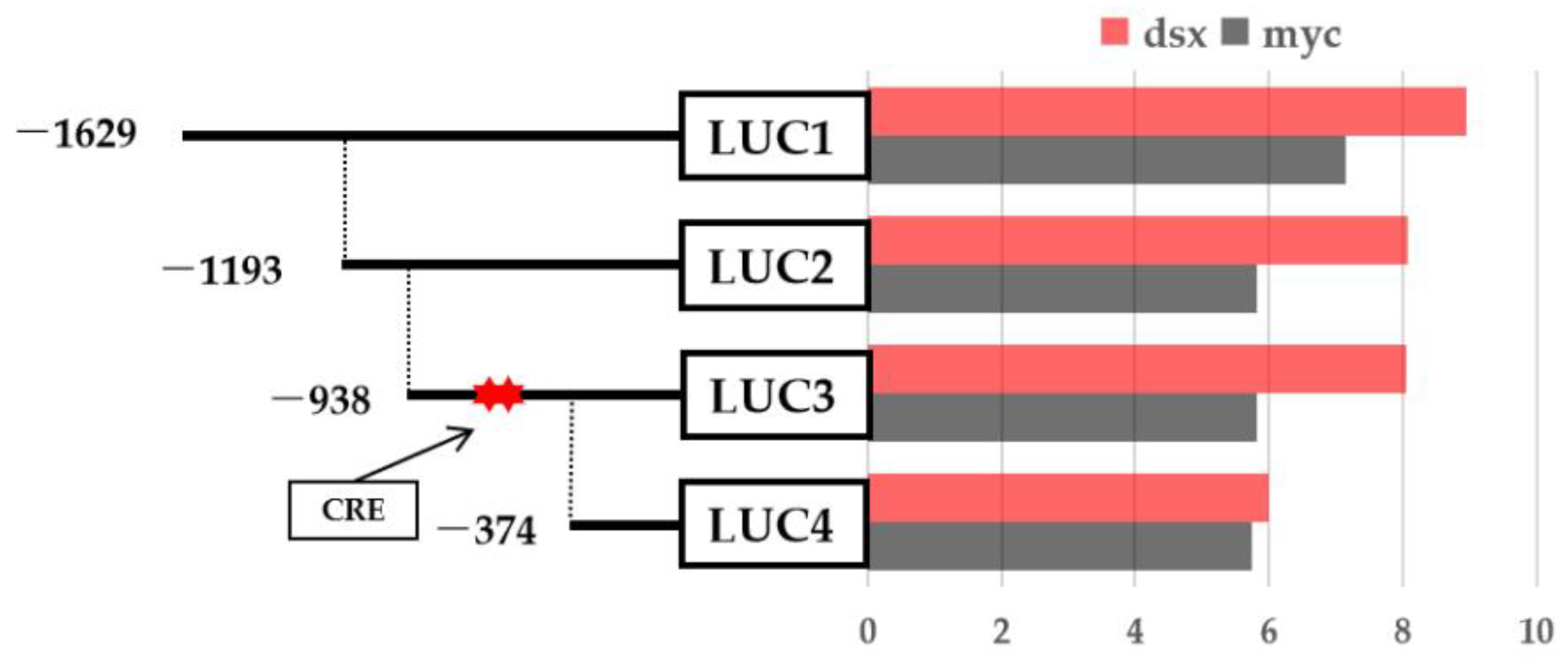
2.5. Prediction of Transcription Factor Binding Sites in the Promoter Region of PmIAG

The promoter containing a 2000 bp sequence upstream of the translation start site of the IAG in the P. monodon genomic sequence (LOC119596132) was obtained in NCBI. Potential transcription factor binding sites were predicted by online software, and the analysis showed that the IAG promoter region contains a series of transcription factors binding sites such as Sox2/B2, Pax5, Myf3, C/EBPbeta, and Dmrt1, and importantly, there are two predicted Dsx binding located at −623 to −633 bp and −692 to −702 bp regions (Figure 7). Based on the location of the binding sites, we then successfully constructed several truncated mutants that contained different regions of promoter sequences (Figure 8 and Figure S1). Four promoter fragments were amplified from P. monodon genomic DNA by PCR using specific primers (Table 1). All cloned PmIAG 5′-flanking sequences were validated and used for dual-luciferase reporter analysis.

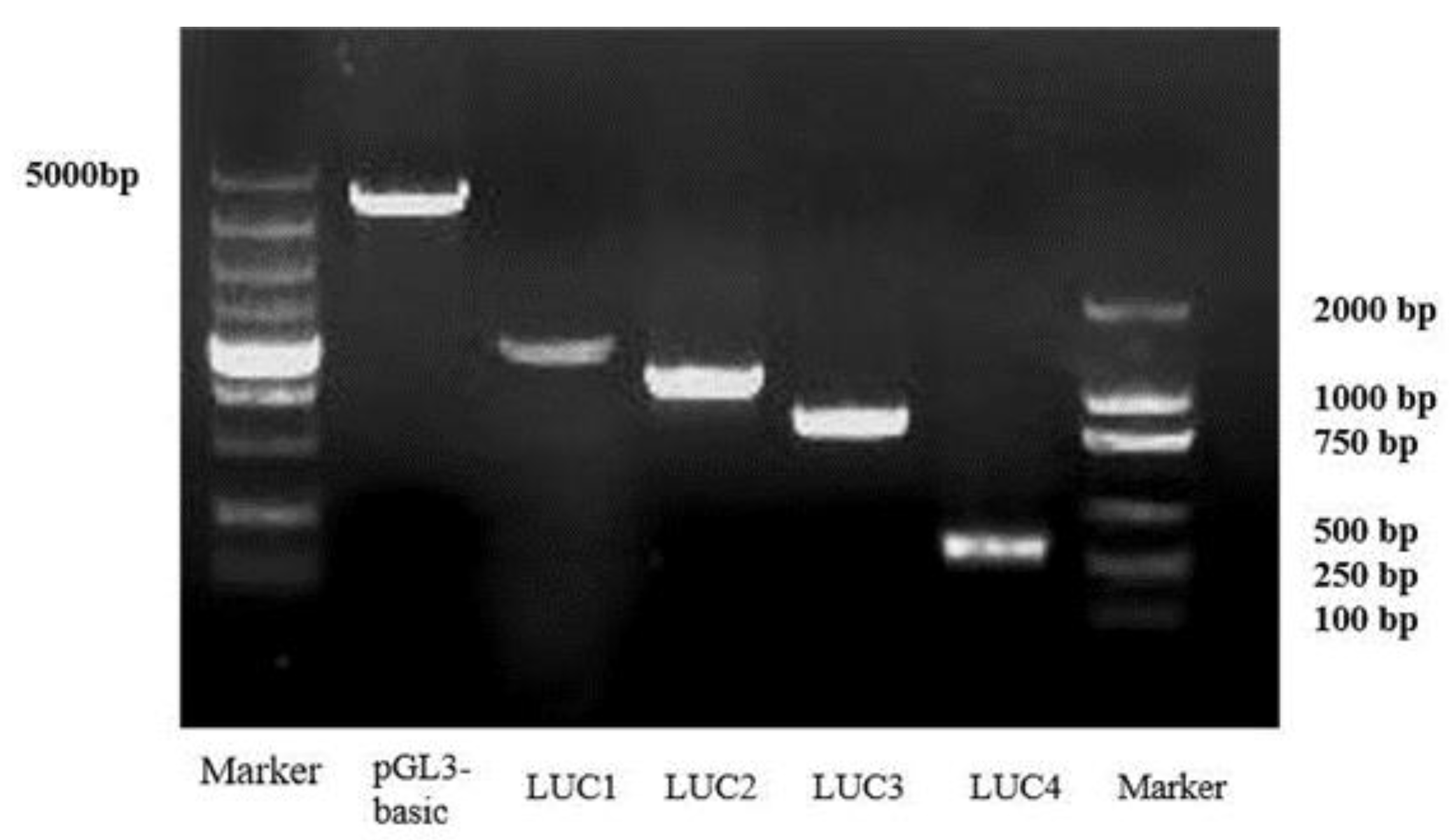
2.6. PmDsx Regulates the Transcriptional Activity of PmIAG

Four truncated mutants were cotransfected into HEK293T cells with the PmDsx constructs to identify the functional transcription factor binding site of PmDsx within the promoter of PmIAG and using the empty vector as a control. Luciferase activity assays confirmed that PmDsx overexpression significantly enhanced the expression activity of the promoter region from −374 to −938 bp compared to the control. However, no significant changes were observed for PmDsx overexpression in other regions. These data suggested that PmDsx is indeed bound to a promoter region between −374 bp to −938 bp that contains two Dsx binding sites to regulate the translation activities of the PmIAG gene in P. monodon (Figure 9).

3. Discussion

Dsx is widely conserved among arthropods as well as vertebrates and was regarded as the master-switch gene in the sex determination cascade in different organisms. PmDsx contained a classic DM structural domain of the Dsx family, which is highly conserved in the Penaeus. The three-dimensional structure confirmed PmDsx had a DNA binding domain (DM domain) in N-terminal and a dimerization domain (Dsx dimer) in C-terminal. The DM domain as a zinc module utilizes its conserved amino acid residues for zinc chelating and promoter binding [27], while the Dsx dimer as an alpha-helical motif improves DNA binding and encourages Dsx dimerization [28]. The DM domain may have specialized and enhanced functions in mediating relevant physiological activities, making the specific role played by Dsx in reproduction.
PmDsx had a high identity and close phylogenetic relationship with F. chinensis, as both contain only one Dsx spliceosome but are far from other branches of arthropods such as Drosophila and insects that have two spliceosomes. The male-specific Dsx M and female-specific Dsx F was directed splicing by tra and the initiation-lethal Sxl protein, and regulate the downstream genes for sex-specific differentiation [29,30]. Since Dsx is highly conserved in the Drosophila sex determination cascade pathway and can determine individual sex, we hypothesize that the Dsx gene in P. monodon and other decapods is involved in the sex determination and differentiation process, but its function is partially lost or altered during the long arthropod genetic differentiation process. This is the reason for the generation of different numbers of Dsx spliceosomes.
The expression of PmDsx has an obvious sex dimorphism that is significantly high in the testis and lowest in the ovary not only in the tissue but also in the different development stages, suggesting its potentially important role in the development and maintenance of the male reproductive system. F.chinensis and Charybdis feriatus also exhibit similar sex differences in that their transcripts are mainly expressed at the highest levels in male-specific structures during embryonic development [25,31]. Interestingly, in C. quadricarinatus, Cqdsx was strongly expressed in the female gonads and a strong hybridization signal was observed mainly in oocytes and ovarian lamellae, and a weaker hybridization signal was detected in spermatocytes suggesting that Cqdsx plays an important role in female ovarian development and differentiation [32]. The Spdsx expression in tissues of the Scylla paramamosain was similar to that of C. quadricarinatus, but different from that of C. feriatus, indicating that the Dsx expression pattern is highly variable among species [33]. Simultaneously, the differential expression of Dsx in individuals of different genders is significant, and in general, the temporal and spatial expression of a gene is tightly linked to its effects. Our study showed that the total expression of PmDsx was always higher in male individuals than in female individuals during different developmental periods, illustrating a sex-specific role of PmDsx in the male development of P. monodon. The regulation of Dsx on downstream somatic sex differentiation of terminal genes is thought to be related to transcriptional regulation, and ultimately leads to the establishment of sexual dimorphism in adult Drosophila [34]; therefore, the study of the regulatory mechanism of Dsx on downstream target structural genes seems to be essential.
Subsequently, we successfully silenced the expression of PmDsx by RNA interference technique. Different from our previous studies [35], we explored not only the positive regulation of PmDsx on PmSox9 and PmIAG but also the negative regulation of PmFoxl2. Some studies have shown that disruption of XY fish Dmrt1 leads to increased expression of Foxl2 and cyp19a1a in Nile tilapia. In contrast, Foxl2 deficiency in XX fish displayed varying degrees of oocyte degeneration or even complete sex reversal, and elevated expression of Dmrt1 and Cyp11b2 illustrated the antagonistic role of Dmrt1 and Foxl2 in estrogen secretion-induced sex differentiation in tilapia [36]. Concurrently, Dmrt1 can positively regulate the transcription of sox9b by binding directly to a specific CRE within the Sox9b promoter in the testis [37], which seems to be consistent with our study. Hence, we can hypothesize that Dmrt, Foxl2, and Sox9 may have a regulatory function in sex determination and differentiation, at least in teleost fishes. Moreover, as with shrimp, MniDMRT11E RNAi leads to significant positive regulation of IAG transcription in the male abdominal ganglion of M. nipponense [38]. Coincidentally, the MroDMRT11E gene of M. rosenbergii has the same positive regulatory effect on IAG [39], which indicates that DMRT might be available to regulate the expression of IAG in shrimp. Considering the important participation of IAG in male crustaceans and the confirmation of several studies [40,41], we hypothesized that PmDsx might be directly or indirectly involved in the upstream pathway of PmIAG.
As a key transcription factor, IAG plays a pivotal role in sex redistribution [12,42], and spermatogenesis [2,41] in reproductive crustaceans. In a study of the relevance of osmoregulatory genes to reproductive development in E. sinensis, the expression of IAG is determined by the phosphorylation of transcription factors such as iDMY and Sox15 by PKA in the gonad, and these phosphorylated transcription factors bind upstream of the promoter of IAG, thereby inducing its transcription [43]. These data suggest that IAG may be a conserved factor downstream of the sexual differentiation process in decapods, which can be regulated by upstream DMRT and Sox family genes. In this study, analysis of the PmIAG promoter sequence predicts two putative Dsx/mab-3 binding sites at −623 bp and −692 bp in the PmIAG gene sequence, a DMRT1 binding site in front of its promoter region start codon, and three Sox family gene binding sites (Sox2, SoxB2, Sox11). In 1999, Yi reported DNA sequences for the Mab-3 and Dsx binding sites, with AATGTTGCGAT (A)NT for the Mab-3 binding site and nTnGT (A)ACAATGTT (A)nCC for the Dsx binding site [44], which overlap with our predicted AATGTAG for the Dsx transcription factor binding site, validating the accuracy of this predicted binding site. As an upstream transcription factor, Dsx could bind to the promoter region of IAG to regulate its transcription in M. rosenbergii [23] and F. chinensis [25]. Therefore, we can tentatively speculate that PmDsx may link to a binding site in the upstream region of the IAG promoter to trigger its transcription.
Several truncated mutants, which contained different promoter regions with different lengths based on the location of transcription factor binding sites, confirmed the regulatory effect of PmDsx on PmIAG expression by a dual-luciferase reporter assay, revealing that PmDsx was successfully bound to PmIAG and triggered its transcriptional activity. Similarly, F. chinensis FcDsx could bind at −434 bp to −416 bp (including the binding site) of the FcIAG gene and regulate its expression [25]; our joint results hypothesize that Dsx and its target gene, IAG, play an integral role in the shrimp sex-regulatory cascade pathway.

4. Materials and Methods

4.1. Experimental Animals and Cell Culture

All healthy cultured P. monodon were collected from the experimental base of the South China Sea Fisheries Research Institute in Shenzhen (Guangdong, China). The shrimps were maintained in recirculating seawater tanks with a salinity of 29‰, a temperature of 24–28 °C, and a pH of 7.5–7.8 before use. The shrimp with a weight of 25.46 ± 2.5 g and a length of 12.63 ± 0.72 cm were used for tissue expression and RNA interference in this study. Moreover, the human embryonic kidney-derived HEK293 cell line was cultured in Dulbecco’s Modified Eagle’s medium (DMEM) (Gibco original, origin Australia), supplemented with 10% heat-inactivated fetal bovine serum (FBS) (Invitrogen, Waltham, MA, USA), 100 U mL−1 penicillin, and 100 µg mL−1 streptomycin at 37 °C in a humidified atmosphere containing 5% CO2, as described previously (Lemaitre et al., 1996). Logarithmic growth phase cells were seeded into 24-well plates and cultured for 24 h, then grown until they reached 70–80% confluency.
The larval development period of P. monodon was determined according to the method of Huang [26]. Starting from the fertilized eggs, the larval development period was confirmed through observation under the microscope. Samples were taken once in each period until the larvae. Thirty sex-determined P. monodon seedlings were selected and observed under an anatomical and electronic microscope and stored in 80% alcohol for subsequent use.

4.2. RNA Isolation and Molecular Cloning

Total RNA from all samples of healthy P. monodon was obtained using the TRIzol reagent (Invitrogen, Carlsbad, CA, USA) and the HiPure Fibrous RNA Plus Kit (Megan, Guangzhou, China). The purity and quantity of total RNA were calculated by measuring the ultraviolet absorbance ratio at 260/280 nm using a NanoDrop 2000 device (Thermo Scientific, Waltham, MA, USA) and 1% agarose gels. The cDNA synthesis was performed using the Evo M-MLV RT Kit with gDNA Clean for qPCR (AG, Hunan, China), and the product was stored at −20 ℃ until subsequent use. Primers were designed using Primer Premier 5.0 software based on partial sequences retrieved from the P. monodon transcriptome library analyzed in our laboratory (Table 1). The PmDsx open reading frame (ORF) was expanded by PCR technology. The final PCR products were cloned into the pMD-19T vector (Takara, Kyoto, Japan), sequencing a positive monoclonal colony.

4.3. Bioinformatics Analysis of PmDsx

The sequencing results were identified using the National Center for Biotechnology Information (NCBI) BLAST search program (http://www.ncbi.nlm.nih.gov/blast, accessed on 12 October 2021). The ORF and its corresponding amino acid sequences of PmDsx were predicted using the ORF Finder (https://www.ncbi.nlm.nih.gov/orffinder, accessed on 12 October 2021). ExPASy-PROSITE (https://prosite.expasy.org/, accessed on 12 October 2021) was used to analyze the functional domain of protein prediction. N-glycosylation sites and phosphorylation sites were predicted by NetNglyc 1.0 server and NetPhos3.1 server Software (https://services.healthtech.dtu.dk, accessed on 13 October 2021). Multiple sequence alignments and phylogenetic analysis of the predicted PmDsx protein with known Dsx proteins were analyzed using the HiPlot online software (www.hiplot.com.cn/, accessed on 13 October 2021). The three-dimensional protein structure of PmDsx was constructed by SWISS-MODEL online software.

4.4. Real-Time Quantitative PCR Analysis

The qRT-PCR amplification was performed on a Roche 480 machine using SYBR® Premix Ex Taq™ II (Takara, Kyoto, Japan) as the fluorescent dye. The translation elongation factor 1-alpha (EF1α) was used as a reference gene, and all reactions were performed in triplicate. The PCR assay was carried out in a total volume of 12.5 μL, containing 6.25 μL of 2 × SYBR Premix Ex Taq (Takara, Kyoto, Japan), 1 μL of the diluted cDNA, 0.5 μL of each primer (10 μM), and 4.25 μL of sterile distilled H2O. The cycle conditions were described as follows: 95 °C for 3 min, followed by 40 cycles of 95 °C for 5 s, 60 °C for 30 s, and 72 °C for 30 s. Amplification curve analysis of the amplification products at the end of each thermal cycling reaction was performed to confirm the specificity of the amplification and to ensure successful amplification and detection, and fold change for the target gene relative expression of mRNA relative to controls was calculated using the 2△△Ct method [45].

4.5. RNA Interference

The double-stranded RNA (dsRNA) for PmDsx and pEGFP-N1 were used as a linearized DNA template for in vitro transcription reactions to synthesize dsRNA by the T7 RiboMAX™ Expressed RNAi System Kit (Promega, WI, USA). The specific primers containing the T7 promoter were designed for transcriptional amplification, and dsRNA formation was quantified by a 1.2% agarose gel electrophoresis. The final dsRNA product was stored at −80 °C. The synthetic dsRNA was diluted to 3 μg/μL with PBS. Before the formal injection experiments, we performed a 48-h pre-experiment to test the silencing efficiency. Each shrimp was injected intramuscularly into the second abdominal segment. Based on the results, we determined that 3 μg of dsRNA was injected per gram of shrimp. The experiment consisted of three groups (30 shrimps per group): the dsPmDsx injection group (experimental group), the PBS injection group (negative control group), and the dsGFP injection group (positive control group). The shrimp were injected every two days, and the testis of three shrimps from each group was collected at 0, 2, 4, and 6 days before each injection. Then, the total RNA was extracted from the testis of shrimps by the same method mentioned above.

4.6. Promoter Cloning and Plasmid Construction

The Transcription Factor Binding Site Prediction (TFBS)-JASPAR database (http://JASPAR.genereg.net/, accessed on 23 November 2021) and ALGGEN-PROMO (http://alggen.lsi.upc.es, accessed on 23 November 2021) were used to search for potential binding sites to determine the potential function of the PmDsx binding site on the core PmIAG promoter. The full-length ORF of PmDsx was amplified and inserted into the pCMV-C-myc vector (Beyotime, China) containing the Hind III/Bgl I site, called pCMV-PmDsx. In addition, four truncated mutants from the PmIAG promoter were designed based on the location of the PmDsx predicted binding site. These fragments have a common 3’-end at −3 bp (translation start codon defined as + 1) and 5’-ends at −1629, −1193, −938, and −371 were amplified using specific primers (Table 1). The vectors were named pGL3-basic-Luc1, pGL3-basic-Luc2, pGL3-basic-Luc3, and pGL3-basic-Luc4, and each fragment was cloned into the pGL3-Basic firefly luciferase expression vector (Promega, USA) for the dual-luciferase reporter assay. The pRL-TK luciferase reporter vector was considered an internal control.

4.7. Dual-Luciferase Reporter Assay

The luciferase activity was measured according to the protocol for the Dual-Luciferase Reporter Assay System (Promega, Madison, WI, USA). The pCMV-PmDsx overexpression vector was separately cotransfected into HEK293 cells with a series of constructs containing luciferase reporter genes driven by different species of P. monodon IAG promoters. Cells were collected and lysed in reporter lysis buffer (Promega, USA) after 48 h of transfection. Then, 100 μL of LARII firefly luciferase assay reagent was added to 20 μL of the cell lysate to be measured. The mixture was then quickly placed into a luminescence detector (Thermo Fisher Scientific, Waltham, MA, USA) and the fluorescence value emitted by the substrate excited by firefly luciferase was read A. An amount of 100 μL of 1× Stop & Glo renilla luciferase assay was then added, and the fluorescence value emitted by mesothelial luciferase of the test samples was read B. The ratio of each sample Ratio = A / B value is the relative luciferase activity of cells transfected with the expression plasmid constructed for the fragment. If the ratio of the experimental group is significantly higher than the ratio of the empty group, it proves that the target gene is activated. All experiments were repeated three times independently.

4.8. Statistical Analysis

The statistical significance of data differences between genders was assessed using one-way ANOVA with GraphPad Prism 8 software. Gene expression differences between different tissues were evaluated using GraphPad Prism 8 software. Data about the luciferase activities were analyzed as mean + SE, and p < 0.05 was statistically significant.

5. Conclusions

In summary, this study characterized the Dsx gene in P. monodon, which exhibits a male-biased expression pattern in different tissues. The putative Dsx binding site was identified in the promoter region of the male sexual differentiation effector gene PmIAG. In addition, PmDsx positively regulated the transcription of the PmIAG gene by binding directly to a specific binding site in the PmIAG gene promoter, suggesting that PmDsx may be an upstream regulator in the process of sexual differentiation in shrimp. Our results provide a reference for exploring the molecular mechanisms of sex determination and differentiation in P. monodon and even crustaceans.

Supplementary Materials

The following supporting information can be downloaded at: https://www.mdpi.com/article/10.3390/ijms232012701/s1.

Author Contributions

W.-Y.W., S.-G.J. and L.-S.Y. conceived and designed the experiments; W.-Y.W., L.-S.Y. and J.-H.H. performed the experiments; W.-Y.W., Q.-B.Y., F.-L.Z., S.J. and Y.-D.L. contributed to sample collection; W.-Y.W. analyzed the data and wrote the paper; S.-G.J. and L.-S.Y. assisted with writing and proofreading. The entire study has not been published, accepted for publication, or is under consideration for publication elsewhere. All authors have read and agreed to the published version of the manuscript.

Funding

This research was funded by The Shenzhen Science and Technology Plan of China (JCYJ20180504170233070); China Agriculture Research System-48 (CARS-48); National Key R&D Program of China (2018YFD0900303-05); Natural Science Foundation of Guangdong Province (2021A1515012519); Financial Fund of Ministry of Agriculture and Rural Affairs of China (NHYYSWZZZYKZX2020); Joint Project of Hainan Province Science and Technology Plan and Scientific Technology Town Sanya Yazhou Bay (320LH008); Hainan Provincial Association for Science and Technology of Young Science and Technology Talents Innovation Plan Project (QCQTXM202206); Guangdong Basic and Applied Basic Research Foundation (2020A1515110200).

Informed Consent Statement

Not applicable.

Data Availability Statement

Data are contained within the article or Supplementary Materials (Table S1).

Acknowledgments

We thank all the staff in the Laboratory of Aquaculture and Biotechnology, Nanhai Fisheries Research Institute, for their help during the experiments.

Conflicts of Interest

The authors declare no conflict of interest.

References

  1. Cronin, L.E. Anatomy and histology of the male reproductive system of Callinectes sapidus Rathbun. J. Morphol. 1947, 81, 209–239. [Google Scholar] [CrossRef]
  2. Ventura, T.; Manor, R.; Aflalo, E.D.; Weil, S.; Raviv, S.; Glazer, L.; Sagi, A. Temporal silencing of an androgenic gland-specific insulin-like gene affecting phenotypical gender differences and spermatogenesis. Endocrinology 2009, 150, 1278–1286. [Google Scholar] [CrossRef] [PubMed] [Green Version]
  3. Ventura, T.; Rosen, O.; Sagi, A. From the discovery of the crustacean androgenic gland to the insulin-like hormone in six decades. Gen. Comp. Endocrinol. 2011, 173, 381–388. [Google Scholar] [CrossRef] [PubMed]
  4. Ma, K.Y.; Lin, J.Y.; Guo, S.Z.; Chen, Y.; Li, J.-L.; Qiu, G.F. Molecular characterization and expression analysis of an insulin-like gene from the androgenic gland of the oriental river prawn, Macrobrachium nipponense. Gen. Comp. Endocrinol. 2013, 185, 90–96. [Google Scholar] [CrossRef] [PubMed]
  5. Li, F.; Bai, H.; Xiong, Y.; Fu, H.; Jiang, S.; Jiang, F.; Jin, S.; Sun, S.; Qiao, H.; Zhang, W. Molecular characterization of insulin-like androgenic gland hormone-binding protein gene from the oriental river prawn Macrobrachium nipponense and investigation of its transcriptional relationship with the insulin-like androgenic gland hormone gene. Gen. Comp. Endocrinol. 2015, 216, 152–160. [Google Scholar] [CrossRef]
  6. Mareddy, V.R.; Rosen, O.; Thaggard, H.B.; Manor, R.; Kuballa, A.V.; Aflalo, E.D.; Sagi, A.; Paterson, B.; Elizur, A. Isolation and characterization of the complete cDNA sequence encoding a putative insulin-like peptide from the androgenic gland of Penaeus monodon. Aquaculture 2011, 318, 364–370. [Google Scholar] [CrossRef]
  7. Li, S.; Li, F.; Sun, Z.; Xiang, J. Two spliced variants of insulin-like androgenic gland hormone gene in the Chinese shrimp, Fenneropenaeus chinensis. Gen. Comp. Endocrinol. 2012, 177, 246–255. [Google Scholar] [CrossRef]
  8. Sroyraya, M.; Chotwiwatthanakun, C.; Stewart, M.J.; Soonklang, N.; Kornthong, N.; Phoungpetchara, I.; Hanna, P.J.; Sobhon, P. Bilateral eyestalk ablation of the blue swimmer crab, Portunus pelagicus, produces hypertrophy of the androgenic gland and an increase of cells producing insulin-like androgenic gland hormone. Tissue Cell 2010, 42, 293–300. [Google Scholar] [CrossRef]
  9. Jin, S.; Fu, Y.; Hu, Y.; Fu, H.; Jiang, S.; Xiong, Y.; Qiao, H.; Zhang, W.; Gong, Y.; Wu, Y. Identification of candidate genes from androgenic gland in Macrobrachium nipponense regulated by eyestalk ablation. Sci. Rep. 2021, 11, 1–16. [Google Scholar] [CrossRef]
  10. Liu, F.; Shi, W.; Ye, H.; Zeng, C.; Zhu, Z. Insulin-like androgenic gland hormone 1 (IAG1) regulates sexual differentiation in a hermaphrodite shrimp through feedback to neuroendocrine factors. Gen. Comp. Endocrinol. 2021, 303, 113706. [Google Scholar] [CrossRef]
  11. Tan, K.; Zhou, M.; Jiang, H.; Jiang, D.; Li, Y.; Wang, W. siRNA-Mediated MrIAG Silencing Induces Sex Reversal in Macrobrachium rosenbergii. Mar. Biotechnol. 2020, 22, 456–466. [Google Scholar] [CrossRef] [PubMed]
  12. Ventura, T.; Manor, R.; Aflalo, E.D.; Weil, S.; Rosen, O.; Sagi, A. Timing sexual differentiation: Full functional sex reversal achieved through silencing of a single insulin-like gene in the prawn, Macrobrachium rosenbergii. Biol. Reprod. 2012, 86, 1–13. [Google Scholar] [CrossRef] [PubMed]
  13. Fu, C.; Li, F.; Wang, L.; Wu, F.; Wang, J.; Fan, X.; Liu, T. Molecular characteristics and abundance of insulin-like androgenic gland hormone and effects of RNA interference in Eriocheir sinensis. Anim. Reprod. Sci. 2020, 215, 106332. [Google Scholar] [CrossRef] [PubMed]
  14. Jiang, Q.; Lu, B.; Wang, G.; Ye, H. Transcriptional inhibition of sp-iag by crustacean female sex hormone in the mud crab, scylla paramamosain. Int. J. Mol. Sci. 2020, 21, 5300. [Google Scholar] [CrossRef]
  15. Burtis, K.C.; Baker, B.S. Drosophila doublesex gene controls somatic sexual differentiation by producing alternatively spliced mRNAs encoding related sex-specific polypeptides. Cell 1989, 56, 997–1010. [Google Scholar] [CrossRef]
  16. Ruiz, M.F.; Alvarez, M.; Eirín-López, J.M.; Sarno, F.; Kremer, L.; Barbero, J.L.; Sánchez, L. An unusual role for doublesex in sex determination in the dipteran Sciara. Genetics 2015, 200, 1181–1199. [Google Scholar] [CrossRef] [Green Version]
  17. Shirangi, T.R.; Wong, A.M.; Truman, J.W.; Stern, D.L. Doublesex Regulates the Connectivity of a Neural Circuit Controlling Drosophila Male Courtship Song. Dev. Cell 2016, 37, 533–544. [Google Scholar] [CrossRef] [Green Version]
  18. Xu, J.; Zhan, S.; Chen, S.; Zeng, B.; Li, Z.; James, A.A.; Tan, A.; Huang, Y. Sexually dimorphic traits in the silkworm, Bombyx mori, are regulated by doublesex. Insect Biochem. Mol. Biol. 2017, 80, 42–51. [Google Scholar] [CrossRef] [Green Version]
  19. Rideout, E.J.; Dornan, A.J.; Neville, M.C.; Eadie, S.; Goodwin, S.F. Control of sexual differentiation and behavior by the doublesex gene in Drosophila melanogaster. Nat. Neurosci. 2010, 13, 458–466. [Google Scholar] [CrossRef] [Green Version]
  20. Coschigano, K.T.; Wensink, P.C. Sex-specific transcriptional regulation by the male and female doublesex proteins of Drosophila. Genes Dev. 1993, 7, 42–54. [Google Scholar] [CrossRef]
  21. Xu, J.; Wang, Y.; Li, Z.; Ling, L.; Zeng, B.; James, A.A.; Tan, A.; Huang, Y. Transcription activator-like effector nuclease (TALEN)-mediated female-specific sterility in the silkworm, Bombyx mori. Insect Mol. Biol. 2014, 23, 800–807. [Google Scholar] [CrossRef] [PubMed] [Green Version]
  22. Kato, Y.; Kobayashi, K.; Watanabe, H.; Iguchi, T. Environmental sex determination in the branchiopod crustacean Daphnia magna: Deep conservation of a Doublesex gene in the sex-determining pathway. PLoS Genet. 2011, 7, e1001345. [Google Scholar] [CrossRef] [PubMed] [Green Version]
  23. Zhong, P.; Zhou, T.; Zhang, Y.; Chen, Y.; Yi, J.; Lin, W.; Guo, Z.; Xu, A.; Yang, S.; Chan, S. Potential involvement of a DMRT family member (Mr-Dsx) in the regulation of sexual differentiation and moulting in the giant river prawn Macrobrachium rosenbergii. Aquac. Res. 2019, 50, 3037–3049. [Google Scholar] [CrossRef]
  24. Yu, Y.Q.; Ma, W.M.; Zeng, Q.G.; Qian, Y.Q.; Yang, J.S.; Yang, W.J. Molecular Cloning and Sexually Dimorphic Expression of Two Dmrt Genes in the Giant Freshwater Prawn, Macrobrachium rosenbergii. Agric. Res. 2014, 3, 181–191. [Google Scholar] [CrossRef]
  25. Li, S.; Li, F.; Yu, K.; Xiang, J. Identification and characterization of a doublesex gene which regulates the expression of insulin-like androgenic gland hormone in Fenneropenaeus chinensis. Gene 2018, 649, 1–7. [Google Scholar] [CrossRef]
  26. Zhikang, H.; Shigui, J.; Falin, Z.; Jianhua, H.; Qibin, Y. Establishment of a sex identification method for black tiger shrimp (Penaeus monodon) in embryonic and early postembryonic development based on an InDel molecular marker. S. China Fish. Sci. 2020, 16. [Google Scholar] [CrossRef]
  27. Zhu, L.; Wilken, J.; Phillips, N.B.; Narendra, U.; Chan, G.; Stratton, S.M.; Kent, S.B.; Weiss, M.A. Sexual dimorphism in diverse metazoans is regulated by a novel class of intertwined zinc fingers. Genes Dev. 2000, 14, 1750–1764. [Google Scholar] [CrossRef]
  28. Bayrer, J.R.; Zhang, W.; Weiss, M.A. Dimerization of doublesex is mediated by a cryptic ubiquitin-associated domain fold: Implications for sex-specific gene regulation. J. Biol. Chem. 2005, 280, 32989–32996. [Google Scholar] [CrossRef] [Green Version]
  29. Boggs, R.T.; Gregor, P.; Idriss, S.; Belote, J.M.; McKeown, M. Regulation of sexual differentiation in D. melanogaster via alternative splicing of RNA from the transformer gene. Cell 1987, 50, 739–747. [Google Scholar] [CrossRef]
  30. Inoue, K.; Hoshijima, K.; Higuchi, I.; Sakamoto, H.; Shimura, Y. Binding of the Drosophila transformer and transformer-2 proteins to the regulatory elements of doublesex primary transcript for sex-specific RNA processing. Proc. Natl. Acad. Sci. USA 1992, 89, 8092–8096. [Google Scholar] [CrossRef]
  31. Zhang, Y.; Waiho, K.; Ikhwanuddin, M.; Ma, H. Identification of sex-related genes from the three-spot swimming crab portunus sanguinolentus and comparative analysis with the crucifix crab charybdis feriatus. Animals 2021, 11, 1946. [Google Scholar] [CrossRef]
  32. Zheng, J.; Cai, L.; Jia, Y.; Chi, M.; Cheng, S.; Liu, S.; Li, F.; Gu, Z. Identification and functional analysis of the doublesex gene in the redclaw crayfish, Cherax quadricarinatus. Gene Expr. Patterns 2020, 37, 119129. [Google Scholar] [CrossRef] [PubMed]
  33. Wan, H.; Zhong, J.; Zhang, Z.; Sheng, Y.; Wang, Y. Identification and functional analysis of the doublesex gene in the mud crab Scylla paramamosain. Comp. Biochem. Physiol. Part A Mol. Integr. Physiol. 2022, 266, 111150. [Google Scholar] [CrossRef] [PubMed]
  34. Chatterjee, S.S.; Uppendahl, L.D.; Chowdhury, M.A.; Ip, P.L.; Siegal, M.L. The female-specific Doublesex isoform regulates pleiotropic transcription factors to pattern genital development in Drosophila. Development 2011, 138, 1099–1109. [Google Scholar] [CrossRef] [PubMed] [Green Version]
  35. Wei, W.-Y.; Huang, J.-H.; Yang, Q.-B.; Zhou, F.-L.; Jiang, S.; Li, Y.-D.; Jiang, S.-G.; Yang, L.-S. Molecular characterization and functional analysis of DMRT11E in black tiger shrimp (Penaeus monodon). Aquac. Reports J. 2022, 22, 100982. [Google Scholar] [CrossRef]
  36. Li, M.H.; Yang, H.H.; Li, M.R.; Sun, Y.L.; Jiang, X.L.; Xie, Q.P.; Wang, T.R.; Shi, H.J.; Sun, L.N.; Zhou, L.Y. Antagonistic roles of Dmrt1 and Foxl2 in sex differentiation via estrogen production in tilapia as demonstrated by TALENs. Endocrinology 2013, 154, 4814–4825. [Google Scholar] [CrossRef]
  37. Wei, L.; Li, X.; Li, M.; Tang, Y.; Wei, J.; Wang, D. Dmrt1 directly regulates the transcription of the testis-biased Sox9b gene in Nile tilapia (Oreochromis niloticus). Gene 2019, 687, 109–115. [Google Scholar] [CrossRef]
  38. Wang, Y.; Jin, S.; Fu, H.; Qiao, H.; Sun, S.; Zhang, W.; Jiang, S.; Gong, Y.; Xiong, Y.; Wu, Y. Identification and characterization of the DMRT11E gene in the oriental river prawn macrobrachium nipponense. Int. J. Mol. Sci. 2019, 20, 1734. [Google Scholar] [CrossRef] [Green Version]
  39. Yang, G.; Lu, Z.; Qin, Z.; Zhao, L.; Pan, G.; Shen, H.; Zhang, M.; Liang, R.; Lin, L.; Zhang, K. Insight into the regulatory relationships between the insulin-like androgenic gland hormone gene and the insulin-like androgenic gland hormone-binding protein gene in giant freshwater prawns (Macrobrachium rosenbergii). Int. J. Mol. Sci. 2020, 21, 4207. [Google Scholar] [CrossRef]
  40. Chen, K.; Li, S.; Xiang, J.; Sagi, A.; Li, F. Transcriptome analysis reveals the endocrine regulation on the expression of iag in litopenaeus vannamei. J. Mar. Sci. Eng. 2021, 9, 677. [Google Scholar] [CrossRef]
  41. Shi, L.; Han, S.; Fei, J.; Zhang, L.; Ray, J.W.; Wang, W.; Li, Y. Molecular Characterization and Functional Study of Insulin-Like Androgenic Gland Hormone Gene in the Red Swamp Crayfish, Procambarus clarkii. Genes 2019, 10, 645. [Google Scholar] [CrossRef] [PubMed] [Green Version]
  42. Priyadarshi, H.; Das, R.; Pavan-Kumar, A.; Gireesh-Babu, P.; Javed, H.; Kumar, S.; Marappan, M.; Somdutt; Krishna, G.; Chaudhari, A. Silencing and augmentation of IAG hormone transcripts in adult Macrobrachium rosenbergii males affects morphotype transformation. J. Exp. Biol. 2017, 220, 4101–4108. [Google Scholar] [CrossRef] [PubMed] [Green Version]
  43. Cui, Z.; Liu, Y.; Yuan, J.; Zhang, X.; Ventura, T.; Ma, K.Y.; Sun, S.; Song, C.; Zhan, D.; Yang, Y. The Chinese mitten crab genome provides insights into adaptive plasticity and developmental regulation. Nat. Commun. 2021, 12, 1–13. [Google Scholar] [CrossRef] [PubMed]
  44. Yi, W.; Zarkower, D. Similarity of DNA binding and transcriptional regulation by Caenorhabditis elegans MAB-3 and Drosophila melanogaster DSX suggests conservation of sex determining mechanisms. Development 1999, 126, 873–881. [Google Scholar] [CrossRef] [PubMed]
  45. Livak, K.J.; Schmittgen, T.D. Analysis of Relative Gene Expression Data Using Real-Time Quantitative PCR and the 2−ΔΔCT Method. Methods 2001, 25, 402–408. [Google Scholar] [CrossRef]
Figure 1. Nucleotide and amino acid sequences of PmDsx. The initiation codon (ATG) and a termination codon (TAA) are boxed in black, the (ORF) is shaded in blue, the DM domain is underlined by the red line, and the phosphorylation sites are circled in black.
Ijms 23 12701 g001
Figure 2. Three-dimensional protein structure of PmDsx. The protein of PmDsx is composed of zinc finger structures, containing α-helix, anti-parallel β-sheets, and Zn2+.
Ijms 23 12701 g002
Figure 3. Multiple sequence alignment and phylogenetic analysis of the proteins of PmDsx and other species. (A)The Dark purple areas are exactly the same, and the blue areas indicate that the sequence has more than 50% similarity. The DM domain is boxed in black. (B) The neighbor-joining phylogenetic tree of PmDsx on their amino acid sequences in different organisms was constructed by MEGA 6.0, and the confidence in each node was assessed by 2000 bootstrap replicates. The position of the P. monodon Dsx protein on the phylogenetic tree is marked by a red triangle. Species clustered into one group are indicated by boxes of the same color.
Ijms 23 12701 g003aIjms 23 12701 g003b
Figure 4. Relative expression of PmDsx in different tissues of P. monodon. Relative expression levels of PmDsx in 12 tissues were detected by qRT-PCR, the statistics were calculated by the 2ΔΔCt method, and expressed as mean ± standard deviation (n ≥ 3). The same letters mean the difference is not significant, and different letters mean the difference is significant (p ≤ 0.001).
Ijms 23 12701 g004
Figure 5. Expression of PmDsx in males and females at different developmental stages. qRT-PCR was used to detect differential expression in different developmental stages (2 cm: post larval; 13~15 cm: gonadogenesis stage; 21~26 cm: gonadal maturation stage). Data were calculated by the 2-ΔΔCt method and expressed as mean ± standard deviation (n ≥ 3), and different asterisks indicate statistically significant differences (*** is p ≤ 0.0001, ** is p ≤ 0.001).
Ijms 23 12701 g005
Figure 6. Time-dependent silencing effect of PmDsx, PmIAG, PmSox9, and PmFoxl2 in testis. The interference efficiency of PmDsx (A) and the expression of PmDsx (B), PmIAG (C), PmSox9 (D), and PmFoxl2 (E) at 0, 2, 4, and 6 days after dsPmDsx injection were analyzed by qRT-PCR. The PBS and dsGFP groups were the control group. Three individuals were pooled as one sample and three replicates were used for analysis. The bars represent the mean ± standard deviation (n = 3), and different asterisks indicate statistically significant differences (*** is p ≤ 0.0001, ** is p ≤ 0.001, * is ≤ 0.01).
Ijms 23 12701 g006
Figure 7. Sequence analysis and fragment amplification of IAG promoter in P. monodon. Bioinformatic prediction of potential cis-regulatory elements of transcription factors within the IAG gene promoter of P. monodon. Binding sites for binding various transcription factors are underlined, translation initiation sites are circled by boxes, and Dsx binding sites are shaded in gray.
Ijms 23 12701 g007
Figure 8. Amplification of different segments of the P. monodon IAG promoter sequence. The PCR amplification products were verified and purified by agarose gel electrophoresis.
Ijms 23 12701 g008
Figure 9. Effects of PmDsx overexpression on the activity of different 5’ flanking regions of the IAG promoter in P. monodon. The red pentagram represents the binding site of PmDsx in the PmIAG promoter region. The data represent the means ± SE (n = 3). The different symbols above the error bars in the figure indicate statistical differences at p < 0.05, as determined by one-way ANOVA followed by the post hoc test compared with the control.
Ijms 23 12701 g009
Table 1. Primers and sequences used in the study.
PrimerPrimer Sequence(5′–3′)Purpose
PmDsx-FGTGGAACCTGAGGCAAACGORF Validation
PmDsx-RAGACGGCGGAGGAAAAGG ORF Validation
PmDsx-qFTCCAACGACGCTACTACACGGqPCR
PmDsx-qRTTGATGCGGGCAAACAGGqPCR
dsPmDsx-T7-FTAATACGACTCACTATAGGGAGAGTACCAGCCACAGAAGCACGRNAi
dsPmDsx-T7-RTAATACGACTCACTATAGGGAGAAGACGGCGGAGGAAAAGGRNAi
dsPmDsx-FGTACCAGCCACAGAAGCACGRNAi
dsPmDsx-RAGACGGCGGAGGAAAAGGRNAi
dsGFP-T7-FTAATACGACTCACTATAGGGAGACCGACAAGCAGAAGAACGGCATCARNAi
dsGFP-T7-RTAATACGACTCACTATAGGGAGATCACGAACTCCAGCAGGACCATGTGARNAi
dsGFP-FCCGACAAGCAGAAGAACGGCATCARNAi
dsGFP-RTCACGAACTCCAGCAGGACCATGTGARNAi
PmSox9-qFGCGAGGGACTTGGTAAATGTGqPCR
PmSox9-qRTGGTGGCTAGGATTGGTCTGAqPCR
PmIAG-qFGCCTTGAATCCGATGCGATATqPCR
PmIAG-qRGCAGCACTCATCCTGTACGTTGTqPCR
PmFoxl2-qFAGGGAAGGGGAATTTCTGGqPCR
PmFoxl2-qRAAGCGTCGGGGTAGGTGTAqPCR
EF-1α-qFAAGCCAGGTATGGTTGTCAACTTTReference gene
EF-1α-qRCGTGGTGCATCTCCACAGACTReference gene
LUC1-FTAAAGGTGCCACGGGCCACTCAGGSequence Validation
LUC1-RCATTTTTTTCTTTGTGTCTATGAGSequence Validation
LUC2-FCAAGTGAAATACATCGGCATGTCACSequence Validation
LUC2-RCATTTTTTTCTTTGTGTCTATGAGSequence Validation
LUC3-FATTGGTACGAAGCGTGTCTGTTATSequence Validation
LUC3-RCATTTTTTTCTTTGTGTCTATGAGSequence Validation
LUC4-FGATGAATTGCTGTGAGACGAGAGTSequence Validation
LUC4-RCATTTTTTTCTTTGTGTCTATGAGSequence Validation
Publisher’s Note: MDPI stays neutral with regard to jurisdictional claims in published maps and institutional affiliations.

