Article

A Temporal Comparative RNA Transcriptome Profile of the Annexin Gene Family in the Salivary versus Lacrimal Glands of the Sjögren’s Syndrome-Susceptible C57BL/6.NOD-Aec1Aec2 Mouse

by
Ammon B. Peck
1,* and
Julian L. Ambrus, Jr.
2
1
Department of Infectious Diseases and Immunology, College of Veterinary Medicine, University of Florida, P.O. Box 100125, Gainesville, FL 32610, USA
2
Division of Allergy, Immunology and Rheumatology, SUNY Buffalo School of Medicine, 875 Ellicott Street, Buffalo, NY 14203, USA
*
Author to whom correspondence should be addressed.
Int. J. Mol. Sci. 2022, 23(19), 11709; https://doi.org/10.3390/ijms231911709
Submission received: 17 August 2022 / Revised: 29 September 2022 / Accepted: 29 September 2022 / Published: 3 October 2022
(This article belongs to the Collection Feature Papers in Molecular Immunology)

Abstract

:
A generally accepted hypothesis for the initial activation of an immune or autoimmune response argues that alarmins are released from injured, dying and/or activated immune cells, and these products complex with receptors that activate signal transduction pathways and recruit immune cells to the site of injury where the recruited cells are stimulated to initiate immune and/or cellular repair responses. While there are multiple diverse families of alarmins such as interleukins (IL), heat-shock proteins (HSP), Toll-like receptors (TLR), plus individual molecular entities such as Galectin-3, Calreticulin, Thymosin, alpha-Defensin-1, RAGE, and Interferon-1, one phylogenetically conserved family are the Annexin proteins known to promote an extensive range of biomolecular and cellular products that can directly and indirectly regulate inflammation and immune activities. For the present report, we examined the temporal expression profiles of the 12 mammalian annexin genes (Anxa1-11 and Anxa13), applying our temporal genome-wide transcriptome analyses of ex vivo salivary and lacrimal glands from our C57BL/6.NOD-Aec1Aec2 mouse model of Sjögren’s Syndrome (SS), a human autoimmune disease characterized primarily by severe dry mouth and dry eye symptoms. Results indicate that annexin genes Anax1-7 and -11 exhibited upregulated expressions and the initial timing for these upregulations occurred as early as 8 weeks of age and prior to any covert signs of a SS-like disease. While the profiles of the two glands were similar, they were not identical, suggesting the possibility that the SS-like disease may not be uniform in the two glands. Nevertheless, this early pre-clinical and concomitant upregulated expression of this specific set of alarmins within the immune-targeted organs represents a potential target for identifying the pre-clinical stage in human SS as well, a fact that would clearly impact future interventions and therapeutic strategies.

1. Introduction

Sjögren’s syndrome (SS) is a human systemic autoimmune disease characterized clinically as the loss of normal salivary and/or lacrimal gland functions leading, respectively, to severe dry mouth and dry eye symptoms [1,2,3,4,5,6,7,8,9,10,11], and occasionally associated pathologies, including lymphomagenesis [12,13,14]. For more than a decade, we have utilized temporal global genome-wide transcriptome analyses in a variety of mouse models of SS to identify molecular entities and signal transduction pathways associated with the cellular autoimmunity and pathological responses underlying the onset and progressive development of SS disease per se [15,16,17,18,19,20,21,22]. It is now well accepted that one of the earliest bioprocesses underlying inflammation and a subsequent autoimmune response involves functional activation of molecules known as alarmins [23,24,25,26] that are rapidly released from injured, dying and/or activated immune cells in response to recognition of PAMPs (pathogen-associated molecular patterns) and DAMPs (damage-associated molecular patterns) [27,28,29]. While there are multiple molecular entities that can be classified as an alarmin, one critical family of alarmins is the proteins known as annexins [27,28,29].
Annexins are calcium (Ca2+) and phospholipid binding proteins characterized by their rapid translocation in cells from the cytosol to intracellular and plasma membranes where their specific binding affinities for a wide range of different phospholipids define their novel specificities as alarmins in regulating cellular activities (excellently reviewed in [30]). Furthermore, annexins undergo post-translational modifications that expand their capacity to interact with a variety of cellular components, thereby diversifying their interactions with and regulations of cellular products and functions that include vesicle trafficking, cell growth, cell division, cell differentiation, cell function, stress and programmed cell death [22,23,24,25,26,27,28,29,30]. Not surprising, therefore, that annexins are found ubiquitously throughout various phyla from microorganisms to plants and animals. The mammalian family of annexins consists of 12 functional molecules, Anxa1-11 plus Anxa13, each encoded by their corresponding Anxa gene and act as environmental sensors by responding to physiological changes in cells, primarily undergoing structural conformational changes [27,30]. These responses not only alter annexins‘ structural conformations, but also their expression levels. These novel characteristics are thought to permit changes in binding capacity, cell mobility, cell repair and survival.
Multiple studies have reported associations between annexins and autoimmune/auto-inflammatory pathologies [25,26,29,31,32,33,34,35,36], including the development of anti-annexin autoantibodies [37,38,39,40,41,42,43], but especially toward Anxa1, Anxa2, Anxa5, Anxa7, and Anxa11. Both over- and under-expressions of annexins are found in development and organ systems as well as pathological conditions ranging from cancers to auto-immune diseases, especially Rheumatoid arthritis (RA), Systemic lupus erythematosus (SLE), and Sjögren’s syndrome (SS). Furthermore, overexpression of Anxa2 has been identified in parotid tissues of both primary SS (pSS) patients [44] and pSS patients with mucosal-associated lymphoid tissue lymphoma (pSS-MALT) by antibodies and Western blotting [42]. With Anxa2 being shown to gradually upregulate throughout the evolution of pSS to pSS/MALT, Cui et al. [42] have suggested that this protein most likely contributes to the pathophysiology of SS development, thereby representing a potential biomarker of disease progression and lymphoproliferation in SS patients, as well as progression to MALT. Similarly, levels of anti-Anxa5 antibodies have been reported in individuals with fibromyalgia, particularly those with a SS co-morbidity [40]. In addition, anti-Anxa11 autoantibodies have been proposed to enhance the vulnerability to inflammatory or autoimmune disorders by interfering with Anxa11’s ability to regulate apoptosis [35]. However, as pointed out by Grewal et al. [30], no definitive correlations between anti-annexin antibodies and clinical or serologic characteristics have been definitively identified.
In the current study, we examined the temporal transcriptome expression profiles expressed in the salivary glands of the twelve known annexin molecules in our SS-susceptible (SSS) C57BL/6.NOD-Aec1Aec2 mouse model [45,46,47,48], then compared these profiles with the profiles of SS-non-susceptible (SSNS) C57BL/6J mice. This was followed by a direct comparison to the annexin profiles in the lacrimal glands using the identical aptamer symbols identified in the salivary glands. Lastly, we examined the expression profiles of a non-specific control, i.e., the pannexin gene (Panx) family members [49].

2. Results

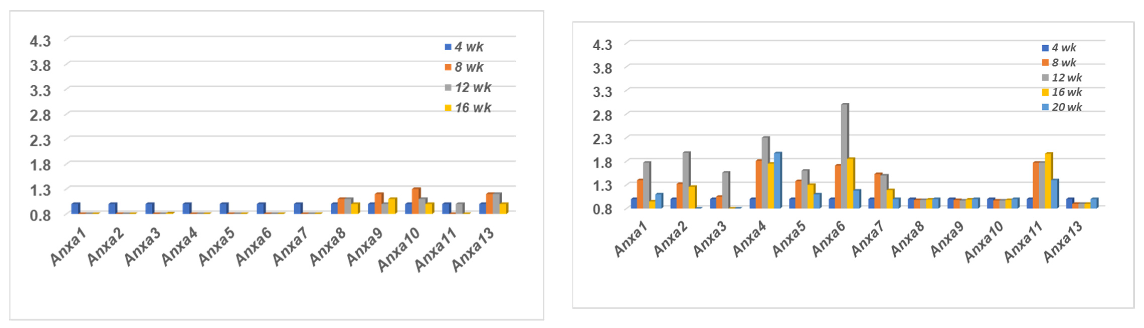
2.1. Comparison of the Annexin Transcriptome Profiles Expressed in the Salivary Glands of SSNS C57BL/6J and SSS C57BL/6.NOD-Aec1Aec2 Mice

A temporal transcriptomic comparison of the twelve annexin genes expressed in the salivary glands of SSNS C57BL/6J versus SSS C57BL/6.NOD-Aec1Aec2 mice is presented in Figure 1. The profile exhibited within the salivary glands of C57BL/6J mice (left panel) shows a complete lack of any upregulated gene expressions for Anxa1-7 and Anxa11 between the ages of 4 to 20 weeks of age. In contrast, a weak temporal upregulated expression was exhibited for Anxa8-10 and Anxa13 with maximum expression occurring around 8 and 12 weeks of age. On the other hand, the profile exhibited within the salivary glands of the C57BL/6.NOD-Aec1Aec2 mice (right panel) revealed a completely opposite result with upregulated temporal expressions for annexin genes Anxa1-7 and Anxa11, but a lack of any changes in gene expressions for Anxa8-10 and Anxa13. In addition, while each of these upregulations were observed starting at 8 weeks of age, maximum expressions of annexins Anxa1-7 occurred around 12 weeks of age, with annexin Anxa11 apparently exhibiting a more prolonged upregulated expression.

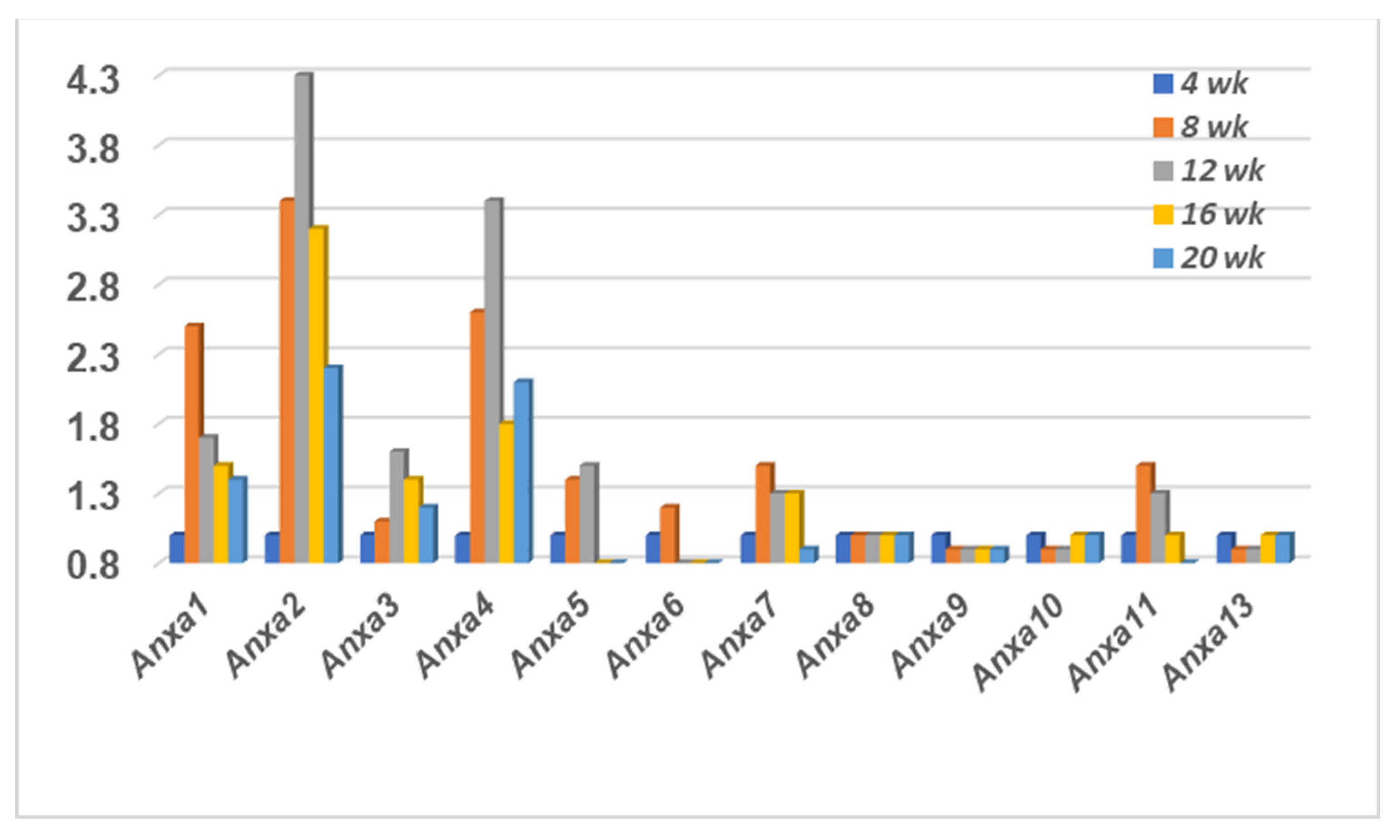
2.2. The Annexin Transcriptome Profile Expressed in the Lacrimal Glands of SSS C57BL/6.NOD-Aec1Aec2 Mice

To determine the temporal transcriptome profile of the annexin family genes in the lacrimal glands of the SSS C57BL/6.NOD-Aec1Aec2 mice, the same gene aptamers selected for the salivary gland data presented in Figure 1 were specifically selected from the microarray data from the C57BL/6.NOD-Aec1Aec2 lacrimal glands, thus permitting a direct comparison of identical aptamers/probes. As shown in Figure 2, the profile exhibited within the lacrimal glands of the C57BL/6.NOD-Aec1Aec2 mice revealed a strongly similar result to those of the salivary glands with upregulated temporal expressions for annexin genes Anxa1-7 and Anxa11, yet again a lack of any changes in gene expressions for Anxa8-10 and Anxa13. Although maximum gene expressions of Anxa1-7 and Anxa11 appear to occur around 12 weeks of age in the lacrimal glands, the majority of the activated genes are also exhibiting an upregulated expression by 8 weeks of age, prior to any obvious autoimmunity or lymphocytic foci. In addition, several of these genes (e.g., Anxa1, 5, 6, 11) show maximum upregulation at the 8 weeks’ time point. While this profile suggests that upregulated annexin genes are observed earlier in the lacrimal glands than in the salivary glands of C57BL/6.NOD-Aec1Aec2 mice, this may be deceptive due to the 4 weeks between data points.

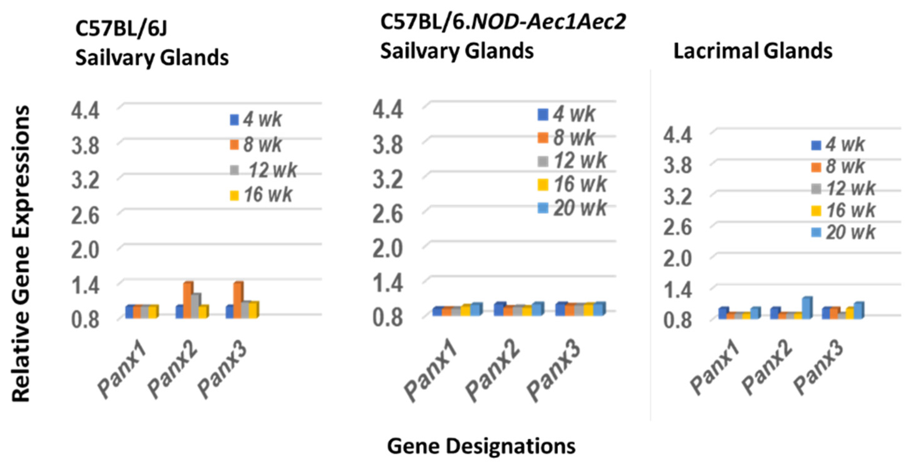
2.3. The Pannexin Transcription Profile in Salivary and Lacrimal Glands of SSNS C57BL/6J and SSS C57BL/6.NOD-Aec1Aec2 Mice during the Early Covert Phase of SS

The three-member family of mammalian pannexins was chosen as a comparative control for the annexin data. Pannexins are glycoproteins that form single membrane associated ATP-release channels that are ubiquitously expressed in most cells, including the salivary and lacrimal glands, and functionally active in both normal and disease states, including inflammation [49]. As presented in Figure 3, a slightly upregulated gene expression is observed for two of the three pannexins, Panx2 and Panx3, in the salivary glands of SSNS C57BL/6J mice, but none of the three genes are upregulated in either the salivary or lacrimal glands of SSS C57BL/6.NOD-Aec1Aec2 mice.

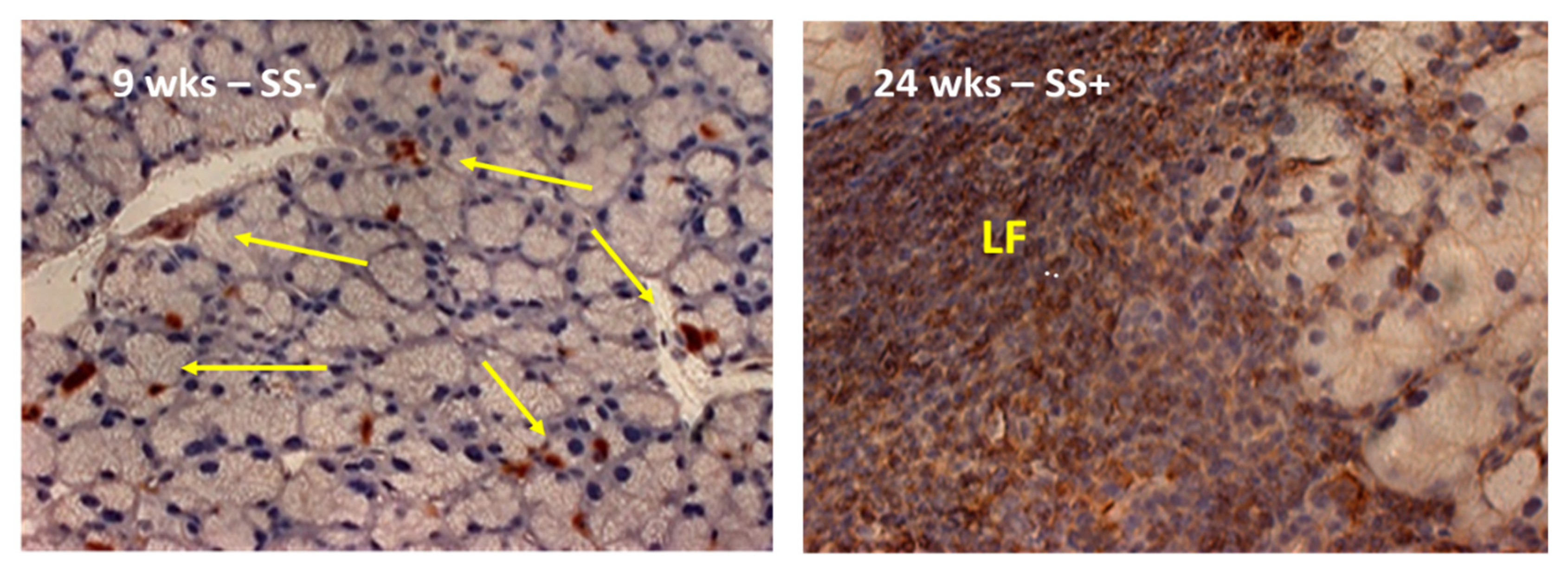
2.4. A Visual Comparison between the Anax Gene Expression Profiling and the Histological Landscape Showing the Distinct Increased Levels of Leukocytic Infiltrations of the Lacrimal Glands between Early-Stage versus Late-Stage Development of the SS-like Disease in the C57BL/6.NOD-Aec1Aec2 Mice

As presented in Figure 4, SSS C57BL.NOD-Aec1Aec2 mice do not exhibit organized leukocytic infiltration of their glands prior to 9 weeks of age, the time point at which the Anxa genes are clearly initiating their upregulated expressions. In contrast, at 24 weeks of age, when the gland is heavily infiltrated with cells of the adaptive autoimmune response, the Anxa genes are no longer expressing an upregulated profile. A similar profile is seen in the salivary glands [20].

3. Discussion

As stated above, annexins are a family of soluble Ca++-regulated phospholipid-binding hydrophobic proteins whose functional behavior classifies them as alarmins [27,28,29,30]. Annexins are known to act as scaffolding proteins capable of anchoring proteins to cellular membranes, thus promoting an extensive array of biomolecular products that in turn can directly and indirectly regulate normal cell functions, such as endocytotic processes, clathrin-coated budding, cholesterol ester internalization, and biogenesis of vesicular endosomes, but also inflammation and innate immune activities [29,31,32,33,34,35,36]. Although the major cellular activities of annexin proteins are internal to cells, several annexins have been observed externally in free form and complexed with phospholipid entities, but whether this external presence is responsible for production of anti-annexin autoantibodies in patients remains unclear. Nevertheless, production of anti-annexin autoantibodies have been proposed to enhance the vulnerability for inflammation and autoimmune disorders by interfering with annexins’ ability to regulate apoptosis [37,38,39,40,41,42,43]. In this sense, it is interesting to note that both NOD/Shi and C57BL/6.NOD-Aec1Aec2 mice have increased levels of cellular apoptosis in the salivary and lacrimal glands prior to onset of clinical SS pathology [46,51].
Perhaps the most important results of the current study are two-fold: first, the overall transcriptome profiles of the 12 annexin genes in the salivary and lacrimal glands of SSS C57BL/6.NOD-Aec1Aec2 mice proved quite similar, and second, the gene profile of the SSNS C57BL/6J mice versus those of SSS C57BL/6.NOD-Aec1Aec2 salivary and lacrimal glands are totally opposite to each other in gene expressions. Why there are minimal, yet positive, upregulated transcriptome gene expressions in the salivary glands of C57BL/6J mice for Anxa8-10 and Anxa13 remain unknown, but because the annexins are known to regulate early inflammation, one can speculate that this activity may be capable of down-regulating any early inflammation in the C57BL/6J mice which can occasionally appear as weak leukocytic infiltrations within their salivary glands. Lastly, although the annexin gene profiles in the salivary and lacrimal glands of the SSS mice are generally similar, there are important differences in individual gene expressions, both in the time at which annexin genes initiate upregulation, the relative strength of expression, and the length of time a gene remains upregulated. However, these data are consistent with the concept fact that the individual genes have marked functional differences, many of which are still to be defined, and activated in two functionally different organ environments.
Despite considerable shared homology, the individual annexin molecules demonstrate multiple unique phenotypes and functions. Anxa1, for example, restricts pro-inflammatory innate responses by binding with the formyl peptide receptor (FPR) to form a complex that inhibits neutrophil recruitment [52,53]. Additionally, corticosteroid stimulation can release Anxa1 from cells to further mitigate inflammation [54,55]. Not surprising, therefore, Anxa1 gene knockout (KO) mice have been shown to be protected from viral infection [56]. In this regard, our previously published data predict the autoimmune response in our SSS C57BL/6.NOD-Aec1Aec2 mice involves a dsRNA virus [57]. Anxa2, the strongest upregulated annexin gene in the lacrimal gland, is expressed in multiple tissues, including macrophages, monocytes, endothelium and epithelium and plays a role in cell growth and differentiation, apoptosis, and cell migrations [58,59]. Like Anxa1, Anxa2 is known to form complexes, e.g., with S100A10 [60,61], that are then translocated to the cell surface where they modulate receptor and ion channel presentations. Studies of Zang et al. [62] indicate that Anxa2 gene KO mice have enhanced TLR4 signaling, reduced TLR4 endocytosis, dysregulated autophagy, and increased production of IL-17, ROS and neutrophil infiltrations, factors previously shown to be critical in the SS-like pathology of the SSS C57BL/6.NOD-Aec1Aec2 mice [63]. Like Anxa1 and Anxa2, Anxa11 can also form complexes, e.g., with S100A6, which then bind to the nuclear envelope, suggesting a functional role in cell growth and differentiation [64,65]. Furthermore, Anxa11 is a ubiquitously expressed protein present in the cytoplasm and nucleus of multiple cell types and shows a propensity to translocate from the nucleus to other organelles due to its ability to bind secretory vesicles that transport to Golgi bodies. Of interest, and not surprising, is the fact that multiple mutations present in Anxa11 are increasingly being identified in various autoimmune diseases [66,67,68].
An interesting aspect of the transcriptome profile of the annexins can be seen in the gene profiles of Anxa6. While this gene exhibited the strongest upregulated expression in the salivary gland, its upregulated expression in the lacrimal gland was the weakest of those genes showing a positive response. Anxa6 is a ubiquitously expressed annexin present in the plasma membrane, mitochondria, endocytic and exocytic vesicles, as well as lipid droplets. This protein interacts with both proteins and lipids in the regulation of cholesterol transport, signal complex disassembly, cytoskeleton rearrangements, the stress response, cell growth and motility, differentiation, lipid and glucose homeostasis, endocytosis, exocytosis and viral infections [69,70,71,72]. Interestingly, Anxa6 is upregulated during B and T cell differentiation, along with IL-2 [73]. Furthermore, annexin Anxa6 has been linked to lipoprotein and cholesterol uptake and transport, lipid and glucose homeostasis, membrane repair, as well as cytokine secretion [74].
Different from Anxa6, Anxa4 shows the second highest transcriptome expressions in both the salivary and lacrimal glands. This gene has three isoforms and is present primarily in the secretory epithelia of the lung, intestine, stomach and kidney with two of the three isoforms present only in the digestive tract [75]. In response to Ca++, Anxa4 can translocate to the plasma and nuclear membranes. Functionally, Anxa4 can inhibit adenyl cyclase 5, which, in turn, controls the conversion of ATP to cAMP [76], suggesting a role in metabolic activities. Additionally, Anxa4 has been reported to influence chemosensitivity [77].
Annexin Anxa7 is a protein found in virtually all tissues, but which undergoes alternate splicing that gives rise to two isoforms, both of which exhibit a distinct functional profile. Anxa7 is predominantly associated with secretory vesicles, plasma membranes, and the nuclear envelope [78,79,80,81,82]. Functionally, this annexin appears to have an important role in Ca++ homeostasis [81,82], especially in cardiomyocytes with respect to cardiac muscle contraction. Interestingly, the studies of Kuijpers et al. [78] have shown that phosphorylation of Anxa7 promotes membrane fusion between exocytic vesicles and plasma membranes following internal interactions with SNARE proteins. In addition, Anxa7 appears to have GTPase activity, suggesting a role in migration [82]. Interestingly, studies of Anxa7-deficient mice have revealed that this annexin may exacerbate multiple disease states, including cardiac and neuronal injury, malignant cells and death, as well as inflammation, pathologies that can be seen in SSS C57BL/6.NOD-Aec1Aec2 mice [17].
Despite the fact that annexin Anxa5 is considered the most abundant annexin of this protein family and present in cells associated with the plasma membrane, nucleus, Golgi, endoplasmic reticulum, endosomes, phagosomes, and mitochondria [83,84], it was not a highly upregulated gene in either the salivary or lacrimal glands of our SSS C57Bl/6.NOD-Aec1Aec2 mice. Importantly, Anxa5 functions as a regulator of membrane transport, Ca++-influx and signaling, ion channels, apoptosis and phagocytosis [85,86,87,88,89,90]. In addition, Anxa5 interacts with Dectin-1, thereby facilitating apoptotic cell phagocytosis by dendritic cells that, in turn, can lead to peripheral tolerance to self-antigens [91]. The last of the upregulated annexin genes, Anxa3, is present in many organ systems and cell types, active in membrane and ion transport, cell signaling, endothelial migration, adipocyte differentiation and inflammation [92,93,94,95,96,97,98]. While elevated Anxa3 levels have been shown to suppress apoptosis and autophagy through suppression of PKCδ-p38Mapk, depletion of Anxa3 apparently inhibits HIF1a-VEGF-dependent cell signaling [99]. Hif1a is a master gene that regulates cellular and homeostatic states in response to hypoxia via regulation of genes such as Infβ, Tnfα and Il6 [100,101], each of which can severely alter homeostasis of MZB cells, a cell population now recognized as an early innate immune mediator of SS [20,22].
Finally, as an internal control for the current study, we chose to examine the transcriptome profile for the pannexin family of genes in the salivary and lacrimal glands of the SSNS C57BL/6J and SSS C57BL/6.NOD-Aec1Aec2 mice. The pannexin family consists of only three proteins, but these glycoproteins have important roles in regulating ATP-release channels and are active during both normal homeostatic states and disease states, especially inflammation [49]. Surprisingly, while the profiles for the Panx genes in the salivary glands of SSNS C57BL/6J mice revealed that two of the three genes, Panx2 and Panx3, showed an early, but relatively weak, upregulation, none of the three Panx genes in the salivary and lacrimal glands of SSS C57BL/6.NOD-Aec1Aec2 mice showed any activation over the microarray study’s 20-week time frame. Considering the potential importance of the Panx molecules in cell regulations, why this particular gene family is not activated during early development of its SS pathology is intriguing but, at the same time, clearly increases the importance of the annexins as definitive early markers of impending disease. Furthermore, these data support the specificity of the annexin activations. Similarly, why annexins Anxa7-10 and -13 show no activation may also provide important information into SS disease initiation. Nevertheless, the strong upregulation of multiple annexin genes capable of carrying out multiple biomolecular functions associated with the onset of early-stage SS disease, together with recent appreciation of these molecules in autoimmune diseases, opens a new focus in the underlying bases for the autoimmune SS response.

4. Materials and Methods

4.1. Animal Model

C57BL/6.NOD-Aec1Aec2 mice, a diabetes-free model of primary SS-like disease, were bred and maintained under specific pathogen free (SPF) conditions within the College of Medicine’s Department of Pathology’s Mouse Facility with oversight by Animal Care Services at the University of Florida, Gainesville. The C57BL/6.NOD-Aec1Aec2 mouse is a well-studied and well-characterized model of SS that spontaneously develops features of SS observed in patients [45], e.g., lymphocytic foci (LF) concomitantly with SS pathology (as shown in Figure 4). Typically, C57BL/6.NOD-Aec1Aec2 mice exhibit: (a) progressive aberrant apoptosis of glandular epithelial cells by 2 months of age, (b) transient macrophage and marginal zone B cell infiltrations of the salivary and lacrimal glands between 2 and 3 months of age, (c) autoantibody production by 3–4 months of age, (d) progressive lymphocytic infiltrations and formation of lymphocytic foci comprised primarily of adaptive immune cells starting between 3 and 4 months of age, and (e) a full-blown SS-like disease with decreased glandular secretions by 4–5 months of age. This mouse does not develop lymphomagenesis. Mice were maintained on a 12-hr light-dark schedule and provided food and acidified water ad libitum. Any mouse showing signs of eye dryness were treated daily with salve ointment. For the present study, breeding pairs (one male and one female) were paired per cage and offspring were weaned at 3–4 weeks of age. Weaned mice were caged maximum of n = 5 per cage per sex. The experimental mice were raised to a maximum of 20 weeks of age, a time prior to visible signs of any overt disease, but beginning of histologically significant glandular leukocytic infiltration. Euthanization was carried out at the appropriate timepoints by cervical dislocation after deep anesthetization as stipulated by the Panel on Euthanasia of the American Veterinary Association. There were no indications that this procedure affected subsequent preparation of RNA specimens. Both the breeding and use of these animals for the present studies were approved by the University of Florida’s Institutional Animal Care and Use Committee (IACUC) under protocols 2008011756 and 201004828.

4.2. RNA Preparations

Procedures for the isolation, preparation, and quality testing of RNA samples are described in detail elsewhere [16,17,19]. In brief, salivary and extra-orbital lacrimal glands, free of lymph nodes, were excised in parallel from male C57BL/6.NOD-Aec1Aec2 and C57BL/6J mice euthanized at 4, 8, 12, 16 or 20 weeks of age (n = 5 per age group, n = 25 per experimental group, n = 50 total for the overall study), snap-frozen in liquid nitrogen and stored at −80 °C until all samples were obtained. Using one salivary and one lacrimal gland from each mouse, total RNA from each specimen was isolated concurrently using the RNeasy Mini-Kit (Qiagen, Valencia, CA, USA), as per the manufacturer’s recommended protocol. Each RNA sample was hybridized to an Affymetrix 3′ Expression Array GeneChip Mouse Genome 430 2.0 array and annotated (build 32; 6 September 2011). Each GeneChip contained 45,102 DNA sequences. Heatmaps of differentially expressed genes are published elsewhere [16,17]. To verify relative microarray gene expressions, numerous genes were randomly selected for comparative expressions to real-time polymerase chain reaction (rt-PCR) analyses. The rt-PCR data confirmed both differential and temporal gene expressions. Full microarray data libraries are deposited with Gene Expression Omnibus, Accession number GSE15640.

4.3. Microarray Data Analyses

Gene expression analyses have been detailed elsewhere [16,17,19]. In brief, microarray data were normalized using the robust multiarray average (GCRMA) algorithm, followed by Linear Models for Microarray Analysis (LIMMA) (http://www.r-project.org accessed on 15 August 2022) for differential expression determinations. The fdr (false discovery rate) method of Benjamini and Hochberg [102] was used to adjust the p-values for multiple testing. The original data represent 5 equally spaced timepoints; thus, multiple models were used to identify temporal patterns of gene expression, i.e., the linear fit (degree = 1), quadratic fit (degree = 2), cubic fit (degree = 3) and quartic fit (degree = 4) regression models. At this point, each gene has a single statistical value based on n = 5 data points at each of the 5 timepoints. B-statistics were calculated for each gene providing odds that a gene shows either positive or negative trends over time [16,17]. In the current study, these temporal changes in an individual gene’s expression relative to its value at the 4 week timepoint were considered in addition to statistical significant; thus, data presented show the differential expression of an individual gene’s value at the 8, 12, 16 or 20 weeks timepoints relative to that gene’s value at the 4 weeks timepoint set as 1.0. The 4 week time point is a time when the exocrine glands have reached functional maturity, yet considered in a pre-diseased state with no observed leukocyte infiltration via histological examinations. The presented data compares the microarray data from salivary glands of Sjögren’s syndrome susceptible C57BL/6.NOD-Aec1Aec2 mice to that obtained from Sjögren’s syndrome non-susceptible C57BL/6J mice. The differentially expressed genes in this initial comparison were then specifically selected from the microarray data of the C57BL/6.NOD-Aec1Aec2 lacrimal glands thereby permitting a direct comparison of identical aptamers/probes.

4.4. Histology

Extra-orbital lacrimal glands, surgically removed from each mouse at time of euthanasia, were placed in 10% phosphate buffered formalin for 24 h. Fixed tissues were embedded in paraffin and sectioned at 5 µm thickness. Paraffin-embedded slides were de-paraffinized by immersing in xylene, then dehydrated in ethanol. Following a 5 min wash with PBS at 25 °C, the sections were incubated 1 h with blocking solution containing normal rabbit serum diluted 1:50 in PBS. Each section was incubated with rat anti-mouse anti-phosphorylated paxillin antibody (Santa Cruz Biotechnology, Santa Cruz, CA, USA). Slides were washed 3× in PBS and visualized microscopically at 200×. Paxillin antibodies are specific for staining migration-associated focal adherens.

5. Conclusions, Summary and Contribution to the Field

Sjögren’s syndrome (SS), a human systemic rheumatoid autoimmune disease that severely affects lacrimal and salivary gland functions, is considered an under-studied disease appearing most frequently in middle-aged women. Patients generally present in clinic only after the adaptive autoimmune phase is active and irreversible pathology in these glands has occurred. Furthermore, the time between disease onset and a correct diagnosis can range from 4 to 10 or more years, permitting the pathology to progress unchecked without intervention therapy, plus seriously slowing generation of effective therapy development. To address these issues, numerous mouse models exhibiting SS-like disease have been developed that permit one to focus on identification of pre-disease phases of the autoimmune processes. Nevertheless, many details of the underlying molecular and cellular events remain unidentified, especially those associated with the early-stage events. Here, we provide evidence that unique biomolecular and physiological events can be identified in the salivary and lacrimal glands of the C57BL/6.NOD-Aec1Aec2 mouse model prior to detection of any overt development of its SS-like disease via measurement of an alarmin protein family, specifically the calcium-dependent binding annexin proteins encoded by Anxa genes. Interestingly, the specific set of Anxa genes that are suddenly upregulated (i.e., time wise) in both salary and lacrimal glands reveal similar, yet partially different profiles between the two glands, but still represent a common biological process that appears to identify a potential marker of initial onset of disease, especially if present in human specimens. Nevertheless, in summary, using temporal global transcription analyses of the C57BL/6.NOD-Aec1Aec2 mouse model, we are identifying molecular profiles for critical bioprocesses involved in its early SS-like disease development and onset. Importantly, the different profiles emerging for salivary and lacrimal glands are suggesting potential autoimmune processes that could impact how one approaches future design and development of potential therapies that might transition to treatment of human SS disease.

Author Contributions

Conceptualization, writing and editing, A.B.P. and J.L.A.J.; Methodology, validation, data analyses, and curation, A.B.P. All authors have read and agreed to the published version of the manuscript.

Funding

Creation of the microarray data was supported in part by the National Institute of Dental and Craniofacial Research (NIDCR) grant R01 DE-014344 and the National Institute of Allergy and Infectious Diseases (NIAID) grant AI-0819529. The NIH had no role in the design of the study; data verification, analyses, or interpretation; in the writing of the manuscript, nor in the decision to publish the results. Publication of the manuscript was supported by authors’ personal funds with no help from the NIH.

Data Availability Statement

Gene Expression Omnibus, Accession numbers GSE15640 and GSE36378.

Acknowledgments

The authors wish to strongly acknowledge T. Grewal, C. Rentero, C. Enrich, M. Wahba, C.A. Raabe, and U. Rescher for their recent publication (Annexin Animal Models—From Fundamental Principles to Translational Research [Int. J. Mol. Sci. 2021]. That publication provided the basis for our current data analysis and final write-up of the possible role annexin molecules play in the early development of SS in our SS-susceptible mouse model.

Conflicts of Interest

The authors have no conflict of interest with regard to this publication.

Abbreviations

AnxaAnnexin gene
Ca++Calcium ion
DAMPsDanger-associated molecular patterns
dsRNAdouble-stranded RNA
FRPFormyl peptide receptor
GCMRARobust multiarray average
GTPaseGTP catalyzing enzymes
KOKnock-out (gene)
LFLymphocytic focus
LIMMALinear model for microarray analysis
NODNon-obese diabetic (mouse)
PanxPannexin gene
PAMPSPathogen-assocated molecular patterns
PRRPathogen recognition receptor
pSS-MALTPrimary Sjögren’s syndrome—mucosal-associated lymphoid tissue lymphoma
RARheumatoid arthritis
RNARibonuclear acid
SLESystemic l erythmetosus
SSSjögren’s syndrome
SSSSjögren’s syndrome—susceptible
SSNSSjögren’s syndrome—non-susceptible

References

  1. Jonsson, R.; Dowman, S.; Gordon, T. Sjogren’s Syndrome. In Arthritis and Allied Conditions—A Textbook in Rheumatology; Koopman, W., Moreland, L., Eds.; Lippincott WIlliams & Wilkins: Philadelphia, PA, USA, 2004; pp. 1681–1705. [Google Scholar]
  2. Fox, R.I. Sjogren’s syndrome. Lancet 2005, 366, 321–331. [Google Scholar] [CrossRef]
  3. Routsias, J.G.; Tzioufas, A.G. Sjogren’s syndrome—Study of autoantigens and autoantibodies. Clin. Rev. Allergy Immunol. 2007, 32, 238–251. [Google Scholar] [CrossRef] [PubMed]
  4. Nordmark, G.; Eloranta, M.L.; Ronnblom, L. Primary Sjogren’s syndrome and the type I interferon system. Curr. Pharm. Biotechnol. 2012, 13, 2054–2062. [Google Scholar] [CrossRef]
  5. Shiboski, S.C.; Shiboski, C.H.; Criswell, L.A.; Baer, A.N.; Challacombe, S.; Lanfranchi, H.; Schiodt, M.; Umehara, H.; Vivino, F.; Zhao, Y.; et al. American College of Rheumatology classification criteria for Sjogren’s syndrome: A data-driven, expert consensus approach in the Sjogren’s International Collaborative Clinical Alliance Cohort. Arthritis Care Res. 2012, 64, 475–487. [Google Scholar] [CrossRef]
  6. Shen, L.; Suresh, L.; Lindemann, M.; Xuan, J.; Kowal, P.; Malyavantham, K.; Ambrus, J.L., Jr. Novel autoantibodies in Sjogren’s syndrome. Clin. Immunol. 2012, 145, 251–255. [Google Scholar] [CrossRef] [PubMed]
  7. Maier-Moore, J.S.; Koelsch, K.A.; Smith, K.; Lessard, C.J.; Radfar, L.; Lewis, D.; Kurien, B.T.; Wolska, N.; Deshmukh, U.; Rasmussen, A.; et al. Antibody-Secreting Cell Specificity in Labial Salivary Glands Reflects the Clinical Presentation and Serology in Patients with Sjogren’s Syndrome. Arthritis Rheumatol. 2014, 66, 3445–3456. [Google Scholar] [CrossRef] [PubMed] [Green Version]
  8. Goules, A.V.; Tzioufas, A.G.; Moutsopoulos, H.M. Classification criteria of Sjogren’s syndrome. J. Autoimmun. 2014, 48–49, 42–45. [Google Scholar] [CrossRef]
  9. Reksten, T.R.; Jonsson, M.V. Sjogren’s Syndrome An Update on Epidemiology and Current Insights on Pathophysiology. Oral Maxillofac. Surg. Clin. N. Am. 2014, 26, 1–12. [Google Scholar] [CrossRef]
  10. Goules, A.V.; Tzioufas, A.G. Primary Sjogren’s syndrome: Clinical phenotypes, outcome and the development of biomarkers. Autoimmun. Rev. 2016, 15, 695–703. [Google Scholar] [CrossRef]
  11. Harris, V.M.; Scofield, R.H.; Sivils, K.L. Genetics in Sjogren’s syndrome: Where we are and where we go. Clin. Exp. Rheumatol. 2019, 37, S234–S239. [Google Scholar]
  12. Liang, Y.; Yang, Z.X.; Qin, B.D.; Zhong, R.Q. Primary Sjogren’s syndrome and malignancy risk: A systematic review and meta-analysis. Ann. Rheum. Dis. 2014, 73, 1151–1156. [Google Scholar] [CrossRef] [PubMed]
  13. Nezos, A.; Gravani, F.; Tassidou, A.; Kapsogeorgou, E.K.; Voulgarelis, M.; Koutsilieris, M.; Crow, M.K.; Mavragani, C.P. Type I and II interferon signatures in Sjogren’s syndrome pathogenesis: Contributions in distinct clinical phenotypes and Sjogren’s related lymphomagenesis. J. Autoimmun. 2015, 63, 47–58. [Google Scholar] [CrossRef] [PubMed] [Green Version]
  14. Nocturne, G.; Pontarini, E.; Bombardieri, M.; Mariette, X. Lymphomas complicating primary Sjogren’s syndrome: From autoimmunity to lymphoma. Rheumatology 2019, 60, 3513–3521. [Google Scholar] [CrossRef] [PubMed]
  15. Killedar, S.J.; Eckenrode, S.E.; McIndoe, R.A.; She, J.X.; Nguyen, C.Q.; Peck, A.B.; Cha, S. Early pathogenic events associated with Sjogren’s syndrome (SjS)-like disease of the NOD mouse using microarray analysis. Lab. Investig. 2006, 86, 1243–1260. [Google Scholar] [CrossRef] [Green Version]
  16. Nguyen, C.Q.; Sharma, A.; Lee, B.H.; She, J.X.; McIndoe, R.A.; Peck, A.B. Differential gene expression in the salivary gland during development and onset of xerostomia in Sjogren’s syndrome-like disease of the C57BL/6.NOD-Aec1Aec2 mouse. Arthritis Res. Ther. 2009, 11, R56. [Google Scholar] [CrossRef] [Green Version]
  17. Nguyen, C.Q.; Sharma, A.; She, J.X.; McIndoe, R.A.; Peck, A.B. Differential gene expressions in the lacrimal gland during development and onset of keratoconjunctivitis sicca in Sjogren’s syndrome (SJS)-like disease of the C57BL/6.NOD-Aec1Aec2 mouse. Exp. Eye Res. 2009, 88, 398–409. [Google Scholar] [CrossRef] [Green Version]
  18. Peck, A.B.; Nguyen, C.Q. Transcriptome analysis of the interferon-signature defining the autoimmune process of Sjogren’s syndrome. Scand. J. Immunol. 2012, 76, 237–245. [Google Scholar] [CrossRef] [Green Version]
  19. Delaleu, N.; Nguyen, C.Q.; Tekle, K.M.; Jonsson, R.; Peck, A.B. Transcriptional landscapes of emerging autoimmunity: Transient aberrations in the targeted tissue’s extracellular milieu precede immune responses in Sjogren’s syndrome. Arthritis Res. Ther. 2013, 15, R174. [Google Scholar] [CrossRef] [Green Version]
  20. Peck, A.B.; Nguyen, C.Q.; Ambrus, J. Early Covert Appearance of Marginal Zone B Cells in Salivary Glands of Sjogren’s Syndrome-Susceptible Mice: Initiators of Subsequent Overt Clinical Disease. Int. J. Mol. Sci. 2021, 22, 1919. [Google Scholar] [CrossRef]
  21. Peck, A.B.; Nguyen, C.Q.; Ambrus, J.L. Upregulated Chemokine and Rho-GTPase Genes Define Immune Cell Emigration into Salivary Glands of Sjogren’s Syndrome-Susceptible C57BL/6.NOD-Aec1Aec2 Mice. Int. J. Mol. Sci. 2021, 22, 7176. [Google Scholar] [CrossRef]
  22. Peck, A.B.; Nguyen, C.Q.; Ambrus, J.L. A MZB Cell Activation Profile Present in the Lacrimal Glands of Sjogren’s Syndrome-Susceptible C57BL/6.NOD-Aec1Aec2 Mice Defined by Global RNA Transcriptomic Analyses. Int. J. Mol. Sci. 2022, 23, 6106. [Google Scholar] [CrossRef] [PubMed]
  23. Jentho, E.; Weis, S. DAMPs and Innate Immune Training. Front. Immunol. 2021, 12, 699563. [Google Scholar] [CrossRef] [PubMed]
  24. Zorro, M.M.; Aguirre-Gamboa, R.; Mayassi, T.; Ciszewski, C.; Barisani, D.; Hu, S.; Weersma, R.K.; Withoff, S.; Li, Y.; Wijmenga, C.; et al. Tissue alarmins and adaptive cytokine induce dynamic and distinct transcriptional responses in tissue-resident intraepithelial cytotoxic T lymphocytes. J. Autoimmun. 2020, 108, 102422. [Google Scholar] [CrossRef] [PubMed]
  25. Giovannetti, A.; Straface, E.; Rosato, E.; Casciaro, M.; Pioggia, G.; Gangemi, S. Role of Alarmins in the Pathogenesis of Systemic Sclerosis. Int. J. Mol. Sci. 2020, 21, 4985. [Google Scholar] [CrossRef]
  26. Sarrand, J.; Baglione, L.; Parisis, D.; Soyfoo, M. The Involvement of Alarmins in the Pathogenesis of Sjogren’s Syndrome. Int. J. Mol. Sci. 2022, 23, 5671. [Google Scholar] [CrossRef]
  27. Moss, S.E.; Morgan, R.O. The annexins. Genome Biol. 2004, 5, 219. [Google Scholar] [CrossRef] [Green Version]
  28. Gerke, V.; Creutz, C.E.; Moss, S.E. Annexins: Linking Ca2+ signalling to membrane dynamics. Nat. Rev. Mol. Cell Biol. 2005, 6, 449–461. [Google Scholar] [CrossRef]
  29. Mirsaeidi, M.; Gidfar, S.; Vu, A.; Schraufnagel, D. Annexins family: Insights into their functions and potential role in pathogenesis of sarcoidosis. J. Transl. Med. 2016, 14, 89. [Google Scholar] [CrossRef] [Green Version]
  30. Grewal, T.; Rentero, C.; Enrich, C.; Wahba, M.; Raabe, C.A.; Rescher, U. Annexin Animal Models-From Fundamental Principles to Translational Research. Int. J. Mol. Sci. 2021, 22, 3439. [Google Scholar] [CrossRef]
  31. Moreno-Angarita, A.; Aragon, C.C.; Tobon, G.J. Cathelicidin LL-37: A new important molecule in the pathophysiology of systemic lupus erythematosus. J. Transl. Autoimmun. 2020, 3, 100029. [Google Scholar] [CrossRef]
  32. Martin, M.; Leffler, J.; Blom, A.M. Annexin A2 and A5 serve as new ligands for C1q on apoptotic cells. J. Biol. Chem. 2012, 287, 33733–33744. [Google Scholar] [CrossRef] [PubMed]
  33. Pianta, A.; Drouin, E.E.; Crowley, J.T.; Arvikar, S.; Strle, K.; Costello, C.E.; Steere, A.C. Annexin A2 is a target of autoimmune T and B cell responses associated with synovial fibroblast proliferation in patients with antibiotic-refractory Lyme arthritis. Clin. Immunol. 2015, 160, 336–341. [Google Scholar] [CrossRef] [PubMed] [Green Version]
  34. Bruschi, M.; Petretto, A.; Vaglio, A.; Santucci, L.; Candiano, G.; Ghiggeri, G.M. Annexin A1 and Autoimmunity: From Basic Science to Clinical Applications. Int. J. Mol. Sci. 2018, 19, 1348. [Google Scholar] [CrossRef] [PubMed] [Green Version]
  35. Hua, K.; Li, Y.; Zhao, Q.; Fan, L.; Tan, B.; Gu, J. Downregulation of Annexin A11 (ANXA11) Inhibits Cell Proliferation, Invasion, and Migration via the AKT/GSK-3beta Pathway in Gastric Cancer. Med. Sci. Monit. 2018, 24, 149–160. [Google Scholar] [CrossRef]
  36. Lim, H.I.; Hajjar, K.A. Annexin A2 in Fibrinolysis, Inflammation and Fibrosis. Int. J. Mol. Sci. 2021, 22, 6836. [Google Scholar] [CrossRef]
  37. Iaccarino, L.; Ghirardello, A.; Canova, M.; Zen, M.; Bettio, S.; Nalotto, L.; Punzi, L.; Doria, A. Anti-annexins autoantibodies: Their role as biomarkers of autoimmune diseases. Autoimmun. Rev. 2011, 10, 553–558. [Google Scholar] [CrossRef]
  38. van Venrooij, W.J.; Wodzig, K.W.; Habets, W.J.; de Rooij, D.J.; van de Putte, L.B. Anti-56K: A novel, frequently occurring autoantibody specificity in connective tissue disease. Clin. Exp. Rheumatol. 1989, 7, 277–282. [Google Scholar]
  39. Rodriguez-Garcia, M.I.; Fernandez, J.A.; Rodriguez, A.; Fernandez, M.P.; Gutierrez, C.; Torre-Alonso, J.C. Annexin V autoantibodies in rheumatoid arthritis. Ann. Rheum. Dis. 1996, 55, 895–900. [Google Scholar] [CrossRef] [Green Version]
  40. Gheita, T.A.; El Sisi, R.W.; Raafat, H.A.; Khalil, H.M. Anti-annexin V antibodies in primary fibromyalgia syndrome: Relation to associated Sjogren’s syndrome. J. Clin. Immunol. 2013, 33, 311–312. [Google Scholar] [CrossRef]
  41. Dema, B.; Charles, N. Autoantibodies in SLE: Specificities, Isotypes and Receptors. Antibodies 2016, 5, 2. [Google Scholar] [CrossRef] [Green Version]
  42. Cui, L.; Elzakra, N.; Xu, S.; Xiao, G.G.; Yang, Y.; Hu, S. Investigation of three potential autoantibodies in Sjogren’s syndrome and associated MALT lymphoma. Oncotarget 2017, 8, 30039–30049. [Google Scholar] [CrossRef] [PubMed]
  43. Scholz, P.; Auler, M.; Ruthard, J.; Brachvogel, B.; Klatt, A.R.; Streichert, T. Detection of annexin A8 antibodies in serum of patients with antiphospholipid syndrome. Biochem. Med. 2018, 28, 030703. [Google Scholar] [CrossRef] [PubMed]
  44. Finamore, F.; Cecchettini, A.; Ceccherini, E.; Signore, G.; Ferro, F.; Rocchiccioli, S.; Baldini, C. Characterization of Extracellular Vesicle Cargo in Sjogren’s Syndrome through a SWATH-MS Proteomics Approach. Int. J. Mol. Sci. 2021, 22, 4864. [Google Scholar] [CrossRef] [PubMed]
  45. Cha, S.; Nagashima, H.; Brown, V.B.; Peck, A.B.; Humphreys-Beher, M.G. Two NOD Idd-associated intervals contribute synergistically to the development of autoimmune exocrinopathy (Sjogren’s syndrome) on a healthy murine background. Arthritis Rheum. 2002, 46, 1390–1398. [Google Scholar] [CrossRef] [PubMed]
  46. Nguyen, C.Q.; Peck, A.B. Unraveling the pathophysiology of Sjogren syndrome-associated dry eye disease. Ocul. Surf. 2009, 7, 11–27. [Google Scholar] [CrossRef] [Green Version]
  47. Horvath, S.; Nazmul-Hossain, A.N.; Pollard, R.P.; Kroese, F.G.; Vissink, A.; Kallenberg, C.G.; Spijkervet, F.K.; Bootsma, H.; Michie, S.A.; Gorr, S.U.; et al. Systems analysis of primary Sjogren’s syndrome pathogenesis in salivary glands identifies shared pathways in human and a mouse model. Arthritis Res. Ther. 2012, 14, R238. [Google Scholar] [CrossRef] [PubMed] [Green Version]
  48. Peck, A.B.; Nguyen, C.Q. What can Sjogren’s syndrome-like disease in mice contribute to human Sjogren’s syndrome? Clin. Immunol. 2017, 182, 14–23. [Google Scholar] [CrossRef] [Green Version]
  49. Bond, S.R.; Naus, C.C. The pannexins: Past and present. Front. Physiol. 2014, 5, 58. [Google Scholar] [CrossRef] [Green Version]
  50. Peck, A.B.; Saylor, B.T.; Nguyen, L.; Sharma, A.; She, J.X.; Nguyen, C.Q.; McIndoe, R.A. Gene expression profiling of early-phase Sjögren’s syndrome in C57BL/6.NOD-Aec1Aec2 mice identifies focal adhesion maturation associated with infiltrating leukocytes. Investig. Ophthalmol. Vis. Sci. 2011, 29, 5647–5655. [Google Scholar] [CrossRef] [Green Version]
  51. Humphreys-Beher, M.G.; Peck, A.B.; Dang, H.; Talal, N. The role of apoptosis in the initiation of the autoimmune response in Sjogren’s syndrome. Clin. Exp. Immunol. 1999, 116, 383–387. [Google Scholar] [CrossRef]
  52. Ernst, S.; Lange, C.; Wilbers, A.; Goebeler, V.; Gerke, V.; Rescher, U. An annexin 1 N-terminal peptide activates leukocytes by triggering different members of the formyl peptide receptor family. J. Immunol. 2004, 172, 7669–7676. [Google Scholar] [CrossRef] [PubMed] [Green Version]
  53. Gavins, F.N.; Hickey, M.J. Annexin A1 and the regulation of innate and adaptive immunity. Front. Immunol. 2012, 3, 354. [Google Scholar] [CrossRef] [PubMed]
  54. Perretti, M.; D’Acquisto, F. Annexin A1 and glucocorticoids as effectors of the resolution of inflammation. Nat. Rev. Immunol. 2009, 9, 62–70. [Google Scholar] [CrossRef] [PubMed]
  55. Hannon, R.; Croxtall, J.D.; Getting, S.J.; Roviezzo, F.; Yona, S.; Paul-Clark, M.J.; Gavins, F.N.; Perretti, M.; Morris, J.F.; Buckingham, J.C.; et al. Aberrant inflammation and resistance to glucocorticoids in annexin 1-/- mouse. FASEB J. 2003, 17, 253–255. [Google Scholar] [CrossRef] [Green Version]
  56. Arora, S.; Lim, W.; Bist, P.; Perumalsamy, R.; Lukman, H.M.; Li, F.; Welker, L.B.; Yan, B.; Sethi, G.; Tambyah, P.A.; et al. Influenza A virus enhances its propagation through the modulation of Annexin-A1 dependent endosomal trafficking and apoptosis. Cell Death Differ. 2016, 23, 1243–1256. [Google Scholar] [CrossRef]
  57. Nguyen, C.Q.; Peck, A.B. The Interferon-Signature of Sjogren’s Syndrome: How Unique Biomarkers Can Identify Underlying Inflammatory and Immunopathological Mechanisms of Specific Diseases. Front. Immunol. 2013, 4, 142. [Google Scholar] [CrossRef] [Green Version]
  58. Seidah, N.G.; Poirier, S.; Denis, M.; Parker, R.; Miao, B.; Mapelli, C.; Prat, A.; Wassef, H.; Davignon, J.; Hajjar, K.A.; et al. Annexin A2 is a natural extrahepatic inhibitor of the PCSK9-induced LDL receptor degradation. PLoS ONE 2012, 7, e41865. [Google Scholar] [CrossRef] [Green Version]
  59. Bharadwaj, A.; Bydoun, M.; Holloway, R.; Waisman, D. Annexin A2 heterotetramer: Structure and function. Int. J. Mol. Sci. 2013, 14, 6259–6305. [Google Scholar] [CrossRef] [Green Version]
  60. Rescher, U.; Gerke, V. S100A10/p11: Family, friends and functions. Pflug. Arch. 2008, 455, 575–578. [Google Scholar] [CrossRef]
  61. Hessner, F.; Dlugos, C.P.; Chehab, T.; Schaefer, C.; Homey, B.; Gerke, V.; Weide, T.; Pavenstadt, H.; Rescher, U. CC chemokine receptor 10 cell surface presentation in melanocytes is regulated by the novel interaction partner S100A10. Sci. Rep. 2016, 6, 22649. [Google Scholar] [CrossRef]
  62. Zhang, S.; Yu, M.; Guo, Q.; Li, R.; Li, G.; Tan, S.; Li, X.; Wei, Y.; Wu, M. Annexin A2 binds to endosomes and negatively regulates TLR4-triggered inflammatory responses via the TRAM-TRIF pathway. Sci. Rep. 2015, 5, 15859. [Google Scholar] [CrossRef] [PubMed] [Green Version]
  63. Nguyen, C.Q.; Hu, M.H.; Li, Y.; Stewart, C.; Peck, A.B. Salivary gland tissue expression of interleukin-23 and interleukin-17 in Sjogren’s syndrome: Findings in humans and mice. Arthritis Rheum. 2008, 58, 734–743. [Google Scholar] [CrossRef] [PubMed]
  64. Tomas, A.; Moss, S.E. Calcium- and cell cycle-dependent association of annexin 11 with the nuclear envelope. J. Biol. Chem. 2003, 278, 20210–20216. [Google Scholar] [CrossRef] [PubMed] [Green Version]
  65. Tomas, A.; Futter, C.; Moss, S.E. Annexin 11 is required for midbody formation and completion of the terminal phase of cytokinesis. J. Cell Biol. 2004, 165, 813–822. [Google Scholar] [CrossRef] [PubMed] [Green Version]
  66. Boutboul, D.; Vince, N.; Mahevas, M.; Bories, J.C.; Fieschi, C.; Def, I.S.G. TNFA, ANXA11 and BTNL2 Polymorphisms in CVID Patients with Granulomatous Disease. J. Clin. Immunol. 2016, 36, 110–112. [Google Scholar] [CrossRef]
  67. Zhou, H.; Diao, M.; Zhang, M. The Association between ANXA11 Gene Polymorphisms and Sarcoidosis: A Meta-Analysis and systematic review. Sarcoidosis Vasc. Diffus. Lung Dis. 2016, 33, 102–111. [Google Scholar]
  68. Hofmann, S.; Franke, A.; Fischer, A.; Jacobs, G.; Nothnagel, M.; Gaede, K.I.; Schurmann, M.; Muller-Quernheim, J.; Krawczak, M.; Rosenstiel, P.; et al. Genome-wide association study identifies ANXA11 as a new susceptibility locus for sarcoidosis. Nat. Genet. 2008, 40, 1103–1106. [Google Scholar] [CrossRef]
  69. Grewal, T.; Hoque, M.; Conway, J.R.W.; Reverter, M.; Wahba, M.; Beevi, S.S.; Timpson, P.; Enrich, C.; Rentero, C. Annexin A6-A multifunctional scaffold in cell motility. Cell Adhes. Migr. 2017, 11, 288–304. [Google Scholar] [CrossRef] [Green Version]
  70. Ma, H.; Kien, F.; Maniere, M.; Zhang, Y.; Lagarde, N.; Tse, K.S.; Poon, L.L.; Nal, B. Human annexin A6 interacts with influenza a virus protein M2 and negatively modulates infection. J. Virol. 2012, 86, 1789–1801. [Google Scholar] [CrossRef] [Green Version]
  71. Musiol, A.; Gran, S.; Ehrhardt, C.; Ludwig, S.; Grewal, T.; Gerke, V.; Rescher, U. Annexin A6-balanced late endosomal cholesterol controls influenza A replication and propagation. mBio 2013, 4, e00608-13. [Google Scholar] [CrossRef] [Green Version]
  72. Kuhnl, A.; Musiol, A.; Heitzig, N.; Johnson, D.E.; Ehrhardt, C.; Grewal, T.; Gerke, V.; Ludwig, S.; Rescher, U. Late Endosomal/Lysosomal Cholesterol Accumulation Is a Host Cell-Protective Mechanism Inhibiting Endosomal Escape of Influenza A Virus. mBio 2018, 9, e01345-18. [Google Scholar] [CrossRef] [PubMed] [Green Version]
  73. Clark, D.M.; Moss, S.E.; Wright, N.A.; Crumpton, M.J. Expression of annexin VI (p68, 67 kDa-calelectrin) in normal human tissues: Evidence for developmental regulation in B- and T-lymphocytes. Histochemistry 1991, 96, 405–412. [Google Scholar] [CrossRef] [PubMed]
  74. Enrich, C.; Rentero, C.; de Muga, S.V.; Reverter, M.; Mulay, V.; Wood, P.; Koese, M.; Grewal, T. Annexin A6-Linking Ca2+ signaling with cholesterol transport. Biochim. Biophys. Acta 2011, 1813, 935–947. [Google Scholar] [CrossRef]
  75. Li, B.; Dedman, J.R.; Kaetzel, M.A. Intron disruption of the annexin IV gene reveals novel transcripts. J. Biol. Chem. 2003, 278, 43276–43283. [Google Scholar] [CrossRef] [Green Version]
  76. Heinick, A.; Pluteanu, F.; Hermes, C.; Klemme, A.; Domnik, M.; Husser, X.; Gerke, V.; Schmitz, W.; Muller, F.U. Annexin A4 N-terminal peptide inhibits adenylyl cyclase 5 and limits beta-adrenoceptor-mediated prolongation of cardiac action potential. FASEB J. 2020, 34, 10489–10504. [Google Scholar] [CrossRef] [PubMed]
  77. Matsuzaki, S.; Enomoto, T.; Serada, S.; Yoshino, K.; Nagamori, S.; Morimoto, A.; Yokoyama, T.; Kim, A.; Kimura, T.; Ueda, Y.; et al. Annexin A4-conferred platinum resistance is mediated by the copper transporter ATP7A. Int. J. Cancer 2014, 134, 1796–1809. [Google Scholar] [CrossRef] [PubMed]
  78. Kuijpers, G.A.; Lee, G.; Pollard, H.B. Immunolocalization of synexin (annexin VII) in adrenal chromaffin granules and chromaffin cells: Evidence for a dynamic role in the secretory process. Cell Tissue Res. 1992, 269, 323–330. [Google Scholar] [CrossRef] [PubMed]
  79. Gerelsaikhan, T.; Vasa, P.K.; Chander, A. Annexin A7 and SNAP23 interactions in alveolar type II cells and in vitro: A role for Ca2+ and PKC. Biochim. Biophys. Acta 2012, 1823, 1796–1806. [Google Scholar] [CrossRef] [Green Version]
  80. Guo, C.; Liu, S.; Greenaway, F.; Sun, M.Z. Potential role of annexin A7 in cancers. Clin. Chim. Acta 2013, 423, 83–89. [Google Scholar] [CrossRef]
  81. Lin, Q.S.; Wang, W.X.; Lin, Y.X.; Lin, Z.Y.; Yu, L.H.; Kang, Y.; Kang, D.Z. Annexin A7 induction of neuronal apoptosis via effect on glutamate release in a rat model of subarachnoid hemorrhage. J. Neurosurg. 2019, 132, 777–787. [Google Scholar] [CrossRef]
  82. Li, H.; Liu, N.; Wang, S.; Wang, L.; Zhao, J.; Su, L.; Zhang, Y.; Zhang, S.; Xu, Z.; Zhao, B.; et al. Identification of a small molecule targeting annexin A7. Biochim. Biophys. Acta 2013, 1833, 2092–2099. [Google Scholar] [CrossRef] [PubMed] [Green Version]
  83. Boersma, H.H.; Kietselaer, B.L.; Stolk, L.M.; Bennaghmouch, A.; Hofstra, L.; Narula, J.; Heidendal, G.A.; Reutelingsperger, C.P. Past, present, and future of annexin A5: From protein discovery to clinical applications. J. Nucl. Med. 2005, 46, 2035–2050. [Google Scholar] [PubMed]
  84. Dubois, T.; Mira, J.P.; Feliers, D.; Solito, E.; Russo-Marie, F.; Oudinet, J.P. Annexin V inhibits protein kinase C activity via a mechanism of phospholipid sequestration. Biochem. J. 1998, 330 Pt 3, 1277–1282. [Google Scholar] [CrossRef] [PubMed]
  85. Hawkins, T.E.; Das, D.; Young, B.; Moss, S.E. DT40 cells lacking the Ca2+-binding protein annexin 5 are resistant to Ca2+-dependent apoptosis. Proc. Natl. Acad. Sci. USA 2002, 99, 8054–8059. [Google Scholar] [CrossRef] [Green Version]
  86. Wang, W.; Xu, J.; Kirsch, T. Annexin-mediated Ca2+ influx regulates growth plate chondrocyte maturation and apoptosis. J. Biol. Chem. 2003, 278, 3762–3769. [Google Scholar] [CrossRef] [Green Version]
  87. Monastyrskaya, K.; Babiychuk, E.B.; Hostettler, A.; Rescher, U.; Draeger, A. Annexins as intracellular calcium sensors. Cell Calcium 2007, 41, 207–219. [Google Scholar] [CrossRef]
  88. Faria, D.; Dahimene, S.; Alessio, L.; Scott-Ward, T.; Schreiber, R.; Kunzelmann, K.; Amaral, M.D. Effect of Annexin A5 on CFTR: Regulated traffic or scaffolding? Mol. Membr. Biol. 2011, 28, 14–29. [Google Scholar] [CrossRef]
  89. Ghislat, G.; Aguado, C.; Knecht, E. Annexin A5 stimulates autophagy and inhibits endocytosis. J. Cell Sci. 2012, 125 Pt 1, 92–107. [Google Scholar] [CrossRef] [Green Version]
  90. Yu, C.; Munoz, L.E.; Mallavarapu, M.; Herrmann, M.; Finnemann, S.C. Annexin A5 regulates surface αvβ5 integrin for retinal clearance phagocytosis. J. Cell Sci. 2019, 132, jcs232439. [Google Scholar] [CrossRef]
  91. Bode, K.; Bujupi, F.; Link, C.; Hein, T.; Zimmermann, S.; Peiris, D.; Jaquet, V.; Lepenies, B.; Weyd, H.; Krammer, P.H. Dectin-1 Binding to Annexins on Apoptotic Cells Induces Peripheral Immune Tolerance via NADPH Oxidase-2. Cell Rep. 2019, 29, 4435–4446.e9. [Google Scholar] [CrossRef] [Green Version]
  92. Hofmann, A.; Raguenes-Nicol, C.; Favier-Perron, B.; Mesonero, J.; Huber, R.; Russo-Marie, F.; Lewit-Bentley, A. The annexin A3-membrane interaction is modulated by an N-terminal tryptophan. Biochemistry 2000, 39, 7712–7721. [Google Scholar] [CrossRef] [PubMed]
  93. Du, R.; Liu, B.; Zhou, L.; Wang, D.; He, X.; Xu, X.; Zhang, L.; Niu, C.; Liu, S. Downregulation of annexin A3 inhibits tumor metastasis and decreases drug resistance in breast cancer. Cell Death Dis. 2018, 9, 126. [Google Scholar] [CrossRef] [PubMed] [Green Version]
  94. Pan, Q.Z.; Pan, K.; Wang, Q.J.; Weng, D.S.; Zhao, J.J.; Zheng, H.X.; Zhang, X.F.; Jiang, S.S.; Lv, L.; Tang, Y.; et al. Annexin A3 as a potential target for immunotherapy of liver cancer stem-like cells. Stem Cells 2015, 33, 354–366. [Google Scholar] [CrossRef] [PubMed]
  95. Watanabe, T.; Ito, Y.; Sato, A.; Hosono, T.; Niimi, S.; Ariga, T.; Seki, T. Annexin A3 as a negative regulator of adipocyte differentiation. J. Biochem. 2012, 152, 355–363. [Google Scholar] [CrossRef] [PubMed]
  96. Meadows, S.M.; Cleaver, O. Annexin A3 Regulates Early Blood Vessel Formation. PLoS ONE 2015, 10, e0132580. [Google Scholar] [CrossRef] [Green Version]
  97. Huang, K.; Crist, A.M.; Patel, N.R.; Blanks, A.; Carter, K.; Cleaver, O.; Meadows, S.M. Annexin A3 is necessary for parallel artery-vein alignment in the mouse retina. Dev. Dyn. 2020, 249, 666–678. [Google Scholar] [CrossRef]
  98. Tong, M.; Che, N.; Zhou, L.; Luk, S.T.; Kau, P.W.; Chai, S.; Ngan, E.S.; Lo, C.M.; Man, K.; Ding, J.; et al. Efficacy of annexin A3 blockade in sensitizing hepatocellular carcinoma to sorafenib and regorafenib. J. Hepatol. 2018, 69, 826–839. [Google Scholar] [CrossRef]
  99. Zhang, Z.; Deng, M.; Huang, J.; Wu, J.; Li, Z.; Xing, M.; Wang, J.; Guo, Q.; Zou, W. Microglial annexin A3 downregulation alleviates bone cancer-induced pain through inhibiting the Hif-1alpha/vascular endothelial growth factor signaling pathway. Pain 2020, 161, 2750–2762. [Google Scholar] [CrossRef]
  100. Malkov, M.I.; Lee, C.T.; Taylor, C.T. Regulation of the Hypoxia-Inducible Factor (HIF) by Pro-Inflammatory Cytokines. Cells 2021, 10, 2340. [Google Scholar] [CrossRef]
  101. Xing, J.; Lu, J. HIF-1alpha Activation Attenuates IL-6 and TNF-alpha Pathways in Hippocampus of Rats Following Transient Global Ischemia. Cell. Physiol. Biochem. 2016, 39, 511–520. [Google Scholar] [CrossRef]
  102. Benjamini, Y.; Hochberg, Y. Controlling the False Discovery Rate—A Practical and Powerful Approach to Multiple Testing. J. R. Stat. Soc. Ser. B-Methodol. 1995, 57, 289–300. [Google Scholar] [CrossRef]
Figure 1. Transcriptome profiles of genes encoding the annexin family proteins. Temporal transcriptomic expressions of annexin genes in the salivary glands of SSNS C57BL/6J (left panel) and SSS C57BL/6.NOD-Aec1Aec2 mice (right panel) showing completely opposing profiles.
Ijms 23 11709 g001
Figure 2. Transcriptome profile of genes encoding the annexin family proteins. Temporal transcriptomic expressions of annexin genes in the lacrimal glands of SSS C57BL/6.NOD-Aec1Aec2 mice.
Ijms 23 11709 g002
Figure 3. Transcriptome profile of genes encoding the pannexin family proteins. Temporal transcriptomic expression profiles of the pannexin genes in the salivary glands of SSNS C57BL/6J mice (left panel) and SSS C57BL/6.NOD-Aec1Aec2 mice (middle panel), plus the lacrimal glands of SSS C57BL/6.NOD-Aec1Aec2 mice (right panel).
Ijms 23 11709 g003
Figure 4. Temporal development of lymphocytic foci (LF) in the lacrimal glands of C57BL/6.NOD-Aec1Aec2 mice. A histological comparison of leukocytic infiltrations within the lacrimal glands revealing the first signs of periductal infiltrations occurring at 9 weeks (left panel, arrows) versus the presence of highly developed lymphocytic foci (LF) at 24 weeks of age (right panel). Mice at 24 weeks of age exhibit high levels of SS pathology and clinical disease. Note: These photomicrographs reflect the general histology of the SSS mice and are not of the same glandular tissue explanted from the experimental mice used for the RNA microarrays. This figure is a modification of Figure 3 published previously in the journal of Investigative Ophthalmology & Visual Sciences (Peck, A.B.; Saylor, B.T.; Nguyen, L.; Sharma, A.; She, J.X.; Nguyen, C.Q.; McIndoe, R.A. Gene expression profiling of early-phase Sjögren’s syndrome in C57BL/6.NOD-Aec1Aec2 mice identifies focal adhesion maturation associated with infiltrating leukocytes. Investig. Ophthalmol. Vis. Sci. 2011, 29, 5647–5655) [50].
Ijms 23 11709 g004
Publisher’s Note: MDPI stays neutral with regard to jurisdictional claims in published maps and institutional affiliations.

