Article

Comparison between Immunocytochemistry, FISH and NGS for ALK and ROS1 Rearrangement Detection in Cytological Samples

1
Aix Marseille Univ, APHM, INSERM, MMG, Hôpital la Timone, Service de Biologie Cellulaire, 13005 Marseille, France
2
APHM, Faculté de Médecine Nord, Service d’Oncobiologie, 13015 Marseille, France
3
Aix Marseille Univ, CNRS, INP, Inst Neurophysiopathol, 13005 Marseille, France
4
Multidisciplinary Oncology and Therapeutic Innovations, Marseille University Hospital (APHM), Aix Marseille University, 13015 Marseille, France
5
Department of Thoracic Oncology, Pleural Diseases and Interventional Pulmonology, APHM, 13015 Marseille, France
*
Author to whom correspondence should be addressed.
Int. J. Mol. Sci. 2022, 23(18), 10556; https://doi.org/10.3390/ijms231810556
Submission received: 2 August 2022 / Revised: 30 August 2022 / Accepted: 6 September 2022 / Published: 12 September 2022

Abstract

:
The detection of ROS1 and ALK rearrangements is performed for advanced-stage non-small cell lung cancer. Several techniques can be used on cytological samples, such as immunocytochemistry (ICC), fluorescence in situ hybridization (FISH) and, more recently, next-generation sequencing (NGS), which is gradually becoming the gold standard. We performed a retrospective study to compare ALK and ROS1 rearrangement results from immunocytochemistry, FISH and NGS methods from 131 cytological samples. Compared to NGS, the sensitivity and specificity of ICC were 0.79 and 0.91, respectively, for ALK, and 1 and 0.87 for ROS1. Regarding FISH, the sensitivity and specificity were both at 1 for ALK and ROS1 probes. False-positive cases obtained by ICC were systematically corrected by FISH. When using ICC and FISH techniques, results are very close to NGS. The false-positive cases obtained by ICC are corrected by FISH, and the true-positive cases are confirmed. NGS has the potential to improve the detection of ALK and ROS1 rearrangements in cytological samples; however, the cost of this technique is still much higher than the sequential use of ICC and FISH.

1. Introduction

Lung cancer is the most frequently diagnosed cancer across the world, and the leading cause of cancer death in both men and women [1]. Non-small cell lung cancer (NSCLC) represents 85% of lung cancers, among which 40% are adenocarcinomas. The emergence of tyrosine kinase inhibitors (TKI) has changed the diagnosis and therapeutic management of NSCLC. When an advanced stage is diagnosed, EGFR mutational status and ALK and ROS1 rearrangements are now routinely performed [2]. ROS1 and ALK rearrangements account, respectively, for 2–3% and 3–7% of non-small cell lung cancer cases [3,4,5,6,7]. The classical method for screening these rearrangements is immunohistochemistry (IHC). Positive results need to be confirmed by fluorescent in situ hybridization (FISH) for ROS1, regardless of the intensity level, and for ALK when the positivity is less than 3+ [8,9,10]. Next-generation sequencing (NGS) examines several cancer–driver–gene alterations, thereby providing a mutational portrait. The European Society for Medical Oncology (ESMO) has published recommendations for the use of NGS in tumors from patients with advanced NSCLC based on the ESMO Scale for Clinical Actionability of molecular Targets (ESCAT) [11]. NGS can be performed on DNA for mutational status (i.e., for EGFR, BRAF, MET, KRAS, ERBB2, BRCA1/2 and PIK3CA) and on RNA for the detection of fusion transcripts (i.e., for ALK, ROS1, NTRK, RET and NRG1 rearrangements).
As we have previously shown, cytological sample are a pertinent alternative for detecting ALK and ROS1 rearrangements [12,13]. According to the ESMO recommendations, patients diagnosed with advanced NSCLC in Assistance Publique des Hôpitaux de Marseille (APHM) are tested to detect molecular alterations in tumors. In cytological samples, ALK and ROS1 rearrangements were performed by FISH since 2015, immunocytochemistry (ICC) since 2017, and by NGS fusion panel from 2018 onward. In this study, we compared the results of ALK and ROS1 rearrangements on cytological samples obtained by ICC and/or FISH and NGS panel fusion.

2. Results

Between November 2018 and December 2021, a total of 142 samples were sent from the Cell Biology Laboratory to the Oncobiology Laboratory for panel fusion testing. Eleven were excluded from this analysis because the diagnosis was neither lung adenocarcinoma nor NSCLC not otherwise specified (NOS). Among the 131 patients included, 73 were males and 58 were females. The average age was 67.4 years old. The majority of the patients were diagnosed with stage IV lung adenocarcinoma (76.3% out of a total of 80.2% of patients with lung adenocarcinoma), and were former or active smokers (70.2%) (Table 1).
Among the 131 samples, 59 (45.0%) were lymph nodes collected by endobronchial ultrasound transbronchial aspiration (EBUS-TBNA), 45 (34.3%) were pleural effusions, nine (6.9%) were mediastinal or pulmonary masses collected by EBUS-TBNA, seven (5.3%) were pericardial effusions, five (3.8%) were bronchial brushings, four (3.1%) were bronchoalveolar lavage fluids (BAL) and two (1.5%) were cerebrospinal fluids (CSF) (Table S1). For NGS analysis, 114 samples were sent as frozen cell pellets and 17 as stained slides. The RNA extraction failed for eight samples (frozen cell pellets), thus preventing the NGS panel testing. All the 17 slides used allowed extraction of enough RNA to perform the NGS panel. The absence of rearrangement of both ALK and ROS1 was found in 105 samples using the NGS panel fusion, including those from the 17 slides.

2.1. NGS Results

ALK exon 20 was rearranged in 15 samples and the partner concerned six different exons of EML4: EML4(6)-ALK(20) in six cases, EML4(13)-ALK(20) in four cases, EML4(2)-ALK(20) in two cases, EML4(14)-ALK(20) in one case, EML4(20)-ALK(20) in one case and EML4(18)-ALK(20) in one case. These samples concerned nine females and six males, with a mean age of 64.7 ± 14.4 years old. For one patient, two samples were tested 2 months apart. The first one was obtained from a stained slide, and no fusion was detected, whereas the second one obtained from a frozen cell pellet led to identification of EML4(6)-ALK(20) fusion.
ROS1 rearrangements with different fusion partners were found in three samples: SDC4(4)-ROS1(34), SLC34A2(13)-ROS1(32) and CD74(6)-ROS1(34). The fusion panel testing revealed two other types of rearrangements: a MET rearrangement in one patient and a RET rearrangement in another.

2.2. Immunohistochemistry and FISH Results

Concerning the ICC technique, 30 samples could not be tested because they had been entirely used for diagnosis and NGS, or because the percentage of malignant cells were <5%, preventing a reliable result. A positive or doubtful result was obtained for 18 samples with ALK antibody and for 15 samples with ROS1 antibody. These samples were tested by FISH, and only 13 were positive for ALK rearrangement and two for ROS1 rearrangement, respectively (Figure 1A–L). It is to note that one of the three samples with ROS1 rearrangement found by NGS, the result was non-interpretable. In one pericardial effusion, ROS1, was positive in ICC, but the FISH result showed a loss of signal in 66% of the nuclei. In seven samples, ALK and ROS1 were both doubtful with the ICC technique. The FISH showed a gene copy number gain for both probes with at least three signals per probe, probably due to a hyperdiploid karyotype or gene amplification in malignant cells (Figure 1M–R).

2.3. ICC and FISH Sensitivity and Specificity

Using NGS as a reference and counting only samples with contributing results for both ICC or FISH techniques, the sensitivity and specificity of ICC were 0.79 and 0.91, respectively for ALK, and 1 and 0.87 for ROS1. Regarding FISH technique, the sensitivity and specificity were both at 1 for ALK and ROS1 probes (Table 2). The concordance between the three methods is shown in the Venn diagram in Figure 2.

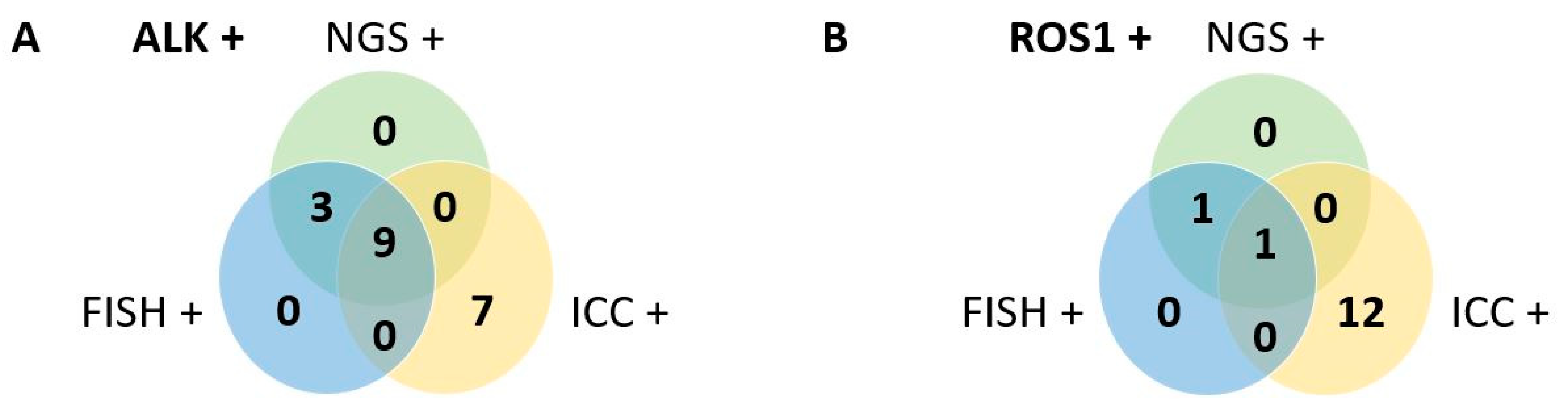
2.4. Additive Results

In addition to the panel fusion, a second NGS DNA mutation panel was systematically performed. Among 123 samples tested, at least one mutation was found in 94 samples. The most frequent was TP53 in 60 (48.8%) samples, followed by KRAS in 40 (32.5%) samples, EGFR in 19 (15.4%), BRAF in eight (6.5%), STK11 in seven (5.7%), MET in three (2.4%) samples, ERBB2 in two (1.6%) samples, as well as CTNNB1, FGFR3, PIK3CA, RET and SMAD4. Furthermore, ALK, ERBB4, PDGFRA and POLE were individually found in one sample (0.8% each). ERBB2 amplification was found in one (0.8%) sample (Figure S1A).
Three patients with ALK rearrangement presented co-occurring mutations. One patient had a mutation in BRAF and SMAD4, one had a mutation CTNNB1 and one had a mutation in TP53. The presence of both ALK rearrangement and TP53 mutation has already been described by Kron et al. [14]. Concerning the three patients with ROS1 rearrangement, two of them had a mutation in TP53. Concerning other mutations, 42 patients had a mutation in TP53 associated with another mutation, and among them, 29 patients exhibited a KRAS mutation (Figure S1B).

3. Discussion

As cytological samples are often the only samples available for advanced NSCLC diagnosis and biomarker testing, cytology research must establish protocols for ALK, ROS1 and EGFR testing [15,16]. The use of cytological samples for the identification of ALK and ROS1 rearrangements, using ICC, FISH and NGS, has been validated and is recommended by expert consensus [2,13,17,18].
In the Cell Biology Laboratory of APHM, we chose to analyze the ICC and FISH results on cytospins rather than on cell blocks. Indeed, the use of cytospins allows one to preserve cellular integrity and circumvents nuclear truncation. We performed the screening of ALK and ROS1 rearrangements by ICC. This procedure is cost-effective and easily integrated into routine diagnosis, and has a quick turnaround of results as the rearrangements are relatively rare. When the result is positive or doubtful, the FISH technique is performed to confirm, or disprove, the rearrangement.
For ALK rearrangements, the NGS found 15 positive cases among 123 tested, which represent 12.2%. This result is above those described in the literature using FISH or immunohistochemistry (IHC) [6,7]. Using ICC, we found 18 positive or doubtful results that were confirmed in 13 cases by FISH. However, in three cases, ICC technique was negative whereas EML4(13)–ALK(20) rearrangement was found by NGS. This fusion type is one of the most commonly identified and was found in 27.3 to 41% of patients presenting ALK fusion analyzed by NGS [19,20,21]. A perfect correlation was obtained between NGS and FISH for the 27 cases tested by the two techniques (13 positives and 14 negatives).
Concerning ROS1, NGS found three patients presenting a rearrangement representing 2.4%. This is in accordance with the percentage described in the literature using IHC and FISH [3,4,5]. The ICC technique detected 15 positive or doubtful cases, among which only two were confirmed by FISH. For one patient, the result was non-interpretable. In all the false-positive results obtained by ICC, an abnormal copy number was found by FISH (copy number gain on 11 cases and loss of signal in one case). The sensitivity and the specificity of ROS1 IHC compared to FISH have been calculated in several studies with a mean of 0.96 and 0.94, respectively, [2], which is in concordance with the results obtained in our study.
Most studies have compared the results obtained on histological samples between IHC and FISH [12]. The sensitivity and specificity mean of 15 studies representing 3919 samples, are at 0.97 and 0.99, respectively. Clavé et al. compared the results of ALK and ROS1 rearrangements obtained on 38 paraffin-embedded samples from non-small cell lung cancer between FISH and NGS. They found discordance in five samples. Among them, four had an isolated 3′ signal FISH pattern. This discrepancy has also been described by Liu et al., who compared results derived by IHC, FISH and NGS [22]. They found one case of an IHC false-negative result confirmed positive by FISH, but not by NGS. A recent study compared IHC, FISH and NGS to detect ALK rearrangement from formalin-fixed paraffin-embedded (FFPE) samples. They found a concordance of 75.9% between IHC and FISH and 87.5% with NGS [23]. In our study, we had strong concordance between results obtained by FISH and NGS.
The false-positive results we obtained by ICC concerned, in most cases, the positivity of both ALK and ROS1. The FISH found an increase of signals for ALK and ROS1 with at least three signals, probably corresponding to a hyperdiploidy or a copy gain number as already described for ALK [24].
NGS is a technique with a turn-around time and a cost higher than ICC and FISH but has the advantage of evaluating a panel of gene rearrangements in a single analysis. NGS can identify the partner gene concerned when a rearrangement is found, even for ALK rearrangement, the tyrosine kinase inhibitor (TKI) can be prescribed irrespective of the gene fusion partner [2]. The feasibility of using cytological samples to detect gene fusion is relatively recent. Before detecting gene fusion, Baum et al. analyzed EGFR and KRAS mutations in 30 cytological samples from lung adenocarcinoma patients, either on cell blocks or on smears. The NGS was successful in 90% when more than 100 malignant cells were present in the sample [25]. In our study, NGS was successful in 93.8% of cases. Among 108 malignant pleural effusions, Ruan et al. identified six with ALK/EML4 rearrangements and one with ROS1/CD74 rearrangement [26]. Yamamoto et al. tested 111 cytological samples (e.g., pleural effusion, cerebrospinal fluid, ascitic fluid, sputum and pericardial effusion) and obtained a result in 90% of cases. ALK was rearranged (EML4–ALK) in three of them [27]. The tumor cell quantity is a parameter that has to be considered for the management of molecular testing. We have already shown that a very small number of malignant cells can be sufficient to detect mutations in cerebrospinal fluid from meningeal carcinoma [28].
The cost of the analysis is also a parameter that must be considered. In our institution, the cost of NGS compared to ICC alone is four times higher, and when it is combined with FISH it is still two to three times higher. Schluckebier et al. evaluated the cost effectiveness of sequential testing, first for EGFR mutations by RT-PCR and then ALK and ROS1 by FISH, compared to NGS [29]. They showed that the NGS was not cost-effective, and displayed a higher probability of correct diagnoses. However, a Canadian study showed that ROS1 screening by IHC with confirmation by FISH remains less expensive than NGS [30]. NGS is a technology that requires a level of expertise that most of pathologists do not possess. A recent Spanish publication showed that IHC and FISH remain the most widely used techniques compared to NGS, with only 1 out of 44 centers using the latter [31].

4. Materials and Methods

4.1. Sample Collection

The samples included in this study were obtained from patients attending the Assistance Publique des Hôpitaux de Marseille for diagnosis and treatment. The only inclusion criteria was the availability of a cytological sample for biomarker studies. This project was approved by the local ethics committee (PADS22-31). The conventional cytological diagnosis was performed by the Cell Biology Laboratory, and the molecular testing was performed by the Cell Biology Laboratory (ROS1 and ALK by ICC and FISH) and the Oncobiology Laboratory (NGS). NGS was performed from frozen cell pellets or, alternatively, stained slides if the entire sample was used for the conventional cytological diagnosis. Results of the molecular testing and clinical data were retrospectively analyzed.

4.2. Immunocytochemistry

Samples were prepared as previously described [32], following manufacturer’s instructions. At least one wash was performed between each step. Slides were fixed with paraformaldehyde (PAF) 4% for 10 min, and then incubated with the peroxidase-blocking solution for 30 min. After being washed, slides were incubated with SensiTEK HRP Kit (ScyTek) for 10 min. Primary antibodies (ALK antibody: clone 5A4, 1/25, ab17127, Abcam; ROS1 antibody: clone D4D6, 1/250, #3287, Cell signaling) were incubated for 30 min. Then, the biotinylated secondary antibody was incubated for 15 min, followed by Streptavidin/HRP for 20 min and DAB Quanto chromogen for 5 min. Nuclei were counterstained with Mayer’s hemalun solution. Slides were mounted with Aquatex®. Negative controls (total non-immune mouse or rabbit IgG as primary antibody) and positive H2228 (ALK rearranged, Figure 1S) and HCC78 (ROS1 rearranged, Figure 1T) cell lines were added to each experiment to validate the results. Slides were observed under optical microscope (Leica, Wetzlar, Germany).

4.3. FISH

Slides were incubated for 10 min in HCl 0.1N/pepsin (0.5%) at 37 °C, and then washed in PBS. Slides were incubated in formaldehyde (1%) for 10 min, dehydrated in ethanol 70°, 90° and 100° (3 min each) and dried. Denaturation and hybridization were performed with a thermobrite (Leica Biosystems, Wetzlar, Germany). Denaturation was performed following manufacturer’s instructions: 10 min at 75 °C for ROS1 probe (zytolight SPEC ROS1 Dual-Color Break-Apart probe (PL101), zytovision, Bremerhaven, Germany), 4 min at 75 °C for ALK probe (Vysis LSI ALK Dual-Color Break-Appart, rearrangement probe, Abbott molecular, Des Plaines, IL, USA). Hybridization was performed overnight at 37 °C. Slides were washed in a buffered bath (saline–sodium citrate buffer 0.4Xat 72 °C for 2 min, then twice at room temperature for 30 s). Nuclei were stained with DAPI for 10 min at room temperature (DAPI II, Abbott, Chicago, IL, USA). Slides were mounted using FluorSave™ reagent (Merck Millipore, Burlington, USA) and observed using a fluorescence microscope (Leica, Wetzlar, Germany).

4.4. Next-Generation Sequencing

The total nucleic acids were extracted with the Maxwell RSC DNA FFPE Kit (Promega, Madison, USA) for the stained slides or with the Maxwell RSC Cell DNA Kit (Promega) for the frozen cell pellets. The pellet was resuspended in PBS solution at the rate of 1 × 10⁶ cells per 100 μL of PBS before extraction. The nucleic acids were recovered in 70 μL of elution buffer and quantified by real-time quantitative PCR for NGS analysis (screening for mutations and fusion transcripts).
The RNAs were extracted specifically from the Maxwell using the Maxwell RSC Simply RNA Blood Kit (Promega) after re-suspension in 200 µL of homogenization solution/thioglycerol and treatment with DNAse. The RNAs were eluted at 50 μL and quantified by spectrophotometry on the Nanodrop 8000 (Thermo Fisher Scientific, Waltham, MA, USA).
The RNAs (ideally 10 ng) were then reverse transcribed into complementary DNA using the SuperScript IV VILO enzyme (Thermo Fisher Scientific). The detection for rearrangements were carried out using next-generation sequencing (NGS) technology on the Ion Torrent S5XL with the Oncomine Focus RNA assay Kit (Thermo Fisher Scientific) (see Table S2 for the 5× Solid Tumor Fusion Transcript Panel). The analysis was performed with the Ion Reporter software (Thermo Fisher Scientific). A minimum of 50,000 mapped reads was required to allow interpretation of the result.
For mutation screening, we used our custom panel Oncomine Solid Tumor and Oncomine Solid Tumor+ (OST/OST+), which includes hotspots of target regions of 28 genes of interest in oncogenesis (Table S3). For all clinical samples, we performed sequencing with Ion Torrent S5XL (Thermo Fisher, Bourgoin, France) with a sensitivity of 5% for the minimum coverage of 500X. Then, sequencing data were analyzed through two pipelines. The first pipeline was developed by Thermo Fisher on the Ion Torrent Suite + Ion Reporter. Ion Torrent Suite generates FASTQ data and ensures BAM (binary alignment mapping) alignment with the hg19 reference genome by using the TMAP (Torrent Mapping Alignment Program). Ion Reporter makes variant caller and variant annotations. The second pipeline was developed in our laboratory and runs open-source software such as BWA-MEM for alignment, SAMtools for mpileup, VarScan2 as variant caller and VEP Ensemble for annotations. All data are stored in our local MySQL database.

5. Conclusions

Cytological samples are adequate for molecular testing. When using ICC combined with FISH technique, the results are very close to those of NGS for ALK and ROS1 rearrangement detection. The false-positive cases obtained by ICC are corrected by FISH and the true-positive cases are confirmed, which demonstrates high specificity and sensitivity. NGS has the potential to improve the detection of ALK and ROS1 rearrangements in cytological samples, and may become the gold standard for molecular genotyping of patients with advanced non-small cell lung cancer. However, due to the cost and the required expertise of biologists and/or bioinformaticians to use this technique, ICC and FISH are still the most commonly used techniques at the moment.

Supplementary Materials

The following supporting information can be downloaded at: https://www.mdpi.com/article/10.3390/ijms231810556/s1.

Author Contributions

L.G., H.D. and P.A. have recruited and followed patients. D.F., E.K. and P.R. have performed the cytological analysis (ICC and FISH). E.P. and N.B. have performed and analyzed the NGS results. I.N., L.O. and C.C. have interpreted the NGS results. D.F. and I.N. have collected the data. D.F. wrote the manuscript. D.F., E.K. and P.R. analyzed the data collected. All the co-authors reviewed the manuscript. All authors have read and agreed to the published version of the manuscript.

Funding

This research received no external funding.

Institutional Review Board Statement

The study was conducted in accordance with the Declaration of Helsinki, and approved by the Institutional Ethics Committee of Assistance Publique des Hôpitaux de Marseille (PADS22-31).

Informed Consent Statement

All biological results collected in this article were performed for the diagnosis and the therapeutic management.

Data Availability Statement

Not applicable.

Acknowledgments

We thank Nathalie Boitano, Corinne Derrien, Christian Brundu and Laetitia Bus for their help and continuous support.

Conflicts of Interest

The authors declare no conflict of interest.

References

  1. Cancer Today. Available online: http://gco.iarc.fr/today/home (accessed on 29 April 2022).
  2. Lindeman, N.I.; Cagle, P.T.; Aisner, D.L.; Arcila, M.E.; Beasley, M.B.; Bernicker, E.H.; Colasacco, C.; Dacic, S.; Hirsch, F.R.; Kerr, K.; et al. Updated Molecular Testing Guideline for the Selection of Lung Cancer Patients for Treatment with Targeted Tyrosine Kinase Inhibitors: Guideline From the College of American Pathologists, the International Association for the Study of Lung Cancer, and the Association for Molecular Pathology. Arch. Pathol. Lab. Med. 2018, 142, 321–346. [Google Scholar] [CrossRef]
  3. Bergethon, K.; Shaw, A.T.; Ou, S.-H.I.; Katayama, R.; Lovly, C.M.; McDonald, N.T.; Massion, P.P.; Siwak-Tapp, C.; Gonzalez, A.; Fang, R.; et al. ROS1 Rearrangements Define a Unique Molecular Class of Lung Cancers. J. Clin. Oncol. 2012, 30, 863–870. [Google Scholar] [CrossRef]
  4. Cai, W.; Li, X.; Su, C.; Fan, L.; Zheng, L.; Fei, K.; Zhou, C.; Manegold, C.; Schmid-Bindert, G. ROS1 Fusions in Chinese Patients with Non-Small-Cell Lung Cancer. Ann. Oncol. 2013, 24, 1822–1827. [Google Scholar] [CrossRef]
  5. Chen, Y.-F.; Hsieh, M.-S.; Wu, S.-G.; Chang, Y.-L.; Shih, J.-Y.; Liu, Y.-N.; Tsai, M.-F.; Tsai, T.-H.; Yu, C.-J.; Yang, J.C.-H.; et al. Clinical and the Prognostic Characteristics of Lung Adenocarcinoma Patients with ROS1 Fusion in Comparison with Other Driver Mutations in East Asian Populations. J. Thorac. Oncol. 2014, 9, 1171–1179. [Google Scholar] [CrossRef]
  6. Rodig, S.J.; Mino-Kenudson, M.; Dacic, S.; Yeap, B.Y.; Shaw, A.; Barletta, J.A.; Stubbs, H.; Law, K.; Lindeman, N.; Mark, E.; et al. Unique Clinicopathologic Features Characterize ALK-Rearranged Lung Adenocarcinoma in the Western Population. Clin. Cancer Res. 2009, 15, 5216–5223. [Google Scholar] [CrossRef]
  7. Soda, M.; Choi, Y.L.; Enomoto, M.; Takada, S.; Yamashita, Y.; Ishikawa, S.; Fujiwara, S.; Watanabe, H.; Kurashina, K.; Hatanaka, H.; et al. Identification of the Transforming EML4-ALK Fusion Gene in Non-Small-Cell Lung Cancer. Nature 2007, 448, 561–566. [Google Scholar] [CrossRef]
  8. Bozzetti, C.; Nizzoli, R.; Tiseo, M.; Squadrilli, A.; Lagrasta, C.; Buti, S.; Gasparro, D.; Zanoni, D.; Majori, M.; De Filippo, M.; et al. ALK and ROS1 Rearrangements Tested by Fluorescence in Situ Hybridization in Cytological Smears from Advanced Non-Small Cell Lung Cancer Patients. Diagn. Cytopathol. 2015, 43, 941–946. [Google Scholar] [CrossRef]
  9. Rogers, T.-M.; Russell, P.A.; Wright, G.; Wainer, Z.; Pang, J.-M.; Henricksen, L.A.; Singh, S.; Stanislaw, S.; Grille, J.; Roberts, E.; et al. Comparison of Methods in the Detection of ALK and ROS1 Rearrangements in Lung Cancer. J. Thorac. Oncol. 2015, 10, 611–618. [Google Scholar] [CrossRef]
  10. Marchetti, A.; Di Lorito, A.; Pace, M.V.; Iezzi, M.; Felicioni, L.; D’Antuono, T.; Filice, G.; Guetti, L.; Mucilli, F.; Buttitta, F. ALK Protein Analysis by IHC Staining after Recent Regulatory Changes: A Comparison of Two Widely Used Approaches, Revision of the Literature, and a New Testing Algorithm. J. Thorac. Oncol. 2016, 11, 487–495. [Google Scholar] [CrossRef]
  11. Mosele, F.; Remon, J.; Mateo, J.; Westphalen, C.B.; Barlesi, F.; Lolkema, M.P.; Normanno, N.; Scarpa, A.; Robson, M.; Meric-Bernstam, F.; et al. Recommendations for the Use of Next-Generation Sequencing (NGS) for Patients with Metastatic Cancers: A Report from the ESMO Precision Medicine Working Group. Ann. Oncol. 2020, 31, 1491–1505. [Google Scholar] [CrossRef]
  12. Pisapia, P.; Lozano, M.D.; Vigliar, E.; Bellevicine, C.; Pepe, F.; Malapelle, U.; Troncone, G. ALK and ROS1 Testing on Lung Cancer Cytologic Samples: Perspectives. Cancer Cytopathol. 2017, 125, 817–830. [Google Scholar] [CrossRef] [PubMed]
  13. Frankel, D.; Bourlard, D.; Garcia, S.; Robaglia-Schlupp, A.; Peker, E.; Groliere, A.; Kaspi, E.; Roll, P. Mise en évidence du réarrangement d’ALK et ROS1 en immunocytochimie sur liquides de ponction. Ann. Pathol. 2019, 39, 227–236. [Google Scholar] [CrossRef] [PubMed]
  14. Kron, A.; Alidousty, C.; Scheffler, M.; Merkelbach-Bruse, S.; Seidel, D.; Riedel, R.; Ihle, M.A.; Michels, S.; Nogova, L.; Fassunke, J.; et al. Impact of TP53 Mutation Status on Systemic Treatment Outcome in ALK-Rearranged Non-Small-Cell Lung Cancer. Ann. Oncol. 2018, 29, 2068–2075. [Google Scholar] [CrossRef] [PubMed]
  15. Roy-Chowdhuri, S.; Stewart, J. Preanalytic Variables in Cytology: Lessons Learned From Next-Generation Sequencing-The MD Anderson Experience. Arch. Pathol. Lab. Med. 2016, 140, 1191–1199. [Google Scholar] [CrossRef] [PubMed]
  16. Hagiwara, K.; Kobayashi, K. Importance of the Cytological Samples for the Epidermal Growth Factor Receptor Gene Mutation Test for Non-Small Cell Lung Cancer. Cancer Sci. 2013, 104, 291–297. [Google Scholar] [CrossRef]
  17. Velizheva, N.P.; Rechsteiner, M.P.; Wong, C.E.; Zhong, Q.; Rössle, M.; Bode, B.; Moch, H.; Soltermann, A.; Wild, P.J.; Tischler, V. Cytology Smears as Excellent Starting Material for Next-Generation Sequencing-Based Molecular Testing of Patients with Adenocarcinoma of the Lung. Cancer Cytopathol. 2017, 125, 30–40. [Google Scholar] [CrossRef] [PubMed]
  18. Conde, E.; Rojo, F.; Gómez, J.; Enguita, A.B.; Abdulkader, I.; González, A.; Lozano, D.; Mancheño, N.; Salas, C.; Salido, M.; et al. Molecular Diagnosis in Non-Small-Cell Lung Cancer: Expert Opinion on ALK and ROS1 Testing. J. Clin. Pathol. 2022, 75, 145–153. [Google Scholar] [CrossRef]
  19. McCoach, C.E.; Le, A.T.; Gowan, K.; Jones, K.; Schubert, L.; Doak, A.; Estrada-Bernal, A.; Davies, K.D.; Merrick, D.T.; Bunn, P.A.; et al. Resistance Mechanisms to Targeted Therapies in ROS1+ and ALK+ Non-Small Cell Lung Cancer. Clin. Cancer Res. 2018, 24, 3334–3347. [Google Scholar] [CrossRef] [PubMed]
  20. Zhao, R.; Zhang, J.; Han, Y.; Shao, J.; Zhu, L.; Xiang, C.; Zhang, Q.; Teng, H.; Qin, G.; Zhao, L.; et al. Clinicopathological Features of ALK Expression in 9889 Cases of Non-Small-Cell Lung Cancer and Genomic Rearrangements Identified by Capture-Based Next-Generation Sequencing: A Chinese Retrospective Analysis. Mol. Diagn. Ther. 2019, 23, 395–405. [Google Scholar] [CrossRef]
  21. Sabir, S.R.; Yeoh, S.; Jackson, G.; Bayliss, R. EML4-ALK Variants: Biological and Molecular Properties, and the Implications for Patients. Cancers 2017, 9, 118. [Google Scholar] [CrossRef] [Green Version]
  22. Liu, Y.; Wu, S.; Shi, X.; Liang, Z.; Zeng, X. ALK Detection in Lung Cancer: Identification of Atypical and Cryptic ALK Rearrangements Using an Optimal Algorithm. J. Cancer Res. Clin. Oncol. 2020, 146, 1307–1320. [Google Scholar] [CrossRef] [PubMed]
  23. Batra, U.; Nathany, S.; Sharma, M.; Pasricha, S.; Bansal, A.; Jain, P.; Mehta, A. IHC versus FISH versus NGS to Detect ALK Gene Rearrangement in NSCLC: All Questions Answered? J. Clin. Pathol. 2022, 75, 405–409. [Google Scholar] [CrossRef] [PubMed]
  24. Salido, M.; Pijuan, L.; Martínez-Avilés, L.; Galván, A.B.; Cañadas, I.; Rovira, A.; Zanui, M.; Martínez, A.; Longarón, R.; Sole, F.; et al. Increased ALK Gene Copy Number and Amplification Are Frequent in Non-Small Cell Lung Cancer. J. Thorac. Oncol. 2011, 6, 21–27. [Google Scholar] [CrossRef] [PubMed]
  25. Baum, J.E.; Zhang, P.; Hoda, R.S.; Geraghty, B.; Rennert, H.; Narula, N.; Fernandes, H.D. Accuracy of Next-Generation Sequencing for the Identification of Clinically Relevant Variants in Cytology Smears in Lung Adenocarcinoma. Cancer Cytopathol. 2017, 125, 398–406. [Google Scholar] [CrossRef] [PubMed]
  26. Ruan, X.; Sun, Y.; Wang, W.; Ye, J.; Zhang, D.; Gong, Z.; Yang, M. Multiplexed Molecular Profiling of Lung Cancer with Malignant Pleural Effusion Using next Generation Sequencing in Chinese Patients. Oncol. Lett. 2020, 19, 3495–3505. [Google Scholar] [CrossRef]
  27. Yamamoto, G.; Kikuchi, M.; Kobayashi, S.; Arai, Y.; Fujiyoshi, K.; Wakatsuki, T.; Kakuta, M.; Yamane, Y.; Iijima, Y.; Mizutani, H.; et al. Routine Genetic Testing of Lung Cancer Specimens Derived from Surgery, Bronchoscopy and Fluid Aspiration by next Generation Sequencing. Int. J. Oncol. 2017, 50, 1579–1589. [Google Scholar] [CrossRef] [PubMed]
  28. Frankel, D.; Nanni-Metellus, I.; Robaglia-Schlupp, A.; Tomasini, P.; Guinde, J.; Barlesi, F.; Astoul, P.; Ouafik, L.; Amatore, F.; Secq, V.; et al. Detection of EGFR, KRAS and BRAF Mutations in Metastatic Cells from Cerebrospinal Fluid. Clin. Chem. Lab. Med. 2018, 56, 748–753. [Google Scholar] [CrossRef]
  29. Schluckebier, L.; Caetano, R.; Garay, O.U.; Montenegro, G.T.; Custodio, M.; Aran, V.; Gil Ferreira, C. Cost-Effectiveness Analysis Comparing Companion Diagnostic Tests for EGFR, ALK, and ROS1 versus next-Generation Sequencing (NGS) in Advanced Adenocarcinoma Lung Cancer Patients. BMC Cancer 2020, 20, 875. [Google Scholar] [CrossRef]
  30. Makarem, M.; Ezeife, D.A.; Smith, A.C.; Li, J.J.N.; Law, J.H.; Tsao, M.-S.; Leighl, N.B. Reflex ROS1 IHC Screening with FISH Confirmation for Advanced Non-Small Cell Lung Cancer-A Cost-Efficient Strategy in a Public Healthcare System. Curr. Oncol. 2021, 28, 3268–3279. [Google Scholar] [CrossRef]
  31. Martín-López, J.; Rojo, F.; Martínez-Pozo, A.; Hernández-Iglesias, T.; Carcedo, D.; de Alda, L.R.; García, J.F.; Salas, C. Biomarker Testing Strategies in Non-Small Cell Lung Cancer in the Real-World Setting: Analysis of Methods in the Prospective Central Lung Cancer Biomarker Registry (LungPath) from the Spanish Society of Pathology (SEAP). J. Clin. Pathol. 2021, 13, 208034. [Google Scholar] [CrossRef]
  32. Frankel, D.; Kaspi, E.; Roll, P. Immunocytochemical Detection of ALK and ROS1 Rearrangements in Lung Cancer Cytological Samples. Methods Mol. Biol. 2021, 2279, 157–164. [Google Scholar] [CrossRef] [PubMed]
Figure 1. Example of malignant cells with or without ALK and ROS1 rearrangements. (AD) Represent the cytology of pleural effusion (P1) containing ALK rearranged malignant cells. (A) Papanicolaou stain, (B) May–Grünwald–Giemsa (MGG) stain, (C) immunocytochemistry (ICC) (peroxidase staining) using antibody against ALK (clone 5A4), (D) ALK FISH probe. (EH) Represent the cytology of pleural effusion (P2) containing ROS1 rearranged malignant cells. (E) Papanicolaou stain, (F) May–Grünwald–Giemsa stain, (G) immunocytochemistry (peroxidase staining) using antibody against ROS1 (clone D4D6), (D) ROS1 FISH probe. (IL) Represent the cytology of a lymph node (P3) containing ALK rearranged malignant cells. (I) Papanicolaou stain, (J) May–Grünwald–Giemsa stain, (K) immunocytochemistry (peroxidase staining) using antibody against ALK (clone 5A4), (L) ALK FISH probe. Of note, the false-negative result was obtained with ICC, whereas the rearrangement is observed with FISH. (MR) Represent the cytology of pleural effusion (P4) containing malignant cells without ALK and ROS1 rearrangements. (M) Papanicolaou stain, (N) May–Grünwald–Giemsa stain, (O) immunocytochemistry (peroxidase staining) using antibody against ALK (clone 5A4), (P) ALK FISH probe. (Q) Immunocytochemistry using antibody against ROS1 (clone D4D6), (R) ROS1 FISH probe. Of note, the false-positive results were obtained with ICC using antibodies against ALK and ROS1, whereas the FISH shows more than two signals without rearrangement using ALK and ROS1 probes. (S) H2228 cell line showing positive staining against anti-ALK antibody (positive control, C+). (T) HCC78 cell line showing a positive staining against anti-ROS1 antibody (positive control, C+). Black scale bar represents 20 µm. White scale bar represents 10 µm.
Ijms 23 10556 g001
Figure 2. A Venn diagram of ALK-positive (A) and ROS1-positive (B) cases detected by NGS, ICC and FISH in cytological samples. Four patients and one patient were excluded for ALK or ROS1 results, respectively, because of non-interpretable results in one of the methods.
Ijms 23 10556 g002
Table 1. Patient demographics.
Parametern (%)
Age (years)Mean ± SD67.4 ± 12
Range36–90
GenderMale73 (55.7)
Female58 (44.3)
Histopathological typeLung adenocarcinoma105 (80.2)
NSCLC NOS26 (19.8)
Smoking statusNever33 (25.2)
Current/former92 (70.2)
Unknown6 (4.6)
StageI2 (1.5)
II5 (3.8)
III21 (16.0)
IV100 (76.3)
Unknown3 (2.3)
Table 2. Sensitivity and specificity of ICC and FISH compared with NGS. FN, false-negative; FP, false-positive; TN, true-negative; TP, true-positive; Se: sensitivity; Spe: specificity.
TechniqueALKROS1
TPFPFNTNSeSpeTPFPFNTNSeSpe
ICC1173690.790.9131207810.87
FISH130014112001311
Publisher’s Note: MDPI stays neutral with regard to jurisdictional claims in published maps and institutional affiliations.

