
Toxicologic Concerns with Current Medical Nanoparticles

 , , ,
, , , 
Abstract
:1. Background
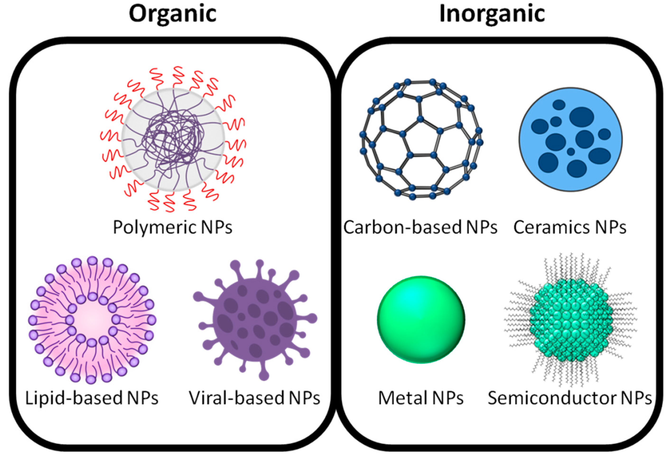
2. Sources of Nanoparticles

| Type | Formation and Compositions | Applications | References |
|---|---|---|---|
| Carbon-based NPs | Fullerenes Carbon nanotubes (CNTs) | Carbon nanotubes are used widely in biomedical applications because of their multipurpose properties. They have been applied for carrying anticancer drugs or genes and proteins for chemotherapy. | [24,25,26,27] |
| Metal NPs | Alkali and noble metals such as Cu, Ag, and Au. | Noble metal-based NPs are applied in medical fields that needed high biocompatibility, stability, and large-scale production with the possibility of avoiding organic solvents. | [28,29,30,31] |
| Ceramics NPs | They are amorphous, polycrystalline, dense, porous, or hollow forms. | Medical technologies use nanoceramics for bone repair. In addition, they have been used in catalysis, photocatalysis, photodegradation of dyes, and imaging applications. | [32] |
| Semiconductor NPs | They possess properties between metals and nonmetals. Semiconductor NPs used in biosensing generally contain metals with nonmetallic elements. | Photocatalysis, photo optics, and electronic devices. | [33] |
| Polymeric NPs | They normally are organic-based nanospheres or nanocapsules primarily. The former are matrix particles that are generally solid, and other molecules are adsorbed to the outer boundary of the sphere. Nanocapsules are completely encapsulated mass particles. | Polymers with superior biocompatibility do not induce immune reactions or stimulate inflammation in contact with the human body. The advantages of synthetic polymers are their stability, excellent mechanical properties, and degradability. Polymers are biocompatible, biodegradable, non-toxic, and popular in medical applications such as drug delivery, wound plug dressings, stents, and tissue engineering. | [34,35,36,37,38] |
| Lipid-based NPs | Lipid-based NPs classified as lipid moieties including liposomes, solid lipid nanoparticles (SLN), and nanostructured lipid carriers (NLC). | Lipid-based NPs are used effectively in biomedical applications. They are used for various applications such as drug carriers, delivery, and RNA release in cancer therapy and COVID-19 vaccines. | [16,39,40] |
| Viral-NPs | Genetically engineered VNPs and chemically engineered VNPs. | Viral NPs serve as multipurpose tools for medical applications. Genetically engineered VNPs are used as vaccines. Chemically engineered VNPs are for targeted drug delivery and biomedical imaging. | [11,41] |
3. Routes of Nanoparticle Uptake to the Human Body
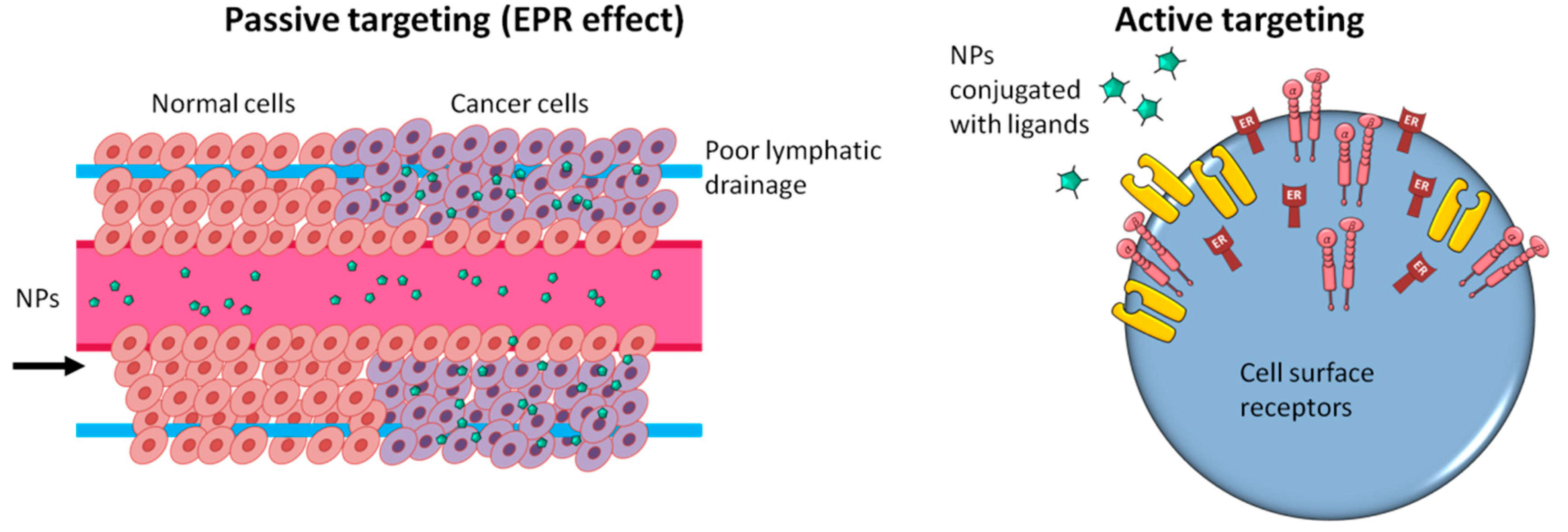
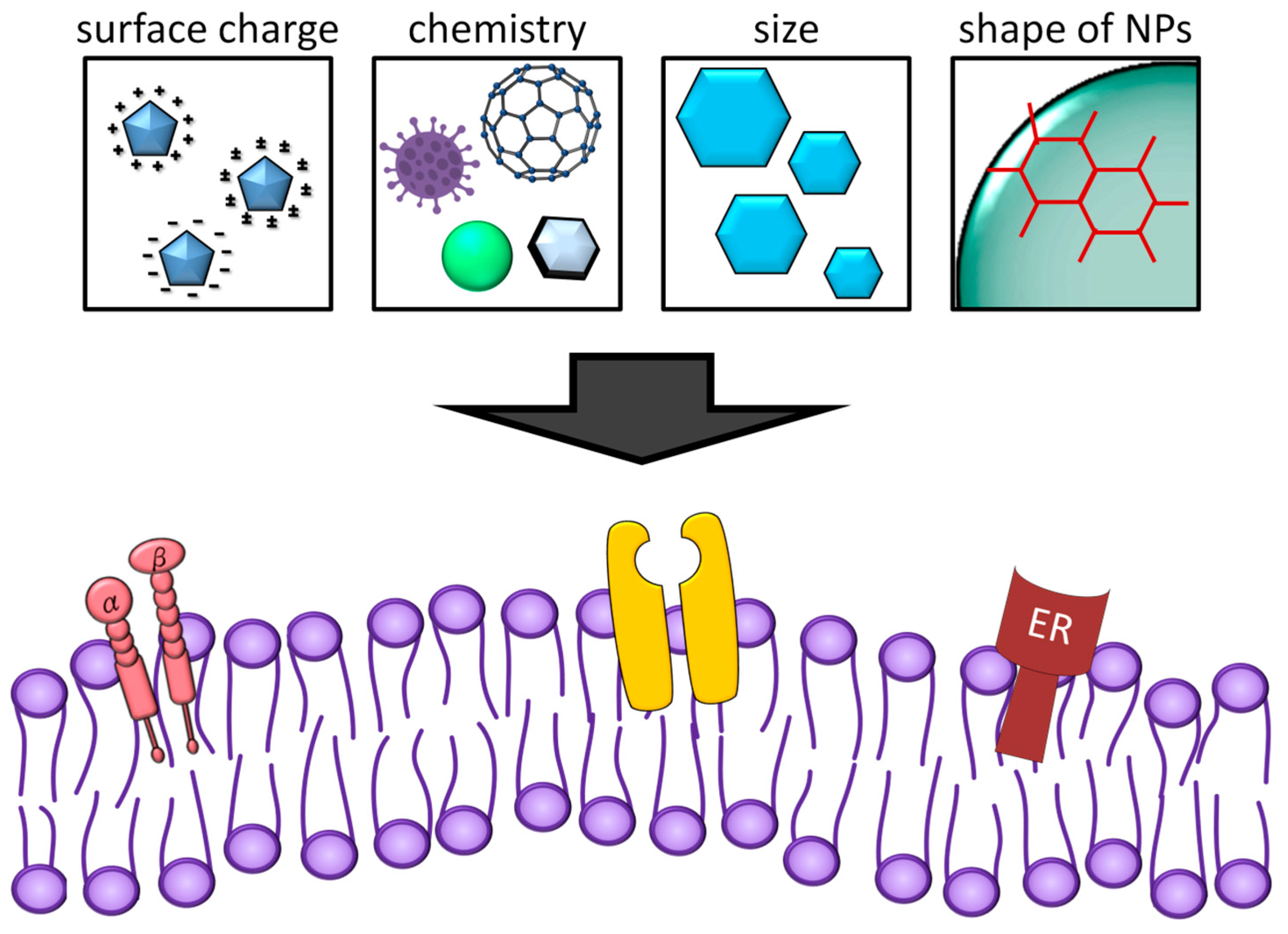
4. Therapeutic Nano-Delivery Systems
5. Nanotoxicology
5.1. Damage to Cells Caused by Nanoparticles
5.2. Effect of Nanoparticles in Different Organs
5.2.1. Nanoparticles on Skin
5.2.2. Nanoparticles in Brain
5.2.3. Nanoparticles in Eye
5.2.4. Nanoparticles in Lung
5.2.5. Nanoparticles in Liver
5.2.6. Nanoparticles in Kidney
5.2.7. Nanoparticles in Reproductive System
5.2.8. Nanoparticles in the Immune System
6. Conclusions Remark
Author Contributions
Funding
Institutional Review Board Statement
Informed Consent Statement
Data Availability Statement
Conflicts of Interest
References
- Yingchoncharoen, P.; Kalinowski, D.S.; Richardson, D.R. Lipid-based drug delivery systems in cancer therapy: What is available and what is yet to come. Pharmacol. Rev. 2016, 68, 701–787. [Google Scholar] [CrossRef] [PubMed] [Green Version]
- Zielińska, A.; Costa, B.; Ferreira, M.V.; Miguéis, D.; Louros, J.; Durazzo, A.; Lucarini, M.; Eder, P.; V Chaud, M.; Morsink, M. Nanotoxicology and nanosafety: Safety-by-design and testing at a glance. Int. J. Environ. Res. Public Health 2020, 17, 4657. [Google Scholar] [CrossRef] [PubMed]
- Carro, C.E.; Pilozzi, A.R.; Huang, X. Nanoneurotoxicity and potential nanotheranostics for Alzheimer’s disease. EC Pharmacol. Toxicol. 2019, 7, 1. [Google Scholar] [PubMed]
- Stone, V.; Miller, M.R.; Clift, M.J.; Elder, A.; Mills, N.L.; Møller, P.; Schins, R.P.; Vogel, U.; Kreyling, W.G.; Alstrup Jensen, K. Nanomaterials versus ambient ultrafine particles: An opportunity to exchange toxicology knowledge. Environ. Health Perspect. 2017, 125, 106002. [Google Scholar] [CrossRef]
- Raftis, J.B.; Miller, M.R. Nanoparticle translocation and multi-organ toxicity: A particularly small problem. Nano Today 2019, 26, 8–12. [Google Scholar] [CrossRef]
- Bhatnagar, A. Environmental determinants of cardiovascular disease. Circ. Res. 2017, 121, 162–180. [Google Scholar] [CrossRef]
- Perez, L.; Wolf, K.; Hennig, F.; Penell, J.; Basagaña, X.; Foraster, M.; Aguilera, I.; Agis, D.; Beelen, R.; Brunekreef, B. Air pollution and atherosclerosis: A cross-sectional analysis of four European cohort studies in the ESCAPE study. Environ. Health Perspect. 2015, 123, 597–605. [Google Scholar] [CrossRef] [Green Version]
- Spurgeon, D.; Lahive, E.; Robinson, A.; Short, S.; Kille, P. Species sensitivity to toxic substances: Evolution, ecology and applications. Front. Environ. Sci. 2020, 8, 588380. [Google Scholar] [CrossRef]
- Sarma, A.; Bania, R.; Devi, J.R.; Deka, S. Therapeutic nanostructures and nanotoxicity. J. Appl. Toxicol. 2021, 41, 1494–1517. [Google Scholar] [CrossRef]
- Jeevanandam, J.; Barhoum, A.; Chan, Y.S.; Dufresne, A.; Danquah, M.K. Review on nanoparticles and nanostructured materials: History, sources, toxicity and regulations. Beilstein J. Nanotechnol. 2018, 9, 1050–1074. [Google Scholar] [CrossRef] [Green Version]
- Khan, I.; Saeed, K.; Khan, I. Nanoparticles: Properties, applications and toxicities. Arab. J. Chem. 2019, 12, 908–931. [Google Scholar] [CrossRef]
- Wen, A.M.; Steinmetz, N.F. Design of virus-based nanomaterials for medicine, biotechnology, and energy. Chem. Soc. Rev. 2016, 45, 4074–4126. [Google Scholar] [CrossRef] [PubMed] [Green Version]
- Daniyal, M.; Liu, B.; Wang, W. Comprehensive review on graphene oxide for use in drug delivery system. Curr. Med. Chem. 2020, 27, 3665–3685. [Google Scholar] [CrossRef] [PubMed]
- Castro-Rojas, M.A.; Vega-Cantu, Y.I.; Cordell, G.A.; Rodriguez-Garcia, A. Dental applications of carbon nanotubes. Molecules 2021, 26, 4423. [Google Scholar] [CrossRef]
- Khan, S.; Jawlikar, P.; Lahoti, S.; Bhusnure, O.; Chitlange, S.; Sangshetti, J. Application of Carbon Nanotubes In Drug Delivery of Non-cancerous Diseases: A Review. Curr. Pharm. Des. 2021, 27, 2454–2467. [Google Scholar] [CrossRef]
- Wani, T.U.; Mohi-Ud-Din, R.; Wani, T.A.; Mir, R.H.; Itoo, A.M.; Sheikh, F.A.; Khan, N.A.; Pottoo, F.H. Green synthesis, spectroscopic characterization and biomedical applications of carbon nanotubes. Curr. Pharm. Biotechnol. 2021, 22, 793–807. [Google Scholar] [CrossRef]
- García-Pinel, B.; Porras-Alcalá, C.; Ortega-Rodríguez, A.; Sarabia, F.; Prados, J.; Melguizo, C.; López-Romero, J.M. Lipid-based nanoparticles: Application and recent advances in cancer treatment. Nanomaterials 2019, 9, 638. [Google Scholar] [CrossRef] [Green Version]
- Hu, X.; Zhang, Y.; Ding, T.; Liu, J.; Zhao, H. Multifunctional gold nanoparticles: A novel nanomaterial for various medical applications and biological activities. Front. Bioeng. Biotechnol. 2020, 8, 990. [Google Scholar] [CrossRef]
- Sukhanova, A.; Bozrova, S.; Sokolov, P.; Berestovoy, M.; Karaulov, A.; Nabiev, I. Dependence of nanoparticle toxicity on their physical and chemical properties. Nanoscale Res. Lett. 2018, 13, 44. [Google Scholar] [CrossRef] [Green Version]
- Mitchell, M.J.; Billingsley, M.M.; Haley, R.M.; Wechsler, M.E.; Peppas, N.A.; Langer, R. Engineering precision nanoparticles for drug delivery. Nat. Rev. Drug Discov. 2021, 20, 101–124. [Google Scholar] [CrossRef]
- Dubaj, T.; Kozics, K.; Sramkova, M.; Manova, A.; Bastús, N.G.; Moriones, O.H.; Kohl, Y.; Dusinska, M.; Runden-Pran, E.; Puntes, V. Pharmacokinetics of PEGylated Gold Nanoparticles: In Vitro—In Vivo Correlation. Nanomaterials 2022, 12, 511. [Google Scholar] [CrossRef] [PubMed]
- Amiri, M.; Imanzadeh, H.; Sefid-Sefidehkhan, Y. An Overview on Electrochemical Sensors Based on Nanomaterials for the Determination of Drugs of Abuse. Curr. Drug Deliv. 2021, 18, 162–183. [Google Scholar] [CrossRef] [PubMed]
- Sabourian, P.; Yazdani, G.; Ashraf, S.S.; Frounchi, M.; Mashayekhan, S.; Kiani, S.; Kakkar, A. Effect of physico-chemical properties of nanoparticles on their intracellular uptake. Int. J. Mol. Sci. 2020, 21, 8019. [Google Scholar] [CrossRef] [PubMed]
- Hoshyar, N.; Gray, S.; Han, H.; Bao, G. The effect of nanoparticle size on in vivo pharmacokinetics and cellular interaction. Nanomedicine 2016, 11, 673–692. [Google Scholar] [CrossRef] [Green Version]
- Adeli, M.; Soleyman, R.; Beiranvand, Z.; Madani, F. Carbon nanotubes in cancer therapy: A more precise look at the role of carbon nanotube–polymer interactions. Chem. Soc. Rev. 2013, 42, 5231–5256. [Google Scholar] [CrossRef] [PubMed]
- Amin, S.A.; Adhikari, N.; Jha, T. Development of decision trees to discriminate HDAC8 inhibitors and non-inhibitors using recursive partitioning. J. Biomol. Struct. Dyn. 2021, 39, 1–8. [Google Scholar] [CrossRef] [PubMed]
- Amenta, V.; Aschberger, K. Carbon nanotubes: Potential medical applications and safety concerns. Wiley Interdiscip. Rev. Nanomed. Nanobiotechnology 2015, 7, 371–386. [Google Scholar] [CrossRef]
- Hwang, Y.; Park, S.-H.; Lee, J.W. Applications of functionalized carbon nanotubes for the therapy and diagnosis of cancer. Polymers 2017, 9, 13. [Google Scholar] [CrossRef] [Green Version]
- Chatterjee, K.; Sarkar, S.; Rao, K.J.; Paria, S. Core/shell nanoparticles in biomedical applications. Adv. Colloid Interface Sci. 2014, 209, 8–39. [Google Scholar] [CrossRef]
- Chelnokov, E.; Cuba, V.; Simeone, D.; Guigner, J.-M.; Schmidhammer, U.; Mostafavi, M.; Le Caër, S. Electron transfer at oxide/water interfaces induced by ionizing radiation. J. Phys. Chem. C 2014, 118, 7865–7873. [Google Scholar] [CrossRef]
- Hamblin, M.R.; Chiang, L.Y.; Lakshmanan, S.; Huang, Y.-Y.; Garcia-Diaz, M.; Karimi, M.; de Souza Rastelli, A.N.; Chandran, R. Nanotechnology for photodynamic therapy: A perspective from the laboratory of Dr. Michael R. Hamblin in the Wellman Center for Photomedicine at Massachusetts General Hospital and Harvard Medical School. Nanotechnol. Rev. 2015, 4, 359–372. [Google Scholar] [CrossRef] [PubMed]
- Klębowski, B.; Depciuch, J.; Parlińska-Wojtan, M.; Baran, J. Applications of noble metal-based nanoparticles in medicine. Int. J. Mol. Sci. 2018, 19, 4031. [Google Scholar] [CrossRef] [PubMed] [Green Version]
- Kaushik, S. Polymeric and Ceramic Nanoparticles: Possible Role in Biomedical Applications. In Handbook of Polymer and Ceramic Nanotechnology; Springer: Cham, Switzerland, 2020. [Google Scholar]
- Massadeh, S.; Alaamery, M. Biological applications of Semiconductor Nanoparticles. In Nanomedicine; Seifalian, A., Ed.; One Central Press: Manchester, UK, 2014; pp. 357–373. [Google Scholar]
- Mansha, M.; Khan, I.; Ullah, N.; Qurashi, A. Synthesis, characterization and visible-light-driven photoelectrochemical hydrogen evolution reaction of carbazole-containing conjugated polymers. Int. J. Hydrogen Energy 2017, 42, 10952–10961. [Google Scholar] [CrossRef]
- Abd Ellah, N.H.; Abouelmagd, S.A. Surface functionalization of polymeric nanoparticles for tumor drug delivery: Approaches and challenges. Expert Opin. Drug Deliv. 2017, 14, 201–214. [Google Scholar] [CrossRef] [PubMed]
- Abouelmagd, S.A.; Meng, F.; Kim, B.-K.; Hyun, H.; Yeo, Y. Tannic acid-mediated surface functionalization of polymeric nanoparticles. ACS Biomater. Sci. Eng. 2016, 2, 2294–2303. [Google Scholar] [CrossRef] [Green Version]
- Wang, X.; Feng, J.; Bai, Y.; Zhang, Q.; Yin, Y. Synthesis, properties, and applications of hollow micro-/nanostructures. Chem. Rev. 2016, 116, 10983–11060. [Google Scholar] [CrossRef]
- Mir, M.; Ahmed, N.; ur Rehman, A. Recent applications of PLGA based nanostructures in drug delivery. Colloids Surf. B Biointerfaces 2017, 159, 217–231. [Google Scholar] [CrossRef]
- Guevara, M.L.; Persano, F.; Persano, S. Advances in lipid nanoparticles for mRNA-based cancer immunotherapy. Front. Chem. 2020, 8, 589959. [Google Scholar] [CrossRef]
- Thi, T.T.H.; Suys, E.J.; Lee, J.S.; Nguyen, D.H.; Park, K.D.; Truong, N.P. Lipid-based nanoparticles in the clinic and clinical trials: From cancer nanomedicine to COVID-19 vaccines. Vaccines 2021, 9, 359. [Google Scholar] [CrossRef]
- Chen, C.C.; Baikoghli, M.A.; Cheng, R.H. Tissue targeted nanocapsids for oral insulin delivery via drink. Pharm. Pat. Anal. 2018, 7, 121–127. [Google Scholar] [CrossRef]
- Ajdary, M.; Moosavi, M.A.; Rahmati, M.; Falahati, M.; Mahboubi, M.; Mandegary, A.; Jangjoo, S.; Mohammadinejad, R.; Varma, R.S. Health concerns of various nanoparticles: A review of their in vitro and in vivo toxicity. Nanomaterials 2018, 8, 634. [Google Scholar] [CrossRef] [PubMed] [Green Version]
- Kong, F.-Y.; Zhang, J.-W.; Li, R.-F.; Wang, Z.-X.; Wang, W.-J.; Wang, W. Unique roles of gold nanoparticles in drug delivery, targeting and imaging applications. Molecules 2017, 22, 1445. [Google Scholar] [CrossRef] [PubMed] [Green Version]
- Conte, R.; Marturano, V.; Peluso, G.; Calarco, A.; Cerruti, P. Recent advances in nanoparticle-mediated delivery of anti-inflammatory phytocompounds. Int. J. Mol. Sci. 2017, 18, 709. [Google Scholar] [CrossRef] [PubMed] [Green Version]
- Ganesan, P.; Ramalingam, P.; Karthivashan, G.; Ko, Y.T.; Choi, D.-K. Recent developments in solid lipid nanoparticle and surface-modified solid lipid nanoparticle delivery systems for oral delivery of phyto-bioactive compounds in various chronic diseases. Int. J. Nanomed. 2018, 13, 1569. [Google Scholar] [CrossRef] [Green Version]
- Bayer, I.S. Hyaluronic acid and controlled release: A review. Molecules 2020, 25, 2649. [Google Scholar] [CrossRef]
- Trombino, S.; Servidio, C.; Curcio, F.; Cassano, R. Strategies for hyaluronic acid-based hydrogel design in drug delivery. Pharmaceutics 2019, 11, 407. [Google Scholar] [CrossRef] [Green Version]
- Chang, Y.-L.; Liao, P.-B.; Wu, P.-H.; Chang, W.-J.; Lee, S.-Y.; Huang, H.-M. Cancer Cytotoxicity of a Hybrid Hyaluronan-Superparamagnetic Iron Oxide Nanoparticle Material: An In-Vitro Evaluation. Nanomaterials 2022, 12, 496. [Google Scholar] [CrossRef]
- Gupta, R.; Xie, H. Nanoparticles in daily life: Applications, toxicity and regulations. J. Environ. Pathol. Toxicol. Oncol. 2018, 37, 209–230. [Google Scholar] [CrossRef]
- Rizvi, S.A.; Saleh, A.M. Applications of nanoparticle systems in drug delivery technology. Saudi Pharm. J. 2018, 26, 64–70. [Google Scholar] [CrossRef]
- Yah, C.S.; Iyuke, S.E.; Simate, G.S. A review of nanoparticles toxicity and their routes of exposures. Iran. J. Pharm. Sci. 2012, 8, 299–314. [Google Scholar]
- Araujo, J.A.; Nel, A.E. Particulate matter and atherosclerosis: Role of particle size, composition and oxidative stress. Part. Fibre Toxicol. 2009, 6, 24. [Google Scholar] [CrossRef] [PubMed] [Green Version]
- Weinberg, H.; Galyean, A.; Leopold, M. Evaluating engineered nanoparticles in natural waters. TrAC Trends Anal. Chem. 2011, 30, 72–83. [Google Scholar] [CrossRef]
- Lewinski, N.; Colvin, V.; Drezek, R. Cytotoxicity of nanoparticles. Small 2008, 4, 26–49. [Google Scholar] [CrossRef]
- Sani, A.; Cao, C.; Cui, D. Toxicity of gold nanoparticles (AuNPs): A review. Biochem. Biophys. Rep. 2021, 26, 100991. [Google Scholar] [CrossRef] [PubMed]
- Kuschner, W.G.; Wong, H.; D’Alessandro, A.; Quinlan, P.; Blanc, P.D. Human pulmonary responses to experimental inhalation of high concentration fine and ultrafine magnesium oxide particles. Environ. Health Perspect. 1997, 105, 1234–1237. [Google Scholar] [CrossRef]
- Stanta, G.; Bonin, S. Overview on clinical relevance of intra-tumor heterogeneity. Front. Med. 2018, 5, 85. [Google Scholar] [CrossRef] [Green Version]
- Chakravarthi, B.V.; Nepal, S.; Varambally, S. Genomic and epigenomic alterations in cancer. Am. J. Pathol. 2016, 186, 1724–1735. [Google Scholar] [CrossRef] [Green Version]
- Crintea, A.; Dutu, A.G.; Samasca, G.; Florian, I.A.; Lupan, I.; Craciun, A.M. The nanosystems involved in treating lung cancer. Life 2021, 11, 682. [Google Scholar] [CrossRef]
- Wang, H.; Mu, X.; He, H.; Zhang, X.-D. Cancer radiosensitizers. Trends Pharmacol. Sci. 2018, 39, 24–48. [Google Scholar] [CrossRef]
- Gadducci, A.; Cosio, S. Neoadjuvant chemotherapy in locally advanced cervical cancer: Review of the literature and perspectives of clinical research. Anticancer. Res. 2020, 40, 4819–4828. [Google Scholar] [CrossRef]
- Yang, Y.-C.S.; Ko, P.-J.; Pan, Y.-S.; Lin, H.-Y.; Whang-Peng, J.; Davis, P.J.; Wang, K. Role of thyroid hormone-integrin αvβ3-signal and therapeutic strategies in colorectal cancers. J. Biomed. Sci. 2021, 28, 24. [Google Scholar] [CrossRef] [PubMed]
- Cheng, T.-M.; Chang, W.-J.; Chu, H.-Y.; De Luca, R.; Pedersen, J.Z.; Incerpi, S.; Li, Z.-L.; Shih, Y.-J.; Lin, H.-Y.; Wang, K. Nano-strategies targeting the integrin αvβ3 network for cancer therapy. Cells 2021, 10, 1684. [Google Scholar] [CrossRef]
- Narvekar, M.; Xue, H.Y.; Eoh, J.Y.; Wong, H.L. Nanocarrier for poorly water-soluble anticancer drugs—barriers of translation and solutions. AAPS PharmSciTech 2014, 15, 822–833. [Google Scholar] [CrossRef] [PubMed]
- Loftsson, T.; Brewster, M.E. Pharmaceutical applications of cyclodextrins: Basic science and product development. J. Pharm. Pharmacol. 2010, 62, 1607–1621. [Google Scholar] [CrossRef] [PubMed]
- Ismael, G.F.; Rosa, D.D.; Mano, M.S.; Awada, A. Novel cytotoxic drugs: Old challenges, new solutions. Cancer Treat. Rev. 2008, 34, 81–91. [Google Scholar] [CrossRef]
- Asad, A.S.; Moreno Ayala, M.A.; Gottardo, M.F.; Zuccato, C.; Nicola Candia, A.J.; Zanetti, F.A.; Seilicovich, A.; Candolfi, M. Viral gene therapy for breast cancer: Progress and challenges. Expert Opin. Biol. Ther. 2017, 17, 945–959. [Google Scholar] [CrossRef]
- Hromic-Jahjefendic, A.; Lundstrom, K. Viral vector-based melanoma gene therapy. Biomedicines 2020, 8, 60. [Google Scholar] [CrossRef] [Green Version]
- Dixit, K.; Kumthekar, P. Gene delivery in neuro-oncology. Curr. Oncol. Rep. 2017, 19, 69. [Google Scholar] [CrossRef]
- Santana-Armas, M.L.; de Ilarduya, C.T. Strategies for cancer gene-delivery improvement by non-viral vectors. Int. J. Pharm. 2021, 596, 120291. [Google Scholar] [CrossRef]
- Negri, V.; Pacheco-Torres, J.; Calle, D.; López-Larrubia, P. Carbon nanotubes in biomedicine. In Surface-Modified Nanobiomaterials for Electrochemical and Biomedicine Applications; Springer Nature: Cham, Switzerland, 2020; pp. 177–217. [Google Scholar]
- Lin, L.; Wong, H. Predicting oral drug absorption: Mini review on physiologically-based pharmacokinetic models. Pharmaceutics 2017, 9, 41. [Google Scholar] [CrossRef] [Green Version]
- Zamay, T.N.; Zamay, G.S.; Kolovskaya, O.S.; Zukov, R.A.; Petrova, M.M.; Gargaun, A.; Berezovski, M.V.; Kichkailo, A.S. Current and prospective protein biomarkers of lung cancer. Cancers 2017, 9, 155. [Google Scholar] [CrossRef] [PubMed] [Green Version]
- Boldrin, A.; Hansen, S.F.; Baun, A.; Hartmann, N.I.B.; Astrup, T.F. Environmental exposure assessment framework for nanoparticles in solid waste. J. Nanopart. Res. 2014, 16, 2394. [Google Scholar] [CrossRef] [PubMed] [Green Version]
- Wang, X.; Jin, N.; Wang, Q.; Liu, T.; Liu, K.; Li, Y.; Bai, Y.; Chen, X. MiRNA delivery system based on stimuli-responsive gold nanoparticle aggregates for multimodal tumor therapy. ACS Appl. Bio Mater. 2019, 2, 2833–2839. [Google Scholar] [CrossRef] [PubMed]
- Albanese, A.; Tang, P.S.; Chan, W.C. The effect of nanoparticle size, shape, and surface chemistry on biological systems. Annu. Rev. Biomed. Eng. 2012, 14, 1–16. [Google Scholar] [CrossRef] [Green Version]
- Shin, S.W.; Song, I.H.; Um, S.H. Role of physicochemical properties in nanoparticle toxicity. Nanomaterials 2015, 5, 1351–1365. [Google Scholar] [CrossRef] [Green Version]
- Slavin, Y.N.; Asnis, J.; Häfeli, U.O.; Bach, H. Metal nanoparticles: Understanding the mechanisms behind antibacterial activity. J. Nanobiotechnol. 2017, 15, 65. [Google Scholar] [CrossRef]
- Oberdörster, G.; Castranova, V.; Asgharian, B.; Sayre, P. Inhalation exposure to carbon nanotubes (CNT) and carbon nanofibers (CNF): Methodology and dosimetry. J. Toxicol. Environ. Health Part B 2015, 18, 121–212. [Google Scholar] [CrossRef] [Green Version]
- Pahuja, R.; Seth, K.; Shukla, A.; Shukla, R.K.; Bhatnagar, P.; Chauhan, L.K.S.; Saxena, P.N.; Arun, J.; Chaudhari, B.P.; Patel, D.K. Trans-blood brain barrier delivery of dopamine-loaded nanoparticles reverses functional deficits in parkinsonian rats. ACS Nano 2015, 9, 4850–4871. [Google Scholar] [CrossRef]
- Grady, M.E.; Parrish, E.; Caporizzo, M.A.; Seeger, S.C.; Composto, R.J.; Eckmann, D.M. Intracellular nanoparticle dynamics affected by cytoskeletal integrity. Soft Matter 2017, 13, 1873–1880. [Google Scholar] [CrossRef] [Green Version]
- Gustafson, H.H.; Holt-Casper, D.; Grainger, D.W.; Ghandehari, H. Nanoparticle uptake: The phagocyte problem. Nano Today 2015, 10, 487–510. [Google Scholar] [CrossRef] [Green Version]
- Baranov, M.V.; Kumar, M.; Sacanna, S.; Thutupalli, S.; Van den Bogaart, G. Modulation of immune responses by particle size and shape. Front. Immunol. 2021, 11, 607945. [Google Scholar] [CrossRef] [PubMed]
- Zhou, J.; Wang, M.; Ying, H.; Su, D.; Zhang, H.; Lu, G.; Chen, J. Extracellular matrix component shelled nanoparticles as dual enzyme-responsive drug delivery vehicles for cancer therapy. ACS Biomater. Sci. Eng. 2018, 4, 2404–2411. [Google Scholar] [CrossRef] [PubMed]
- Wei, F.; Neal, C.J.; Sakthivel, T.S.; Seal, S.; Kean, T.; Razavi, M.; Coathup, M. Cerium oxide nanoparticles protect against irradiation-induced cellular damage while augmenting osteogenesis. Mater. Sci. Eng. C 2021, 126, 112145. [Google Scholar] [CrossRef] [PubMed]
- Yong, J.M.; Fu, L.; Tang, F.; Yu, P.; Kuchel, R.P.; Whitelock, J.M.; Lord, M.S. ROS-Mediated Anti-Angiogenic Activity of Cerium Oxide Nanoparticles in Melanoma Cells. ACS Biomater. Sci. Eng. 2022, 8, 512–525. [Google Scholar] [CrossRef] [PubMed]
- Nqakala, Z.B.; Sibuyi, N.R.; Fadaka, A.O.; Meyer, M.; Onani, M.O.; Madiehe, A.M. Advances in Nanotechnology towards Development of Silver Nanoparticle-Based Wound-Healing Agents. Int. J. Mol. Sci. 2021, 22, 11272. [Google Scholar] [CrossRef]
- Zhao, C.-Y.; Cheng, R.; Yang, Z.; Tian, Z.-M. Nanotechnology for cancer therapy based on chemotherapy. Molecules 2018, 23, 826. [Google Scholar] [CrossRef] [Green Version]
- Walker, L.C. Aβ plaques. Free. Neuropathol. 2020, 1, 31. [Google Scholar]
- Iqbal, K.; Liu, F.; Gong, C.-X.; Alonso, A.d.C.; Grundke-Iqbal, I. Mechanisms of tau-induced neurodegeneration. Acta Neuropathol. 2009, 118, 53–69. [Google Scholar] [CrossRef] [Green Version]
- Dugger, B.N.; Dickson, D.W. Pathology of neurodegenerative diseases. Cold Spring Harb. Perspect. Biol. 2017, 9, a028035. [Google Scholar] [CrossRef] [Green Version]
- Amanzadeh, E.; Esmaeili, A.; Rahgozar, S.; Nourbakhshnia, M. Application of quercetin in neurological disorders: From nutrition to nanomedicine. Rev. Neurosci. 2019, 30, 555–572. [Google Scholar] [CrossRef]
- Holbrook, J.A.; Jarosz-Griffiths, H.H.; Caseley, E.; Lara-Reyna, S.; Poulter, J.A.; Williams-Gray, C.H.; Peckham, D.; McDermott, M.F. Neurodegenerative disease and the NLRP3 inflammasome. Front. Pharmacol. 2021, 12, 193. [Google Scholar] [CrossRef] [PubMed]
- Horie, M.; Kato, H.; Fujita, K.; Endoh, S.; Iwahashi, H. In vitro evaluation of cellular response induced by manufactured nanoparticles. Chem. Res. Toxicol. 2012, 25, 605–619. [Google Scholar] [CrossRef] [PubMed]
- Elder, A.; Gelein, R.; Silva, V.; Feikert, T.; Opanashuk, L.; Carter, J.; Potter, R.; Maynard, A.; Ito, Y.; Finkelstein, J. Translocation of inhaled ultrafine manganese oxide particles to the central nervous system. Environ. Health Perspect. 2006, 114, 1172–1178. [Google Scholar] [CrossRef]
- Borisova, T. Nervous system injury in response to contact with environmental, engineered and planetary micro-and nano-sized particles. Front. Physiol. 2018, 9, 728. [Google Scholar] [CrossRef]
- Bahadar, H.; Maqbool, F.; Niaz, K.; Abdollahi, M. Toxicity of nanoparticles and an overview of current experimental models. Iran. Biomed. J. 2016, 20, 1. [Google Scholar]
- Attarilar, S.; Yang, J.; Ebrahimi, M.; Wang, Q.; Liu, J.; Tang, Y.; Yang, J. The toxicity phenomenon and the related occurrence in metal and metal oxide nanoparticles: A brief review from the biomedical perspective. Front. Bioeng. Biotechnol. 2020, 8, 822. [Google Scholar] [CrossRef] [PubMed]
- Jia, J.; Zhang, Y.; Xin, Y.; Jiang, C.; Yan, B.; Zhai, S. Interactions between nanoparticles and dendritic cells: From the perspective of cancer immunotherapy. Front. Oncol. 2018, 8, 404. [Google Scholar] [CrossRef]
- Heitbrink, W.A.; Lo, L.-M.; Dunn, K.H. Exposure controls for nanomaterials at three manufacturing sites. J. Occup. Environ. Hyg. 2015, 12, 16–28. [Google Scholar] [CrossRef] [Green Version]
- Yang, H.; Liu, C.; Yang, D.; Zhang, H.; Xi, Z. Comparative study of cytotoxicity, oxidative stress and genotoxicity induced by four typical nanomaterials: The role of particle size, shape and composition. J. Appl. Toxicol. 2009, 29, 69–78. [Google Scholar] [CrossRef]
- Wang, L.; Ai, W.; Zhai, Y.; Li, H.; Zhou, K.; Chen, H. Effects of nano-CeO2 with different nanocrystal morphologies on cytotoxicity in HepG2 cells. Int. J. Environ. Res. Public Health 2015, 12, 10806–10819. [Google Scholar] [CrossRef] [Green Version]
- Orest, V.; Leclerc, L.; Hochepied, J.-F.; Trouvé, A.; Sarry, G.; Pourchez, J. Impact of cerium oxide nanoparticles shape on their in vitro cellular toxicity. Toxicol. In Vitro 2017, 38, 136–141. [Google Scholar] [CrossRef] [PubMed] [Green Version]
- Dekkers, S.; Miller, M.R.; Schins, R.P.; Römer, I.; Russ, M.; Vandebriel, R.J.; Lynch, I.; Belinga-Desaunay, M.-F.; Valsami-Jones, E.; Connell, S.P. The effect of zirconium doping of cerium dioxide nanoparticles on pulmonary and cardiovascular toxicity and biodistribution in mice after inhalation. Nanotoxicology 2017, 11, 794–808. [Google Scholar] [CrossRef] [PubMed]
- Khanal, D.; Kondyurin, A.; Hau, H.; Knowles, J.C.; Levinson, O.; Ramzan, I.; Fu, D.; Marcott, C.; Chrzanowski, W. Biospectroscopy of nanodiamond-induced alterations in conformation of intra-and extracellular proteins: A nanoscale IR study. Anal. Chem. 2016, 88, 7530–7538. [Google Scholar] [CrossRef] [PubMed] [Green Version]
- Nel, A.E.; Mädler, L.; Velegol, D.; Xia, T.; Hoek, E.; Somasundaran, P.; Klaessig, F.; Castranova, V.; Thompson, M. Understanding biophysicochemical interactions at the nano–bio interface. Nat. Mater. 2009, 8, 543–557. [Google Scholar] [CrossRef] [PubMed]
- Martin, A.L.; Bernas, L.M.; Rutt, B.K.; Foster, P.J.; Gillies, E.R. Enhanced cell uptake of superparamagnetic iron oxide nanoparticles functionalized with dendritic guanidines. Bioconjugate Chem. 2008, 19, 2375–2384. [Google Scholar] [CrossRef]
- Augustine, R.; Hasan, A.; Primavera, R.; Wilson, R.J.; Thakor, A.S.; Kevadiya, B.D. Cellular uptake and retention of nanoparticles: Insights on particle properties and interaction with cellular components. Mater. Today Commun. 2020, 25, 101692. [Google Scholar] [CrossRef]
- Anastasiadis, S.H.; Chrissopoulou, K.; Stratakis, E.; Kavatzikidou, P.; Kaklamani, G.; Ranella, A. How the Physicochemical Properties of Manufactured Nanomaterials Affect Their Performance in Dispersion and Their Applications in Biomedicine: A Review. Nanomaterials 2022, 12, 552. [Google Scholar] [CrossRef]
- Jaurand, M.-C.F.; Renier, A.; Daubriac, J. Mesothelioma: Do asbestos and carbon nanotubes pose the same health risk? Part. Fibre Toxicol. 2009, 6, 16. [Google Scholar] [CrossRef] [Green Version]
- Boyles, M.S.; Young, L.; Brown, D.M.; MacCalman, L.; Cowie, H.; Moisala, A.; Smail, F.; Smith, P.J.; Proudfoot, L.; Windle, A.H. Multi-walled carbon nanotube induced frustrated phagocytosis, cytotoxicity and pro-inflammatory conditions in macrophages are length dependent and greater than that of asbestos. Toxicol. In Vitro 2015, 29, 1513–1528. [Google Scholar] [CrossRef]
- Westphal, G.A.; Rosenkranz, N.; Brik, A.; Weber, D.; Föhring, I.; Monsé, C.; Kaiser, N.; Hellack, B.; Mattenklott, M.; Brüning, T. Multi-walled carbon nanotubes induce stronger migration of inflammatory cells in vitro than asbestos or granular particles but a similar pattern of inflammatory mediators. Toxicol. In Vitro 2019, 58, 215–223. [Google Scholar] [CrossRef]
- Yanagisawa, H.; Seki, Y.; Yogosawa, S.; Takumi, S.; Shimizu, H.; Suka, M. Potential role of mitochondrial damage and S9 mixture including metabolic enzymes in ZnO nanoparticles-induced oxidative stress and genotoxicity in Chinese hamster lung (CHL/IU) cells. Mutat. Res./Genet. Toxicol. Environ. Mutagenesis 2018, 834, 25–34. [Google Scholar] [CrossRef] [PubMed]
- Roberta, P.; Elena Maria, S.; Carmelo, I.; Fabiano, C.; Maria Teresa, R.; Sara, I.; Antonio, S.; Roberto, F.; Giuliana, I.; Maria Violetta, B. Toxicological assessment of CeO2 nanoparticles on early development of zebrafish. Toxicol. Res. 2021, 10, 570–578. [Google Scholar] [CrossRef] [PubMed]
- Ernst, L.M.; Casals, E.; Italiani, P.; Boraschi, D.; Puntes, V. The Interactions between Nanoparticles and the Innate Immune System from a Nanotechnologist Perspective. Nanomaterials 2021, 11, 2991. [Google Scholar] [CrossRef] [PubMed]
- Qie, Y.; Yuan, H.; Von Roemeling, C.A.; Chen, Y.; Liu, X.; Shih, K.D.; Knight, J.A.; Tun, H.W.; Wharen, R.E.; Jiang, W. Surface modification of nanoparticles enables selective evasion of phagocytic clearance by distinct macrophage phenotypes. Sci. Rep. 2016, 6, 26269. [Google Scholar] [CrossRef] [Green Version]
- Yoo, C.-J.; Lee, U.; Kim, Y.-J.; Park, J.; Yoo, Y.-M. Dose-dependent cytotoxicity of gold nanoparticles on human neural progenitor cells and rat brain. J. Nanosci. Nanotechnol. 2019, 19, 5441–5447. [Google Scholar] [CrossRef]
- Shin, T.H.; Lee, D.Y.; Manavalan, B.; Basith, S.; Na, Y.-C.; Yoon, C.; Lee, H.-S.; Paik, M.J.; Lee, G. Silica-coated magnetic nanoparticles activate microglia and induce neurotoxic d-serine secretion. Part. Fibre Toxicol. 2021, 18, 30. [Google Scholar] [CrossRef]
- Samak, D.H.; El-Sayed, Y.S.; Shaheen, H.M.; Ali, H.; Onoda, A.; Abdel-Daim, M.M.; Umezawa, M. In-ovo exposed carbon black nanoparticles altered mRNA gene transcripts of antioxidants, proinflammatory and apoptotic pathways in the brain of chicken embryos. Chem.-Biol. Interact. 2018, 295, 133–139. [Google Scholar] [CrossRef]
- Wang, Z.; Zhang, C.; Huang, F.; Liu, X.; Wang, Z.; Yan, B. Breakthrough of ZrO2 nanoparticles into fetal brains depends on developmental stage of maternal placental barrier and fetal blood-brain-barrier. J. Hazard. Mater. 2021, 402, 123563. [Google Scholar] [CrossRef]
- Diao, J.; Xia, Y.; Jiang, X.; Qiu, J.; Cheng, S.; Su, J.; Duan, X.; Gao, M.; Qin, X.; Zhang, J. Silicon dioxide nanoparticles induced neurobehavioral impairments by disrupting microbiota–gut–brain axis. J. Nanobiotechnol. 2021, 19, 174. [Google Scholar] [CrossRef]
- Chen, J.; Zhang, S.; Chen, C.; Jiang, X.; Qiu, J.; Qiu, Y.; Zhang, Y.; Wang, T.; Qin, X.; Zou, Z. Crosstalk of gut microbiota and serum/hippocampus metabolites in neurobehavioral impairments induced by zinc oxide nanoparticles. Nanoscale 2020, 12, 21429–21439. [Google Scholar] [CrossRef]
- Zhang, S.; Jiang, X.; Cheng, S.; Fan, J.; Qin, X.; Wang, T.; Zhang, Y.; Zhang, J.; Qiu, Y.; Qiu, J. Titanium dioxide nanoparticles via oral exposure leads to adverse disturbance of gut microecology and locomotor activity in adult mice. Arch. Toxicol. 2020, 94, 1173–1190. [Google Scholar] [CrossRef] [PubMed]
- Su, J.; Duan, X.; Qiu, Y.; Zhou, L.; Zhang, H.; Gao, M.; Liu, Y.; Zou, Z.; Qiu, J.; Chen, C. Pregnancy exposure of titanium dioxide nanoparticles causes intestinal dysbiosis and neurobehavioral impairments that are not significant postnatally but emerge in adulthood of offspring. J. Nanobiotechnol. 2021, 19, 234. [Google Scholar] [CrossRef] [PubMed]
- Adeyemi, O.S.; Uloko, R.A.; Awakan, O.J.; Adeyanju, A.A.; Otohinoyi, D.A. The oral administration of silver nanoparticles activates the kynurenine pathway in rat brain independently of oxidative stress. Chem.-Biol. Interact. 2019, 302, 22–27. [Google Scholar] [CrossRef] [PubMed] [Green Version]
- Roda, E.; Bottone, M.; Biggiogera, M.; Milanesi, G.; Coccini, T. Pulmonary and hepatic effects after low dose exposure to nanosilver: Early and long-lasting histological and ultrastructural alterations in rat. Toxicol. Rep. 2019, 6, 1047–1060. [Google Scholar] [CrossRef]
- Rosário, F.; Hoet, P.; Nogueira, A.J.A.; Santos, C.; Oliveira, H. Differential pulmonary in vitro toxicity of two small-sized polyvinylpyrrolidone-coated silver nanoparticles. J. Toxicol. Environ. Health Part A 2018, 81, 675–690. [Google Scholar] [CrossRef]
- Durantie, E.; Vanhecke, D.; Rodriguez-Lorenzo, L.; Delhaes, F.; Balog, S.; Septiadi, D.; Bourquin, J.; Petri-Fink, A.; Rothen-Rutishauser, B. Biodistribution of single and aggregated gold nanoparticles exposed to the human lung epithelial tissue barrier at the air-liquid interface. Part. Fibre Toxicol. 2017, 14, 49. [Google Scholar] [CrossRef] [Green Version]
- Li, L.; Tian, J.; Wang, X.; Xu, G.; Jiang, W.; Yang, Z.; Liu, D.; Lin, G. Cardiotoxicity of intravenously administered CdSe/ZnS quantum dots in BALB/c mice. Front. Pharmacol. 2019, 10, 1179. [Google Scholar] [CrossRef]
- Nirmal, N.K.; Awasthi, K.K.; John, P.J. Hepatotoxicity of graphene oxide in Wistar rats. Environ. Sci. Pollut. Res. 2021, 28, 46367–46376. [Google Scholar] [CrossRef]
- Bahamonde, J.; Brenseke, B.; Chan, M.Y.; Kent, R.D.; Vikesland, P.J.; Prater, M.R. Gold nanoparticle toxicity in mice and rats: Species differences. Toxicol. Pathol. 2018, 46, 431–443. [Google Scholar] [CrossRef]
- Albrahim, T.; Alonazi, M.A. Role of beetroot (Beta vulgaris) juice on chronic nanotoxicity of silver nanoparticle-induced hepatotoxicity in male rats. Int. J. Nanomed. 2020, 15, 3471. [Google Scholar] [CrossRef]
- Al-Doaiss, A.A.; Jarrar, Q.; Alshehri, M.; Jarrar, B. In vivo study of silver nanomaterials’ toxicity with respect to size. Toxicol. Ind. Health 2020, 36, 540–557. [Google Scholar] [CrossRef] [PubMed]
- Lei, R.; Wu, C.; Yang, B.; Ma, H.; Shi, C.; Wang, Q.; Wang, Q.; Yuan, Y.; Liao, M. Integrated metabolomic analysis of the nano-sized copper particle-induced hepatotoxicity and nephrotoxicity in rats: A rapid in vivo screening method for nanotoxicity. Toxicol. Appl. Pharmacol. 2008, 232, 292–301. [Google Scholar] [CrossRef] [PubMed]
- Fahmy, H.M.; Abd El-Daim, T.M.; Ali, O.A.; Hassan, A.A.; Mohammed, F.F.; Fathy, M.M. Surface modifications affect iron oxide nanoparticles’ biodistribution after multiple-dose administration in rats. J. Biochem. Mol. Toxicol. 2021, 35, e22671. [Google Scholar] [CrossRef] [PubMed]
- Vassal, M.; Rebelo, S.; Pereira, M.D.L. Metal oxide nanoparticles: Evidence of adverse effects on the male reproductive system. Int. J. Mol. Sci. 2021, 22, 8061. [Google Scholar] [CrossRef]
- Deb, S.; Patra, H.K.; Lahiri, P.; Dasgupta, A.K.; Chakrabarti, K.; Chaudhuri, U. Multistability in platelets and their response to gold nanoparticles. Nanomed. Nanotechnol. Biol. Med. 2011, 7, 376–384. [Google Scholar] [CrossRef] [PubMed]
- Bai, G.; Wu, C.; Jin, J.; Yan, M. Structural, electron transportation and magnetic behavior transition of metastable FeAlO granular films. Sci. Rep. 2016, 6, 24410. [Google Scholar] [CrossRef] [Green Version]
- Cai, X.; Lee, A.; Ji, Z.; Huang, C.; Chang, C.H.; Wang, X.; Liao, Y.-P.; Xia, T.; Li, R. Reduction of pulmonary toxicity of metal oxide nanoparticles by phosphonate-based surface passivation. Part. Fibre Toxicol. 2017, 14, 13. [Google Scholar] [CrossRef] [Green Version]
- Li, X.; Yang, H.; Wu, S.; Meng, Q.; Sun, H.; Lu, R.; Cui, J.; Zheng, Y.; Chen, W.; Zhang, R. Suppression of PTPN6 exacerbates aluminum oxide nanoparticle-induced COPD-like lesions in mice through activation of STAT pathway. Part. Fibre Toxicol. 2017, 14, 53. [Google Scholar] [CrossRef] [Green Version]
- Casals, E.; Zeng, M.; Parra-Robert, M.; Fernández-Varo, G.; Morales-Ruiz, M.; Jiménez, W.; Puntes, V.; Casals, G. Cerium oxide nanoparticles: Advances in biodistribution, toxicity, and preclinical exploration. Small 2020, 16, 1907322. [Google Scholar] [CrossRef]
- Gagnon, J.; Fromm, K.M. Toxicity and protective effects of cerium oxide nanoparticles (nanoceria) depending on their preparation method, particle size, cell type, and exposure route. Eur. J. Inorg. Chem. 2015, 2015, 4510–4517. [Google Scholar] [CrossRef] [Green Version]
- Zhao, H.; Cheng, J.; Cai, J.; Cheng, Z.; Cui, Y.; Gao, G.; Hu, R.; Gong, X.; Wang, L.; Hong, F. Liver injury and its molecular mechanisms in mice caused by exposure to cerium chloride. Arch. Environ. Contam. Toxicol. 2012, 62, 154–164. [Google Scholar] [CrossRef] [PubMed]
- Collins, A.; Nasir, A. Nanotechnology and dermatology: Benefits and pitfalls. G. Ital. Dermatol. Venereol. Organo Uff. Soc. Ital. Dermatol. Sifilogr. 2011, 146, 115–126. [Google Scholar]
- Lademann, J.; Richter, H.; Schanzer, S.; Knorr, F.; Meinke, M.; Sterry, W.; Patzelt, A. Penetration and storage of particles in human skin: Perspectives and safety aspects. Eur. J. Pharm. Biopharm. 2011, 77, 465–468. [Google Scholar] [CrossRef]
- Sguizzato, M.; Esposito, E.; Cortesi, R. Lipid-based nanosystems as a tool to overcome skin barrier. Int. J. Mol. Sci. 2021, 22, 8319. [Google Scholar] [CrossRef] [PubMed]
- Silva, F.A.; Costa-Almeida, R.; Timochenco, L.; Amaral, S.I.; Pinto, S.; Gonçalves, I.C.; Fernandes, J.R.; Magalhães, F.D.; Sarmento, B.; Pinto, A.M. Graphene Oxide Topical Administration: Skin Permeability Studies. Materials 2021, 14, 2810. [Google Scholar] [CrossRef]
- Kim, S.-H.; Lee, D.H.; Choi, S.; Yang, J.-Y.; Jung, K.; Jeong, J.; Oh, J.H.; Lee, J.H. Skin Sensitization Potential and Cellular ROS-Induced Cytotoxicity of Silica Nanoparticles. Nanomaterials 2021, 11, 2140. [Google Scholar] [CrossRef]
- Jankovskaja, S.; Labrousse, A.; Prévaud, L.; Holmqvist, B.; Brinte, A.; Engblom, J.; Rezeli, M.; Marko-Varga, G.; Ruzgas, T. Visualisation of H2O2 penetration through skin indicates importance to develop pathway-specific epidermal sensing. Microchim. Acta 2020, 187, 656. [Google Scholar] [CrossRef]
- Lee, D.-H.; Choi, S.-Y.; Jung, K.-K.; Yang, J.-Y.; Jeong, J.-y.; Oh, J.-H.; Kim, S.-H.; Lee, J.-H. The Research of Toxicity and Sensitization Potential of PEGylated Silver and Gold Nanomaterials. Toxics 2021, 9, 355. [Google Scholar] [CrossRef]
- Huang, Y.-C.; Yang, Y.-C.; Yang, K.-C.; Shieh, H.-R.; Wang, T.-Y.; Hwu, Y.; Chen, Y.-J. Pegylated gold nanoparticles induce apoptosis in human chronic myeloid leukemia cells. BioMed Res. Int. 2014, 2014, 182353. [Google Scholar] [CrossRef] [Green Version]
- Ceña, V.; Játiva, P. Nanoparticle crossing of blood–brain barrier: A road to new therapeutic approaches to central nervous system diseases. Nanomedicine 2018, 13, 1513–1516. [Google Scholar] [CrossRef] [Green Version]
- Gonzalez-Carter, D.; Liu, X.; Tockary, T.A.; Dirisala, A.; Toh, K.; Anraku, Y.; Kataoka, K. Targeting nanoparticles to the brain by exploiting the blood–brain barrier impermeability to selectively label the brain endothelium. Proc. Natl. Acad. Sci. USA 2020, 117, 19141–19150. [Google Scholar] [CrossRef] [PubMed]
- Chang, X.; Li, J.; Niu, S.; Xue, Y.; Tang, M. Neurotoxicity of metal-containing nanoparticles and implications in glial cells. J. Appl. Toxicol. 2021, 41, 65–81. [Google Scholar] [CrossRef] [PubMed]
- Mabrouk, M.; Ibrahim Fouad, G.; El-Sayed, S.A.; Rizk, M.Z.; Beherei, H.H. Hepatotoxic and Neurotoxic Potential of Iron Oxide Nanoparticles in Wistar Rats: A Biochemical and Ultrastructural Study. Biol. Trace Elem. Res. 2021, 200, 3638–3665. [Google Scholar] [CrossRef] [PubMed]
- Ako-Adounvo, A.-M.; C Nagarwal, R.; Oliveira, L.; HS Boddu, S.; S Wang, X.; Dey, S.; K Karla, P. Recent patents on ophthalmic nanoformulations and therapeutic implications. Recent Pat. Drug Deliv. Formul. 2014, 8, 193–201. [Google Scholar] [CrossRef] [Green Version]
- Chen, X.; Zhu, S.; Hu, X.; Sun, D.; Yang, J.; Yang, C.; Wu, W.; Li, Y.; Gu, X.; Li, M. Toxicity and mechanism of mesoporous silica nanoparticles in eyes. Nanoscale 2020, 12, 13637–13653. [Google Scholar] [CrossRef]
- Quan, J.-H.; Gao, F.F.; Ismail, H.A.H.A.; Yuk, J.-M.; Cha, G.-H.; Chu, J.-Q.; Lee, Y.-H. Silver nanoparticle-induced apoptosis in ARPE-19 cells is inhibited by Toxoplasma gondii pre-infection through suppression of NOX4-dependent ROS generation. Int. J. Nanomed. 2020, 15, 3695. [Google Scholar] [CrossRef]
- Sun, J.-G.; Jiang, Q.; Zhang, X.-P.; Shan, K.; Liu, B.-H.; Zhao, C.; Yan, B. Mesoporous silica nanoparticles as a delivery system for improving antiangiogenic therapy. Int. J. Nanomed. 2019, 14, 1489. [Google Scholar] [CrossRef] [Green Version]
- Mehravi, B.; Alizadeh, A.M.; Khodayari, S.; Khodayari, H.; Ashtari, K.; Mohseni, M.; Anaraki, N.I.; Dana, E.A.; Safari, S.; Amanlou, M. Acute toxicity evaluation of glycosylated Gd3+-based silica nanoprobe. Mol. Imaging Biol. 2017, 19, 522–530. [Google Scholar] [CrossRef]
- Chen, Y.; Hu, X.; Sun, J.; Zhou, Q. Specific nanotoxicity of graphene oxide during zebrafish embryogenesis. Nanotoxicology 2016, 10, 42–52. [Google Scholar] [CrossRef]
- De Jesus Ferreira, D.; de Paula Lana, R.; de Moura Zanine, A.; Santos, E.M.; Veloso, C.M.; Ribeiro, G.A. Silage fermentation and chemical composition of elephant grass inoculated with rumen strains of Streptococcus bovis. Anim. Feed. Sci. Technol. 2013, 183, 22–28. [Google Scholar] [CrossRef]
- Cui, L.; Wang, X.; Sun, B.; Xia, T.; Hu, S. Predictive metabolomic signatures for safety assessment of metal oxide nanoparticles. ACS Nano 2019, 13, 13065–13082. [Google Scholar] [CrossRef] [PubMed]
- Yang, Y.-F.; Wang, W.-M.; Chen, C.-Y.; Lu, T.-H.; Liao, C.-M. Assessing human exposure risk and lung disease burden posed by airborne silver nanoparticles emitted by consumer spray products. Int. J. Nanomed. 2019, 14, 1687. [Google Scholar] [CrossRef] [PubMed]
- Yang, L.; Kuang, H.; Zhang, W.; Aguilar, Z.P.; Wei, H.; Xu, H. Comparisons of the biodistribution and toxicological examinations after repeated intravenous administration of silver and gold nanoparticles in mice. Sci. Rep. 2017, 7, 9110. [Google Scholar] [CrossRef] [PubMed]
- Alqahtani, S.; Xia, L.; Jannasch, A.; Ferreira, C.; Franco, J.; Shannahan, J.H. Disruption of pulmonary resolution mediators contribute to exacerbated silver nanoparticle-induced acute inflammation in a metabolic syndrome mouse model. Toxicol. Appl. Pharmacol. 2021, 431, 115730. [Google Scholar] [CrossRef] [PubMed]
- Gambelunghe, A.; Giovagnoli, S.; Di Michele, A.; Boncompagni, S.; Dell’Omo, M.; Leopold, K.; Iavicoli, I.; Talesa, V.N.; Antognelli, C. Redox-Sensitive Glyoxalase 1 Up-Regulation Is Crucial for Protecting Human Lung Cells from Gold Nanoparticles Toxicity. Antioxidants 2020, 9, 697. [Google Scholar] [CrossRef]
- Mohammadinejad, R.; Moosavi, M.A.; Tavakol, S.; Vardar, D.Ö.; Hosseini, A.; Rahmati, M.; Dini, L.; Hussain, S.; Mandegary, A.; Klionsky, D.J. Necrotic, apoptotic and autophagic cell fates triggered by nanoparticles. Autophagy 2019, 15, 4–33. [Google Scholar] [CrossRef] [Green Version]
- Attia, N.; Rostom, D.M.; Mashal, M. The use of cerium oxide nanoparticles in liver disorders: A double-sided coin? Basic Clin. Pharmacol. Toxicol. 2022, 130, 349–363. [Google Scholar] [CrossRef]
- Park, K.; Park, J.; Lee, H.; Choi, J.; Yu, W.-J.; Lee, J. Toxicity and tissue distribution of cerium oxide nanoparticles in rats by two different routes: Single intravenous injection and single oral administration. Arch. Pharmacal Res. 2018, 41, 1108–1116. [Google Scholar] [CrossRef]
- Piao, M.J.; Kang, K.A.; Lee, I.K.; Kim, H.S.; Kim, S.; Choi, J.Y.; Choi, J.; Hyun, J.W. Silver nanoparticles induce oxidative cell damage in human liver cells through inhibition of reduced glutathione and induction of mitochondria-involved apoptosis. Toxicol. Lett. 2011, 201, 92–100. [Google Scholar] [CrossRef]
- Barreto, A.; Carvalho, A.; Silva, D.; Pinto, E.; Almeida, A.; Paíga, P.; Correira-Sá, L.; Delerue-Matos, C.; Trindade, T.; Soares, A. Effects of single and combined exposures of gold (nano versus ionic form) and gemfibrozil in a liver organ culture of Sparus aurata. Mar. Pollut. Bull. 2020, 160, 111665. [Google Scholar] [CrossRef]
- Kozics, K.; Sramkova, M.; Kopecka, K.; Begerova, P.; Manova, A.; Krivosikova, Z.; Sevcikova, Z.; Liskova, A.; Rollerova, E.; Dubaj, T. Pharmacokinetics, Biodistribution, and Biosafety of PEGylated Gold Nanoparticles In Vivo. Nanomaterials 2021, 11, 1702. [Google Scholar] [CrossRef] [PubMed]
- Sadauskas, E.; Danscher, G.; Stoltenberg, M.; Vogel, U.; Larsen, A.; Wallin, H. Protracted elimination of gold nanoparticles from mouse liver. Nanomed. Nanotechnol. Biol. Med. 2009, 5, 162–169. [Google Scholar] [CrossRef] [PubMed]
- Choo, W.H.; Park, C.H.; Jung, S.E.; Moon, B.; Ahn, H.; Ryu, J.S.; Kim, K.-S.; Lee, Y.H.; Yu, I.J.; Oh, S.M. Long-term exposures to low doses of silver nanoparticles enhanced in vitro malignant cell transformation in non-tumorigenic BEAS-2B cells. Toxicol. In Vitro 2016, 37, 41–49. [Google Scholar] [CrossRef] [PubMed]
- Tiwari, R.; Singh, R.D.; Khan, H.; Gangopadhyay, S.; Mittal, S.; Singh, V.; Arjaria, N.; Shankar, J.; Roy, S.K.; Singh, D. Oral subchronic exposure to silver nanoparticles causes renal damage through apoptotic impairment and necrotic cell death. Nanotoxicology 2017, 11, 671–686. [Google Scholar] [CrossRef]
- Tiwari, R.; Singh, R.D.; Binwal, M.; Srivastav, A.K.; Singh, N.; Khan, H.; Gangopadhyay, S.; Argaria, N.; Saxena, P.N.; Roy, S.K. Perinatal exposure to silver nanoparticles reprograms immunometabolism and promotes pancreatic beta-cell death and kidney damage in mice. Nanotoxicology 2021, 15, 636–660. [Google Scholar] [CrossRef]
- Wang, R.; Song, B.; Wu, J.; Zhang, Y.; Chen, A.; Shao, L. Potential adverse effects of nanoparticles on the reproductive system. Int. J. Nanomed. 2018, 13, 8487. [Google Scholar] [CrossRef] [Green Version]
- Ong, C.; Lee, Q.Y.; Cai, Y.; Liu, X.; Ding, J.; Yung, L.-Y.L.; Bay, B.-H.; Baeg, G.-H. Silver nanoparticles disrupt germline stem cell maintenance in the Drosophila testis. Sci. Rep. 2016, 6, 20632. [Google Scholar] [CrossRef] [Green Version]
- Dasmahapatra, A.K.; Dasari, T.P.; Tchounwou, P.B. Graphene-based nanomaterials toxicity in fish. Rev. Environ. Contam. Toxicol. 2018, 247, 1–58. [Google Scholar]
- Chen, M.; Zhou, K.; Lu, X.; Li, Y.; Feng, G.; Xu, X.; Chen, Z.; Xu, N. The aggregation and dispersion of anatase and rutile tio2 nanoparticles in the presence of phosphate. Fresenius Environ. Bull. 2015, 24, 3205–3212. [Google Scholar]
- Chekli, L.; Phuntsho, S.; Roy, M.; Shon, H.K. Characterisation of Fe-oxide nanoparticles coated with humic acid and Suwannee River natural organic matter. Sci. Total Environ. 2013, 461, 19–27. [Google Scholar] [CrossRef]
- Sabella, S.; Carney, R.P.; Brunetti, V.; Malvindi, M.A.; Al-Juffali, N.; Vecchio, G.; Janes, S.M.; Bakr, O.M.; Cingolani, R.; Stellacci, F. A general mechanism for intracellular toxicity of metal-containing nanoparticles. Nanoscale 2014, 6, 7052–7061. [Google Scholar] [CrossRef] [PubMed] [Green Version]
- Semerád, J.; Filip, J.; Ševců, A.; Brumovský, M.; Nguyen, N.H.; Mikšíček, J.; Lederer, T.; Filipová, A.; Boháčková, J.; Cajthaml, T. Environmental fate of sulfidated nZVI particles: The interplay of nanoparticle corrosion and toxicity during aging. Environ. Sci. Nano 2020, 7, 1794–1806. [Google Scholar] [CrossRef]


| Organ | Formation | Nanotoxicities | References |
|---|---|---|---|
| Brain | MNPs@SiO2(RITC) | Silica-coated magnetic NPs activate microglia and induce neurotoxic D-serine secretion | [118,119] |
| IONP | Neurotoxic potential of iron oxide NPs in Wistar Rats | [118] | |
| Carbon black nanoparticles (CBNPs) | Exposure of carbon black NPs to chicken embryos | [120] | |
| ZrO2 NP | Breakthrough of ZrO2 NPs into fetal brains depends on developmental stage of maternal placental barrier and fetal blood–brain barrier | [121] | |
| Silicon dioxide NPs | Silicon dioxide NPs induced neurobehavioral impairments by disrupting microbiota–gut–brain axis. | [122,123] | |
| zinc oxide NPs | Crosstalk of gut microbiota and serum/hippocampus metabolites in neurobehavioral impairments induced by zinc oxide NPs. | [122,123] | |
| Silica NPs | Silica NPs promote α-Synuclein aggregation and Parkinson’s disease pathology. | [122,123] | |
| Titanium dioxide nanoparticles | Titanium dioxide NPs via oral exposure leads to locomotor activity in adult mice. | [124] | |
| Titanium dioxide nanoparticles | Titanium dioxide NPs exposure during pregnancy causes neurobehavioral impairments that emerge in offspring adulthood. | [125] | |
| AgNPs | Trolox potentiated oxidative stress in rats following exposure to AgNPs. However, AgNPs did not induce oxidative stress by themselves in brain. | [126] | |
| AuNPs | AuNPs induced dose-dependent cytotoxicity in human neural progenitor cells and rat brain. | [127,128] | |
| Lung | MOx NPs | Toxicities of four different types of MOx NPs (ZnO, SiO2, TiO2, and CeO2) in human bronchial epithelial cells. | [127] |
| AgNPs | The low dose of AgNPs induced early and long-lasting histological and ultrastructural alterations in rats. | [127] | |
| AgNP | Toxicity mediated by small AgNP (≤20 nm) in lung cells is not only dependent on the level of particle internalization, but also on AgNP size and concentration, which may involve varying pathways as targets | [128] | |
| AgNP | Low-dose AgNP exposure induced histological and ultrastructural alterations in rats’ lungs. | [127] | |
| AuNPs | Single as well as aggregated AuNPs show similar translocation rates across the lung barrier model. | [129] | |
| ZnONPs | High-dose (25 μg/mL) ZnO NPs caused severe cytotoxicity. | [127] | |
| Heart | CdSe/ZnS Quantum dots | Quantum dots might build up in the heart and induce some biochemical indicators. The consequence alternated and caused oxidative damage and cardiotoxicity. | [130] |
| Liver | CeO2NP | Iron oxide NPs aggravate hepatic steatosis and liver injury. | [130] |
| Iron oxide NP | Hepatotoxicity of graphene oxide in Wistar rats. | [131] | |
| Graphene oxide | AuNPs induced species-specific differences in their biodistribution, excretion, and potential for toxicity. | [132] | |
| AuNP | AuNPs caused granulomas to develop in the mice’s livers and transiently increased serum levels of the pro-inflammatory cytokine interleukin-18. | [133] | |
| AgNP | AgNPs intoxicated liver by elevating the liver function markers and decreased serum levels of albumin and total proteins. It also disturbed oxidation homeostasis and induced apoptotic reaction. | [127] | |
| AgNP | AgNPs exhibited a marked elevation in liver DNA damage. | [134] | |
| AgNP | The low dose of AgNP induced hepatotoxicity showing early and long-lasting histological and ultrastructural alterations in male rats. | [127,134] | |
| AgNP | In vivo study of silver nanomaterials’ toxicity concerning size. | [134] | |
| Kidney | Nano-copper particle | The nano-sized copper particle induced hepatotoxicity and nephrotoxicity in rats. | [135] |
| IONP | Surface modifications affect iron oxide NP biodistribution in rats. | [136] | |
| AgNP | Single silver nanoparticle instillation induced early and persisting moderate cortical damage in rat kidneys. | [134] | |
| AgNP | AgNPs could interact with the anatomical structures of the kidney to induce injury. | [134] | |
| Reproductive system | Metal oxide NPs (MONPs) | MONPs may induce ROS overproduction, oxidative stress, and lead to germ cells’ toxicity. Eventual, consequence of the impairment of the male reproductive system. | [137] |
| AgNPs | AgNPs could interact with the anatomical structures of testis and induce injury. | [134] | |
| Blood | AuNPs | Trigger platelet aggregation | [138] |
| TiO2NPs Al2O3NPs, Fe2O3NPs | Aggregated NPs increase oxidative stress and immune response. | [139,140,141] | |
| Ag, Fe3O4, CdSe/ZnS, AuNPs | Several metallic NPs such as Ag, Fe3O4, CdSe/ZnS, and AuNPs have been shown to be bio-degradable and produces a high concentration of free radicals that may trigger an inflammatory immune response. | [142,143,144] |
Publisher’s Note: MDPI stays neutral with regard to jurisdictional claims in published maps and institutional affiliations. |
© 2022 by the authors. Licensee MDPI, Basel, Switzerland. This article is an open access article distributed under the terms and conditions of the Creative Commons Attribution (CC BY) license (https://creativecommons.org/licenses/by/4.0/).
Share and Cite
Cheng, T.-M.; Chu, H.-Y.; Huang, H.-M.; Li, Z.-L.; Chen, C.-Y.; Shih, Y.-J.; Whang-Peng, J.; Cheng, R.H.; Mo, J.-K.; Lin, H.-Y.; et al. Toxicologic Concerns with Current Medical Nanoparticles. Int. J. Mol. Sci. 2022, 23, 7597. https://doi.org/10.3390/ijms23147597
Cheng T-M, Chu H-Y, Huang H-M, Li Z-L, Chen C-Y, Shih Y-J, Whang-Peng J, Cheng RH, Mo J-K, Lin H-Y, et al. Toxicologic Concerns with Current Medical Nanoparticles. International Journal of Molecular Sciences. 2022; 23(14):7597. https://doi.org/10.3390/ijms23147597
Chicago/Turabian StyleCheng, Tsai-Mu, Hsiu-Yi Chu, Haw-Ming Huang, Zi-Lin Li, Chiang-Ying Chen, Ya-Jung Shih, Jacqueline Whang-Peng, R. Holland Cheng, Ju-Ku Mo, Hung-Yun Lin, and et al. 2022. "Toxicologic Concerns with Current Medical Nanoparticles" International Journal of Molecular Sciences 23, no. 14: 7597. https://doi.org/10.3390/ijms23147597