Next Issue
Volume 23, August-1
Previous Issue
Volume 23, July-1
 
 
ijms-logo

Journal Browser

Journal Browser

Int. J. Mol. Sci., Volume 23, Issue 14 (July-2 2022) – 566 articles

Cover Story (view full-size image): 
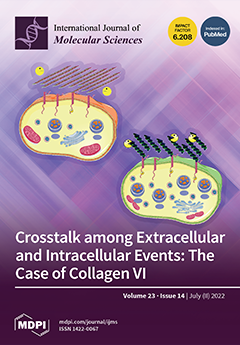
Advanced microscopy techniques reveal the link between extracellular collagen VI and subcellular alterations in fibroblasts derived from patients affected by a severe form of congenital muscular dystrophy associated with a common mutation in the COL6A1 gene. In summary, the analysed mutation in COL6A1 triggers the production of an unorganized collagen VI network, more susceptible to the enzyme collagenase. Consequently, mutated collagen VI molecules are dispersed in the extracellular matrix, and hence are more prone to bind to the CMG2 receptors on the cell membrane. The increased phosphorylation of the CMG2 receptor due to the collagen VI binding triggers the accumulation of endosomes and lysosomes. All the pathological phenotypes observed in human fibroblasts were corrected using the CRISPR/Cas9 technique to silence the common mutation. View this paper
  • Issues are regarded as officially published after their release is announced to the table of contents alert mailing list.
  • You may sign up for e-mail alerts to receive table of contents of newly released issues.
  • PDF is the official format for papers published in both, html and pdf forms. To view the papers in pdf format, click on the "PDF Full-text" link, and use the free Adobe Reader to open them.
Order results
Result details
Section
Select all
Export citation of selected articles as:
10 pages, 3829 KiB  
Article
Peptide Assembly of Al/CuO Nanothermite for Enhanced Reactivity of Nanoaluminum Particles
by Miaomiao Jin, Zhanxin Song, Wei Liu, Zilu Zhou, Guozhen Wang and Mo Xian
Int. J. Mol. Sci. 2022, 23(14), 8054; https://doi.org/10.3390/ijms23148054 - 21 Jul 2022
Viewed by 1568
Abstract
Biological self-assembly procedures, which are generally carried out in an aqueous solution, have been found to be the most promising method for directing the fabrication of diverse nanothermites, including Al/CuO nanothermite. However, the aqueous environment in which Al nanoparticles self-assemble has an impact [...] Read more.
Biological self-assembly procedures, which are generally carried out in an aqueous solution, have been found to be the most promising method for directing the fabrication of diverse nanothermites, including Al/CuO nanothermite. However, the aqueous environment in which Al nanoparticles self-assemble has an impact on their stability. We show that using a peptide to self-assemble Al or CuO nanoparticles considerably improves their durability in phosphate buffer aqueous solution, with Al and CuO nanoparticles remaining intact in aqueous solution for over 2 weeks with minimal changes in the structure. When peptide-assembled Al/CuO nanothermite was compared with a physically mixed sample in phosphate buffer for 30 min, the energy release of the former was higher by 26%. Furthermore, the energy release of peptide-assembled Al/CuO nanocomposite in phosphate buffer showed a 6% reduction by Day 7, while that of the peptide-assembled Al/CuO nanocomposite in ultrapure water was reduced by 75%. Taken together, our study provides an easy method for keeping the thermal activity of Al/CuO nanothermite assembled in aqueous solution. Full article
(This article belongs to the Special Issue Nano-Materials and Methods 3.0)
Show Figures

Figure 1

13 pages, 3353 KiB  
Article
Tumor-Stroma Ratio and Programmed Cell Death Ligand 1 Expression in Preoperative Biopsy and Matched Laryngeal Carcinoma Surgical Specimen
by Lara Alessandrini, Leonardo Franz, Marta Sbaraglia, Tommaso Saccardo, Filippo Cappello, Alessandro Drigo, Anna Chiara Frigo and Gino Marioni
Int. J. Mol. Sci. 2022, 23(14), 8053; https://doi.org/10.3390/ijms23148053 - 21 Jul 2022
Cited by 3 | Viewed by 1783
Abstract
Programmed cell death ligand 1 (PD-L1) seems to rely on close relations between neoplastic and immune cells in the tumor microenvironment. Tumor to stroma ratio (TSR) has been associated with prognosis in different malignancies. The aims of this exploratory investigation were to analyze [...] Read more.
Programmed cell death ligand 1 (PD-L1) seems to rely on close relations between neoplastic and immune cells in the tumor microenvironment. Tumor to stroma ratio (TSR) has been associated with prognosis in different malignancies. The aims of this exploratory investigation were to analyze for the first time the: (i) association between TSR, PD-L1 expression and other clinical–pathological features in laryngeal squamous cell carcinoma (LSCC) biopsies and paired surgical specimens; (ii) prognostic and predictive role of TSR and PD-L1. TSR, PD-L1 expression (in terms of combined positive score [CPS]), and other clinical–pathological features were analyzed in biopsies and surgical specimens of 43 consecutive LSCC cases. A CPS < 1 evaluated on surgical specimens was associated with a low TSR (stroma rich) on both biopsies and surgical specimens (p = 0.0143 and p = 0.0063). Low TSR showed a significant negative prognostic value when evaluated on both biopsies and surgical specimens (HR = 8.808, p = 0.0003 and HR = 11.207, p = 0.0002). CPS ≥ 1 appeared to be a favorable prognostic factor (HR = 0.100, p = 0.0265). The association between bioptic and surgical specimen TSR and PD-L1 expression should be further investigated for a potential impact on targeted treatments, also with regard to immunotherapeutic protocols. Full article
(This article belongs to the Topic Anti-Tumor Immune Responses)
Show Figures

Figure 1

16 pages, 5556 KiB  
Article
The Single-Stranded DNA-Binding Gene Whirly (Why1) with a Strong Pathogen-Induced Promoter from Vitis pseudoreticulata Enhances Resistance to Phytophthora capsici
by Chengchun Lai, Qiuxia Que, Ruo Pan, Qi Wang, Huiying Gao, Xuefang Guan, Jianmei Che and Gongti Lai
Int. J. Mol. Sci. 2022, 23(14), 8052; https://doi.org/10.3390/ijms23148052 - 21 Jul 2022
Cited by 3 | Viewed by 1665
Abstract
Vitis vinifera plants are disease-susceptible while Vitis pseudoreticulata plants are disease-resistant; however, the molecular mechanism remains unclear. In this study, the single-stranded DNA- and RNA-binding protein gene Whirly (VvWhy1 and VpWhy1) were cloned from V. vinifera “Cabernet Sauvignon” and V. pseudoreticulata [...] Read more.
Vitis vinifera plants are disease-susceptible while Vitis pseudoreticulata plants are disease-resistant; however, the molecular mechanism remains unclear. In this study, the single-stranded DNA- and RNA-binding protein gene Whirly (VvWhy1 and VpWhy1) were cloned from V. vinifera “Cabernet Sauvignon” and V. pseudoreticulata “HD1”. VvWhy1 and VpWhy1 promoter sequences (pVv and pVp) were also isolated; however, the identity of the promoter sequences was far lower than that between the Why1 coding sequences (CDSs). Both Why1 gene sequences had seven exons and six introns, and they had a C-terminal Whirly conserved domain and N-terminal chloroplast transit peptide, which was then verified to be chloroplast localization. Transcriptional expression showed that VpWhy1 was strongly induced by Plasmopara viticola, while VvWhy1 showed a low expression level. Further, the GUS activity indicated pVp had high activity involved in response to Phytophthora capsici infection. In addition, Nicotiana benthamiana transiently expressing pVp::VvWhy1 and pVp::VpWhy1 enhanced the P. capsici resistance. Moreover, Why1, PR1 and PR10 were upregulated in pVp transgenic N. benthamiana leaves. This research presented a novel insight into disease resistance mechanism that pVp promoted the transcription of Why1, which subsequently regulated the expression of PR1 and PR10, further enhancing the resistance to P. capsici. Full article
(This article belongs to the Section Molecular Plant Sciences)
Show Figures

Figure 1

12 pages, 299 KiB  
Review
Roles of Keratins in Intestine
by Jeongwon Mun, Whan Hur and Nam-On Ku
Int. J. Mol. Sci. 2022, 23(14), 8051; https://doi.org/10.3390/ijms23148051 - 21 Jul 2022
Cited by 4 | Viewed by 2064
Abstract
Keratins make up a major portion of epithelial intermediate filament proteins. The widely diverse keratins are found in both the small and large intestines. The human intestine mainly expresses keratins 8, 18, 19, and 20. Many of the common roles of keratins are [...] Read more.
Keratins make up a major portion of epithelial intermediate filament proteins. The widely diverse keratins are found in both the small and large intestines. The human intestine mainly expresses keratins 8, 18, 19, and 20. Many of the common roles of keratins are for the integrity and stability of the epithelial cells. The keratins also protect the cells and tissue from stress and are biomarkers for some diseases in the organs. Although an increasing number of studies have been performed regarding keratins, the roles of keratin in the intestine have not yet been fully understood. This review focuses on discussing the roles of keratins in the intestine. Diverse studies utilizing mouse models and samples from patients with intestinal diseases in the search for the association of keratin in intestinal diseases have been summarized. Full article
(This article belongs to the Special Issue Gut and the Liver in Health and Disease)
23 pages, 7070 KiB  
Article
Analysis of Binding Determinants for Different Classes of Competitive and Noncompetitive Inhibitors of Glycine Transporters
by Kamil Łątka and Marek Bajda
Int. J. Mol. Sci. 2022, 23(14), 8050; https://doi.org/10.3390/ijms23148050 - 21 Jul 2022
Cited by 5 | Viewed by 2044
Abstract
Glycine transporters are interesting therapeutic targets as they play significant roles in glycinergic and glutamatergic systems. The search for new selective inhibitors of particular types of glycine transporters (GlyT-1 and GlyT-2) with beneficial kinetics is hampered by limited knowledge about the spatial structure [...] Read more.
Glycine transporters are interesting therapeutic targets as they play significant roles in glycinergic and glutamatergic systems. The search for new selective inhibitors of particular types of glycine transporters (GlyT-1 and GlyT-2) with beneficial kinetics is hampered by limited knowledge about the spatial structure of these proteins. In this study, a pool of homology models of GlyT-1 and GlyT-2 in different conformational states was constructed using the crystal structures of related transporters from the SLC6 family and the recently revealed structure of GlyT-1 in the inward-open state, in order to investigate their binding sites. The binding mode of the known GlyT-1 and GlyT-2 inhibitors was determined using molecular docking studies, molecular dynamics simulations, and MM-GBSA free energy calculations. The results of this study indicate that two amino acids, Gly373 and Leu476 in GlyT-1 and the corresponding Ser479 and Thr582 in GlyT-2, are mainly responsible for the selective binding of ligands within the S1 site. Apart from these, one pocket of the S2 site, which lies between TM3 and TM10, may also be important. Moreover, selective binding of noncompetitive GlyT-1 inhibitors in the intracellular release pathway is affected by hydrophobic interactions with Ile399, Met382, and Leu158. These results can be useful in the rational design of new glycine transporter inhibitors with desired selectivity and properties in the future. Full article
(This article belongs to the Section Molecular Biology)
Show Figures

Figure 1

14 pages, 5906 KiB  
Article
Yohimbine, an α2-Adrenoceptor Antagonist, Suppresses PDGF-BB-Stimulated Vascular Smooth Muscle Cell Proliferation by Downregulating the PLCγ1 Signaling Pathway
by Chih-Wei Chiu, Cheng-Ying Hsieh, Chih-Hao Yang, Jie-Heng Tsai, Shih-Yi Huang and Joen-Rong Sheu
Int. J. Mol. Sci. 2022, 23(14), 8049; https://doi.org/10.3390/ijms23148049 - 21 Jul 2022
Cited by 3 | Viewed by 2293
Abstract
Yohimbine (YOH) has antiproliferative effects against breast cancer and pancreatic cancer; however, its effects on vascular proliferative diseases such as atherosclerosis remain unknown. Accordingly, we investigated the inhibitory mechanisms of YOH in vascular smooth muscle cells (VSMCs) stimulated by platelet-derived growth factor (PDGF)-BB, [...] Read more.
Yohimbine (YOH) has antiproliferative effects against breast cancer and pancreatic cancer; however, its effects on vascular proliferative diseases such as atherosclerosis remain unknown. Accordingly, we investigated the inhibitory mechanisms of YOH in vascular smooth muscle cells (VSMCs) stimulated by platelet-derived growth factor (PDGF)-BB, a major mitogenic factor in vascular diseases. YOH (5–20 μM) suppressed PDGF-BB-stimulated a mouse VSMC line (MOVAS-1 cell) proliferation without inducing cytotoxicity. YOH also exhibited antimigratory effects and downregulated matrix metalloproteinase-2 and -9 expression in PDGF-BB-stimulated MOVAS-1 cells. It also promoted cell cycle arrest in the initial gap/first gap phase by upregulating p27Kip1 and p53 expression and reducing cyclin-dependent kinase 2 and proliferating cell nuclear antigen expression. We noted phospholipase C-γ1 (PLCγ1) but not ERK1/2, AKT, or p38 kinase phosphorylation attenuation in YOH-modulated PDGF-BB-propagated signaling pathways in the MOVAS-1 cells. Furthermore, YOH still inhibited PDGF-BB-induced cell proliferation and PLCγ1 phosphorylation in MOVAS-1 cells with α2B-adrenergic receptor knockdown. YOH (5 and 10 mg/kg) substantially suppressed neointimal hyperplasia in mice subjected to CCA ligation for 21 days. Overall, our results reveal that YOH attenuates PDGF-BB-stimulated VSMC proliferation and migration by downregulating a α2B-adrenergic receptor–independent PLCγ1 pathway and reduces neointimal formation in vivo. Therefore, YOH has potential for repurposing for treating atherosclerosis and other vascular proliferative diseases. Full article
Show Figures

Figure 1

17 pages, 1097 KiB  
Review
Natural Products of Marine Origin for the Treatment of Colorectal and Pancreatic Cancers: Mechanisms and Potential
by Nasrin Fares Amer and Tal Luzzatto Knaan
Int. J. Mol. Sci. 2022, 23(14), 8048; https://doi.org/10.3390/ijms23148048 - 21 Jul 2022
Cited by 5 | Viewed by 2843
Abstract
Gastrointestinal cancer refers to malignancy of the accessory organs of digestion, and it includes colorectal cancer (CRC) and pancreatic cancer (PC). Worldwide, CRC is the second most common cancer among women and the third most common among men. PC has a poor prognosis [...] Read more.
Gastrointestinal cancer refers to malignancy of the accessory organs of digestion, and it includes colorectal cancer (CRC) and pancreatic cancer (PC). Worldwide, CRC is the second most common cancer among women and the third most common among men. PC has a poor prognosis and high mortality, with 5-year relative survival of approximately 11.5%. Conventional chemotherapy treatments for these cancers are limited due to severe side effects and the development of drug resistance. Therefore, there is an urgent need to develop new and safe drugs for effective treatment of PC and CRC. Historically, natural sources—plants in particular—have played a dominant role in traditional medicine used to treat a wide spectrum of diseases. In recent decades, marine natural products (MNPs) have shown great potential as drugs, but drug leads for treating various types of cancer, including CRC and PC, are scarce. To date, marine-based drugs have been used against leukemia, metastatic breast cancer, soft tissue sarcoma, and ovarian cancer. In this review, we summarized existing studies describing MNPs that were found to have an effect on CRC and PC, and we discussed the potential mechanisms of action of MNPs as well as future prospects for their use in treating these cancers. Full article
(This article belongs to the Special Issue Cytotoxicity, Antioxidant and Anticancer Activity of Natural Products)
Show Figures

Figure 1

2 pages, 196 KiB  
Editorial
Frontiers in Antimicrobial Materials
by Ángel Serrano-Aroca, Murtaza M. Tambuwala and Martin Birkett
Int. J. Mol. Sci. 2022, 23(14), 8047; https://doi.org/10.3390/ijms23148047 - 21 Jul 2022
Cited by 2 | Viewed by 1844
Abstract
The aim of this Special Edition is to highlight the exponential work performed in the field of antimicrobial material research from the beginning of the current COVID-19 pandemic [...] Full article
(This article belongs to the Special Issue Frontiers in Antimicrobial Materials)
18 pages, 3065 KiB  
Article
Designing and Synthesis of New Isatin Derivatives as Potential CDK2 Inhibitors
by Przemysław Czeleń, Agnieszka Skotnicka and Beata Szefler
Int. J. Mol. Sci. 2022, 23(14), 8046; https://doi.org/10.3390/ijms23148046 - 21 Jul 2022
Cited by 9 | Viewed by 1820
Abstract
Tumors are still one of the main causes of death; therefore, the search for new therapeutic agents that will enable the implementation of effective treatment is a significant challenge for modern pharmacy. One of the important factors contributing to the development of neoplastic [...] Read more.
Tumors are still one of the main causes of death; therefore, the search for new therapeutic agents that will enable the implementation of effective treatment is a significant challenge for modern pharmacy. One of the important factors contributing to the development of neoplastic diseases is the overexpression of enzymes responsible for the regulation of cell division processes such as cyclin-dependent kinases. Numerous studies and examples of already-developed drugs confirm that isatin is a convenient basis for the development of new groups of inhibitors for this class of enzyme. Therefore, in this work, a new group of potential inhibitors of the CDK2 enzyme, utilizing isatin derivatives and substituted benzoylhydrazines, has been designed based on the application of computational chemistry methods, such as docking and molecular dynamics, and their inhibiting ability was assessed. In the cases of the selected compounds, a synthesis method was developed, and the selected physicochemical properties of the newly synthesized derivatives were estimated. As part of the completed project, new compounds are developed which are potential inhibitors of the CDK2 enzyme. Full article
Show Figures

Figure 1

11 pages, 1349 KiB  
Article
A Bioinformatics Approach to MicroRNA-Sequencing Analysis Based on Human Saliva Samples of Patients with Endometriosis
by Sofiane Bendifallah, Yohann Dabi, Stéphane Suisse, Ludmila Jornea, Delphine Bouteiller, Cyril Touboul, Anne Puchar and Emile Daraï
Int. J. Mol. Sci. 2022, 23(14), 8045; https://doi.org/10.3390/ijms23148045 - 21 Jul 2022
Cited by 13 | Viewed by 3847
Abstract
Endometriosis, defined by the presence of endometrium-like tissue outside the uterus, affects 2–10% of the female population, i.e., around 190 million women, worldwide. The aim of the prospective ENDO-miRNA study was to develop a bioinformatics approach for microRNA-sequencing analysis of 200 saliva samples [...] Read more.
Endometriosis, defined by the presence of endometrium-like tissue outside the uterus, affects 2–10% of the female population, i.e., around 190 million women, worldwide. The aim of the prospective ENDO-miRNA study was to develop a bioinformatics approach for microRNA-sequencing analysis of 200 saliva samples for miRNAome expression and to test its diagnostic accuracy for endometriosis. Among the 200 patients, 76.5% (n = 153) had confirmed endometriosis and 23.5% (n = 47) had no endometriosis (controls). Small RNA-seq of 200 saliva samples yielded ~4642 M raw sequencing reads (from ~13.7 M to ~39.3 M reads/sample). The number of expressed miRNAs ranged from 1250 (outlier) to 2561 per sample. Some 2561 miRNAs were found to be differentially expressed in the saliva samples of patients with endometriosis compared with the control patients. Among these, 1.17% (n = 30) were up- or downregulated. Among these, the F1-score, sensitivity, specificity, and AUC ranged from 11–86.8%, 5.8–97.4%, 10.6–100%, and 39.3–69.2%, respectively. Here, we report a bioinformatic approach to saliva miRNA sequencing and analysis. We underline the advantages of using saliva over blood in terms of ease of collection, reproducibility, stability, safety, non-invasiveness. This report describes the whole saliva transcriptome to make miRNA quantification a validated, standardized, and reliable technique for routine use. The methodology could be applied to build a saliva signature of endometriosis. Full article
(This article belongs to the Special Issue Molecular Research on Endometriosis)
Show Figures

Figure 1

18 pages, 4894 KiB  
Article
Genome-Wide Identification of Eucalyptus Heat Shock Transcription Factor Family and Their Transcriptional Analysis under Salt and Temperature Stresses
by Tan Yuan, Jianxiang Liang, Jiahao Dai, Xue-Rong Zhou, Wenhai Liao, Mingliang Guo, Mohammad Aslam, Shubin Li, Guangqiu Cao and Shijiang Cao
Int. J. Mol. Sci. 2022, 23(14), 8044; https://doi.org/10.3390/ijms23148044 - 21 Jul 2022
Cited by 5 | Viewed by 2372
Abstract
Heat shock transcription factors (HSFs) activate heat shock protein gene expression by binding their promoters in response to heat stress and are considered to be pivotal transcription factors in plants. Eucalyptus is a superior source of fuel and commercial wood. During its growth, [...] Read more.
Heat shock transcription factors (HSFs) activate heat shock protein gene expression by binding their promoters in response to heat stress and are considered to be pivotal transcription factors in plants. Eucalyptus is a superior source of fuel and commercial wood. During its growth, high temperature or other abiotic stresses could impact its defense capability and growth. Hsf genes have been cloned and sequenced in many plants, but rarely in Eucalyptus. In this study, we used bioinformatics methods to analyze and identify Eucalyptus Hsf genes, their chromosomal localization and structure. The phylogenetic relationship and conserved domains of their encoded proteins were further analyzed. A total of 36 Hsf genes were identified and authenticated from Eucalyptus, which were scattered across 11 chromosomes. They could be classified into three classes (A, B and C). Additionally, a large number of stress-related cis-regulatory elements were identified in the upstream promoter sequence of HSF, and cis-acting element analysis indicated that the expression of EgHsf may be regulated by plant growth and development, metabolism, hormones and stress responses. The expression profiles of five representative Hsf genes, EgHsf4, EgHsf9, EgHsf13, EgHsf24 and EgHsf32, under salt and temperature stresses were examined by qRT-PCR. The results show that the expression pattern of class B genes (EgHsf4, EgHsf24 and EgHsf32) was more tolerant to abiotic stresses than that of class A genes (EgHsf9 and EgHsf13). However, the expressions of all tested Hsf genes in six tissues were at different levels. Finally, we investigated the network of interplay between genes, and the results suggest that there may be synergistic effects between different Hsf genes in response to abiotic stresses. We conclude that the Hsf gene family played an important role in the growth and developmental processes of Eucalyptus and could be vital for maintaining cell homeostasis against external stresses. This study provides basic information on the members of the Hsf gene family in Eucalyptus and lays the foundation for the functional identification of related genes and the further investigation of their biological functions in plant stress regulation. Full article
(This article belongs to the Special Issue 21st Anniversary of IJMS: Advances in Molecular Genetics and Genomics)
Show Figures

Figure 1

16 pages, 734 KiB  
Review
Therapeutic Potential of Photobiomodulation for Chronic Kidney Disease
by Ji Bian, Ann Liebert, Brian Bicknell, Xin-Ming Chen, Chunling Huang and Carol A. Pollock
Int. J. Mol. Sci. 2022, 23(14), 8043; https://doi.org/10.3390/ijms23148043 - 21 Jul 2022
Cited by 3 | Viewed by 3851
Abstract
Chronic kidney disease (CKD) is a growing global public health problem. The implementation of evidence-based clinical practices only defers the development of kidney failure. Death, transplantation, or dialysis are the consequences of kidney failure, resulting in a significant burden on the health system. [...] Read more.
Chronic kidney disease (CKD) is a growing global public health problem. The implementation of evidence-based clinical practices only defers the development of kidney failure. Death, transplantation, or dialysis are the consequences of kidney failure, resulting in a significant burden on the health system. Hence, innovative therapeutic strategies are urgently needed due to the limitations of current interventions. Photobiomodulation (PBM), a form of non-thermal light therapy, effectively mitigates mitochondrial dysfunction, reactive oxidative stress, inflammation, and gut microbiota dysbiosis, all of which are inherent in CKD. Preliminary studies suggest the benefits of PBM in multiple diseases, including CKD. Hence, this review will provide a concise summary of the underlying action mechanisms of PBM and its potential therapeutic effects on CKD. Based on the findings, PBM may represent a novel, non-invasive and non-pharmacological therapy for CKD, although more studies are necessary before PBM can be widely recommended. Full article
(This article belongs to the Special Issue Advances in Chronic Kidney Disease: An Update)
Show Figures

Figure 1

19 pages, 5370 KiB  
Article
Exposure to Bisphenol A Caused Hepatoxicity and Intestinal Flora Disorder in Rats
by Ruijing Liu, Boping Liu, Lingmin Tian, Xinwei Jiang, Xusheng Li, Dongbao Cai, Jianxia Sun, Weibin Bai and Yulong Jin
Int. J. Mol. Sci. 2022, 23(14), 8042; https://doi.org/10.3390/ijms23148042 - 21 Jul 2022
Cited by 17 | Viewed by 2806
Abstract
Bisphenol A (BPA) is a globally utilized industrial chemical and is commonly used as a monomer of polycarbonate plastics and epoxy resins. Recent research reveals that BPA could cause potential adverse biological effects and liver dysfunction. However, the underlying mechanisms of BPA-induced hepatoxicity [...] Read more.
Bisphenol A (BPA) is a globally utilized industrial chemical and is commonly used as a monomer of polycarbonate plastics and epoxy resins. Recent research reveals that BPA could cause potential adverse biological effects and liver dysfunction. However, the underlying mechanisms of BPA-induced hepatoxicity and gut dysbiosis remain unclear and deserve further study. In this study, male Sprague Dawley rats were exposed to different doses (0, 30, 90, and 270 mg/kg bw) of BPA by gavage for 30 days. The results showed that the high dose of BPA decreased superoxide dismutase (SOD), glutathione (GSH), and increased malondialdehyde (MDA) levels. Moreover, a high dose of BPA caused a significant increase in serum alanine aminotransferase (ALT), aspartate aminotransferase (AST), total cholesterol (TC), and low-density lipoprotein cholesterol (LDL-C), while high-density lipoprotein cholesterol (HDL-C) was significantly decreased in BPA-treated rats. The gene expression of PGC-1α and Nrf1 were decreased in the liver of high doses of BPA-administrated rats, as well as the protein levels of SIRT1, PGC-1α, Nrf2, and TFAM. However, the protein expression of IL-1β was significantly increased in BPA-treated rats. In addition, BPA weakened the mitochondrial function of hepatocytes and promoted cell apoptosis in the liver by up-regulating the protein levels of Bax, cleaved-Caspase3, and cleaved-PARP1 while down-regulating the Bcl-2 in the liver. More importantly, a high dose of BPA caused a dramatic change in microbiota structure, as characterized at the genus level by increasing the ratio of Firmicutes to Bacteroidetes (F/B), and the relative abundance of Proteobacteria in feces, while decreasing the relative abundance of Prevotella_9 and Ruminococcaceae_UCG-014, which is positively correlated with the content of short-chain fatty acids (SCFAs). In summary, our data indicated that BPA exposure caused hepatoxicity through apoptosis and the SIRT1/PGC-1α pathway. BPA-induced intestinal flora and SCFA changes may be associated with hepatic damage. The results of this study provide a new sight for the understanding of BPA-induced hepatoxicity. Full article
Show Figures

Figure 1

25 pages, 8450 KiB  
Review
Recent Advances in the Photoreactions Triggered by Porphyrin-Based Triplet–Triplet Annihilation Upconversion Systems: Molecular Innovations and Nanoarchitectonics
by Bin Yao, Hongfei Sun, Youzhou He, Song Wang and Xingyan Liu
Int. J. Mol. Sci. 2022, 23(14), 8041; https://doi.org/10.3390/ijms23148041 - 21 Jul 2022
Cited by 6 | Viewed by 3208
Abstract
Triplet–triplet annihilation upconversion (TTA-UC) is a very promising technology that could be used to convert low-energy photons to high-energy ones and has been proven to be of great value in various areas. Porphyrins have the characteristics of high molar absorbance, can form a [...] Read more.
Triplet–triplet annihilation upconversion (TTA-UC) is a very promising technology that could be used to convert low-energy photons to high-energy ones and has been proven to be of great value in various areas. Porphyrins have the characteristics of high molar absorbance, can form a complex with different metal ions and a high proportion of triplet states as well as tunable structures, and thus they are important sensitizers for TTA-UC. Porphyrin-based TTA-UC plays a pivotal role in the TTA-UC systems and has been widely used in many fields such as solar cells, sensing and circularly polarized luminescence. In recent years, applications of porphyrin-based TTA-UC systems for photoinduced reactions have emerged, but have been paid little attention. As a consequence, this review paid close attention to the recent advances in the photoreactions triggered by porphyrin-based TTA-UC systems. First of all, the photochemistry of porphyrin-based TTA-UC for chemical transformations, such as photoisomerization, photocatalytic synthesis, photopolymerization, photodegradation and photochemical/photoelectrochemical water splitting, was discussed in detail, which revealed the different mechanisms of TTA-UC and methods with which to carry out reasonable molecular innovations and nanoarchitectonics to solve the existing problems in practical application. Subsequently, photoreactions driven by porphyrin-based TTA-UC for biomedical applications were demonstrated. Finally, the future developments of porphyrin-based TTA-UC systems for photoreactions were briefly discussed. Full article
(This article belongs to the Special Issue Advances in the Chemistry of Porphyrins and Related Macrocycles 2.0)
Show Figures

Figure 1

21 pages, 6591 KiB  
Article
Characterization and Comparative Genomics Analysis of a New Bacteriophage BUCT610 against Klebsiella pneumoniae and Efficacy Assessment in Galleria mellonella Larvae
by Mingfang Pu, Pengjun Han, Guangye Zhang, Yucong Liu, Yahao Li, Fei Li, Mengzhe Li, Xiaoping An, Lihua Song, Yiming Chen, Huahao Fan and Yigang Tong
Int. J. Mol. Sci. 2022, 23(14), 8040; https://doi.org/10.3390/ijms23148040 - 21 Jul 2022
Cited by 12 | Viewed by 2213
Abstract
The spread of multidrug-resistant Klebsiella pneumoniae (MDR-KP) has become an emerging threat as a result of the overuse of antibiotics. Bacteriophage (phage) therapy is considered to be a promising alternative treatment for MDR-KP infection compared with antibiotic therapy. In this research, a lytic [...] Read more.
The spread of multidrug-resistant Klebsiella pneumoniae (MDR-KP) has become an emerging threat as a result of the overuse of antibiotics. Bacteriophage (phage) therapy is considered to be a promising alternative treatment for MDR-KP infection compared with antibiotic therapy. In this research, a lytic phage BUCT610 was isolated from hospital sewage. The assembled genome of BUCT610 was 46,774 bp in length, with a GC content of 48%. A total of 83 open reading frames (ORFs) and no virulence or antimicrobial resistance genes were annotated in the BUCT610 genome. Comparative genomics and phylogenetic analyses showed that BUCT610 was most closely linked with the Vibrio phage pYD38-A and shared 69% homology. In addition, bacteriophage BUCT610 exhibited excellent thermal stability (4–75 °C) and broad pH tolerance (pH 3–12) in the stability test. In vivo investigation results showed that BUCT610 significantly increased the survival rate of Klebsiella pneumonia-infected Galleria mellonella larvae from 13.33% to 83.33% within 72 h. In conclusion, these findings indicate that phage BUCT610 holds great promise as an alternative agent with excellent stability for the treatment of MDR-KP infection. Full article
(This article belongs to the Special Issue Bacteriophage Biology: From Genomics to Therapy)
Show Figures

Figure 1

28 pages, 3367 KiB  
Review
Phase Separation-Mediated Chromatin Organization and Dynamics: From Imaging-Based Quantitative Characterizations to Functional Implications
by Woei Shyuan Ng, Hendrik Sielaff and Ziqing Winston Zhao
Int. J. Mol. Sci. 2022, 23(14), 8039; https://doi.org/10.3390/ijms23148039 - 21 Jul 2022
Cited by 4 | Viewed by 3429
Abstract
As an effective and versatile strategy to compartmentalize cellular components without the need for lipid membranes, phase separation has been found to underpin a wide range of intranuclear processes, particularly those involving chromatin. Many of the unique physico-chemical properties of chromatin-based phase condensates [...] Read more.
As an effective and versatile strategy to compartmentalize cellular components without the need for lipid membranes, phase separation has been found to underpin a wide range of intranuclear processes, particularly those involving chromatin. Many of the unique physico-chemical properties of chromatin-based phase condensates are harnessed by the cell to accomplish complex regulatory functions in a spatially and temporally controlled manner. Here, we survey key recent findings on the mechanistic roles of phase separation in regulating the organization and dynamics of chromatin-based molecular processes across length scales, packing states and intranuclear functions, with a particular emphasis on quantitative characterizations of these condensates enabled by advanced imaging-based approaches. By illuminating the complex interplay between chromatin and various chromatin-interacting molecular species mediated by phase separation, this review sheds light on an emerging multi-scale, multi-modal and multi-faceted landscape that hierarchically regulates the genome within the highly crowded and dynamic nuclear space. Moreover, deficiencies in existing studies also highlight the need for mechanism-specific criteria and multi-parametric approaches for the characterization of chromatin-based phase separation using complementary techniques and call for greater efforts to correlate the quantitative features of these condensates with their functional consequences in close-to-native cellular contexts. Full article
Show Figures

Figure 1

37 pages, 2323 KiB  
Review
Perivascular Mesenchymal Stem/Stromal Cells, an Immune Privileged Niche for Viruses?
by Grégorie Lebeau, Franck Ah-Pine, Matthieu Daniel, Yosra Bedoui, Damien Vagner, Etienne Frumence and Philippe Gasque
Int. J. Mol. Sci. 2022, 23(14), 8038; https://doi.org/10.3390/ijms23148038 - 21 Jul 2022
Cited by 7 | Viewed by 4303
Abstract
Mesenchymal stem cells (MSCs) play a critical role in response to stress such as infection. They initiate the removal of cell debris, exert major immunoregulatory activities, control pathogens, and lead to a remodeling/scarring phase. Thus, host-derived ‘danger’ factors released from damaged/infected cells (called [...] Read more.
Mesenchymal stem cells (MSCs) play a critical role in response to stress such as infection. They initiate the removal of cell debris, exert major immunoregulatory activities, control pathogens, and lead to a remodeling/scarring phase. Thus, host-derived ‘danger’ factors released from damaged/infected cells (called alarmins, e.g., HMGB1, ATP, DNA) as well as pathogen-associated molecular patterns (LPS, single strand RNA) can activate MSCs located in the parenchyma and around vessels to upregulate the expression of growth factors and chemoattractant molecules that influence immune cell recruitment and stem cell mobilization. MSC, in an ultimate contribution to tissue repair, may also directly trans- or de-differentiate into specific cellular phenotypes such as osteoblasts, chondrocytes, lipofibroblasts, myofibroblasts, Schwann cells, and they may somehow recapitulate their neural crest embryonic origin. Failure to terminate such repair processes induces pathological scarring, termed fibrosis, or vascular calcification. Interestingly, many viruses and particularly those associated to chronic infection and inflammation may hijack and polarize MSC’s immune regulatory activities. Several reports argue that MSC may constitute immune privileged sanctuaries for viruses and contributing to long-lasting effects posing infectious challenges, such as viruses rebounding in immunocompromised patients or following regenerative medicine therapies using MSC. We will herein review the capacity of several viruses not only to infect but also to polarize directly or indirectly the functions of MSC (immunoregulation, differentiation potential, and tissue repair) in clinical settings. Full article
(This article belongs to the Special Issue Stem Cell Biology in Health and Disease)
Show Figures

Figure 1

12 pages, 1355 KiB  
Article
Preclinical Assessment of the Combination of PSMA-Targeting Radionuclide Therapy with PARP Inhibitors for Prostate Cancer Treatment
by Eline A. M. Ruigrok, Nicole S. Verkaik, Erik de Blois, Corrina de Ridder, Debra Stuurman, Stefan J. Roobol, Dik C. Van Gent, Marion de Jong, Wytske M. Van Weerden and Julie Nonnekens
Int. J. Mol. Sci. 2022, 23(14), 8037; https://doi.org/10.3390/ijms23148037 - 21 Jul 2022
Cited by 10 | Viewed by 2548
Abstract
Prostate specific membrane antigen targeted radionuclide therapy (PSMA-TRT) is a promising novel treatment for prostate cancer (PCa) patients. However, PSMA-TRT cannot be used for curative intent yet, thus additional research on how to improve the therapeutic efficacy is warranted. A potential way of [...] Read more.
Prostate specific membrane antigen targeted radionuclide therapy (PSMA-TRT) is a promising novel treatment for prostate cancer (PCa) patients. However, PSMA-TRT cannot be used for curative intent yet, thus additional research on how to improve the therapeutic efficacy is warranted. A potential way of achieving this, is combining TRT with poly ADP-ribosylation inhibitors (PARPi), which has shown promising results for TRT of neuroendocrine tumor cells. Currently, several clinical trials have been initiated for this combination for PCa, however so far, no evidence of synergism is available for PCa. Therefore, we evaluated the combination of PSMA-TRT with three classes of PARPi in preclinical PCa models. In vitro viability and survival assays were performed using PSMA-expressing PCa cell lines PC3-PIP and LNCaP to assess the effect of increasing concentrations of PARPi veliparib, olaparib or talazoparib in combination with PSMA-TRT compared to single PARPi treatment. Next, DNA damage analyses were performed by quantifying the number of DNA breaks by immunofluorescent stainings. Lastly, the potential of the combination treatments was studied in vivo in mice bearing PC3-PIP xenografts. Our results show that combining PSMA-TRT with PARPi did not synergistically affect the in vitro clonogenic survival or cell viability. DNA-damage analysis revealed only a significant increase in DNA breaks when combining PSMA-TRT with veliparib and not in the other combination treatments. Moreover, PSMA-TRT with PARPi treatment did not improve tumor control compared to PSMA-TRT monotherapy. Overall, the data presented do not support the assumption that combining PSMA-TRT with PARPi leads to a synergistic antitumor effect in PCa. These results underline that extensive preclinical research using various PCa models is imperative to validate the applicability of the combination strategy for PCa, as it is for other cancer types. Full article
(This article belongs to the Special Issue Urogenital Tumors: From Molecular Basis to Therapy)
Show Figures

Figure 1

17 pages, 1822 KiB  
Article
Integrated DNA Copy Number and Expression Profiling Identifies IGF1R as a Prognostic Biomarker in Pediatric Osteosarcoma
by Aaron M. Taylor, Jiayi M. Sun, Alexander Yu, Horatiu Voicu, Jianhe Shen, Donald A. Barkauskas, Timothy J. Triche, Julie M. Gastier-Foster, Tsz-Kwong Man and Ching C. Lau
Int. J. Mol. Sci. 2022, 23(14), 8036; https://doi.org/10.3390/ijms23148036 - 21 Jul 2022
Cited by 4 | Viewed by 2065
Abstract
Osteosarcoma is a primary malignant bone tumor arising from bone-forming mesenchymal cells in children and adolescents. Despite efforts to understand the biology of the disease and identify novel therapeutics, the survival of osteosarcoma patients remains dismal. We have concurrently profiled the copy number [...] Read more.
Osteosarcoma is a primary malignant bone tumor arising from bone-forming mesenchymal cells in children and adolescents. Despite efforts to understand the biology of the disease and identify novel therapeutics, the survival of osteosarcoma patients remains dismal. We have concurrently profiled the copy number and gene expression of 226 osteosarcoma samples as part of the Strategic Partnering to Evaluate Cancer Signatures (SPECS) initiative. Our results demonstrate the heterogeneous landscape of osteosarcoma in younger populations by showing the presence of genome-wide copy number abnormalities occurring both recurrently among samples and in a high frequency. Insulin growth factor receptor 1 (IGF1R) is a receptor tyrosine kinase which binds IGF1 and IGF2 to activate downstream pathways involved in cell apoptosis and proliferation. We identify prevalent amplification of IGF1R corresponding with increased gene expression in patients with poor survival outcomes. Our results substantiate previously tenuously associated copy number abnormalities identified in smaller datasets (13q34+, 20p13+, 4q35-, 20q13.33-), and indicate the significance of high fibroblast growth factor receptor 2 (FGFR2) expression in distinguishing patients with poor prognosis. FGFR2 is involved in cellular proliferation processes such as division, growth and angiogenesis. In summary, our findings demonstrate the prognostic significance of several genes associated with osteosarcoma pathogenesis. Full article
(This article belongs to the Special Issue Molecular Biology in Bone Tumors)
Show Figures

Figure 1

23 pages, 5283 KiB  
Article
ATP13A2 Declines Zinc-Induced Accumulation of α-Synuclein in a Parkinson’s Disease Model
by Huiling Gao, Hehong Sun, Nan Yan, Pu Zhao, He Xu, Wei Zheng, Xiaoyu Zhang, Tao Wang, Chuang Guo and Manli Zhong
Int. J. Mol. Sci. 2022, 23(14), 8035; https://doi.org/10.3390/ijms23148035 - 21 Jul 2022
Cited by 4 | Viewed by 2764
Abstract
Parkinson’s disease (PD) is characterized by the presence of Lewy bodies caused by α-synuclein. The imbalance of zinc homeostasis is a major cause of PD, promoting α-synuclein accumulation. ATP13A2, a transporter found in acidic vesicles, plays an important role in Zn2+ homeostasis [...] Read more.
Parkinson’s disease (PD) is characterized by the presence of Lewy bodies caused by α-synuclein. The imbalance of zinc homeostasis is a major cause of PD, promoting α-synuclein accumulation. ATP13A2, a transporter found in acidic vesicles, plays an important role in Zn2+ homeostasis and is highly expressed in Lewy bodies in PD-surviving neurons. ATP13A2 is involved in the transport of zinc ions in lysosomes and exosomes and inhibits the aggregation of α-synuclein. However, the potential mechanism underlying the regulation of zinc homeostasis and α-synuclein accumulation by ATP13A2 remains unexplored. We used α-synuclein-GFP transgenic mice and HEK293 α-synuclein-DsRed cell line as models. The spatial exploration behavior of mice was significantly reduced, and phosphorylation levels of α-synuclein increased upon high Zn2+ treatment. High Zn2+ also inhibited the autophagy pathway by reducing LAMP2a levels and changing the expression of LC3 and P62, by reducing mitochondrial membrane potential and increasing the expression of cytochrom C, and by activating the ERK/P38 apoptosis signaling pathway, ultimately leading to increased caspase 3 levels. These protein changes were reversed after ATP13A2 overexpression, whereas ATP13A2 knockout exacerbated α-synuclein phosphorylation levels. These results suggest that ATP13A2 may have a protective effect on Zn2+-induced abnormal aggregation of α-synuclein, lysosomal dysfunction, and apoptosis. Full article
(This article belongs to the Section Molecular Neurobiology)
Show Figures

Figure 1

20 pages, 4130 KiB  
Article
Transcriptional Regulation and Protein Localization of Zip10, Zip13 and Zip14 Transporters of Freshwater Teleost Yellow Catfish Pelteobagrus fulvidraco Following Zn Exposure in a Heterologous HEK293T Model
by Sheng-Zan Liu, Yi-Chuang Xu, Xiao-Ying Tan, Tao Zhao, Dian-Guang Zhang, Hong Yang and Zhi Luo
Int. J. Mol. Sci. 2022, 23(14), 8034; https://doi.org/10.3390/ijms23148034 - 21 Jul 2022
Cited by 1 | Viewed by 1613
Abstract
Zip family proteins are involved in the control of zinc (Zn) ion homeostasis. The present study cloned the promoters and investigated the transcription responses and protein subcellular localizations of three LIV-1 subfamily members (zip10, zip13, and zip14) from common [...] Read more.
Zip family proteins are involved in the control of zinc (Zn) ion homeostasis. The present study cloned the promoters and investigated the transcription responses and protein subcellular localizations of three LIV-1 subfamily members (zip10, zip13, and zip14) from common freshwater teleost yellow catfish, Pelteobagrus fulvidraco, using in vitro cultured HEK293T model cells. The 2278 bp, 1917 bp, and 1989 bp sequences of zip10, zip13, and zip14 promoters, respectively, were subcloned into pGL3-Basic plasmid for promoter activity analysis. The pcDNA3.1 plasmid coding EGFP tagged pfZip10, pfZip13, and pfZip14 were generated for subsequent confocal microscope analysis. Several potential transcription factors’ binding sites were predicted within the promoters. In vitro promoter analysis in the HEK293T cells showed that high Zn administration significantly reduced the transcriptional activities of the zip10, zip13, and zip14 promoters. The −2017 bp/−2004 bp MRE in the zip10 promoter, the −360 bp/−345 bp MRE in the zip13 promoter, and the −1457 bp/−1442 bp MRE in the zip14 promoter were functional loci that were involved in the regulation of the three zips. The −606 bp/−594 bp KLF4 binding site in the zip13 promoter was a functional locus responsible for zinc-responsive regulation of zip13. The −1383 bp/−1375 bp STAT3 binding site in the zip14 promoter was a functional locus responsible for zinc-responsive regulation of zip14. Moreover, confocal microscope analysis indicated that zinc incubation significantly reduced the fluorescence intensity of pfZip10-EGFP and pfZip14-EGFP but had no significant influence on pfZip13-EGFP fluorescence intensity. Further investigation found that pfZip10 localizes on cell membranes, pfZip14 colocalized with both cell membranes and lysosome, and pfZip13 colocalized with intracellular ER and Golgi. Our research illustrated the transcription regulation of zip10, zip13, and zip14 from P. fulvidraco under zinc administration, which provided a reference value for the mechanisms involved in Zip-family-mediated control of zinc homeostasis in vertebrates. Full article
(This article belongs to the Section Biochemistry)
Show Figures

Figure 1

19 pages, 3244 KiB  
Article
Treatment of Mouse Sperm with a Non-Catalytic Mutant of PLA2G10 Reveals That PLA2G10 Improves In Vitro Fertilization through Both Its Enzymatic Activity and as Ligand of PLA2R1
by Roland Abi Nahed, Magali Dhellemmes, Christine Payré, Emilie Le Blévec, Jean-Philippe Perrier, Sylviane Hennebicq, Jessica Escoffier, Pierre F. Ray, Corinne Loeuillet, Gérard Lambeau and Christophe Arnoult
Int. J. Mol. Sci. 2022, 23(14), 8033; https://doi.org/10.3390/ijms23148033 - 21 Jul 2022
Viewed by 1660
Abstract
The group X secreted phospholipase A2 (PLA2G10) is present at high levels in mouse sperm acrosome. The enzyme is secreted during capacitation and amplifies the acrosome reaction and its own secretion via an autocrine loop. PLA2G10 also improves the rate of fertilization. In [...] Read more.
The group X secreted phospholipase A2 (PLA2G10) is present at high levels in mouse sperm acrosome. The enzyme is secreted during capacitation and amplifies the acrosome reaction and its own secretion via an autocrine loop. PLA2G10 also improves the rate of fertilization. In in vitro fertilization (IVF) experiments, sperm from Pla2g10-deficient mice produces fewer two-cell embryos, and the absence of PLA2G10 is rescued by adding recombinant enzymes. Moreover, wild-type (WT) sperm treated with recombinant PLA2G10 produces more two-cell embryos. The effects of PLA2G10 on mouse fertility are inhibited by sPLA2 inhibitors and rescued by products of the enzymatic reaction such as free fatty acids, suggesting a role of catalytic activity. However, PLA2G10 also binds to mouse PLA2R1, which may play a role in fertility. To determine the relative contribution of enzymatic activity and PLA2R1 binding in the profertility effect of PLA2G10, we tested H48Q-PLA2G10, a catalytically-inactive mutant of PLA2G10 with low enzymatic activity but high binding properties to PLA2R1. Its effect was tested in various mouse strains, including Pla2r1-deficient mice. H48Q-PLA2G10 did not trigger the acrosome reaction but was as potent as WT-PLA2G10 to improve IVF in inbred C57Bl/6 mice; however, this was not the case in OF1 outbred mice. Using gametes from these mouse strains, the effect of H48Q-PLA2G10 appeared dependent on both spermatozoa and oocytes. Moreover, sperm from C57Bl/6 Pla2r1-deficient mice were less fertile and lowered the profertility effects of H48Q-PLA2G10, which were completely suppressed when sperm and oocytes were collected from Pla2r1-deficient mice. Conversely, the effect of WT-PLA2G10 was not or less sensitive to the absence of PLA2R1, suggesting that the effect of PLA2G10 is polymodal and complex, acting both as an enzyme and a ligand of PLA2R1. This study shows that the action of PLA2G10 on gametes is complex and can simultaneously activate the catalytic pathway and the PLA2R1-dependent receptor pathway. This work also shows for the first time that PLA2G10 binding to gametes’ PLA2R1 participates in fertilization optimization. Full article
(This article belongs to the Special Issue Novel Insights into the Biology of Spermatozoa)
Show Figures

Figure 1

13 pages, 11065 KiB  
Review
Different Roles of Dendritic Cells for Chronic Rhinosinusitis Treatment According to Phenotype
by Junhu Tai, Jiwon Kwak, Munsoo Han and Tae Hoon Kim
Int. J. Mol. Sci. 2022, 23(14), 8032; https://doi.org/10.3390/ijms23148032 - 21 Jul 2022
Cited by 1 | Viewed by 2188
Abstract
Dendritic cells (DCs) are antigen-presenting cells derived from the bone marrow that play an important role in the association between the innate and adaptive immune responses. The onset and development of chronic rhinosinusitis (CRS) involve a serious imbalance in immune regulation and mechanical [...] Read more.
Dendritic cells (DCs) are antigen-presenting cells derived from the bone marrow that play an important role in the association between the innate and adaptive immune responses. The onset and development of chronic rhinosinusitis (CRS) involve a serious imbalance in immune regulation and mechanical dysfunction caused by an abnormal remodeling process. Recent studies have shown that an increase in DCs in CRS and their function of shaping the nasal mucosal immune response may play an important role in the pathogenesis of CRS. In this review, we discuss DC subsets in mice and humans, as well as the function of DCs in the nasal sinus mucosa. In addition, the mechanism by which DCs can be used as targets for therapeutic intervention for CRS and potential future research directions are also discussed. Full article
(This article belongs to the Special Issue The Role of Dendritic Cell in Cancers and Immune Diseases)
Show Figures

Figure 1

14 pages, 3674 KiB  
Article
Antimicrobial Efficacy and Permeability of Various Sealing Materials in Two Different Types of Implant–Abutment Connections
by Igor Smojver, Roko Bjelica, Marko Vuletić, Dražena Gerbl, Ana Budimir and Dragana Gabrić
Int. J. Mol. Sci. 2022, 23(14), 8031; https://doi.org/10.3390/ijms23148031 - 21 Jul 2022
Cited by 3 | Viewed by 1589
Abstract
The presence of a microgap along an implant–abutment connection (IAC) is considered the main disadvantage of two-piece implant systems. Its existence may lead to mechanical and biological complications. Different IAC designs have been developed to minimise microleakage through the microgap and to increase [...] Read more.
The presence of a microgap along an implant–abutment connection (IAC) is considered the main disadvantage of two-piece implant systems. Its existence may lead to mechanical and biological complications. Different IAC designs have been developed to minimise microleakage through the microgap and to increase the stability of prosthodontic abutments. Furthermore, different sealing materials have appeared on the market to seal the gap at the IAC. The purpose of this study was to evaluate the antimicrobial efficacy and permeability of different materials designed to seal the microgap, and their behaviour in conical and straight types of internal IACs. One hundred dental implants with original prosthodontic abutments were divided into two groups of fifty implants according to the type of IAC. Three different sealing materials (GapSeal, Flow.sil, and Oxysafe gel) were applied in the test subgroups. The contamination of implant–abutment assemblies was performed by a joint suspension containing Candida albicans and Staphylococcus aureus. It was concluded that the IAC type had no significant influence on microleakage regarding microbial infection. No significant difference was found between the various sealing agents. Only one sealing agent (GapSeal) was found to significantly prevent microleakage. A complete hermetic seal was not achieved with any of the sealing agents tested in this study. Full article
(This article belongs to the Special Issue Materials for Infectious Diseases)
Show Figures

Figure 1

29 pages, 3453 KiB  
Article
In Vivo Bio-Activation of JWH-175 to JWH-018: Pharmacodynamic and Pharmacokinetic Studies in Mice
by Micaela Tirri, Raffaella Arfè, Sabrine Bilel, Giorgia Corli, Beatrice Marchetti, Anna Fantinati, Fabrizio Vincenzi, Fabio De-Giorgio, Cristian Camuto, Monica Mazzarino, Mario Barbieri, Rosa Maria Gaudio, Katia Varani, Pier Andrea Borea, Francesco Botrè and Matteo Marti
Int. J. Mol. Sci. 2022, 23(14), 8030; https://doi.org/10.3390/ijms23148030 - 21 Jul 2022
Cited by 6 | Viewed by 2719
Abstract
3-(1-Naphthalenylmethyl)-1-pentyl-1H-indole (JWH-175) is a synthetic cannabinoid illegally marketed for its psychoactive cannabis-like effects. This study aimed to investigate and compare in vitro and in vivo pharmacodynamic activity of JWH-175 with that of 1-naphthalenyl (1-pentyl-1H-indol-3-yl)-methanone (JWH-018), as well as evaluate the in vitro (human [...] Read more.
3-(1-Naphthalenylmethyl)-1-pentyl-1H-indole (JWH-175) is a synthetic cannabinoid illegally marketed for its psychoactive cannabis-like effects. This study aimed to investigate and compare in vitro and in vivo pharmacodynamic activity of JWH-175 with that of 1-naphthalenyl (1-pentyl-1H-indol-3-yl)-methanone (JWH-018), as well as evaluate the in vitro (human liver microsomes) and in vivo (urine and plasma of CD-1 male mice) metabolic profile of JWH-175. In vitro binding studies showed that JWH-175 is a cannabinoid receptor agonist less potent than JWH-018 on mouse and human CB1 and CB2 receptors. In agreement with in vitro data, JWH-175 reduced the fESPS in brain hippocampal slices of mice less effectively than JWH-018. Similarly, in vivo behavioral studies showed that JWH-175 impaired sensorimotor responses, reduced breath rate and motor activity, and increased pain threshold to mechanical stimuli less potently than JWH-018. Metabolic studies demonstrated that JWH-175 is rapidly bioactivated to JWH-018 in mice blood, suggesting that in vivo effects of JWH-175 are also due to JWH-018 formation. The pharmaco-toxicological profile of JWH-175 was characterized for the first time, proving its in vivo bio-activation to the more potent agonist JWH-018. Thus, it highlighted the great importance of investigating the in vivo metabolism of synthetic cannabinoids for both clinical toxicology and forensic purposes. Full article
(This article belongs to the Special Issue Pharmaco-Toxicological Effects of Novel Psychoactive Substances 2.0)
Show Figures

Figure 1

16 pages, 3930 KiB  
Article
Antibacterial and Antibiofilm Activities of Novel Cyclic Peptides against Methicillin-Resistant Staphylococcus aureus
by Guoxing Wei and Yun He
Int. J. Mol. Sci. 2022, 23(14), 8029; https://doi.org/10.3390/ijms23148029 - 21 Jul 2022
Cited by 12 | Viewed by 1978
Abstract
Methicillin-resistant Staphylococcus aureus (MRSA) has led to serious infections, especially in hospitals and clinics, where treatment and prevention have become more difficult due to the formation of biofilms. Owing to biofilm-derived antibiotic tolerance, the currently available traditional antibiotics have failed to treat MRSA [...] Read more.
Methicillin-resistant Staphylococcus aureus (MRSA) has led to serious infections, especially in hospitals and clinics, where treatment and prevention have become more difficult due to the formation of biofilms. Owing to biofilm-derived antibiotic tolerance, the currently available traditional antibiotics have failed to treat MRSA infections. Hence, there is a urgent need to develop novel antibiotics for treating life-threatening MRSA infections. Lugdunin (cyclic peptide-1), a nonribosomal cyclic peptide produced by Staphylococcus lugdunensis, exhibits potent antimicrobial activity against MRSA. Amazingly, cyclic peptide-1 and its analogues cyclic peptide-11 and cyclic peptide-14 have the ability to disperse mature MRSA biofilms and show anti-clinical MRSA activity, including MRSA persister cells. In addition, these three cyclic peptide compounds have non-toxicity, lower hemolytic activity and lack of resistance development. Our results indicate that cyclic peptide-1, cyclic peptide-11, and cyclic peptide-14 have great potential as new antimicrobial drug candidates for the treatment of clinical MRSA infections. Full article
(This article belongs to the Section Molecular Microbiology)
Show Figures

Figure 1

17 pages, 4649 KiB  
Article
A Novel Isaindigotone Derivative Displays Better Anti-Proliferation Activities and Induces Apoptosis in Gastric Cancer Cells
by Kangjia Du, Chengjie Yang, Zhongkun Zhou, Yunhao Ma, Yanan Tian, Rentao Zhang, Hao Zhang, Xinrong Jiang, Hongmei Zhu, Huanxiang Liu, Peng Chen and Yingqian Liu
Int. J. Mol. Sci. 2022, 23(14), 8028; https://doi.org/10.3390/ijms23148028 - 21 Jul 2022
Cited by 5 | Viewed by 1860
Abstract
Isaindigotone is an alkaloid containing a pyrrolo-[2,1-b]quinazoline moiety conjugated with a benzylidene group and isolated from the root of Isatis indigotca Fort. However, further anticancer activities of this alkaloid and its derivatives have not been fully explored. In this work, a [...] Read more.
Isaindigotone is an alkaloid containing a pyrrolo-[2,1-b]quinazoline moiety conjugated with a benzylidene group and isolated from the root of Isatis indigotca Fort. However, further anticancer activities of this alkaloid and its derivatives have not been fully explored. In this work, a novel isaindigotone derivative was synthesized and three different gastric cell lines and one human epithelial gastric cell line were used to study the anti-proliferation effects of the novel isaindigotone derivative BLG26. HGC27 cells and AGS cells were used to further explore the potential mechanisms. BLG26 exhibited better anti-proliferation activities in AGS cells with a half-maximal inhibitory concentration (IC50) of 1.45 μM. BLG26 caused mitochondrial membrane potential loss and induced apoptosis in both HGC27 cells and AGS cells by suppressing mitochondrial apoptotic pathway and PI3K/AKT/mTOR axis. Acute toxicity experiment showed that LD50 (median lethal dose) of BLG26 was above 1000.0 mg/kg. This research suggested that BLG26 can be a potential candidate for the treatment of gastric cancer. Full article
(This article belongs to the Topic Cancer Cell Metabolism)
Show Figures

Figure 1

14 pages, 1608 KiB  
Article
Regulated Expression of lpxC Allows for Reduction of Endotoxicity in Bordetella pertussis
by Jesús Pérez-Ortega, Ria van Boxtel, Eline F. de Jonge and Jan Tommassen
Int. J. Mol. Sci. 2022, 23(14), 8027; https://doi.org/10.3390/ijms23148027 - 21 Jul 2022
Cited by 4 | Viewed by 2033
Abstract
The Gram-negative bacterium Bordetella pertussis is the causative agent of a respiratory infection known as whooping cough. Previously developed whole-cell pertussis vaccines were effective, but appeared to be too reactogenic mainly due to the presence of lipopolysaccharide (LPS, also known as endotoxin) in [...] Read more.
The Gram-negative bacterium Bordetella pertussis is the causative agent of a respiratory infection known as whooping cough. Previously developed whole-cell pertussis vaccines were effective, but appeared to be too reactogenic mainly due to the presence of lipopolysaccharide (LPS, also known as endotoxin) in the outer membrane (OM). Here, we investigated the possibility of reducing endotoxicity by modulating the LPS levels. The promoter of the lpxC gene, which encodes the first committed enzyme in LPS biosynthesis, was replaced by an isopropyl β-D-1-thiogalactopyranoside (IPTG)-inducible promoter. The IPTG was essential for growth, even when the construct was moved into a strain that should allow for the replacement of LPS in the outer leaflet of the OM with phospholipids by defective phospholipid transporter Mla and OM phospholipase A. LpxC depletion in the absence of IPTG resulted in morphological changes of the cells and in overproduction of outer-membrane vesicles (OMVs). The reduced amounts of LPS in whole-cell preparations and in isolated OMVs of LpxC-depleted cells resulted in lower activation of Toll-like receptor 4 in HEK-Blue reporter cells. We suggest that, besides lipid A engineering, also a reduction in LPS synthesis is an attractive strategy for the production of either whole-cell- or OMV-based vaccines, with reduced reactogenicity for B. pertussis and other Gram-negative bacteria. Full article
(This article belongs to the Special Issue Lipopolysaccharides)
Show Figures

Figure 1

16 pages, 32957 KiB  
Article
Overexpression of AcEXPA23 Promotes Lateral Root Development in Kiwifruit
by Zhiyong Wu, Ming Li, Yunpeng Zhong, Lan Li, Dawei Cheng, Hong Gu, Xizhi Guo, Xiujuan Qi and Jinyong Chen
Int. J. Mol. Sci. 2022, 23(14), 8026; https://doi.org/10.3390/ijms23148026 - 21 Jul 2022
Cited by 4 | Viewed by 1942
Abstract
Kiwifruit is loved by consumers for its unique taste and rich vitamin C content. Kiwifruit are very sensitive to adverse soil environments owing to fleshy and shallow roots, which limits the uptake of water and nutrients into the root system, resulting in low [...] Read more.
Kiwifruit is loved by consumers for its unique taste and rich vitamin C content. Kiwifruit are very sensitive to adverse soil environments owing to fleshy and shallow roots, which limits the uptake of water and nutrients into the root system, resulting in low yield and poor fruit quality. Lateral roots are the key organs for plants to absorb water and nutrients. Improving water and fertilizer use efficiency by promoting lateral root development is a feasible method to improve yield and quality. Expansin proteins plays a major role in lateral root growth; hence, it is important to identify expansin protein family members, screen key genes, and explore gene function in root development. In this study, 41 expansin genes were identified based on the genome of kiwifruit (‘Hongyang’, Actinidia chinensis). By clustering with the Arabidopsis thaliana expansin protein family, the 41 AcExpansin proteins were divided into four subfamilies. The AcExpansin protein family was further analysed by bioinformatics methods and was shown to be evolutionarily diverse and conserved at the DNA and protein levels. Based on previous transcriptome data and quantitative real-time PCR assays, we screened the candidate gene AcEXPA23. Overexpression of AcEXPA23 in kiwifruit increased the number of kiwifruit lateral roots. Full article
(This article belongs to the Section Molecular Plant Sciences)
Show Figures

Figure 1

15 pages, 2919 KiB  
Article
Targeted Co-Delivery of Gefitinib and Rapamycin by Aptamer-Modified Nanoparticles Overcomes EGFR-TKI Resistance in NSCLC via Promoting Autophagy
by Yuhong Liu, Xiaoyong Dai, Shengwei Jiang, Mulan Qahar, Chunyan Feng, Dongdong Guo, Lijun Wang, Shaohua Ma and Laiqiang Huang
Int. J. Mol. Sci. 2022, 23(14), 8025; https://doi.org/10.3390/ijms23148025 - 21 Jul 2022
Cited by 8 | Viewed by 2416
Abstract
Acquired drug resistance decreases the efficacy of gefitinib after approximately 1 year of treatment in non-small-cell lung cancer (NSCLC). Autophagy is a process that could lead to cell death when it is prolonged. Thus, we investigated a drug combination therapy of gefitinib with [...] Read more.
Acquired drug resistance decreases the efficacy of gefitinib after approximately 1 year of treatment in non-small-cell lung cancer (NSCLC). Autophagy is a process that could lead to cell death when it is prolonged. Thus, we investigated a drug combination therapy of gefitinib with rapamycin—a cell autophagy activator—in gefitinib-resistant NSCLC cell line H1975 to improve the therapeutic efficacy of gefitinib in advanced NSCLC cells through acute cell autophagy induction. Cell viability and tumor formation assays indicated that rapamycin is strongly synergistic with gefitinib inhibition, both in vitro and in vivo. Mechanistic studies demonstrated that EGFR expression and cell autophagy decreased under gefitinib treatment and were restored after the drug combination therapy, indicating a potential cell autophagy–EGFR positive feedback regulation. To further optimize the delivery efficiency of the combinational agents, we constructed an anti-EGFR aptamer-functionalized nanoparticle (NP-Apt) carrier system. The microscopic observation and cell proliferation assays suggested that NP-Apt achieved remarkably targeted delivery and cytotoxicity in the cancer cells. Taken together, our results suggest that combining rapamycin and gefitinib can be an efficacious therapy to overcome gefitinib resistance in NSCLC, and targeted delivery of the drugs using the aptamer-nanoparticle carrier system further enhances the therapeutic efficacy of gefitinib. Full article
(This article belongs to the Section Molecular Nanoscience)
Show Figures

Graphical abstract

Previous Issue
Next Issue
Back to TopTop