Article

Roles of AGD2a in Plant Development and Microbial Interactions of Lotus japonicus

1
Key Laboratory of South China Agricultural Plant Molecular Analysis and Genetic Improvement & Guangdong Provincial Key Laboratory of Applied Botany, South China Botanical Garden, Chinese Academy of Sciences, Guangzhou 510650, China
2
University of Chinese Academy of Sciences, Beijing 100049, China
3
Key Laboratory of South Subtropical Fruit Biology and Genetic Resource Utilization, Ministry of Agriculture/Key Laboratory of Tropical and Subtropical Fruit Tree Research of Guangdong Province, Institution of Fruit Tree Research, Guangdong Academy of Agricultural Sciences, Guangzhou 510640, China
*
Author to whom correspondence should be addressed.
Int. J. Mol. Sci. 2022, 23(12), 6863; https://doi.org/10.3390/ijms23126863
Submission received: 13 May 2022 / Revised: 15 June 2022 / Accepted: 16 June 2022 / Published: 20 June 2022
(This article belongs to the Collection Advances in Plant Cell and Organism Development)

Abstract

:
Arabidopsis AGD2 (Aberrant Growth and Death2) and its close homolog ALD1 (AGD2-like defense response protein 1) have divergent roles in plant defense. We previously reported that modulation of salicylic acid (SA) contents by ALD1 affects numbers of nodules produced by Lotus japonicus, but AGD2′s role in leguminous plants remains unclear. A combination of enzymatic analysis and biological characterization of genetic materials was used to study the function of AGD2 (LjAGD2a and LjAGD2b) in L. japonicus. Both LjAGD2a and LjAGD2b could complement dapD and dapE mutants of Escherichia coli and had aminotransferase activity in vitro. ljagd2 plants, with insertional mutations of LjAGD2, had delayed flowering times and reduced seed weights. In contrast, overexpression of LjAGD2a in L. japonicus induced early flowering, with increases in seed and flower sizes, but reductions in pollen fertility and seed setting rates. Additionally, ljagd2a mutation resulted in increased expression of nodulin genes and corresponding increases in infection threads and nodule numbers following inoculation with Rhizobium. Changes in expression of LjAGD2a in L. japonicus also affected endogenous SA contents and hence resistance to pathogens. Our results indicate that LjAGD2a functions as an LL-DAP aminotransferase and plays important roles in plant development. Moreover, LjAGD2a activates defense signaling via the Lys synthesis pathway, thereby participating in legume–microbe interaction.

1. Introduction

The functional significance of lysine (Lys) catabolism in plants is being gradually elucidated. Three routes of Lys catabolism have been described in plants: the cadaverine, saccharopine and N-hydroxypipecolic acid (NHP) pathways [1,2]. In the cadaverine pathway, Lys decarboxylase catalyzes conversion of Lys into the alkaloid cadaverine, a precursor of a large family of plant alkaloids [3]. In the saccharopine pathway, Lys is converted into α-aminoadipate by lysine-ketoglutarate reductase/saccharopine dehydrogenase (LKR/SDH) and α-aminoadipate semialdehyde dehydrogenase. In this pathway (also called the α-amino adipic acid pathway), lysine is catabolized into acetyl-CoA and glutamate [4]. In the NHP pathway, Lys is converted into NHP by three reaction steps. The first, catalyzed by ALD1 (AGD2-like defense response protein 1) [1,2], deaminates Lys into dehydropipecolate (DHP). Then, systemic acquired resistance-deficient 4 (SARD4) reduces DHP to pipecolate, and finally flavin-dependent monooxygenase (FMO1) catalyzes N-hydroxylation of pipecolate, forming NHP [1]. Pipecolic acid (Pip) and NHP play key roles in plant immunity due to their role in the activation of systemic acquired resistance (SAR) upon pathogen attack [1,5,6].
The major pathway of Lys biosynthesis in prokaryotes is the succinyl and acetyl-DAP pathway [7], in which Asp semialdehyde is first converted into tetrahydrodipicolinate (THDPA) through the sequential action of dihydrodipicolinate synthase (DapA) and dihydrodipicolinate reductase (DapB). Then, THDPA is succinylated by a succinylCoA-dependent transferase (DapD), which results in opening of the ring and exposure of a keto group that serves as the acceptor site for the next reaction, a Glu-dependent transamination. At least two gene products, DapC and ArgD, have been shown to catalyze the transamination. After aminotransfer, the succinyl group is removed by a desuccinylase (DapE), forming LL-diaminopimelate (LL-DAP). An epimerase (DapF) then converts LL-DAP to m-DAP. Finally, formation of Lys from m-DAP is catalyzed by meso-diaminopimelate (m-DAP) decarboxylase (LysA) [8]. In plants such as Arabidopsis thaliana, an LL-DAP aminotransferase (LL-DAP-AT) can catalyze direct conversion of THDPA to LL-DAP by transamination, and the catalytic reaction can also be reversed, as shown using O-aminobenzaldehyde (OAB), which forms a colored dihydroquinazolinium adduct with THDPA [8,9]. LL-DAP-AT has also been called AGD2 (Aberrant Growth and Death2). AGD2 plays vital roles in plant development and defenses in Arabidopsis: T-DNA insertion mutation of AGD2 causes embryo lethality, and a point mutation of AGD2 causes changes including pointed leaves, reduced stature and elevated resistance to Pseudomonas syringae in Arabidopsis [8,10,11,12].
We previously reported that LjALD1, an orthologous gene of Arabidopsis ALD1, is highly expressed in root nodules and involved in nodulation through regulation of endogenous SA contents in Lotus japonicus [13]. In this study, we investigated the biochemical characteristics and biological functions of AGD2 related to Lys synthesis in L. japonicus. Our results show that LjAGD2a has LL-DAP-AT activity and plays diverse roles in L. japonicus development. Moreover, LjAGD2a positive regulates resistance to the bacterial pathogen Ralstonia solanacearum and negatively modulates nodulation in L. japonicus by maintaining the balance of Lys metabolism.

2. Results

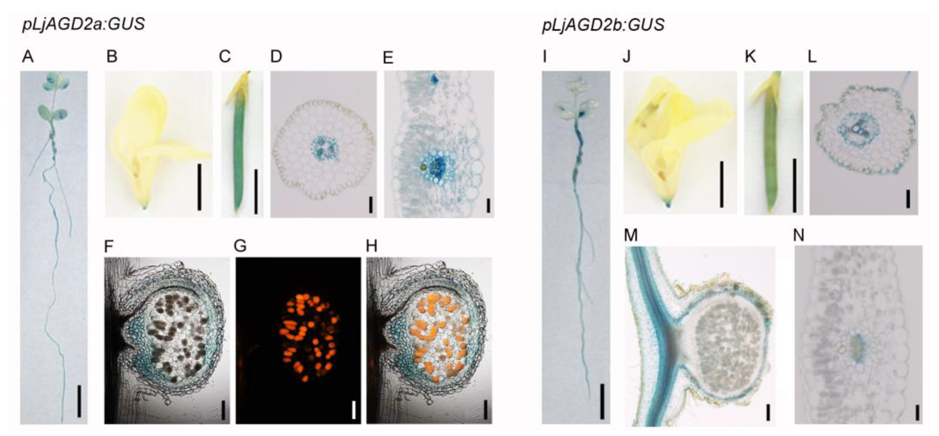
2.1. Expression Pattern of LjAGD2a in L. japonicus

Both AGD2 and ALD1 are encoded by single genes in Arabidopsis, and their amino acid sequences have high homology. However, there are four LjAGD2 homologs in the L. japonicus genome. LjALD1 and LjALD1L belong to the same cluster, while LjAGD2a and LjAGD2b are members of another cluster (Figure S1).
To investigate the function of AGD2 in L. japonicus, expression patterns of LjAGD2 genes were analyzed. Results of real-time PCR analysis showed that LjAGD2a and LjAGD2b were expressed in all the tested tissues (Figure S2). β-glucuronidase (GUS) staining in pLjAGD2a:GUS and pLjAGD2b:GUS plants corroborated the expression of LjAGD2a and LjAGD2b in the leaves, stems, roots, flowers, pods and nodules (Figure 1). In transverse sections, GUS signals were detected mainly in vascular tissues of root and leaf cells in both pLjAGD2b:GUS and pLjAGD2b:GUS plants (Figure 1D,F,L,N), but GUS expression was also present in the epidermis cells in pLjAGD2b:GUS roots (Figure 1L). Examination of nodule sections showed that both LjAGD2a and LjAGD2a were mostly expressed in vascular bundles (Figure 1F–H,M). These results suggest that LjAGD2a and LjAGD2b are largely expressed in the vascular tissues of the various plant organs in L. japonicus.

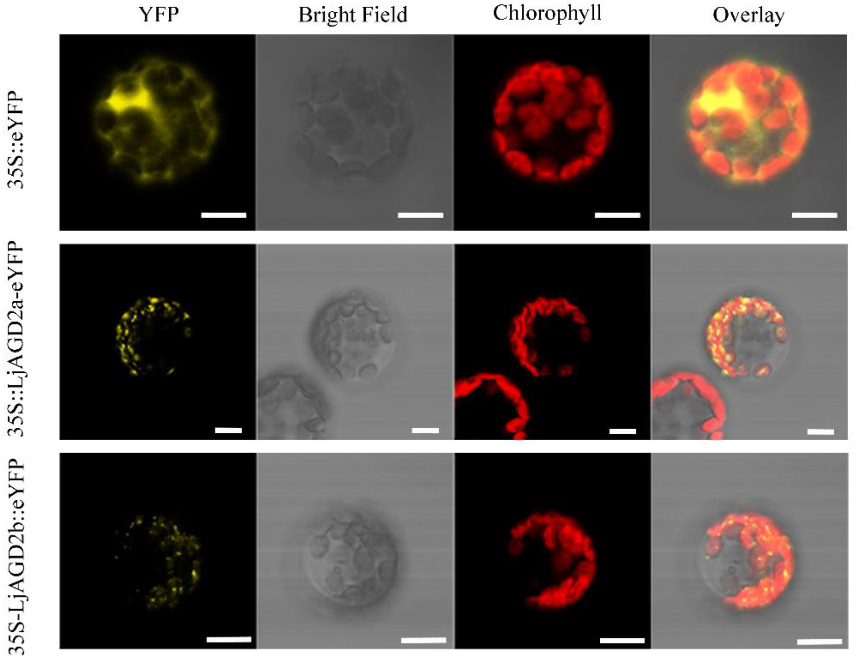
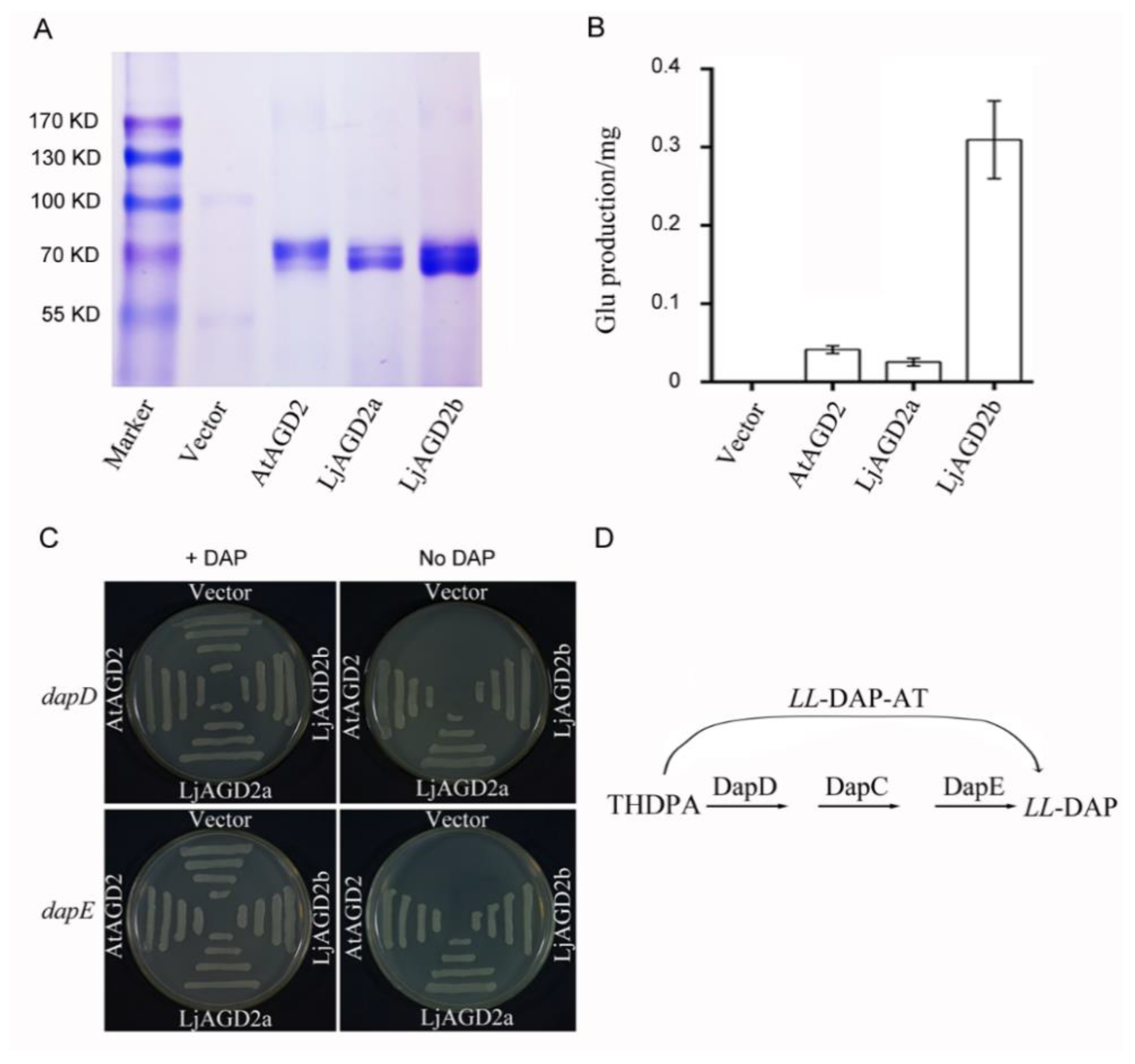
2.2. LjAGD2s Are Localized in Chloroplasts and Function as LL-Diaminopimelate Aminotransferases

To investigate where LjAGD2a might function in cells, LjAGD2a-eYFP (enhanced yellow fluorescent protein) and LjAGD2b-eYFP constructs were transiently expressed in Arabidopsis protoplasts. The results show that LjAGD2a and LjAGD2b were both located in chloroplasts (Figure 2), in accordance with their putative role in Lys synthesis as Lys is known to be synthesized in chloroplasts [14,15].
To determine the biochemical function of LjAGD2, CDSs of the LjAGD2a and LjAGD2b genes were inserted into an Escherichia coli expression vector. Molecular masses of tag-purified/cleaved recombinant proteins estimated by SDS-PAGE analysis matched the predicted sizes (in kDa) of LjAGD2a and LjAGD2b well (Figure 3A). Aminotransferase activities were assayed with these recombinant proteins using L-Lys and 2-oxoglutarate as substrates, with soluble protein extracts of E. coli containing vector alone as negative controls, and AtAGD2 as a positive control. The results showed that LjAGD2 proteins can catalyze formation of Glu from Lys and 2-oxoglutarate (Figure 3B and Figure S3), indicating that they are aminotransferases. To corroborate the conclusion that LjAGD2s are LL-DAP aminotransferase (LL-DAP-ATs) in plants, we used dapD and dapE mutants, which are auxotrophic for DAP in functional complementation assays [8]. The dapD and dapE mutant strains were transformed with either an empty plasmid or expression plasmids with LjAGD2a and LjAGD2b. The results showed that dapD and dapE mutant stains expressing AtAGD2, LjAGD2a or LjAGD2b can grow on medium without DAP, but not the negative controls (Figure 3C). In this experiment, AtAGD2 was used as a positive control. Therefore, LjAGD2a and LjAGD2b can bypass the succinylation and desuccinylation reactions required by E. coli to synthesize LL-DAP from THDPA (Figure 3D), strongly indicating that LjAGD2a and LjAGD2b are aminotransferases involved in Lys biosynthesis.

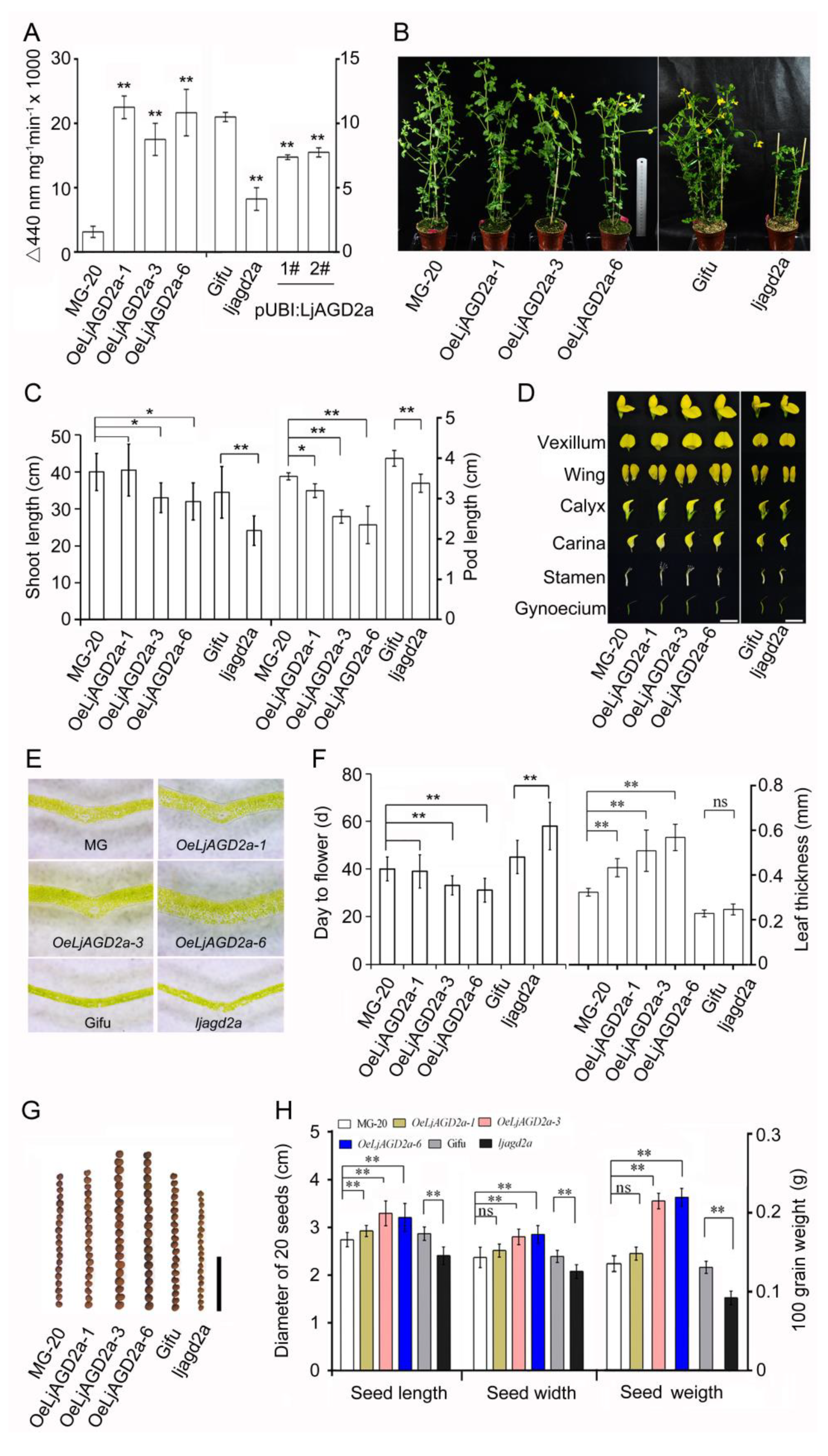
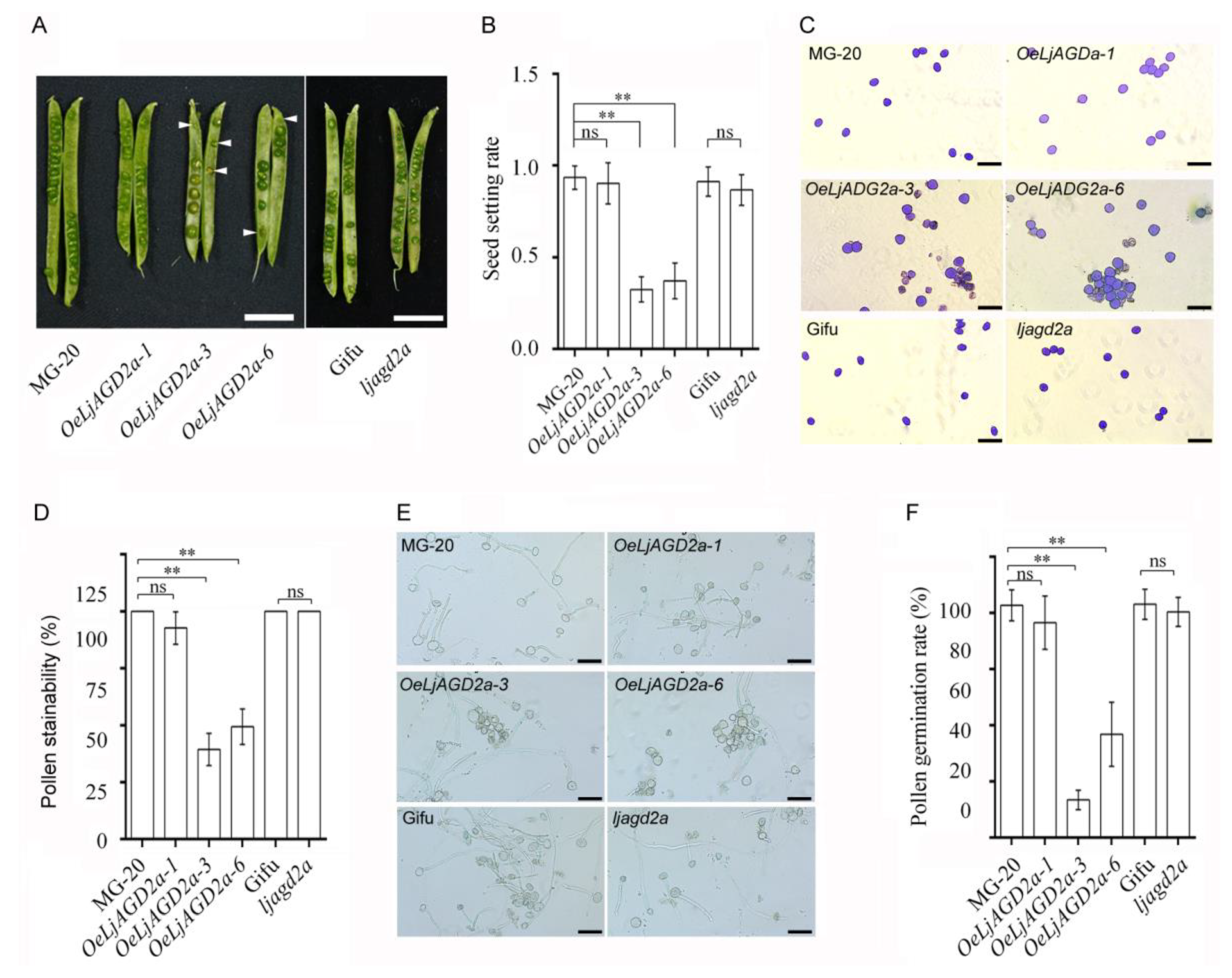
2.3. LjAGD2a Affects Plant Morphology and Development

To assess the effects of changing expression of LjAGD2 on L. japonicus, we obtained an insertional mutant line ljagd2a (LORE ID: 30001979, Figure S4A) and three LjAGD2a overexpression lines (designated OeLjAGD2a-1, OeLjAGD2a-3 and OeLjAGD2a-6). The expression of LjAGD2a in each line was detected by semi-quantitative or quantitative PCR (Figure S4B,C). We assayed LL-DAP-AT activity in leaves of the mutant and transgenic plants. The results showed that overexpression of LjAGD2a significantly increased, and the mutation of LjAGD2a significantly decreased LL-DAP-AT activity in L. japonicus. Overexpression of LjAGD2a in ljagd2a increased the transcription level of LjAGD2a and partially restored LL-DAP-AT activity (Figure 4A).
Compared to wildtype controls, ljagd2a mutants showed retarded growth, delayed flowering and smaller seeds, while overexpression of LjAGD2a in L. japonicus resulted in dwarf shoots, early flowering and larger seeds (Figure 4B,C,F–H). Additionally, OeLjAGD2a lines had shorter pods, larger floral organs and thicker leaves (Figure 4C–F). Owing to the roles of AtAGD2 in embryo development in Arabidopsis, we also assessed the effects of LjAGD2a on the seed setting rate and pollen fertility. No significant differences in seed setting rate and pollen fertility between the wildtype (Gifu B-129) and the ljagd2a mutant were observed (Figure 5). However, OeLjAGD2a-3 and OeLjAGD2a-6 plants had 60–75% lower seed setting rates than wildtype plants (Figure 5A,B). In addition, iodine-potassium iodide staining showed that wildtype pollen grains were round, whereas many OeLjAGD2a pollen grains were shrunken, irregular and wizened (Figure 5C). Nearly 100% of wildtype pollen grains became dark blue upon iodine staining, but the proportion of pollen grains from OeLjAGD2a-3 and OeLjAGD2a-6 was 35–50% (Figure 5D). After incubation on medium for 30 min at 37 °C, the germination rate of pollen from OeLjAGD2a-3 and OeLjAGD2a-6 plants was around 18–40%, whereas the rate of germination of wild type pollen was about 85% (Figure 5E,F). Clearly, overexpression of LjAGD2a substantially reduces the fertility of L. japonicus pollen.

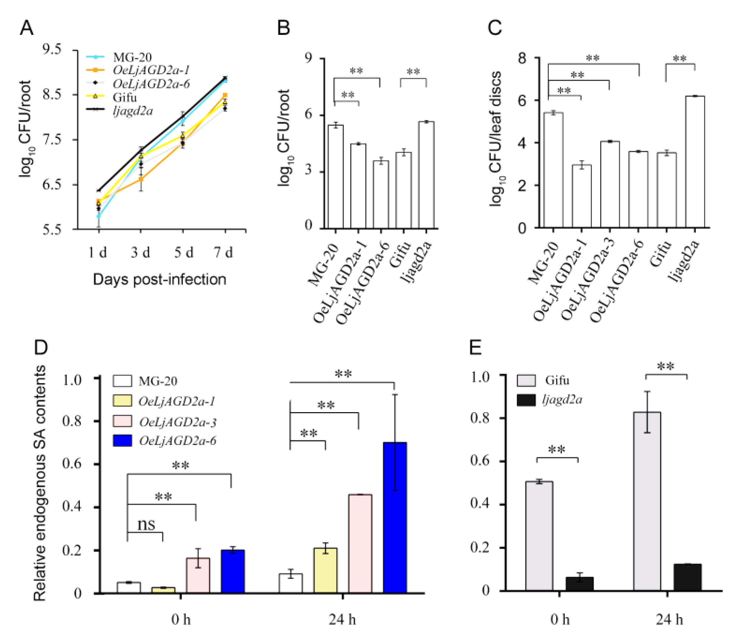
2.4. Alteration of LjAGD2a Expression in L. japonicus Changed Resistance to the Bacterial Pathogen R. Solanacearum

Here, to investigate the function of LjAGD2a in L. japonicus–pathogen interactions, we analyzed resistance phenotypes. Two-day-old seedlings were infected with R. solanacearum and its growth in the roots was monitored by counting the bacteria. We found no significant difference in densities of the bacteria in the overexpressing or mutant lines 1- and 3-days post-infection (dpi), but growth of R. solanacearum was dramatically inhibited on OeLjAGD2a roots at 5 dpi (Figure 6A). Ljagd2a was consistently more susceptible to R. solanacearum than its wildtype (Figure 6A,B). We also assayed the growth of R. solanacearum on detached leaves, and the results showed that OeLjAGD2a plants were more resistant and ljagd2a plants more susceptible to R. solanacearum than wildtype plants (Figure 6C). Simultaneous measurements of SA levels in leaves after infection with R. solanacearum showed that overexpression of LjAGD2a caused increases in SA contents in the leaves while the mutation of LjAGD2a resulted in decreased SA contents (Figure 6D,E). In view of divergent roles of Arabidopsis AGD2 homologs in defense signaling [14], we further investigated the resistance phenotype of ljald1 mutants, and the results showed that ljald1 mutants were more susceptible to R. solanacearum than wildtype plants (Figure S5).

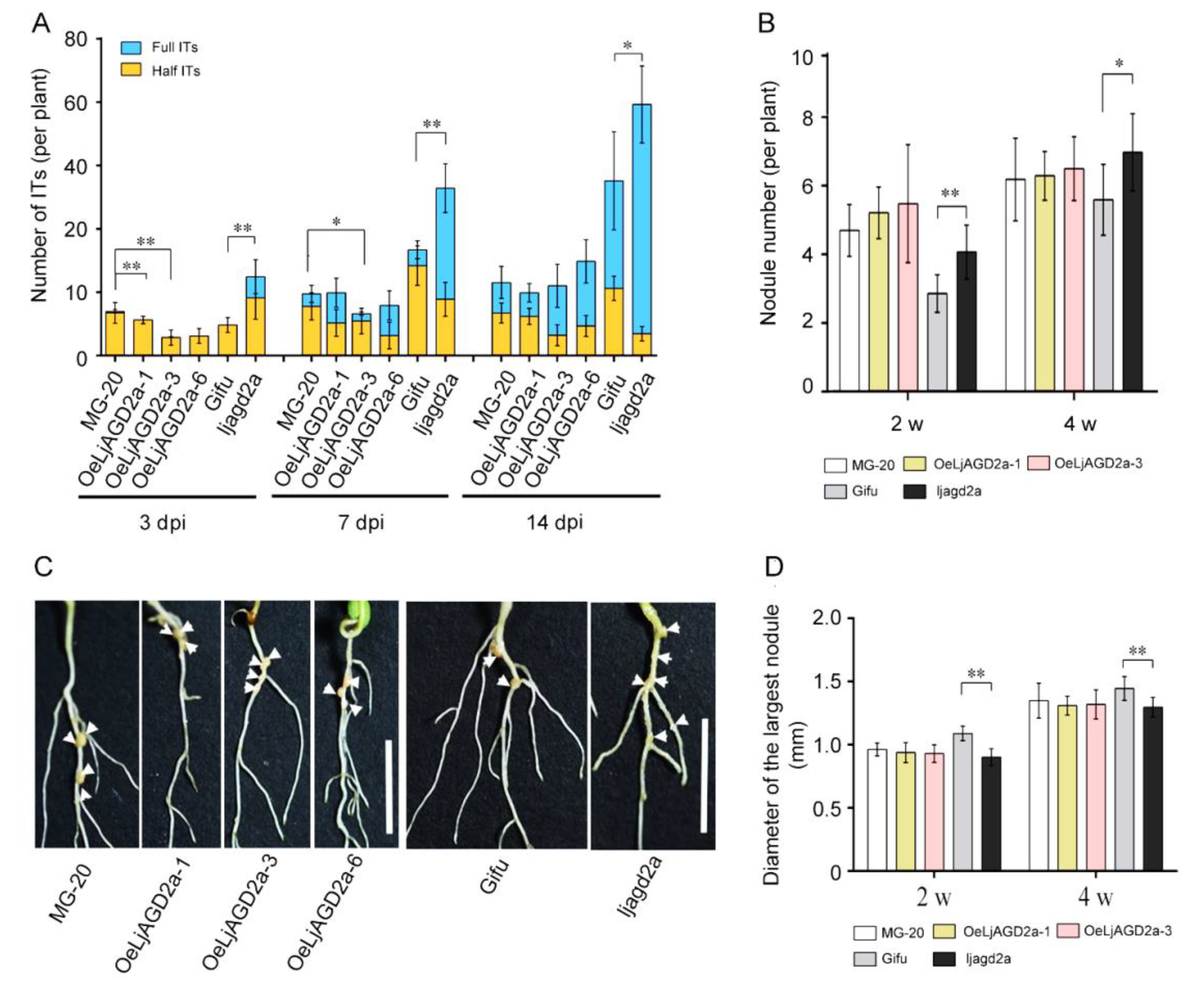
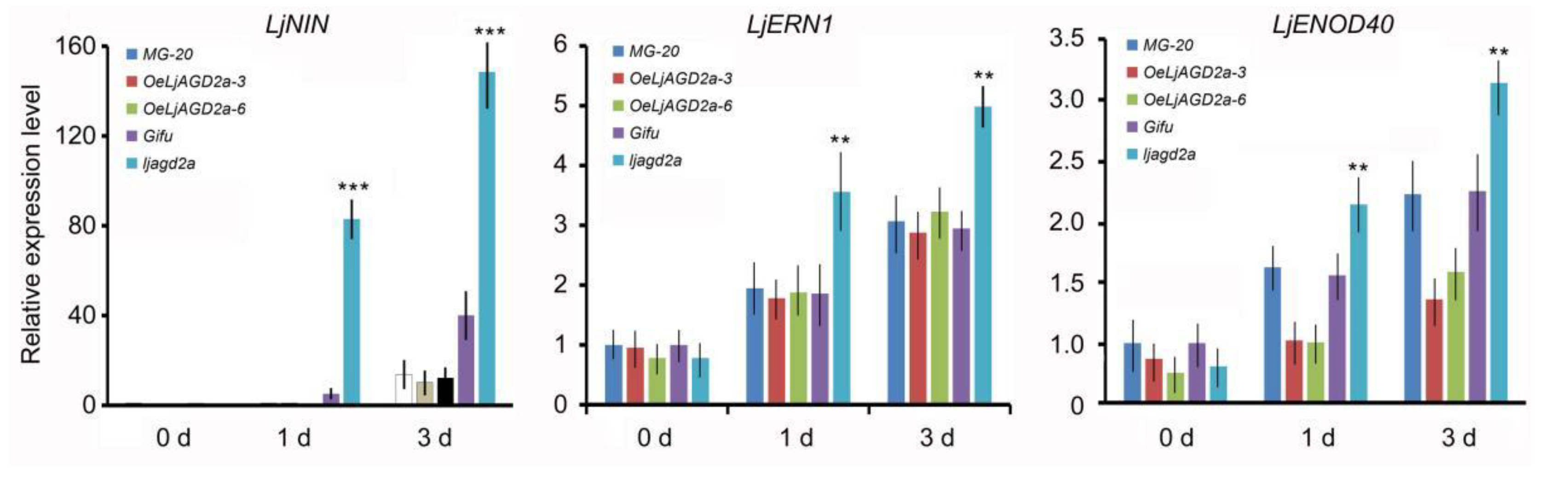
2.5. Knockout Mutation of LjAGD2a Increased Infection Thread and Nodule Numbers in L. japonicus

Next, nodulation phenotypes were observed. Compared to the wildtype, ljagd2a mutants had more infection threads and nodules after inoculation with Mesorhizobium loti (Figure 7A,B). In addition, nodules were smaller in ljagd2a plants than in the wildtype plants (Figure 7D). Although OeLjAGD2a plants had few infection threads at 3 and 7 days after M. loti inoculation, the number of infection threads at 14 days and nodule numbers at 2- and 4-weeks post-infection did not significantly differ from numbers in wildtype plants (Figure 7A–C). Therefore, overexpression of LjAGD2a may only delay infection of M. loti. Subsequent assessment of the expression of nodulin genes after M. loti infection showed that levels of LjENOD40, LjNIN and LjERN1 transcripts were significantly higher in ljagd2 plants but not significantly different in OeLjAGD2a lines, compared to their wildtype (Figure 8).

3. Discussion

Our experiments with ljagd2a insertion mutants and OeLjAGD2a plants showed that the Lys biosynthesis pathway plays an important role in the development and microbial interactions of L. japonicus (and likely other leguminous plants). The insights obtained are summarized in this section.

3.1. LjAGD2s Function as LL-DAP-Ats

AGD2 in Arabidopsis has LL-DAP aminotransferase (LL-DAP-AT) activity, which can directly convert THDPA to LL-DAP and is equivalent to the functions of DapD, DapC and DapE in prokaryotes [8]. Recombinant LjAGD2a and LjAGD2b have aminotransferase activity in vitro (Figure S3) and functional complementation of LjAGD2a and LjAGD2b in dapD and dapE mutants showed that LjAGD2s can function in the ‘forward direction’, towards synthesis of LL-DAP in E. coli. These results suggest that LjAGD2s have LL-DAP-AT activity. Alteration of LjAGD2a expression in L. japonicus also changed the LL-DAP-AT activity, corroborating LjAGD2s’ functionality as aminotransferases involved in the Lys biosynthetic direction in L. japonicus. In addition, LjAGD2s, such as AtAGD2, are located in the chloroplast, which is consistent with their roles in Lys synthesis in chloroplasts [14,16]. Therefore, we propose that LjAGD2s have the same biochemical functions in plants as AtAGD2.

3.2. LjAGD2s May Have Functional Redundancy in Regulation of Plant Development

Lys is an important signaling amino acid that regulates plant growth and responses to the environment [17]. In Arabidopsis, loss of AGD2 function renders embryos inviable [14], and a point mutation of agd2 leads to metabolic disorders and growth inhibition [12]. Here, we found that there are two AGD2 genes in L. japonicus and knockout of LjAGD2a both reduced LL-DAP-AT activity and suppressed growth of shoots. However, ljagd2a plants did not have clear abnormalities in embryo development, seeds or fertility (Figure 5). Therefore, we hypothesize that LjAGD2a and LjAGD2b may be functionally redundant in regulation of plant development. Reductions in LL-DAP-AT activity cause changes in levels of amino acids and other metabolites involved in Lys synthesis and related pathways [12,18,19], thus resulting in metabolic disorders that affect development of L. japonicus.

3.3. AGD2a Affects Disease Resistance and Nodulation via the Lys Biosynthesis Pathway

AGD2 suppresses SA-dependent defenses while ALD1 increases SA contents and defense signaling in Arabidopsis [10,14]. We found that ljald1 mutants were more susceptible than wildtype plants to R. solanacearum (Figure S5). However, contrary to the negative effect of AtAGD2 on defenses, knockout mutation of LjAGD2a reduced resistance of L. japonicus to the bacterial pathogen while its overexpression significantly increased resistance (Figure 6). We speculate that LjAGD2 is involved in plant disease resistance through its participation in Lys biosynthesis and hence regulation of Lys supply and associated metabolites. Similarly, we found elevated numbers of infection threads and nodules in ljagd2 plants following infections with M. loti, as in LjALD1 RNAi plants (Figure 7) [13]. Overexpression of salicylate hydroxylase NahG in M. truncatula and L. japonicus resulted in the reduction of endogenous SA levels and therefore a greater number of infection threads and nodules [19]. Consistent with this result, knockdown of LjALD1 decreased SA content to promote nodulation in L. japonicus [13]. We thus speculate that ljagd2, like ljald1, can affect Lys metabolism by attenuating SA-dependent defense signaling, and hence expression of nodulin genes. Small nodules in ljagd2a plants may result from an insufficient carbon source supply from host plants since mutation of AGD2 in Arabidopsis can reportedly cause a shift in amino acid metabolism and C/N imbalance [12,20].

4. Materials and Methods

4.1. Plant Growth Conditions and Treatments

Two ecotypes of L. japonicus, Gifu B-129 and MG-20, were mainly used in this study. LORE1 insertion mutant lines of ljagd2a (plant IDs in Lotus Base: 30001979) with an L. japonicus Gifu B-129 background were obtained from Lotus Base [21,22] (https://lotus.au.dk/, accessed on 11 November 2020). pLjAGD2a:GUS and pLjAGD2b:GUS lines were obtained by transformation of MG-20 with Agrobacterium tumefaciens strain AGL1. Plants were grown in growth chambers with the temperature maintained at 22 °C (light intensities about 100 µm m−2 s−1). To germinate seeds, they were treated with concentrated sulfuric acid for 7 min, sterilized with 1.5% sodium hypochlorite solution for 10 min, washed six times with sterile water and placed in sterile dishes for 2 d. To induce production of infection threads, 2-day-old seedlings were moved to 0.8% agar plates containing 1/2 B&D solution (Broughton and Dilworth 1971) for 2 days, then infected by adding 1 mL suspension of DsRed-labeled Mesorhizobium loti (OD600 = 0.01) and the excess liquid was sucked off 5 min later. The materials were then cultured in an artificially illuminated growth cabinet providing 22 °C, 16 h light and 18 °C, 8 h dark cycles with a constant relative humidity of 70% (GXZ-380, Ningbo Jiangnan Instrument Factory). Infection threads on the whole root were counted at different time points after inoculation with M. loti expressing DsRED under a fluorescence microscope (DMI4000B, Leica, Germany). Rhizobial strain was Mesorhizobium loti MAFF303099 and cultured in YM liquid medium (containing 10 g/L Mannitol, 3 g/L Yeast extract, 0.25 g/L K2HPO4, 0.2 g/L MgSO47H2O, 0.1 g/L NaCl, pH 6.8). YM liquid medium was added to 15 g/L agar to produce the solid medium.

4.2. Plasmid Constructs and Plant Transformation

Full-length LjAGD2a and LjAGD2b was amplified by PCR with the primers shown in Supplementary Table S1 using L. japonicus cDNA as a template. LjAGD2a fragments were digested with Nco I and EcoR I, then cloned into pSAT6-eYFP-N1 behind the CaMV 35S promoter to form 35S::LjAGD2a-eYFP.
Full-length LjAGD2a and LjAGD2b or fragments lacking signal peptides were amplified by PCR, again with primers shown in Supplementary Table S1 and using L. japonicus cDNA as template, digested with Nco I and Xho I, and cloned into pGEX-KG. The resulting recombinant plastids for functional complementation were transformed into E. coli strains dap AT980 (dapD) and dap AT984 (dapE) (The Coli Genetic Stock Center).
For construction of the pLjAGD2a:GUS and pLjAGD2b:GUS vector, a sequence upstream of the first initial ATG codon of LjAGD2a and LjAGD2b was amplified, respectively. The specific primer sequence is shown in Supplementary Table S1. After digestion by restriction enzymes, the cDNA fragments were cloned into pCAMBIA1391Z before the GUS gene. The resulting construct was introduced into Agrobacterium tumefaciens strain AGL1 by the freeze–thaw procedure. L. japonicus was transformed according to a previously described method [13]. Hypocotyls excised from L. japonicus MG-20 seedlings were infected with A. tumefaciens strain AGL1 harboring the pLjAGD2a:GUS or pLjAGD2b:GUS constructs. Regenerated plants showing hygromycin resistance were grown in vermiculite pots to harvest T1 seeds.

4.3. Functional Complementation of E. coli Dap Mutants and Enzyme Assays

Dap mutants were cultured in LB liquid medium containing DAP and 100 μg/mL ampicillin until their absorbance reached 0.4 to 0.6. Appropriate volumes of bacterial suspension were diluted 100-fold, then 10 μL of diluted bacterial solution was spread on solidified NZY medium (5 g/L NaCl, 2 g/L MgSO4·7H2O, 10 g/L casein hydrolysate, 5 g/L yeast extract, 15 g/L agar) supplemented with 0.2% (w/v) Ara and 100 μg/mL ampicillin with or without 50 mg/mL DAP. All plates were incubated at 37 °C for 12 h.
Recombinant plastids for functional complementation were also used for enzyme assays in vitro. For protein expression and purification, recombinant plastids were transformed into E. coli Rosetta strains. The resulting strains were grown on LB medium at 37 °C to OD600 = 0.5, then protein expression was induced by incubation with 0.1 mM isopropylthio-β-galactoside for 12 h at 20 °C. Cells were lysed by sonication in PBS buffer (8 g/L NaCl, 0.2 g/L KCl, 1.44 g/L Na2HPO4, 0.24 g/L KH2PO4, pH 8.0) with 1 mM PMSF. After centrifugation at 4 °C and 12,000 g for 30 min, the supernatant was filtered with a 0.45 μM filter, then added to 1 mL GST resin. The soluble fraction was washed three times with PBS buffer. Freshly prepared 10 mM reduced glutathione solution in 50 mM Tris-HCl, pH 8.0, was used to elute the target protein. The reaction system consisted of 50 mM Tris HCl (pH 8.0), 50 mM Lys, 50 mM 2-oxoglutarate (2-OG), 1 μM pyridoxal-5-phosphate, 100 mM MgCl2 and purified protein. After reaction at 37 °C for 6 h, anhydrous ethanol was added to a final concentration of 80% (v/v). After standing for 30 min, samples were centrifuged at 12,000 g for 2 min. The supernatant was dried, dissolved in 400 μL 0.01 M HCl and filtered through a 0.45 μm filter. Then, Glu was measured in 50 μL portions of the filtrate using a S-430D amino acid analyzer (Sykam, Germany), mobile phase of lithium citrate with pH sequentially rising from 2.9 to 4.2 and 8.0 (flow rate: 0.45 mL/min) and derivatization reagent flow rate of 0.25 mL/min. The temperatures of the column, post-column reaction equipment and automatic sampler were set at 38, 130 and 5 °C, respectively, and the eluate was monitored at 570 and 440 nm [23].
To assay enzyme activities in crude protein extracts, leaves were ground in liquid nitrogen, soluble proteins were dissolved in 100 mM HEPES.KOH (pH 7.6), and the resulting mixture was centrifuged at 12,000 g for 15 min, then subjected to buffer exchange using an Amicon Ultra 30,000 MWCO filter. The standard curve of protein concentration was obtained by the BSA method when A280 was used, and the extracted leaf protein concentration was obtained by standard curve. A 200 μL reaction system containing 100 mM HEPESKOH (pH 7.6), 0.5 mM amino donor LL-DAP, 2.0 mM 2-OG, 0.25 mg O-aminobenzaldehyde (OAB), and crude soluble protein was used. Reactions were incubated at 30 °C and the ΔA (440 nm) measured [24].

4.4. Phylogenetic Tree Construction

Multiple sequence alignments of amino acid sequences of LjAGD2s and other AGD2 and ALD1 orthologs in different plant species were performed using Clustalx 2.1 and DNAMAN 6.0 software. A phylogenetic tree was constructed by MEGA 6.0 using the neighbor-joining (NJ) method and 1000 bootstraps [25,26].

4.5. RNA Isolation and Expression Analysis

Total RNA was extracted from selected L. japonicus tissues using a HiPure Plant Mini Kit (Guangzhou Magen Biotechnology Co., Ltd., Guangzhou, China) following the manufacturer’s instructions, and the isolated RNA was treated with RNase-free Dnase I (Magen). RNA samples (2 μg) were used for reverse transcription with a GoScript™ Reverse Transcription System (Promega, Madison, WI, USA). Quantitative real-time PCR (qRT-PCR) analysis was performed using the primers listed in Supplementary Table S1, with LjATPase and LjUBC genes as internal controls. qRT-PCR was performed with a LightCycler 480 (Roche Scientific, Basel, Switzerland) and the following amplification conditions: 10 min at 95 °C, followed by 40 cycles of 95 °C for 5 s, 60 °C for 20 s and 72 °C for 20 s. The experiment was performed with three replicates and average measurements are presented.

4.6. Subcellular Localization of LjAGD2s

Protoplasts of Arabidopsis ecotype Columbia were isolated and transformed following a previously described method [26]. Fluorescence images were recorded with a META LSM510 laser scanning confocal microscope (Zeiss10) using a 63 × 1.40 oil objective. eYFP fluorescence was imaged at an excitation wavelength of 514 nm (30% power) and 527 nm emission wavelength. The autofluorescence of chloroplasts was observed using an excitation wavelength of 543 nm and emission wavelength of 562 nm.

4.7. Histochemical Staining and Microscopic Analysis

For histochemical assays, seedlings of pLjAGD2a::GUS or pLjAGD2b::GUS transgenic lines at selected growth stages were stained with GUS staining buffer (1 mM X-Gluc, 0.5 mM K3[Fe(CN)6], 0.5 mM K4[Fe(CN)6], 1 mM EDTA, pH 8.0, 0.1 mM PBS buffer, pH 7.0) at 37 °C for 6–8 h after vacuum infiltration for 15 min. For nodule sectioning, 2-week-old nodules were harvested after inoculation with DsRed-labeled M. loti, and sections (50 μm) of fresh nodules were cut with a Leica VT1200 S vibrating-blade microtome.
For the analysis of leaf thickness, leaves were fixed with 2% agar, and 50 μm slices were obtained, in 0.1× PBS buffer (8 g/L NaCl, 0.2 g/L KCl, 1.44 g/L Na2HPO4, 0.24 g/L KH2PO4, pH 8.0), using a Leica VT1200 S vibrating-blade microtome.

4.8. Observation of Pollen Morphology and Germination

For observations of pollen morphology and fertility, pollen grains were harvested from fully expanded flowers. After staining with an iodine–potassium iodide solution, they were viewed with a bright-field microscope. To assess the pollen germination rate, pollen grains were dispersed in a solution of 10% sucrose, 0.01% H3BO3 and 0.05% Ca(NO3)2·4H2O. After incubation at 37 °C for 30 min, germinated pollen grains were viewed with a bright-field microscope [27].

4.9. Cultivation of Bacteria and Plant Inoculation

Ralstonia solanacearum strain FQY_4 was cultured in SMSA liquid medium (containing 2.5 mL 1% polymyxin, 125 μL 1% crystal violet, 1.25 mL 1% TTC, 625 μL 1% bacitracin, 125 μL 0.1% penicillin, 125 μL 1% chloramphenicol and 5 μg/mL gentamicin per liter) to OD600 = 0.5–0.6, then 100 μL bacterial liquid was coated on SMSA solid medium and grown for 2 days [28]. The bacteria were suspended in sterile 10 mM MgCl2 to OD600 = 0.001. Portions (100 μL) of R. solanacearum suspension were injected into the back of fresh leaves (n > 15) of selected plant lines by a 1 mL sterile syringe. After inoculation, the leaves were stored in agar medium containing 10 ppm imidazole. After cultivation in the dark at 28 °C for 2 days, the leaves were exposed to light for 16 h at 22 °C and dark for 8 h at 18 °C for 2 days. Leaf discs were made with a puncher, washed with sterile water five times and ground with a pestle in 10 mM MgCl2. The resulting homogenates were fully mixed and continuously diluted. A 100 μL diluted bacterial solution was spread on SMSA solid medium, and the number of R. solanacearum colonies was counted 48 h later.

4.10. Measurement of Salicylic Acid and Free Amino Acid Contents

Salicylic acid was extracted as previously described [13]. For the extraction of amino acids, 1 g samples of leaves were taken just before the beginning and end of the photoperiod from the same position of each plant line, ground into homogenate with 10 mL 0.1 N HCl and agitated ultrasonically at 4 °C for 15 min. After a further hour of incubation, the homogenate was centrifuged at 3000× g for 30 min, and then absolute ethanol was added to the supernatant to a final concentration of 80% to precipitate protein. After further centrifugation at 3000× g for 30 min, the supernatant was transferred to another tube and dried under vacuum. The dry matter was dissolved in 400 μL of 0.01 N HCl, and the resulting solution was filtered using a 0.45 μm filter membrane. Finally, 50 μL portions of the filtrate were subjected to analysis with an automatic amino acid analyzer (Sykam S-430D, Munich, Germany) using the C4 free 41AA standard procedure, an LCA K07/Li 4.6 × 150 mm chromatographic column and analyte detection wavelengths of 570 and 440 nm.

4.11. Statistical Significance

The numbers of independent repetitions of each experiment are given in the corresponding figures and tables. Statistical significance was tested as described in the given figures and tables. For all assays, differences between samples or time points with probabilities < 0.05 and 0.01 are indicated in figures by single and double asterisks, respectively. SPSS Statistics V17.0 software was used for all statistical analyses.

5. Conclusions

Our results indicate that LjAGD2a functions as an LL-DAP aminotransferase. We examined how mutation of LjAGD2a and overexpression of LjAGD2a affected shoot, leaf, flower, pod, seed and pollen development, which suggests that LjAGD2a plays important roles in plant development. LjAGD2a, like LjALD1, has a positive role in plant–pathogen interaction and a negative role in legume–rhizobia symbiosis. Our results also indicate that LjAGD2a activates defense signaling via the Lys synthesis pathway, thereby participating in legume–microbe interaction.

Supplementary Materials

The following supporting information can be downloaded at: https://www.mdpi.com/article/10.3390/ijms23126863/s1.

Author Contributions

Conceptualization, Y.C. and G.W.; methodology, P.W.; software, M.Y.; validation, Y.C. and M.H.; formal analysis, H.J.; investigation, M.H. and C.S.; resources, P.W.; writing—original draft preparation, M.H.; writing—review and editing, Y.C.; visualization, M.L.; supervision, Y.C. and G.W.; project administration, Y.C. and G.W.; funding acquisition, Y.C. All authors have read and agreed to the published version of the manuscript.

Funding

This research was funded by National Natural Science Foundation of China (grant number 31570242), Guizhou Provincial Academician Workstation of Microbiology and Health ([2020]4004) and Guizhou Tobacco Company (2020XM03).

Institutional Review Board Statement

Not applicable.

Informed Consent Statement

Not applicable.

Data Availability Statement

Not applicable.

Acknowledgments

We would like to thank Jens Stougaard (Aarhus University) for providing the DsRED-labeled M. loti MAFF303099 and Liuti Cai (Guizhou Academy of Tobacco Science) for providing the pathogen strain Ralstonia solanacearum strain FQY_4.

Conflicts of Interest

The authors declare no conflict of interest.

References

  1. Hartmann, M.; Zeier, J. L-lysine metabolism to N-hydroxypipecolic acid: An integral immune-activating pathway in plants. Plant J. 2018, 96, 5–21. [Google Scholar] [CrossRef] [PubMed] [Green Version]
  2. Arruda, P.; Kemper, E.L.; Papes, F.; Leite, A. Regulation of lysine catabolism in higher plants. Trends Plant Sci. 2000, 5, 324–330. [Google Scholar] [CrossRef]
  3. Bunsupa, S.; Yamazaki, M.; Saito, K. Quinolizidine alkaloid biosynthesis: Recent advances and future prospects. Front. Plant Sci. 2012, 3, 239. [Google Scholar] [CrossRef] [PubMed] [Green Version]
  4. Arruda, P.; Barreto, P. Lysine catabolism through the saccharopine pathway: Enzymes and intermediates involved in plant responses to abiotic and biotic stress. Front. Plant Sci. 2020, 11, 587. [Google Scholar] [CrossRef]
  5. Hartmann, M.; Zeier, T.; Bernsdorff, F.; Reichel-Deland, V.; Kim, D.; Hohmann, M.; Scholten, N.; Schuck, S.; Bräutigam, A.; Hölze, T.; et al. Flavin monooxygenase-generated N-hydroxypipecolic acid is a critical element of plant systemic immunity. Cell 2018, 173, 456.e16–469.e16. [Google Scholar] [CrossRef] [PubMed] [Green Version]
  6. Chen, Y.C.; Holmes, E.C.; Rajniak, J.; Kim, J.G.; Tang, S.; Fischer, C.R.; Mudgett, M.B.; Sattely, E.S. N-hydroxy-pipecolic acid is a mobile metabolite that induces systemic disease resistance in Arabidopsis. Proc. Natl. Acad. Sci. USA 2018, 115, E4920–E4929. [Google Scholar] [CrossRef] [Green Version]
  7. Zhang, X.; Song, Z.Q.; Liu, T.; Guo, L.L.; Li, X.F. De novo assembly and comparative transcriptome analysis provide insight into lysine biosynthesis in Toona sinensis Roem. Int. J. Genom. 2016, 2016, 6735209. [Google Scholar] [CrossRef] [Green Version]
  8. Hudson, A.O.; Singh, B.K.; Leustek, T.; Gilvarg, C. An LL-diaminopimelate aminotransferase defines a novel variant of the lysine biosynthesis pathway in plants. Plant Physiol. 2006, 140, 292–301. [Google Scholar] [CrossRef] [Green Version]
  9. Ding, P.; Rekhter, D.; Ding, Y.; Feussner, K.; Busta, L.; Haroth, S.; Xu, S.; Li, X.; Jetter, R.; Feussner, I.; et al. Characterization of a pipecolic acid biosynthesis pathway required for systemic acquired resistance. Plant Cell 2016, 28, 2603–2615. [Google Scholar] [CrossRef] [Green Version]
  10. Rate, D.N.; Greenberg, J.T. The Arabidopsis aberrant growth and death2 mutant shows resistance to Pseudomonas syringae and reveals a role for NPR1 in suppressing hypersensitive cell death. Plant J. 2001, 27, 203–211. [Google Scholar] [CrossRef]
  11. Hudson, A.O.; Bless, C.; Macedo, P.; Chatterjee, S.P.; Singh, B.K.; Gilvarg, C.; Leustek, T. Biosynthesis of lysine in plants: Evidence for a variant of the known bacterial pathways. BBA 2005, 1721, 27–36. [Google Scholar] [CrossRef] [PubMed]
  12. Cavalcanti, J.H.F.; Kirma, M.; Barros, J.A.S.; Quinhones, C.G.S.; Pereira-Lima, I.A.; Obata, T.; Nunes-Nesi, A.; Galili, G.; Fernie, A.R.; Avin-Wittenberg, T.; et al. An L,L-diaminopimelate aminotransferase mutation leads to metabolic shifts and growth inhibition in Arabidopsis. J. Exp. Bot. 2018, 69, 5489–5506. [Google Scholar] [CrossRef] [PubMed]
  13. Chen, W.; Li, X.; Tian, L.; Wu, P.; Li, M.; Jiang, H.; Chen, Y.; Wu, G. Knockdown of LjALD1, AGD2-like defense response protein 1, influences plant growth and nodulation in Lotus japonicus. J. Integr. Plant Biol. 2014, 56, 1034–1041. [Google Scholar] [CrossRef] [PubMed]
  14. Song, J.T.; Lu, H.; Greenberg, J.T. Divergent roles in Arabidopsis thaliana development and defense of two homologous genes, aberrant growth and death2 and AGD2-LIKE DEFENSE RESPONSE PROTEIN1, encoding novel aminotransferases. Plant Cell 2004, 16, 353–366. [Google Scholar] [CrossRef] [Green Version]
  15. Cecchini, N.M.; Jung, H.W.; Engle, N.L.; Tschaplinski, T.J.; Greenberg, J.T. ALD1 regulates basal immune components and early inducible defense responses in Arabidopsis. Mol. Plant Microbe Interact. 2015, 28, 455–466. [Google Scholar] [CrossRef] [Green Version]
  16. Bryan, J.K. 5—Advances in the biochemistry of amino acid biosynthesis. In Intermediary Nitrogen Metabolism; Miflin, B.J., Lea, P.J., Eds.; Academic Press: San Diego, CA, USA, 1990; Volume 16, pp. 161–195. [Google Scholar]
  17. Galili, G. New insights into the regulation and functional significance of lysine metabolism in plants. Annu. Rev. Plant Biol. 2002, 53, 27–43. [Google Scholar] [CrossRef]
  18. Yang, Q.; Zhao, D.; Liu, Q. Connections between amino acid metabolisms in plants: Lysine as an example. Front. Plant Sci. 2020, 11, 928. [Google Scholar] [CrossRef]
  19. Stacey, G.; McAlvin, C.B.; Kim, S.Y.; Olivares, J.; Soto, M.J. Effects of endogenous salicylic acid on nodulation in the model legumes Lotus japonicus and Medicago truncatula. Plant Physiol. 2006, 141, 1473–1481. [Google Scholar] [CrossRef] [Green Version]
  20. Zhu, X.H.; Galili, G. Lysine metabolism is concurrently regulated by synthesis and catabolism in both reproductive and vegetative tissues. Plant Physiol. 2004, 135, 129–136. [Google Scholar] [CrossRef] [Green Version]
  21. Malolepszy, A.; Mun, T.; Sandal, N.; Gupta, V.; Dubin, M.; Urbanski, D.; Shah, N.; Bachmann, A.; Fukai, E.; Hirakawa, H.; et al. The LORE1 insertion mutant resource. Plant J. 2016, 88, 306–317. [Google Scholar] [CrossRef] [Green Version]
  22. Mun, T.; Bachmann, A.; Gupta, V.; Stougaard, J.; Andersen, S.U. Lotus Base: An integrated information portal for the model legume Lotus japonicus. Sci. Rep. 2016, 6, 39447. [Google Scholar] [CrossRef] [Green Version]
  23. Chen, Y.; Fu, X.; Mei, X.; Zhou, Y.; Cheng, S.; Zeng, L.; Dong, F.; Yang, Z. Proteolysis of chloroplast proteins is responsible for accumulation of free amino acids in dark-treated tea (Camellia sinensis) leaves. J. Proteom. 2017, 157, 10–17. [Google Scholar] [CrossRef] [PubMed]
  24. Tamura, K.; Peterson, D.; Peterson, N.; Stecher, G.; Nei, M.; Kumar, S. MEGA5: Molecular evolutionary genetics analysis using maximum likelihood, evolutionary distance, and maximum parsimony methods. Mol. Biol. Evol. 2011, 28, 2731–2739. [Google Scholar] [CrossRef] [PubMed] [Green Version]
  25. Tian, L.; Liu, L.; Yin, Y.; Huang, M.; Chen, Y.; Xu, X.; Wu, P.; Li, M.; Wu, G.; Jiang, H.; et al. Heterogeneity in the expression and subcellular localization of POLYOL/MONOSACCHARIDE TRANSPORTER genes in Lotus japonicus. PLoS ONE 2017, 12, e0185269. [Google Scholar] [CrossRef] [PubMed] [Green Version]
  26. Yin, Y.; Tian, L.; Li, X.; Huang, M.; Liu, L.; Wu, P.; Li, M.; Jiang, H.; Wu, G.; Chen, Y. The role of endogenous thiamine produced via THIC in root nodule symbiosis in Lotus japonicus. Plant Sci. 2019, 283, 311–320. [Google Scholar] [CrossRef]
  27. Qin, S.; Tang, Y.; Chen, Y.; Wu, P.; Li, M.; Wu, G.; Jiang, H. Overexpression of the starch phosphorylase-like gene (PHO3) in Lotus japonicus has a profound effect on the growth of plants and reduction of transitory starch accumulation. Front. Plant Sci. 2016, 7, 1315. [Google Scholar] [CrossRef]
  28. Tan, X.D.; Dai, X.Q.; Chen, T.; Wu, Y.S.; Yang, D.; Zheng, Y.X.; Chen, H.L.; Wan, X.R.; Yang, Y. Complete genome sequence analysis of Ralstonia solanacearum strain PeaFJ1 provides insights into its strong virulence in peanut plants. Front Microbiol. 2022, 13, 830900. [Google Scholar] [CrossRef]
Figure 1. Expression pattern of LjAGD2s in L. japonicus. (AH) GUS histochemical staining of pLjAGD2a:GUS plants. (A) GUS histochemical staining of a 2-week-old pLjAGD2a:GUS seedling. (B) GUS staining of a flower of a pLjAGD2a:GUS plant. (C) GUS staining of a pod of a pLjAGD2a:GUS plant. (D) Section of a root of a 2-week-old pLjAGD2a:GUS plant. (E) Section of a leaf of a 2-month-old pLjAGD2a:GUS plant. (FH) Section of a nodule of a pLjAGD2a:GUS plant 2 weeks after inoculation with Mesorhizobium loti expressing DsRED. GUS histochemical staining of the nodule in (F), Bacteriods in the infection cells expressing red fluorescence in (G), Overlap of F and G images in (H). (IN) Histochemical staining of pLjAGD2b:GUS plants. (I) GUS histochemical staining of a 2-week-old pLjAGD2b:GUS seedling. (J) GUS staining of a flower of a pLjAGD2b:GUS plant. (K) GUS staining of a pod of a pLjAGD2b:GUS plant. (L) Section of a root of a 2-week-old pLjAGD2b:GUS plant. (M) Section of a nodule of a 2-week-old pLjAGD2b:GUS plant inoculated with M. loti. (N) Section of a leaf of a 2-month-old pLjAGD2b:GUS plant. Sixty seedlings were subjected to GUS staining as illustrated in (A,I), with more than 90% showing the phenotype. Flowers and pods collected from more than 10 individual plants were used for the GUS staining illustrated in (B,C,J,K). For sections, more than six samples were used.
Ijms 23 06863 g001
Figure 2. Subcellular localization of LjAGD2a and LjAGD2b. LjAGD2a-eYFP and LjAGD2b-eYFP images indicating the localization of LjAGD2a and LjAGD2b in the chloroplasts of Arabidopsis protoplasts. The top row of images indicates the control. Scale bar = 10 µm.
Ijms 23 06863 g002
Figure 3. Aminotransferase activities of AGD2s in E. coli. (A) Electrophoretic patterns of recombinant proteins purified using a Ni2+-nitrilotriacetic acid column. (B) Aminotransferase activities of AGD2s, assayed using 50 mM Lys and 50 mM 2-oxoglutarate as substrate and co-substrate, respectively. Aminotransferase activity was determined by measuring the concentration of the reaction product Glu. Bars indicate standard deviations (n = 3). (C) Complementation of dap mutants with AGD2. Strains AT980 (dapD mutant) and AT984 (dapE mutant) were transformed with either the plasmid vector (pGEX-KG) or an LjAGD2a, LjAGD2b and AtAGD2 (At4g33680) expression plasmid (pGEX-KG-AGD2). Colonies were selected on LB medium with 50 µg/mL DAP and 100 µg/mL ampicillin. Individual colonies were then plated onto NZY medium supplemented with 0.2% (w/v) Ara with or without 50 µg/mL DAP. The cultures were grown at 37 °C for 12 h. (D) Diagram of the DAP pathway in E. coli with the reaction catalyzed by LL-DAP-AT indicated.
Ijms 23 06863 g003
Figure 4. Morphological effects of changing expression of LjAGD2a in L. japonicus. (A) LL-diaminopimelate aminotransferase (LL-DAP-AT) activities, showing a drastic reduction in ljagd2a and a significant increase in OeLjAGD2a lines (means with error bars indicating SD of five biological replicates). (B) Representative photograph of 8-week-old plants showing that OeLjAGD2a and ljagd2a plants had shorter roots than wild type plants (scale bar = 10 cm). (C) Shoot lengths, showing retarded growth of OeLjAGD2a and ljagd2a lines (Left). Both OeLjAGD2a and ljagd2a lines also produced shorter pods than wildtype plants (Right). (D) Photograph of 10-week-old plants showing that OeLjAGD2a plants had bigger flowers than wildtype plants, but insertional mutation of LjAGD2a did not change the flower size. Scale bar = 1 cm. (E) Sections showing that OeLjAGD2a plants had thick leaves. Scale bar = 1 mm. (F) OeLjAGD2a lines exhibited early flowering while insertional mutation of LjAGD2a delayed flowering (Left). Bar chart confirming that leaves of OeLjAGD2a lines were significantly thicker than wildtype leaves, but ljagd2a leaves did not significantly differ from wildtype leaves in thickness (Right). (G) Effects of changing LjAGD2a expression on seed morphology. Scale bar = 1 cm. (H) Seed sizes showing that LjAGD2a overexpression led to larger seeds, while ljagd2a seeds were smaller than wildtype seeds (Left). Measurement of 100-kernel weights of seeds showing that LjAGD2a overexpression and mutation resulted in increases and reductions in seed weight, respectively (Right). In the tests used to obtain results presented in (C,F,H), n = 20–30 per treatment. *, ** and ns indicate p < 0.05; p < 0.01 and no significant difference, respectively, according to the Duncan test. Error bars indicate the SD of three biological replicates.
Ijms 23 06863 g004
Figure 5. Effects of changing expression of LjAGD2a on fertility in L. japonicus. (A) Immature seeds in OeLjAGD2a siliques. Aborted dead seeds are indicated by arrows. Scale bar = 1 cm. (B) Measured seed setting rates, which were dramatically decreased in OeLjAGD2a plants, relative to wildtype rates, but not significantly altered in ljagd2a mutants (means obtained from analyses of more than 50 siliques with standard deviations: ** indicate p < 0.01, respectively, according to the Duncan test). (C) Photograph showing morphology of pollen grains. (D) Stainability of pollen by iodine-potassium iodide solution, indicating viability. (E) Photograph showing results of in vitro pollen germination assays. (F) Pollen germination rates, based on analyses of pollen grains from 5 to 7 anthers of each of 8 to 10 plants (** and ns indicate p < 0.01 and no significant difference, respectively, according to the Duncan test). Scale bar = 50 µm in (C,D). Error bars in (B,D,F) indicate the SD of three biological replicates.
Ijms 23 06863 g005
Figure 6. LjAGD2a promotes disease resistance in L japonicus. (A) Growth curves of R. solanacearum strains on the roots of transgenic ljagd2a lines, mutant lines and their respective wildtypes (MG-20 and Gifu129). Plants were infected with R. solanacearum at indicated time points. R. solanacearum grew more rapidly in ljagd2a plants than in their wildtype. In the first 3 days of pathogen infection, there was no significant difference in growth of R. solanacearum between OeLjAGD2a and MG-20 plants, but the pathogen’s growth was inhibited in OeLjAGD2a plants at 5 dpi. (B) Numbers of colony forming units (CFU) showing the disease susceptibility of roots. The indicated genotypes were inoculated with R. solanacearum by including the bacterium at a density equivalent to OD600 = 0.0002 in the growth medium and assaying the bacteria at 5 dpi. (C) Numbers of colony forming units (CFU) showing the disease susceptibility of detached leaves. Overexpression of LjAGD2a suppressed and its insertion mutation promoted growth of the pathogen. Detached leaves from 8-week-old plants were infected with R. solanacearum. (D) Total SA levels in OeLjAGD2a plants 0 and 24 h post-infection. (E) Total SA levels in ljagd2a mutants 0 and 24 h post-infection. Overexpression of LjAGD2a increased and its mutation reduced total SA levels after pathogen infection. 6–10 roots/leaves were used in each replicate and Error bars represent the SD of three replicates per genotype in (AC). In experiments providing results shown in (D,E) each sample had three to four replicates and error bars represent the SD of 3–4 replicates. In A–E, ** indicates p < 0.01 according to the Duncan test.
Ijms 23 06863 g006
Figure 7. Changes in LjAGD2a expression affect L japonicus nodulation. (A) Numbers of infection threads (ITs) in transgenic plants and insertion mutants. Consistently higher numbers of ITs, relative to numbers in wildtype plants, were found in ljagd2a plants after M. loti inoculation. We also found fewer ITs in OeLjAGD2a plants at 3 days post-infection (dpi), but not 7 or 14 dpi. (B) Nodule numbers were higher in ljagd2a plants, but unchanged in OeLjAGD2a lines, relative to numbers in wildtype plants. (C) Nodules on roots 2 weeks after inoculation of plants. White arrows indicate nodules on the roots. (D) Measurements of the largest nodules on plants 4 weeks after inoculation with M. loti showing that mutation of LjAGD2a reduced nodule size. 15–20 plants were used in each replicate and three replicates per genotype were used in experiments providing data presented in A, B and D (±SD). * and ** indicate p < 0.5 and p < 0.01, respectively, according to the Duncan test.
Ijms 23 06863 g007
Figure 8. Levels of transcript of nodulin genes—the nodule inception gene (LjNIN), ERF required for nodulation 1 (LjERN1) and early nodulin gene (LjENOD40)—in OeLjAGD2 plants and mutants. The expression of LjNIN, LjERN1 and LjENOD40 was increased in ljagd2a mutants after inoculation with M. loti, but overexpression of LjAGD2a did not change the expression of nodulin genes. The data were normalized using expression of Actin as an internal control. The Actin-normalized value for the 0-day wildtype sample was set to 1. Presented data are averages of triplicate RNA preparations (n = 15, ±SD). ** and *** indicate p < 0.01 and p < 0.001, respectively, according to the Duncan test.
Ijms 23 06863 g008
Publisher’s Note: MDPI stays neutral with regard to jurisdictional claims in published maps and institutional affiliations.

