1. Introduction
Orthopedic implants, such as prostheses, bone autografts, and allografts, are available in clinical settings to treat bone defects in non-union fractures, trauma, or osteoporosis, and have limitations due to their physical performance in healing the defects. This is a major socioeconomic problem regarding insubstantiality and weak recovery and is associated with high health care costs. In the EU only, clinical failure from non-union bone defects resulting from impaired osteogenesis with existing bone graft transplantations was estimated to be 1 million recipients in 2017. The annual number of fracture casualties is expected to rise from 2.7 million in 2017 to 3.3 million in 2030, corresponding to a 23% increase [
1]. Bone autografts remain the gold standard for treating patients with bone defects and have shown promising outcomes, but they are limited by their availability and donor site morbidity. The field of bone tissue engineering (BTE) is a rapidly developing area in orthopedic regenerative medicine that focuses on alternative treatment options that will solve existing clinical treatment problems over time. Standard approaches in BTE target the route of osteogenesis, utilizing osteoinductive and osteoconductive biomaterials at the site of implantation with osteoprogenitor cells [
2]. However, this strategy is primarily associated with inadequate vascularization and integration with the host tissue. Natural bone healing at the site of the defect involves mostly three vital phases. First, in a reactive phase, blood vessels come in proximity to an injury and constrict to stop the bleeding process, resulting in a clot formation, allowing fibroblasts to expand and form granular tissue. Second, in the reparative phase, fibroblasts from the granular tissue differentiate into primary chondroblasts capable of producing hyaline cartilage. Third, hyaline cartilage formation connects the gap between the fracture site and results in a callus development. The hyaline cartilage at later stages gets replaced by the lamellar bone. This process of replacement is known as endochondral ossification [
3].
The current challenges faced in clinical therapies for the repair of large bone defects are to develop sustainable tissue engineered (TE) constructs that can mimic the natural structure and functional aspects of bone tissue—providing biomechanical and biological signals that can stimulate the mechanism of bone remodeling as occurring in the native state under physiological settings. Several requirements have to be fulfilled when developing clinically viable TE bone constructs, such as biocompatibility, controlled degradation, high porosity, and permeability for cells to infiltrate, vascularize, and integrate into the host when implanted [
4,
5]. The functional BTE paradigm highlights these TE constructs’ fulfillment to perform osteoconduction, osteoinduction, and osseointegration after host implantation, along with the functions as mentioned above.
Over the last decades, substantial attention was given to understand the components of the extracellular matrix (ECM) of skeletal tissues, like bone and cartilage in the area of orthopedic regenerative medicine to mimic and design ECM-like biomaterials [
6,
7,
8]. ECM is the core constituent of connective tissues like cartilage and bone that regulates the tissue structure’s maintenance and organization. ECM controls both mechanical strength and structural rigidity and maintains tissue homeostasis. Engineered bone based on endochondral ossification gained significant interest over the last decade [
9]. The formation of long bones stimulates the idea of reverse engineering bone via endochondral ossification during embryonic development and fracture healing [
10]. During long bone formation, mesenchymal stem cells (MSCs) aggregate and differentiate into chondrocytes, which then produce a cartilaginous template rich in collagen II (COLII) and glycosaminoglycans (GAGs). Eventually, the chondrocytes undergo hypertrophic differentiation and ossify into a mineralized matrix. This denotes that hypertrophic chondrocytes mediate the undeviating path for endochondral bone formation [
11]. However, challenges in tissue-engineered bone constructs include functionality under the mechanical environment inside in vivo conditions. Engineered bone constructs must withstand higher mechanical loads under physiological conditions [
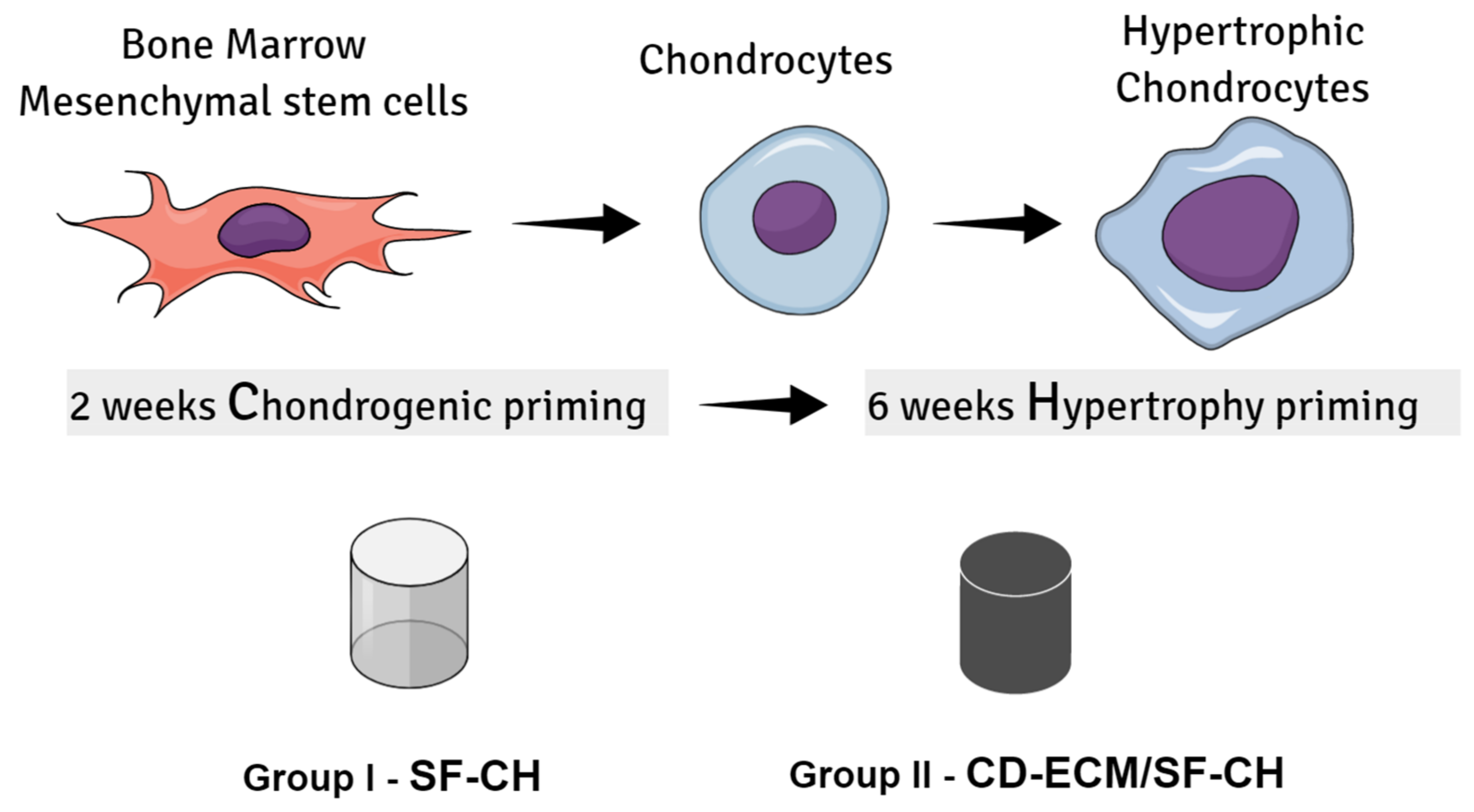
12]. Therefore, engineered bone’s biomechanical properties are an essential factor in the development of functional grafts for clinical therapies. Our current study aimed to modulate endochondral ossification-driven mineralization by human bone marrow-derived mesenchymal stem cells (hBMSCs) in a decellularized cartilage-derived extracellular matrix (CD-ECM), incorporating silk fibroin (SF) as a hybrid scaffold in vitro. RT-qPCR determined the gene expression for hypertrophic and osteogenic markers, and to quantification of the mineralized tissue formation µCT scans was performed (
Figure 1).
3. Discussion
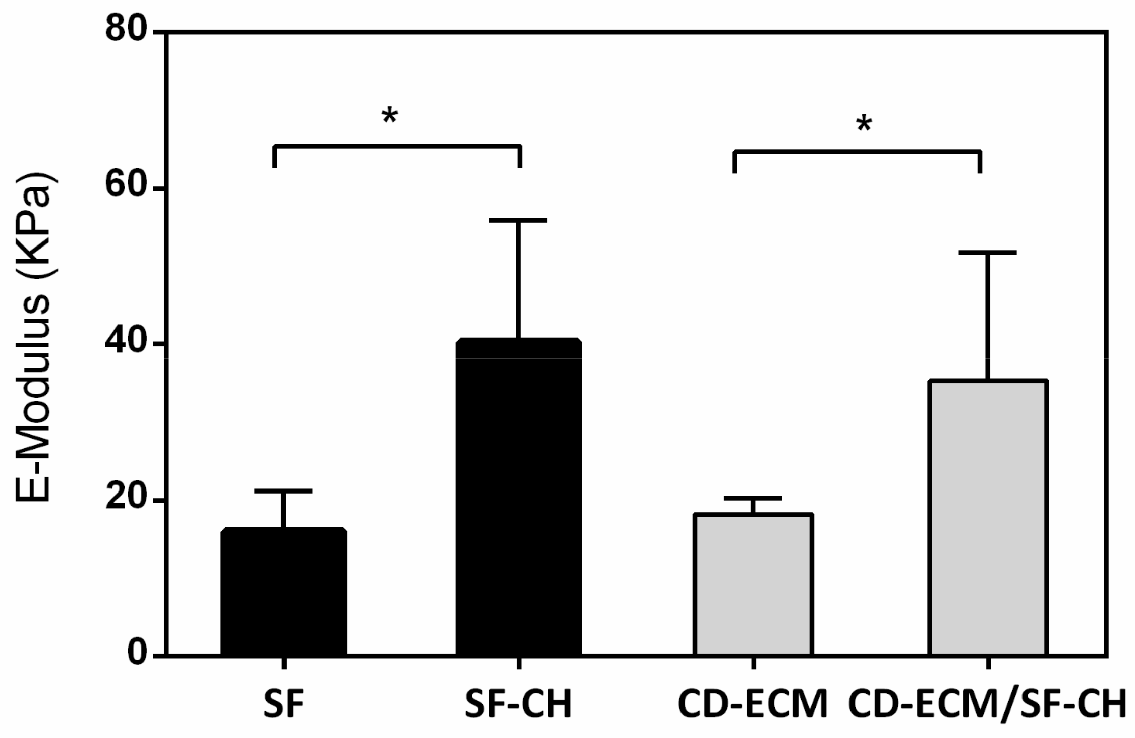
The present study contemplated to develop an in vitro model of bone regeneration mimicking the endochondral ossification process by modulating the extracellular environment in tissue-engineered scaffolds. We investigated this approach by application of a decellularized cartilage-derived extracellular matrix (CD-ECM) onto silk fibroin (SF) as hybrid scaffolds (CD-ECM/SF), with appropriate mechanical properties to favor mineralization of human BMSCs. We found that incorporation of CD-ECM to SF scaffolds had a significant effect on early and late hypertrophy state of differentiation of hBMSC’s and expression of osteogenic markers compared to the same groups cultured without application of CD-ECM. The results indicate that the osteogenic potential, based on calcium phosphate (CaP) crystal nucleation and mineralized tissue formation of hBMSCs, was constructively increased by utilizing chondrogenic priming, followed by hypertrophic priming, and it was sufficient with no further osteogenic induction being required.
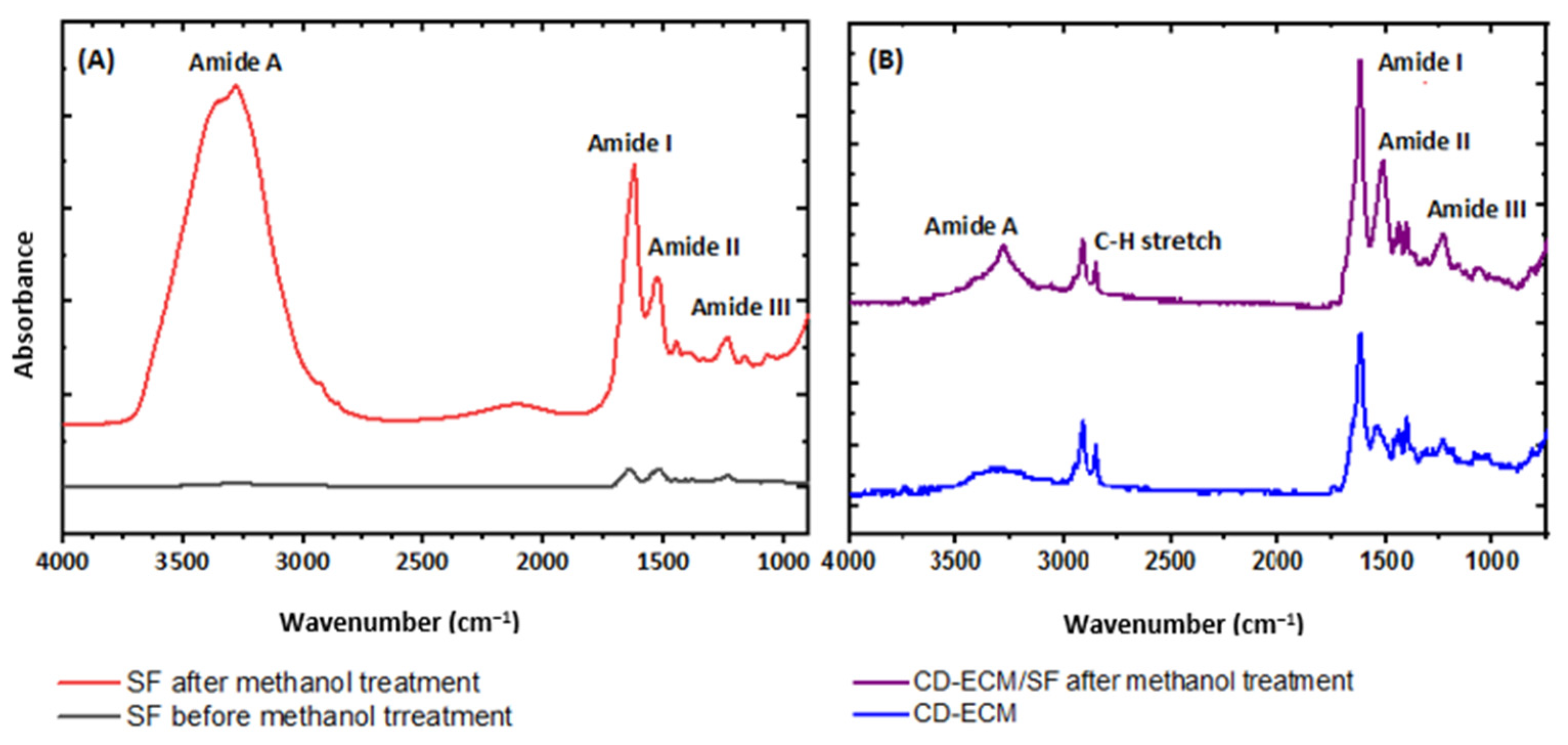
The FTIR findings exposed the effectiveness of methanol treatment for the deposition of CD-ECM to SF scaffold’s void struts by alkene to alkane transitional cross-linking process as retention of the ECM in a scaffold over culture period, and is vital for cell proliferation and for providing signals for cell differentiation. In this study, other than providing an external ECM environment, chondrogenic and hypertrophic factors were supplemented as biochemical cues to direct hBMSC’s towards the specific lineage, but in the case of an in vivo setting, such factors are not present, and the scaffold would stand alone moderating the cells from external cues [
13,
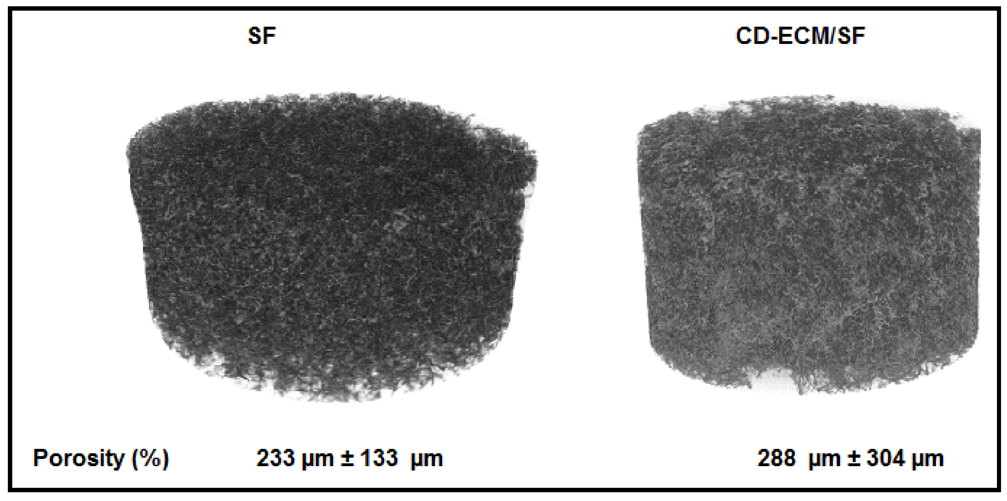
14]. Porosity in the scaffolds was controlled by a particle leaching method between 200 µm to 300 µm, as it was shown that vascularization and osseointegration occur only in bone grafts with pore size more than 200 µm upon implantation for critical size bone defects [
15].
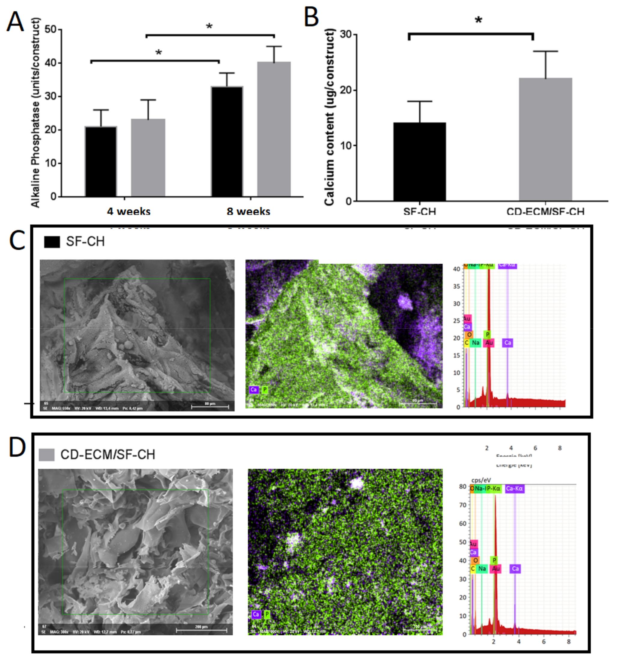
Morphological observation post eight weeks of differentiation by ESEM revealed that the chondrocytes were enlarged in size, indicating a differentiated state of hypertrophic chondrocytes. EDX measurements performed in the same area denoted a higher accumulation of CaP in the CD-ECM/SF-CH group than in the SF-CH group. This was further confirmed by biochemical analysis that calcium content was higher in the CD-ECM/SF-CH, followed by an increase in alkaline phosphatase levels secreted by the cells. Fetal calf serum supplementation in culture media was reported to contribute towards CaP deposition in silk fibroin scaffolds [
16,
17]; however, in our study, serum-free media were utilized to reduce osteogenic growth factors, permitting us to examine the in vitro mineralization ability of hBMSC differentiated hypertrophic chondrocytes.
Hypertrophic chondrocyte mediated mineralization assessed by RT-qPCR for hypertrophic and osteogenic markers revealed that COL10A1 gene expression was higher in the SF-CH group than the CD-ECM/SF-CH group, as in the CD-ECM/SF-CH group the decrease in COL10A1 could have occurred due to remodeling of the cartilaginous template by metalloproteinases towards mineralized tissue formation [
18,
19]. However, only in the CD-ECM/SF-CH group, it decelerated the cartilage template’s maturation and remodeling of the matrix, confirmed by an increased expression of early osteogenic markers ALP, IBSP, OSX, COLIA1, than in the SF-CH group indicating a precursor osteoblast state of differentiation. The late hypertrophic marker TSBS1 was expressed. The early hypertrophic marker CHM1 was absent; this differentiation towards a late hypertrophy stage along VEGF being defined would expedite vascular invasion onto the grafts followed by osseointegration upon implantation in vivo.
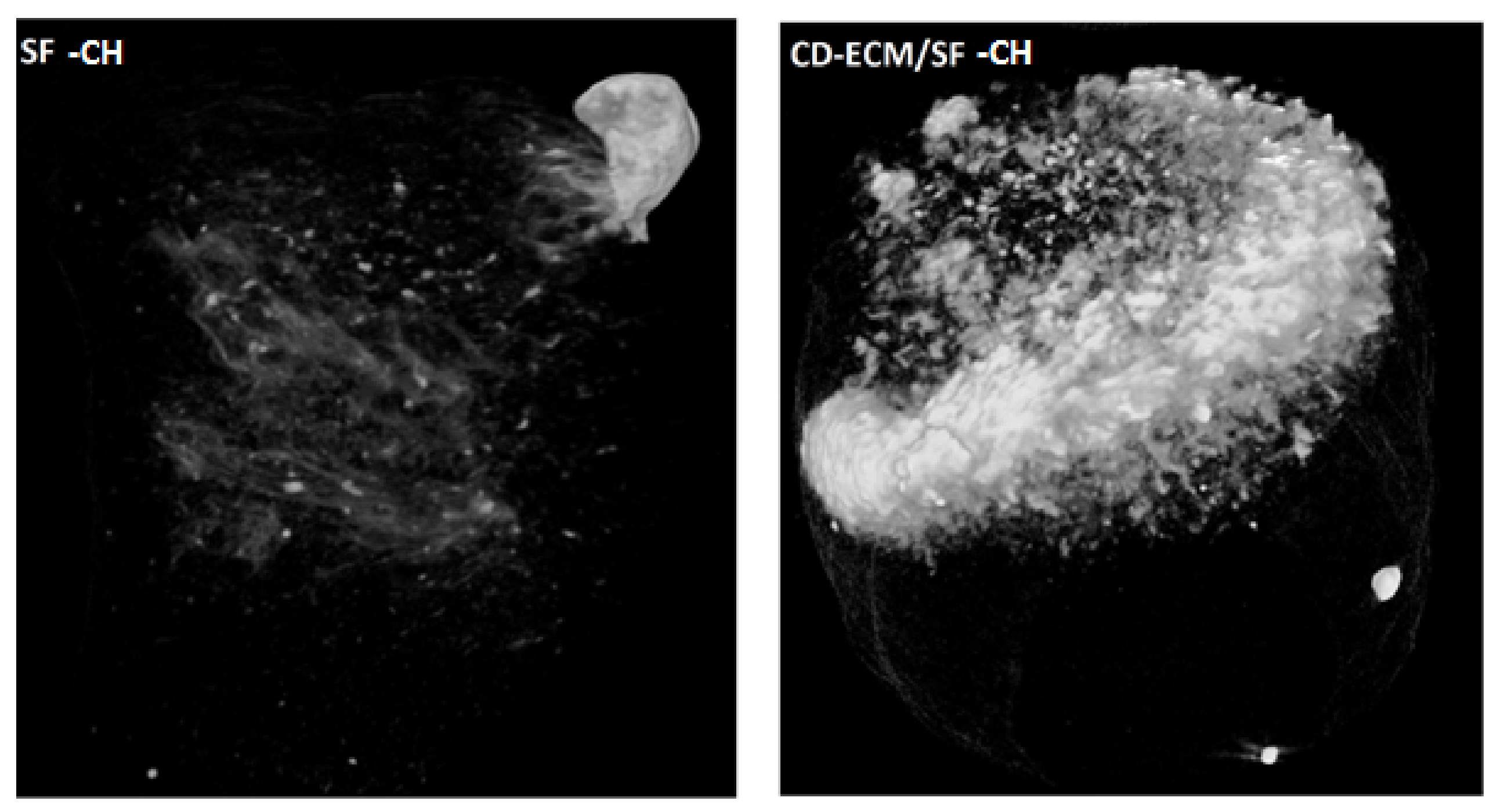
Micro-computed tomography (µCT) offers a non-destructive approach to the visualization of hydroxyapatite formation in tissue-engineered scaffolds [
20] than conventional histological and calcium assays in bone tissue engineering; mineralized tissue formation is deemed to the final product of an in vitro cell culture. µCT scans demonstrated a dense cloud of hydroxyapatite minerals occurring on the CD-ECM/SF-CH group’s surface, whereas in the SF-CH group, it was comparatively less observed. However, the uniform distribution of hydroxyapatite was not observed throughout the scaffold. This could be attributed to the fact that static culture is inefficient in propagating the mineralized ossicles during ECO. On the contrary, dynamic culture systems such as flow perfusion enhanced hypertrophic matrix production but inhibited further bone formation [
20,
21]. Recent studies investigated mimicking the right biomechanical cues favoring ECO, such as hydrostatic pressure and chondrogenic priming enhanced hypertrophic matrix productions and mineralized matrix formation [
22]. Similarly, the higher magnitude of cyclic tensile strain-induced ECO applies to chondrogenically primed MSC’s in fibrin hydrogels [
23].
4. Materials and Methods
4.1. Decellularization and Isolation of Cartilage Derived Extracellular Matrix (CD-ECM)
Bovine knee joints were obtained from the local abattoir proceeding sacrifice of animals. The articular cartilage from both medial and lateral sides of the knee joint was collected with scalpels. The cartilage pieces were minced into fine pieces and frozen at −20 °C and lyophilized overnight. The freeze-dried cartilage was later coarsely ground in a cryomill (Retsch, Germany) for two cycles. The cryogrind powder was decellularized by osmotic shock, detergent, and enzymatic washing under constant agitation at 200 rpm. The ground cartilage powder was subjected to a hypertonic salt solution (HSS) for 12 h to disrupt the cell membrane. After HSS treatment, the cartilage was subjected to two cycles of Triton-X 100 (0.05% v/v) and HSS treatments to break down cellular membranes. The cartilage was further treated with benzonase (0.0625 KU/mL) for 12 h at 37 °C to fragment the nucleic acids. Sodium-lauroyl sarcosine (NLS, 1% v/v) was added for the next 12 h to remove the cells. Then, the cartilage was rinsed with 40% ethanol, and anionic exchange chromatography was performed to remove the organic solvents. The tissue was further rinsed with deionized water before freezing. Post decellularization, the tissue was lyophilized for 48 h and cryo-ground into a fine powder with a cryo-mill.
4.2. Fabrication of Cartilage Derived Extracellular Matrix and Silk Fibroin Hybrid (CD-ECM/SF) Scaffolds
Lyophilized SF was produced as previously described [
24]. Briefly, cocoons of
B. mori (Seidentraum, Germany) were boiled in 0.02 M Na
2CO
3 twice for 1 h, and the resulting fibers were rinsed five times in ddH
2O and dried overnight. The fibers were dissolved in 9.3 M LiBr and dialyzed for three days against ddH
2O. The resulting regenerated silk fibroin solution was lyophilized until further usage. Lyophilized CD-ECM and SF were dissolved in 1,1,1,3,3,3-Hexa- fluoro-2-propanol (HFIP, Sigma-Aldrich, St. Louis, MO, USA) at a ratio of 1:1 to yield a 17% (w/v) solution and cast onto a teflon mold. The resulting scaffolds were cut with a dermal biopsy punch in a dimension of 5 mm h, 5 mm d. The cut scaffolds were immersed in 90% methanol to induce confirmation of secondary structure of β sheets in SF. Post-washes in PBS and sterilization were achieved by autoclaving at 121 °C, and the scaffolds were used for cell culture.
4.3. Cell Culture and Hypertrophic Differentiation of BMSC’s
Human bone marrow derived MSC’s (hBMSCs,) were purchased from Lonza (Lonza, Switzerland). After expansion to passage twp, cells were seeded onto the scaffold’s surface at an initial seeding density of 1 × 10⁶ cells in 20 µL suspensions and let incubated at 37 °C for 2 h. Post seeding, the scaffolds were immersed for the next 24 h in 10% FCS containing basal medium (DMEM F12, GLUTAMAX, 1% ITS, 1.5 mg/mL BSA, 2% penstrep, 2.5 µg/mL amphob) for the cells to be at an equilibrium. The constructs are primed towards chondrogenesis for the next two weeks in serum-free basal medium (1% ITS, 1% BSA, 25 mM HEPES, DMEM high glucose) with chondrogenic supplements (100 nM Dexamethasone, 50 µg/mL Ascorbic-acid-2-phosphate, 10 ng/mL TGF β1). After two weeks, the constructs are primed towards hypertrophic chondrocytes with serum-free basal media and hypertrophy supplements (1nM Dexamethasone, 50 µg/mL Ascorbic-acid-2-phosphate, 50 ng/mL L-Thyroxine, 10mM β-Glycerolphosphate) for the next six weeks under static conditions.
4.4. Biochemical Analysis
4.4.1. sGAG and DNA Assays
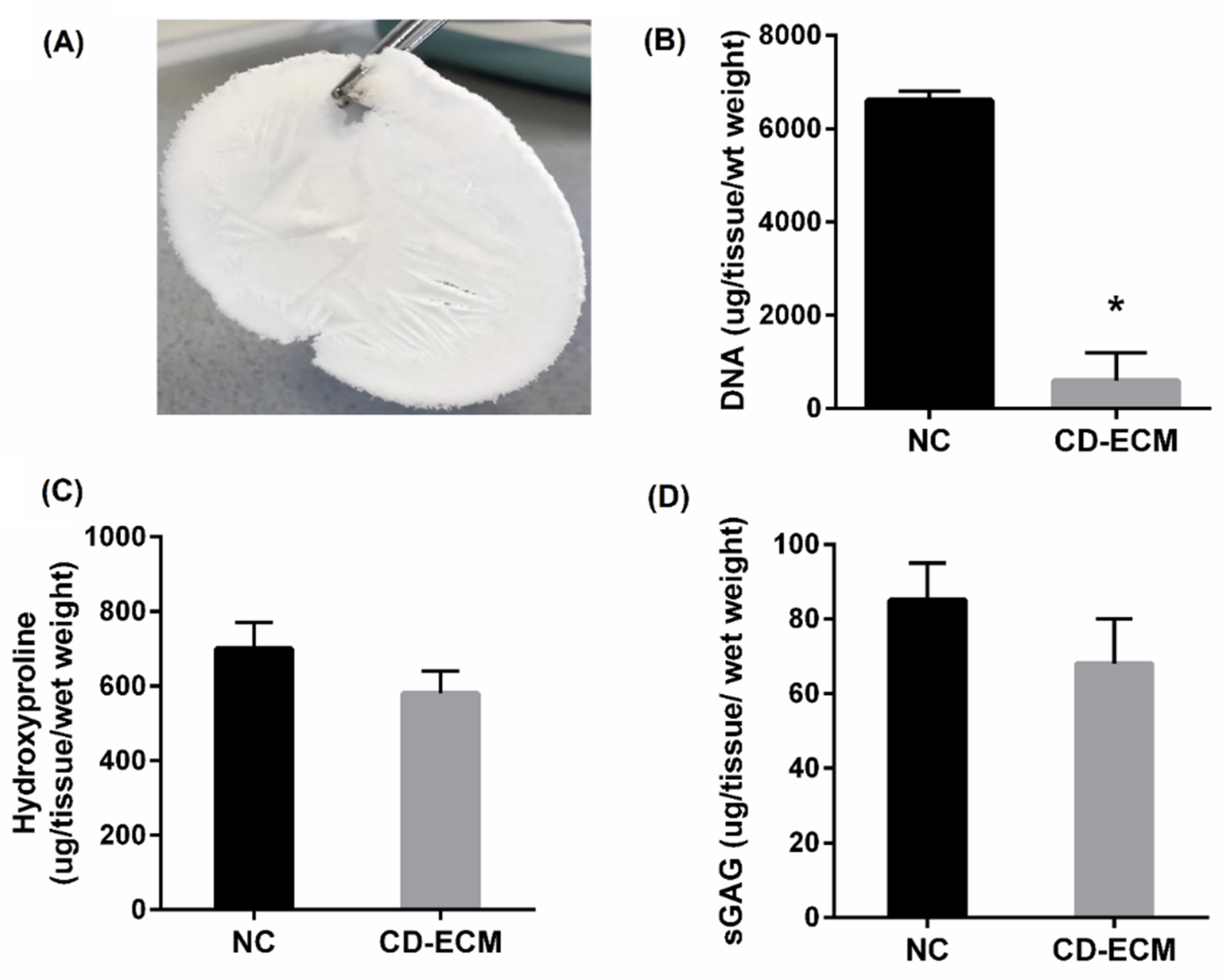
Total sGAG was measured from decellularized cartilage (CD-ECM) by freezing them at −80 °C and lyophilized at 20 °C to evaporate the water content. Quantification was done by treating them overnight with 25u/mL proteinase K enzyme at 56 °C. Enzyme inactivation was then performed at 90 °C for 10 min. The resultant supernatant was transferred to ultra-free filter reaction tubes of 0.1 µm pore size (Millipore, Billerica, MA, USA) and centrifuged at 12,000× g for 10 min. sGAG was measured through complexation and decomplexation with a 1,9 dimethyl methylene blue solution (DMMB). The absorbance was measured at 656 nm in a plate reader. For DNA analysis, n = 6 constructs after the decellularized cartilage (CD-ECM) were compared to the native articular cartilage. Briefly, the supernatant above from the Proteinase K digested constructs were removed before ultrafiltration. The DNA content was measured fluorometrically using the PicoGreen assay (Fisher Scientific), according to the manufacturer’s instructions at an excitation wavelength of 480 nm, emission wavelength of 528 nm.
4.4.2. Hydroxyproline Assay
Total collagen content was determined by quantifying the hydroxyproline content. CD-ECM and native cartilage samples were hydrolyzed in 6 M HCL at 110 °C for 18 h, and the hydroxyproline content from the hydrolyzed solution was measured with a chloramine-T/Ehrlich spectrophotometry-based assay (Sigma-Aldrich, St. Louis, MO, USA). The hydroxyproline content was measured at a wavelength of 560 nm.
4.4.3. Alkaline Phosphatase Assay
Extracellular alkaline phosphatase (ALP) production was determined from the supernatant of cells based on a custom-made biochemical assay in all the cell-scaffold constructs. The samples were briefly measured colorimetrically by converting p-nitrophenyl phosphate to p-nitrophenol in a spectrophotometer at 405 nm.
4.4.4. Calcium Assay
Calcium mineralization was quantitatively determined in cell-scaffold constructs by disintegrating them in a mechanical homogenizer (MagnaLyser) in 5% trichloroacetic acid. The resulting supernatant was measured spectrophotometrically at 575 nm based on the reaction with o-cresolphthalein complexion, according to manufacturer’s instructions (Sigma-Aldrich, St. Louis, MO, USA).
4.5. Scanning Electron Microscopy (SEM) and Energy Dispersive X-ray (EDX) Analysis
Cell attachment and distribution after eight weeks were observed using a Hitachi ESEM-FEG. Cell-scaffold constructs were rinsed twice with PBS and fixed in 10% formalin for 2 hr. Subsequently, the samples were cut in progressive sections and dehydrated in sequential ethanol series (50%, 60%, 70%, 80%, 90%, 96%, and 100%), 30 min for each concentration. For the final dehydration step, scaffolds were immersed in hexamethyldisilazane, and the solvent was left to evaporate overnight. Finally, samples were gold sputter costed prior to SEM analysis. SEM images were obtained under a high vacuum with an acceleration voltage of 10 kV and a working distance of 25 mm. An EDX (Bruker) system integrated into the SEM was expended to analyze the possible co-localization of calcium (Ca) and phosphate (P) in the extracellular matrix. The spectra were taken under a high vacuum with an acceleration voltage of 10 kV and a working distance of 10 mm.
4.6. Attenuated Total Reflection Fourier-Transform Infrared Spectroscopy (ATR-FTIR)
ATR-FTIR spectra were collected using a Spectrum Two FT-IR Spectrometer (PerkinElmer Inc.) equipped with a LiTaO3 detector and with a MIRacl single reflection ATR (ZnSe) accessory (PIKE Technologies). Additionally, 4 to 64 scans were co-added at a nominal resolution of 4 cm−1. After each data acquisition, ATR correction was performed; for all spectral manipulation, the Spectrum 10 software package (PerkinElmer Inc.) was used.
4.7. Real-Time Quantitative PCR
Cell-scaffold constructs were minced into fine pieces in lysis buffer and added to the MagNA Lyser tubes. The tubes were frozen in liquid nitrogen and homogenized with the MagNA Lyser four times at 6500 rpm. The samples were treated by Proteinase K for 30 min at 55 °C and centrifuged to pellet the debris. The resulting supernatant was used for RNA isolation with the Qiagen Eneasy Fibrous Tissue Kit, followed by elution with 30 µl of RNase-free water. cDNA synthesis was performed using Transcriptor First Strand cDNA Synthesis Kit (Roche, Basel, Switzerland), and additionally, RNA from bacteriophage MS2 was added to stabilize the isolated RNA during cDNA synthesis. Probe-based real-time quantitative polymerase chain reaction (RTqPCR) was performed in triplicates in the LightCycler 96 using Fast Start Essential DNA Probes Master. (Roche, Basel, Switzerland). Probe-primer pairs were designed for hypertrophic and osteogenic specific markers COLX, RUNX2, TSBS1, CHM1, VEGF, COLI, ALPL, IBSP, OSX (
Table 1),with IDT Real-Time qPCR software and synthesized by IDT (Integrated DNA Technologies, Leuven, Belgium). As a reference gene, we chose TBP [
25] Annealing temperature, which was experimentally determined for reference. Each target gene and the relative expression was evaluated with the 2
−∆∆Ct method.
4.8. Biomechanical Analysis
Cell-scaffold constructs were tested under compression load using the Zwick test machine (ZwickRoell GmbH, Ulm, Germany), equipped with a 10 N load cell and carried out with a 1 mm/min cross-head speed. All samples were wet during testing. The displacement was tracked using a three-dimensional Digital Image Correlation (DIC) system (GOM GmbH, Germany) equipped with two CCD cameras (6 Megapixels). Thirty scaffolds with a 5 mm diameter and 5 mm in height were evaluated. The compressive force-displacement curves were converted into stress and strain curves. Compressive elastic moduli were calculated from the slopes of the stress−strain curves. The average compressive modulus and standard deviation of different sample groups were calculated.
4.9. Micro-Computed Tomography (µCT) Analysis
The wet constructs were scanned with a calibrated micro CT scanner (Bruker Skyscan 1173, Bruker, Kontich, Belgium) at 30 kV, 180 μA, integration time 3200 ms, nominal resolution of 5 μm, and without a filter. Scaffolds were scanned in wet conditions, except empty scaffolds for porosity analysis were scanned dry. Using medtool (Version 4.3; Dr. Pahr Ingenieurs e.U., Pfaffstätten, Austria), the µCT scans were processed. First, raw image files from Brucker were imported into Medtool, and the region of interest was cropped. Second, registration was performed using the iterative selection method to find a single level threshold at the minimum between the scaffold constructs and calcified regions. Third, the equivalent bone volume to total volume was calculated as the number of calcified voxels divided by the voxels’ total number in one scaffold.
4.10. Statistical Analysis
A non-parametric Mann–Whitney two-tailed U-test was performed for comparisons between two datasets at a time. Multiple comparisons were performed using the non-parametric Kruskal–Wallis test followed by Dunn’s multiple comparisons test. All data are presented as the mean ± SEM. The significance level was set at p < 0.05. All statistical analyses were performed using the GraphPad Prism software (Graphpad Prism Software Inc., San Diego, CA, USA).


 ,
, 


