Article

A Novel Sulfonamide, 4-FS, Reduces Ethanol Drinking and Physical Withdrawal Associated With Ethanol Dependence

1
Abbottabad Campus, COMSATS University Islamabad, Abbottabad, Khyber Pakhtunkhawa 22060, Pakistan
2
VA San Diego Healthcare System, San Diego, CA 92161, USA
3
Department of Anesthesiology, University of California San Diego, San Diego, CA 92161, USA
4
Department of Pharmacy, Capital University of Science & Technology, Islamabad 44000, Pakistan
5
Department of Chemistry, Forman Christian College, A Chartered University, Ferozepur Road, Lahore 54600, Pakistan
*
Authors to whom correspondence should be addressed.
Int. J. Mol. Sci. 2020, 21(12), 4411; https://doi.org/10.3390/ijms21124411
Submission received: 22 May 2020 / Revised: 13 June 2020 / Accepted: 17 June 2020 / Published: 21 June 2020
(This article belongs to the Special Issue Protease and Carbonic Anhydrase Inhibitors, II)

Abstract

:
Carbonic anhydrase (CA) is abundant in glial cells in the brain and CA type II isoform (CA II) activity in the hippocampus plays an important role in buffering extracellular pH transients produced by neural activity. Chronic ethanol exposure results in respiratory and metabolic acidosis, producing shifts in extracellular pH in the brain and body. These neurophysiological changes by ethanol are hypothesized to contribute to the continued drinking behavior and physical withdrawal behavior in subjects consuming ethanol chronically. We explored whether chronic ethanol self-administration (ethanol drinking, 10% v/v; ED) without or under the influence of chronic intermittent ethanol vapor (CIE-ED) experience alters the expression of CA II in the hippocampus. Postmortem hippocampal tissue analyses demonstrated that CA II levels were enhanced in the hilus region of the hippocampus in ED and CIE-ED rats. We used a novel molecule—4-fluoro-N-(4-sulfamoylphenyl) benzenesulfonamide (4-FS)—a selective CA II inhibitor, to determine whether CA II plays a role in ethanol self-administration in ED and CIE-ED rats and physical withdrawal behavior in CIE-ED rats. 4-FS (20 mg/kg, i.p.) reduced ethanol self-administration in ED rats and physical withdrawal behavior in CIE-ED rats. Postmortem hippocampal tissue analyses demonstrated that 4-FS reduced CA II expression in ED and CIE-ED rats to control levels. In parallel, 4-FS enhanced GABAA receptor expression, reduced ratio of glutamatergic GluN2A/2B receptors and enhanced the expression of Fos, a marker of neuronal activation in the ventral hippocampus in ED rats. These findings suggest that 4-FS enhanced GABAergic transmission and increased activity of neurons of inhibitory phenotypes. Taken together, these findings support the role of CA II in assisting with negative affective behaviors associated with moderate to severe alcohol use disorders (AUD) and that CA II inhibitors are a potential therapeutic target to reduce continued drinking and somatic withdrawal symptoms associated with moderate to severe AUD.

1. Introduction

Alcohol use disorder (AUD) affects a significant population in the United States [1], and is associated with a plethora of neurological deficits. Particularly notable from clinical findings are the deficits dependent on the hippocampus, including, but not limited to, loss of brain volume and cognitive impairments [2,3,4,5,6]. Even more interesting is the fact that these impairments have been replicated in several widely accepted rodent models of alcohol dependence [7,8,9,10]. The chronic intermittent ethanol vapor exposure (CIE) model, is a widely used model that implements daily cycles of intoxication via ethanol vapors and withdrawal to induce clinical signs of alcoholism, such as somatic withdrawal symptoms and escalated ethanol drinking in rats [11,12]. Neurobiological alterations in the hippocampus produced by CIE could be associated with unregulated drinking patterns observed in CIE animals [13,14,15,16,17,18,19,20,21].
In the context of the above hypothesis, ethanol disrupts the delicate balance between γ-aminobutyric acid (GABA) and glutamate in the hippocampus. These neuroadaptations induced by ethanol could lead to behavioral deficits, including unregulated patterns of drinking and physical withdrawal behaviors [19]. For example, in vitro studies have demonstrated excitotoxicity in the hippocampus after withdrawal from chronic ethanol exposure and not during ethanol exposure [22,23,24]. Mechanistic studies using in vitro models have further shown that excessive release of glutamate and polyamines and corresponding activation of N-methyl-D-aspartate type glutamatergic receptors (GluNs) contribute to the excitotoxicity [25]. Supporting the in vitro studies, in vivo studies also show neurotoxicity in the hippocampus [26,27,28,29,30]. For example, during ethanol withdrawal, glutamate release is increased in the hippocampus [26,27], and the changes in glutamate levels are also associated with increased number of functional GluNs [31,32], suggesting that these effects may induce ethanol withdrawal hyperexcitability and may also lead to increased susceptibility to somatic withdrawal symptoms [33].
The neuroadaptations by ethanol also could produce neurophysiological effects via GABA/glutamate imbalance, including changes in extracellular pH, and alkaline transients in the brain, especially in the hippocampus [34,35]. Particularly interesting is the presence of extracellular carbonic anhydrases (CA) in the hippocampus, enzymes that regulate pH transients associated with glutamatergic and GABAergic transmission [34]. Notably, inhibition of CA activity is anticonvulsive [36,37]. For example, in humans, the CA inhibitor acetazolamide is used for adjuvant antiepileptic therapy [38,39], and in animals reduces the convulsant effects in a kindling model of partial epilepsy [40]. Furthermore, topiramate, a drug used to treat seizures, psychiatric and neurological conditions is a CA inhibitor, and has shown beneficial effects in reducing craving and drinking obsessions in subjects with AUD [41]. Given that chronic ethanol experience results in epileptiform activity in the hippocampus [42], we tested the hypothesis that systemic administration of a novel CA inhibitor 4-fluoro-N-(4-sulfamoylphenyl)benzenesulfonamide (4-FS; CA II inhibitor [43]) will reduce physical withdrawal and unregulated drinking associated with alcohol dependence in CIE rats. We also tested the subhypothesis that the reduced behaviors by 4-FS will be associated with reduced expression of CA II and enhanced expression of GABA receptors in the hippocampus.

2. Results

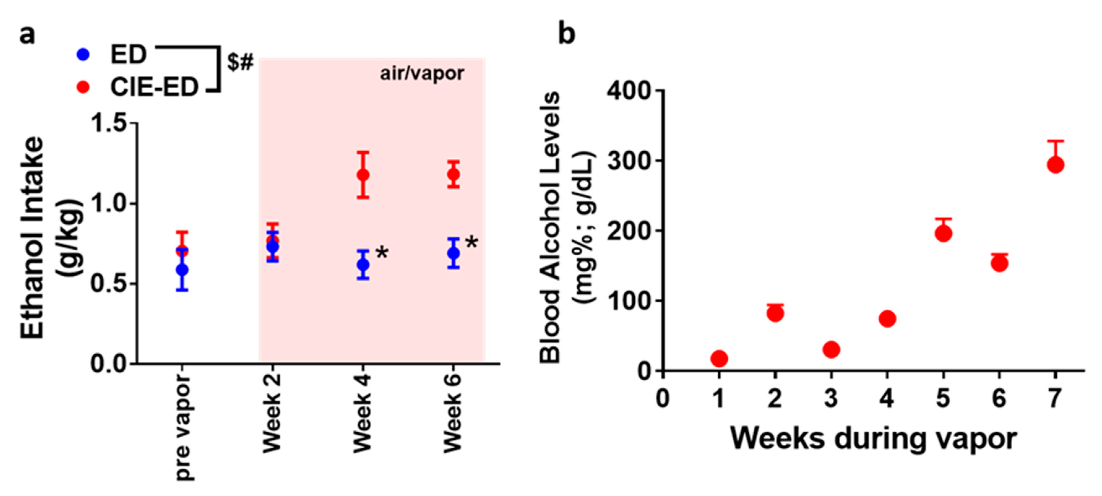
2.1. CIE-ED Rats Consume More Ethanol Than ED Rats

Experimental design, timeline of vapor exposure and treatment groups are indicated in Figure 1. Ethanol (10% v/v) consumption was determined in CIE-ED and ED rats prior to the onset of vapor exposure and during vapor exposure weeks (Figure 2a). The amount of alcohol experienced by CIE by CIE-ED rats reached the desired range by the fourth week of vapor exposure as indicated by their BALs (Figure 2b). With respect to drinking sessions, a significant sessions × ethanol groups interaction (F(3,39) = 6.1, p = 0.001), main effect of session (F(3,39) = 6.4, p = 0.001) and main effect of group (F(1,13) = 6.4, p = 0.02) was obtained for ethanol consumed over the six weeks of vapor exposure. Further investigation of the interaction revealed that in CIE-ED rats, consumption of ethanol increased during weeks 4–7 compared to pre-vapor responding (ps < 0.05; Figure 2a). No difference in ethanol consumption was observed across weeks in ED rats. Also, from week 4 onwards, CIE-ED rats exhibited higher consumption of ethanol compared to ED rats (ps < 0.05).

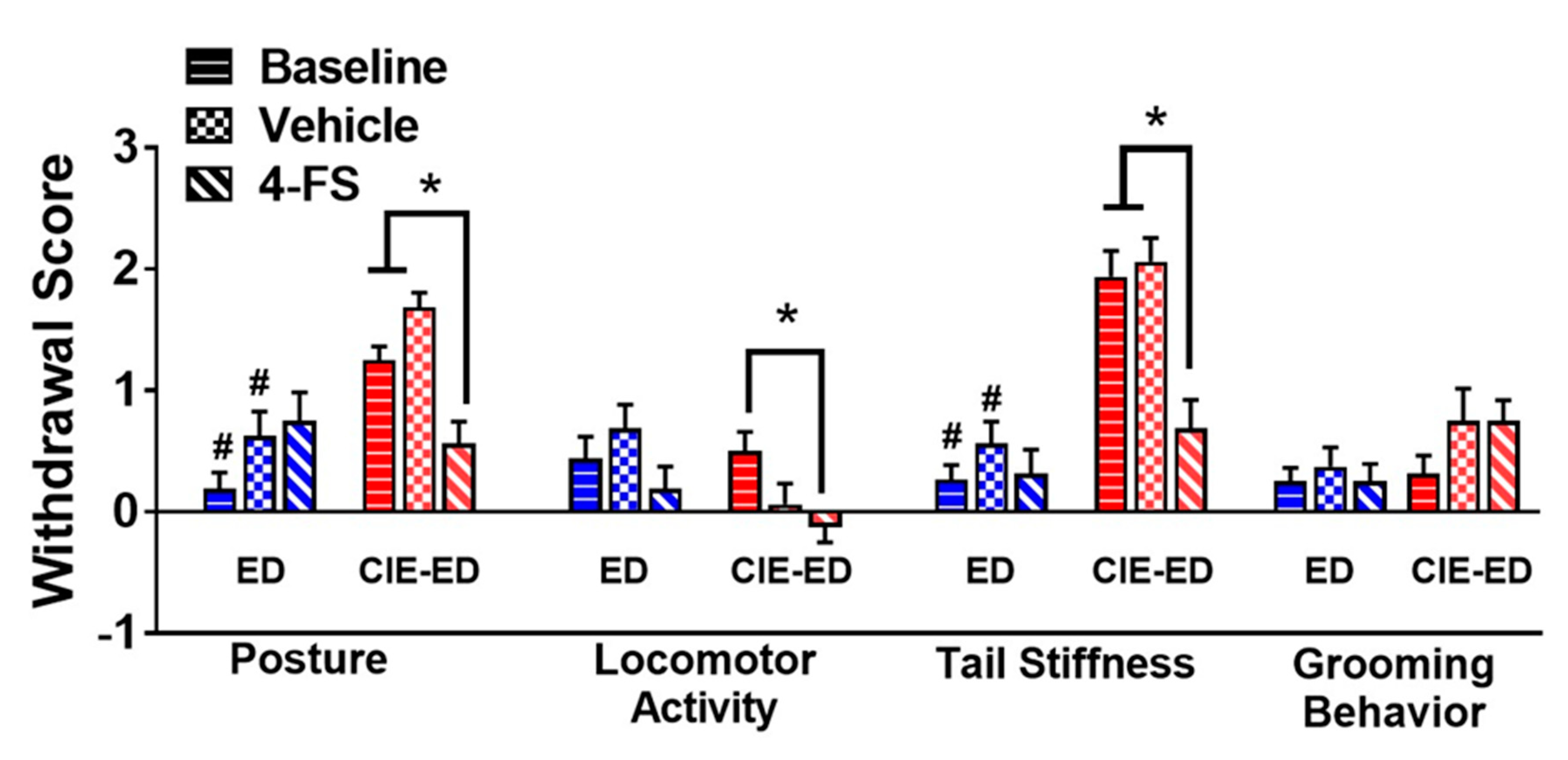
2.2. CIE-ED Rats Demonstrate Somatic Withdrawal Symptoms

Withdrawal scores of body posture and tail stiffness were higher in CIE-ED rats compared with ED rats (ps < 0.05; Figure 4). Locomotor activity and grooming behavior were not different between the two groups.

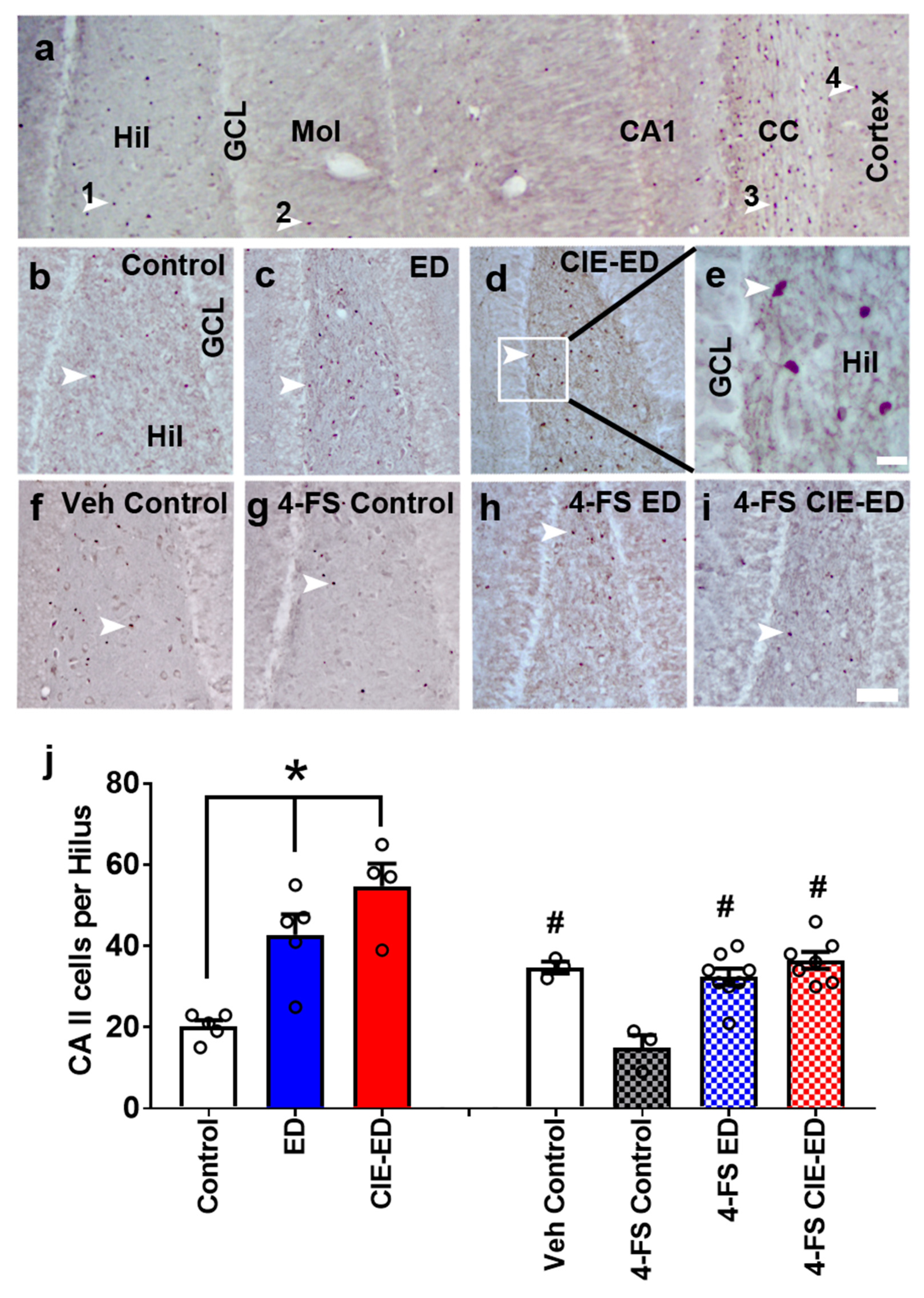
2.3. ED and CIE-ED Rats Show Increased Number of CA II Immunoreactive Cells in the Hilus and 4-FS Reduces This Effect

CA II immunoreactive cells were detected in the hilus and the corpus callosum regions (Figure 3a). Separate analyses were performed on controls, ED and CIE-ED rats that did not receive any 4-FS treatment. ED and CIE-ED rats that did not experience 4-FS treatment show increased number of CA II immunoreactive cells in the hilus compared to controls by one-way ANOVA (F(2,11) = 17.0, p = 0.0004). Post hoc analyses demonstrated higher number of CA II cells in ED and CIE-ED rats compared to their controls (ps < 0.01; Figure 3b–e,j).
4-FS treatment in ethanol-naïve rats reduced the number of CA II immunoreactive cells in the hilus and 4-FS treated CIE-ED and ED rats had the same number of CA II immunoreactive cells in the hilus compared with controls as shown by one-way ANOVA (F(3,17) = 11.9, p = 0.0002). Post hoc analyses demonstrated reduced number of CA II cells in 4-FS treated ethanol naïve rats compared with controls, 4-FS treated ED and CIE-ED rats (p < 0.01; Figure 3j).

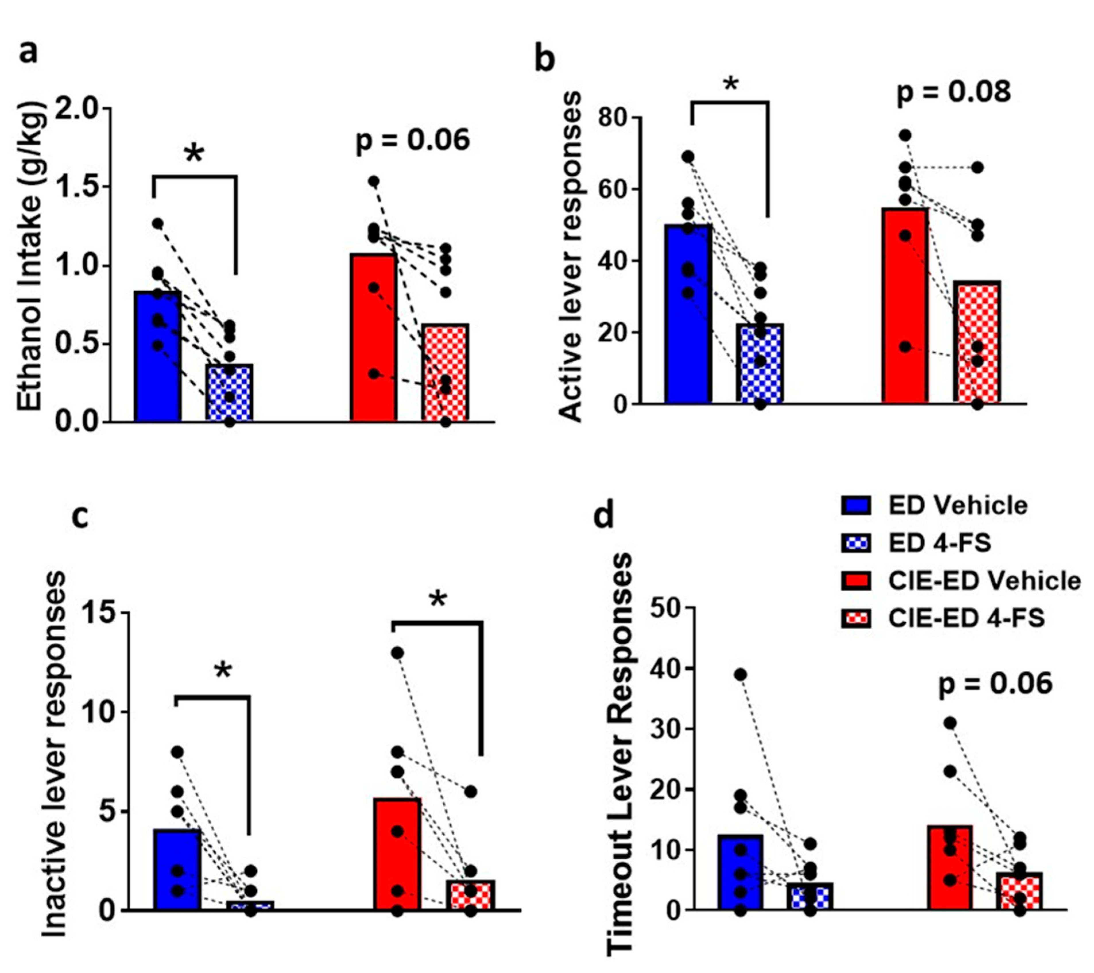
2.4. 4-FS reduces Withdrawal Behavior in CIE-ED Rats and Reduces Drinking in ED Rats

The effect of vehicle and 4-FS on physical withdrawal and drinking during withdrawal in CIE-ED and ED rats were determined as a within subject design during week 7 of ethanol sessions. 4-FS did not alter withdrawal scores in ED rats. 4-FS reduced withdrawal scores of posture and tail stiffness in CIE-ED rats bringing them to the levels of ED rats (ps < 0.05; Figure 4).
The effect of 4-FS on ethanol self-administration in ED and CIE-ED rats were determined as a within subject effect (Figure 5). 4-FS reduced the amount of ethanol consumed and the associated active lever responses in ED rats (ethanol intake: p = 0.0004; active lever responses: p = 0.001; by paired t test Figure 5a,b). 4-FS did not significantly alter the amount of ethanol consumed or active lever responses in CIE-ED rats, however, showed a strong trend towards decrease compared with vehicle treatment (ethanol intake: p = 0.06; active lever responses: p = 0.08). 4-FS reduced inactive lever responses in CIE-ED and ED rats (CIE-ED: p = 0.04; ED: p = 0.01; Figure 5c). 4-FS did not alter lever responses during timeout in CIE-ED and ED rats (CIE-ED: p = 0.06; ED: p = 0.13; Figure 5d).

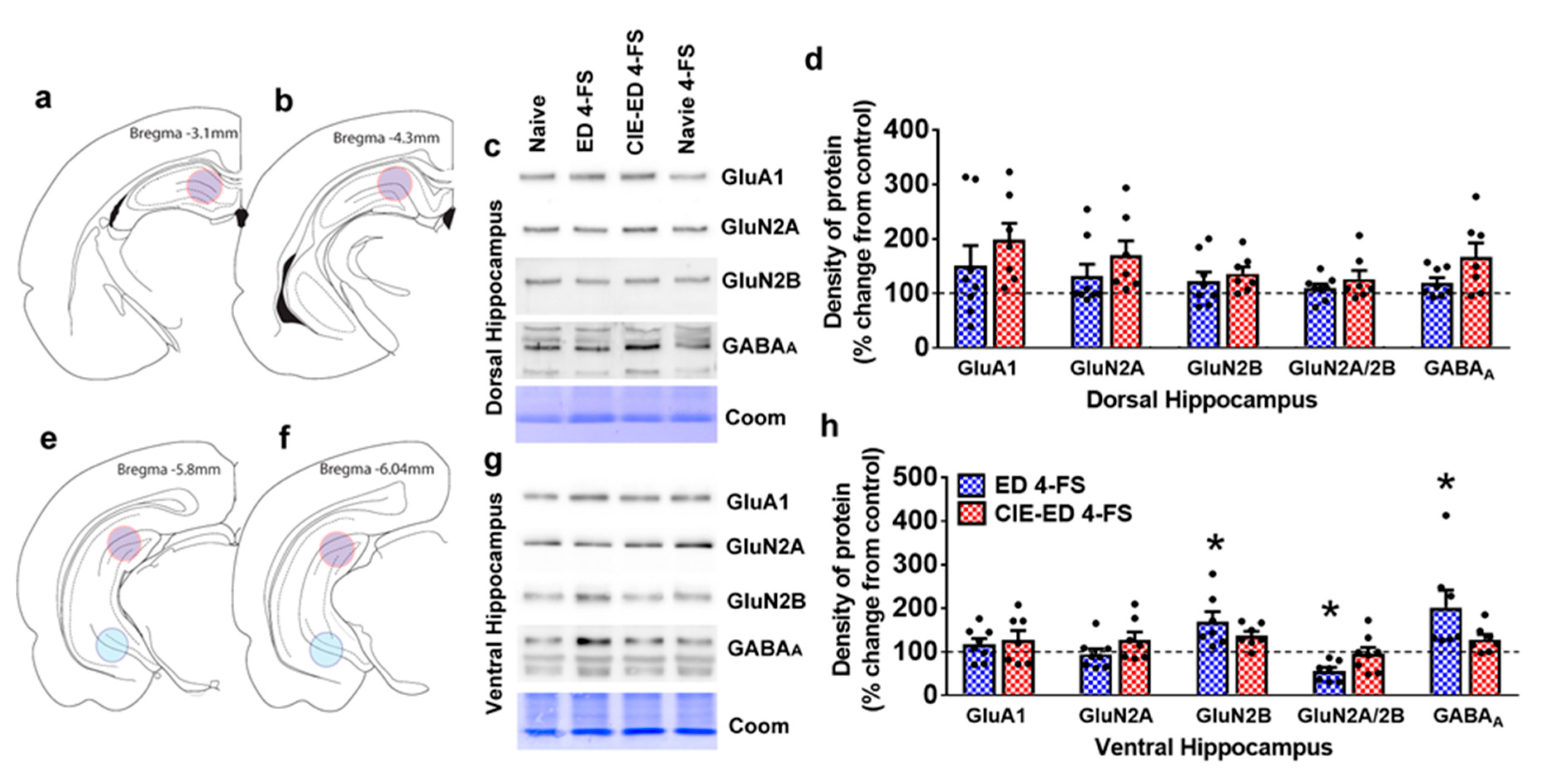
2.5. 4-FS Alters Expression of GluRs and GABAARs in the Ventral Hippocampus

Protein expression between vehicle controls and 4-FS treated controls did not differ and therefore they were combined as controls and used for analyses.
In the dorsal hippocampus, there was a trend towards increase in GABAA expression (one-way ANOVA GABAA: F(2,18) = 3.0; p = 0.07; Figure 6d). GluA1, GluN2A, GluN2B and the ratio of GluN2A/2B were unaltered (Figure 6d).
In the ventral hippocampus, GluN2B and GABAA expression were increased (one-way ANOVA, GluN2B: F(2,18) = 1.5; p = 0.03; GABAA: F(2,17) = 1.6; p = 0.04). Post hoc analysis indicated higher expression of GluN2B in 4-FS treated ED rats compared with controls (p = 0.02), and higher expression of GABAA in 4-FS treated ED rats compared with controls (p = 0.05). The ratio of GluN2A/2B expression was reduced (one-way ANOVA GluN2A/2B: F(2,18) = 1.0; p = 0.03; Figure 6h). Post hoc analysis did not detect any group differences in GluN2A/2B expression.

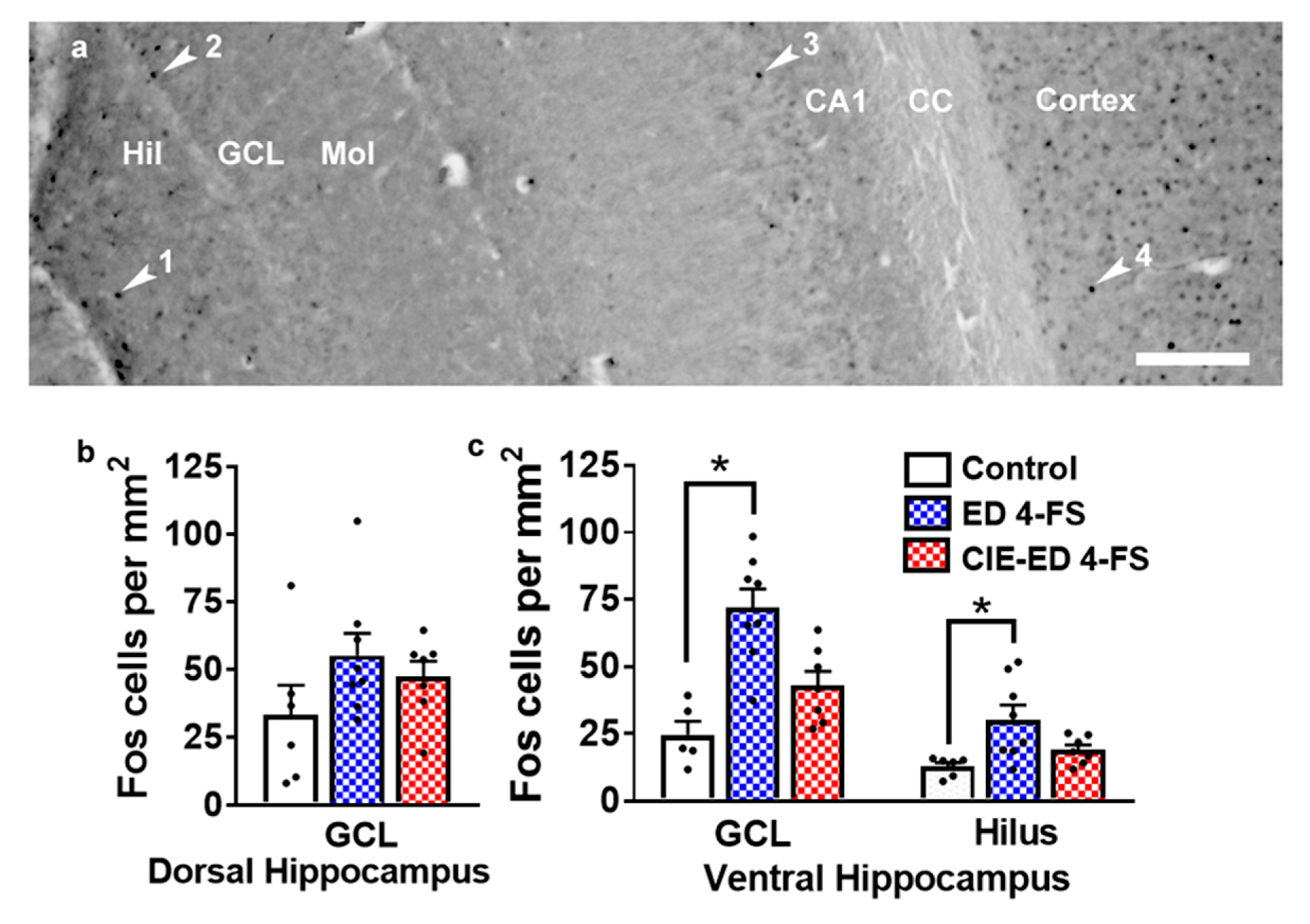
2.6. 4-FS Alters the Number of Fos Cells in the Ventral Hippocampus

The number of Fos cells did not differ between vehicle controls and 4-FS treated controls and therefore, they were combined as controls and used for analyses. The number of Fos cells did not differ in the dorsal hippocampus in the granule cell layer (GCL); (F(2,18) = 1.7; p = 0.19; Figure 7b). In the ventral hippocampus, the number of Fos cells increased in the GCL and hilus regions (one-way ANOVA, GCL: F(2,18) = 12.2; p = 0.0004; hilus: F(2,18) = 5.9; p = 0.009; Figure 7c). Post hoc analyses indicate higher number of Fos cells in the GCL in 4-FS treated ED rats compared to controls (p = 0.0007) and 4-FS treated CIE-ED rats (p = 0.004) (Figure 7c). Post hoc analyses indicate higher number of Fos cells in the hilus in 4-FS treated ED rats compared to controls (p = 0.01) and 4-FS treated CIE-ED rats (p = 0.04) (Figure 7c).

3. Discussion

Our study demonstrates the expression of CA II in the adult rat hippocampus and corpus callosum, and that systemic 4-FS treatment reduces CA II expression in the hippocampus. In this study we also demonstrate a novel role of CA II in ethanol self-administration associated with non-dependent drinking and somatic withdrawal symptoms associated with ethanol dependence. For example, 4-FS reduced drinking in ED rats. 4-FS also demonstrated a strong trend towards reduction in drinking in CIE-ED rats, although this effect did not reach statistical significance. These findings imply that the dose of 4-FS required for reducing drinking in ED vs. CIE-ED rats may be different, and additional studies are required to address this issue. A more notable finding is that 4-FS reduced physical withdrawal behavior in CIE-ED rats without producing any non-specific effects in the ED rats. Postmortem tissue analyses revealed significant neurobiological changes in the hippocampus. Specifically in the ventral hippocampus, we show that 4-FS enhanced GABAA receptor expression and Fos expression, and that these changes were associated with reduced ethanol consumption in ED rats. These findings highlight the importance of studying the role of CAs in AUD and may offer a potential new therapeutic approach to treat AUD.
The role of CAs in CNS disorders are fairly unexplored and needs investigation [44,45,46]. This is because physiological studies have demonstrated the expression and activity of CAs in the rodent and human CNS, particularly in the hippocampus [34,35,36,46,47]. Notably, the expression of CAs is abundant in oligodendroglia [48,49], and our immunohistochemical findings in adult rat tissue indicate CA II immunoreactivity in the hilus and molecular layer of the dentate gyrus and the corpus callosum, areas rich in oligodendrocytes. In addition, the lack of CA II expression in the granule cell layer and CA1 and CA3 pyramidal layers of the hippocampus supports the oligodendroglial expression of the protein. Given the abundant non-neuronal expression of CA II in the hippocampus, it is interesting to note that extracellular CA II can regulate neuronal transmission in the hippocampus [36,47,50]. For example, activity of CAs, specifically, CA II enables the rapid, reversible hydration of carbon dioxide, replenishing H+ ions, limiting interstitial alkalosis by preventing amplification of GluN and GABAA receptor-mediated, bicarbonate-dependent alkaline shifts [34,51,52]. This is important because, neural activity, particularly GluN and GABAA receptor-mediated activity in the hippocampus is accompanied by shifts in extracellular pH, which can influence normal and pathological brain function [53,54,55].
In the context of these studies, ethanol experience in rodents produces respiratory acidosis and metabolic acidosis that is accompanied by respiratory depression [56]. For example, it is likely that chronic ethanol experience will lead to brain tissue acidification and carbon dioxide retention [56,57]. These physiological changes by ethanol may induce increased tissue CA activity to maintain acid-base balance by rapid catalytic hydration of carbon dioxide to carbonic acid [58]. Additionally, it is possible that ethanol in vivo induces glutamate and GABA evoked alkaline shifts in the hippocampus, such that glutamate and GABA-induced efflux of bicarbonate from cells increases CA expression and activity to maintain generation of H+, and production of carbonic acid [59,60]. Acute withdrawal following high levels of intoxication produces respiratory alkalosis and hyperventilation, probably due to reduced levels of carbon dioxide – a physiological response that is observed in rodents and humans [57,61,62]. Interestingly, hyperventilation is associated with enhanced CA function [58,63]. These studies suggest that the acid-base imbalance in the brain in ED and CIE-ED animals are probably significant, and that the accompanying pathophysiological changes may induce aberrant levels of CA in the brain, particularly the hippocampus. Our results supports this hypothesis.
The enhanced expression of CA in the hippocampus could modulate GluN and GABA receptor function in ED and CIE-ED rats [64,65]. For example, evidence from in vitro and in vivo studies suggests that glutamatergic and GABAergic neurotransmission are critical mediators of synaptic plasticity that may underlie alcohol dependence [19,64]. Particularly interesting is the increases in GluN receptor subunit expression and downregulation of GABA receptors in the hippocampus during CIE [14,18,64] and continued loss of GABAA receptors and alterations in GABAA receptor composition during withdrawal, which parallels the increased GluN subunit and reduced GABAA expression in the hippocampus during epileptogenesis [64,66,67]. Although the neuroplasticity (imbalance of GluN and GABA) in the hippocampus may not directly regulate excessive drinking associated with dependence, the withdrawal symptomatology manifested as somatic symptoms could be driven by the hyperglutamatergic state in the hippocampus [68]. Moreover, the alterations in the hippocampus may be driven by the hyperglutamatergic state in the basolateral amygdala and other limbic regions which play a direct role in excessive drinking during dependence [69,70,71,72]. Taken together, the hyperactivity in the hippocampus stemming from the extended amygdala and the other limbic regions may be decisive factors for the maintenance of dependence in the long term [66,67,69,73].
As discussed above, the enhanced CA activity in the hippocampus in CIE-ED and ED rats may be occurring as a rebound effect in response to ethanol-induced acid-base imbalance in the hippocampus. Inhibitors of CAs are antiepileptic and reduce hyperexcitability induced by altered GluN and GABAergic signaling in the hippocampus [37,50]. Therefore, we used 4-FS, a novel and selective CA II inhibitor to determine whether inhibition of CA II would coincide with reduced drinking and physical withdrawal behavior in ED and CIE-ED rats. Our results show that 4-FS produced beneficial effects in ED and CIE-ED rats, indicating that this novel compound can be used to treat symptoms associated with moderate to severe AUD. Our findings are the first to demonstrate the protective effects of a CA II inhibitor in drinking behaviors in an animal model of moderate AUD and physical withdrawal behaviors in an animal model of severe AUD. A potential limitation in the interpretation of our findings is that only one type of CA II inhibitor (a sulfonamide) was tested in our study, and testing additional classes of CA II inhibitors may offer complete support to mechanisms underlying the protective effects of such inhibitors. However, we believe that these studies are beyond the scope of the current study. Nevertheless, our findings support other preclinical studies conducted with other sulfonamide anticonvulsants and nonselective CA II inhibitors, which have indicated protective effects of this drug class in ethanol drinking behaviors [74,75]. Our findings also support several clinical studies conducted with sulfonamides and their therapeutic effects in reducing risky drinking in alcohol dependent individuals [76,77,78,79]. Additionally, they support a previous publication performed in ex vivo hippocampal slice cultures, where a nonselective inhibitor of CA prevented binge ethanol-induced edema and neurodegeneration [80]. These findings suggest that CA II inhibitors are a promising therapeutic target to treat cellular toxicity and behavioral deficits associated with moderate to severe AUD.
We next attempted to determine the mechanism underlying the behavioral effects mediated by 4-FS. For example, pharmacological targets of clinically used CA inhibitors include the GABAA receptors, glutamatergic receptors, sodium channels, potassium channels and calcium channels [81]. Notably, sulfonamides exert their effects by blocking sodium channels, reducing glutamate neurotransmission via direct actions on excitatory amino acid transporters and potassium channels and indirectly by enhancing GABAergic neurotransmission [77,82,83,84,85]. We therefore determined whether 4-FS treatment altered the expression of GABAA and glutamatergic receptors in the hippocampus in ED and CIE-ED rats, due to the contribution of these receptor systems in the development and maintenance of alcohol dependence [19,64]. Furthermore we evaluated the expression of proteins along the dorsal/ventral gradient of the hippocampus. This is because, neuroanatomical studies in the hippocampus support segregation of neuronal outputs along the dorso-ventral axis whose connectivity may influence the expression of behavior dependent on the hippocampus [86,87]. The dorsal hippocampus is more plastic and is vital for spatial learning, and is particularly critical in mediating contextual discrimination [88]. However, the ventral hippocampus is less plastic and is strongly associated with negative affective symptoms, including craving, drug-seeking and increased consumption [89,90]. Similar functional differences along the septo-temporal axis of the hippocampus have been noted in humans, with ventral hippocampus demonstrating greater activity in response to negative affective symptoms [91]. Our findings demonstrate 4-FS-induced higher GABAA expression, higher GluN2B expression and lower ratio of GluN2A to 2B in the ventral hippocampus in ED rats compared with control conditions, and these changes coincided with reduced drinking in ED rats. Therefore, enhanced GABAA expression in the ventral hippocampus could have significant implications on GABAergic transmission and its role in regulating reduced drinking in 4-FS treated ED rats [64]. In addition, our results demonstrate altered expression of the GluN2B subunits and altered ratio of 2A to 2B in the ventral hippocampus in 4-FS treated ED rats. Altered expression of GluN2A been linked to impaired hippocampal-sensitive cognitive and electrophysiological function [92], and chronic ethanol experience alters expression of hippocampal GluN2A and GluN2B, findings which were attributed to the increases in the ratio of 2A/2B [18,93,94]. Therefore, reduced 2A/2B ratio in the ventral hippocampus could have significant implications on glutamatergic transmission and its role in regulating reduced drinking in 4-FS treated ED rats [95]. 4-FS did not produce any significant changes in the levels of GABAA and GluNs in the hippocampus in CIE-ED rats. Therefore reduced physical withdrawal behavior observed in 4-FS treated CIE-ED rats could be occurring via mechanisms in the extended amygdala region, and investigating these mechanisms would be an important future pursuit [19].
We next investigated alterations in the expression of Fos, a marker for neuronal activity. The increases in GABAA expression in ED rats in the ventral hippocampus paralleled the increases in the number of Fos immunoreactive cells in the GCL and hilus in the ventral hippocampus. Given that GABAA receptor expression in somatostatin and parvalbumin neurons in the granule cell layer and hilus of the dentate gyrus controls inhibitory network activity in the hippocampus [96,97], it is possible that enhanced Fos activity occurred in either of the neuronal populations. Taken together, our findings demonstrate a new mechanism associated with ethanol consumption in nondependent subjects and physical withdrawal associated with ethanol dependence. Given that certain classes of CA inhibitors have been used for over six decades to treat various central and peripheral disorders, safely, and with minimal side effects [81], it is tempting to speculate that CA II inhibitors such as 4-FS will be a promising therapeutic approach to treat negative affective symptoms associated with moderate to severe AUDs.

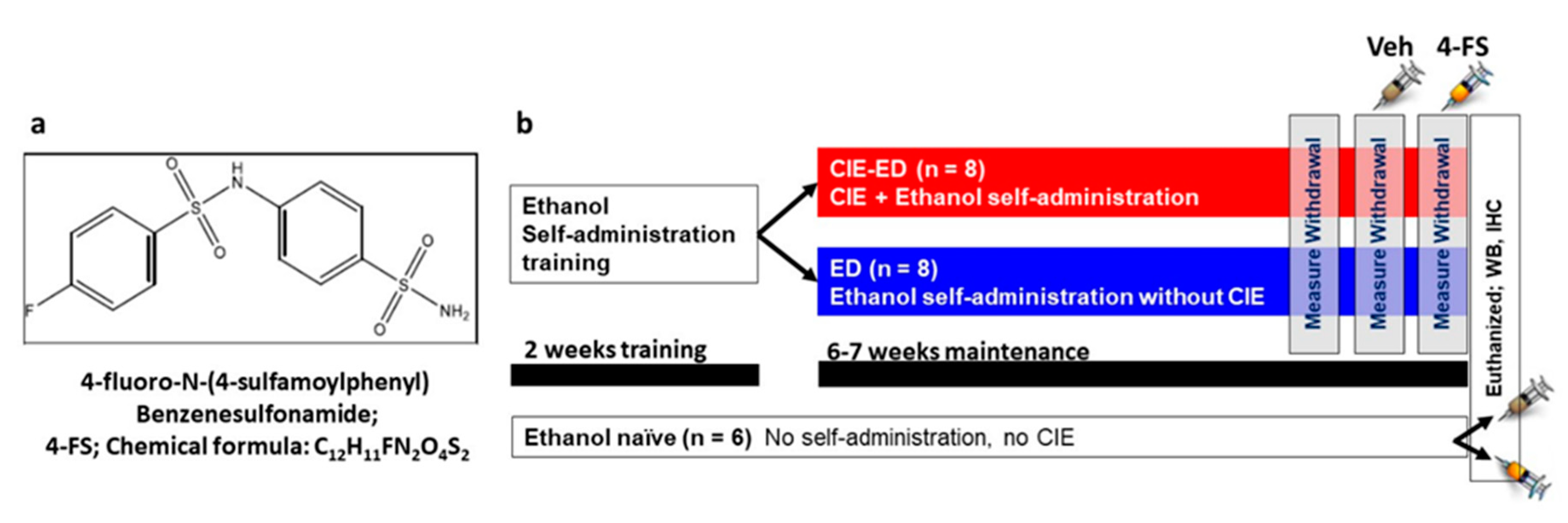
4. Materials and Methods

4.1. Animals

Twenty-two adult male Long Evans rats (Charles River, Wilmington, MA, USA) were 8 weeks old at the beginning of the study, and weighed approximately 220–250 g. The rats were housed in reverse 12 h light-12 h dark cycle rooms and two/cage. Food and water were available ad libitum. All experimental procedures were approved by the Institutional Animal Care and Use Committee (protocol #A16-000, approved on 29 April 2016) at VA San Diego Healthcare System.

4.2. Ethanol Self-Administration

The behavioral experiments conducted herein are presented as a detailed schematic in Figure 1a. Sixteen experimentally-naive rats were given two 14-h lever-responding training sessions in the operant conditioning boxes (Med Associates Inc, St. Albans, VT, USA), on an fixed-ratio 1 schedule (FR1) for water followed by 30 min sessions of ethanol (10% v/v) that lasted for 10 days. These self-administration sessions of ethanol (training and maintenance) was performed according to our previous publication [20]. Following training and maintenance of ethanol self-administration, the rats were divided into two groups; one group experienced chronic intermittent ethanol vapor exposure (CIE; details of this procedure are available in our previous publication [20]) while the other group was exposed to air in their normal housing condition (did not experience ethanol vapors) for a duration of 6–7 weeks. Self-administration of ethanol continued during the 6–7 weeks of vapor or air exposure; all rats received two 30-min FR1 sessions per week (Tuesdays and Thursdays) during these 6–7 weeks. Based on these experimental conditions, these rats belonged to CIE-ED (n = 8) or ED (n = 8) groups, respectively. Lever responses were analyzed to determine escalation of self-administration compared to pre-vapor stable responding.

4.3. Tail Bleeding for Determination of Blood Alcohol Levels (BAL)

To measure and ensure consistent BALs, tail bleeding was performed on the CIE-ED rats, once a week (every Wednesday), between hours 13–14 of vapor exposure as detailed in our previous publication [20]. When plasma samples were outside the target range (125–250 mg/dL), vapor levels were adjusted accordingly. One CIE-ED rat was excluded from the study as the rat did not meet the criteria for ethanol dependence.

4.4. 4-FS Synthesis and Treatment

4-FS an inhibitor of CA type II was synthesized via a green synthetic method as reported in a recent publication [43]. 4-FS was dissolved in 6% DMSO in sterile water. 4-FS or equal volume of vehicle was injected at a dose of 20 mg/kg i.p., 20 min before physical withdrawal scoring. Few ethanol naïve rats (n = 3) received 4-FS 1 h before euthanasia.

4.5. Scoring of Physical Withdrawal

During the 7th week, for CIE-ED rats (or time matched for ED rats), physical withdrawal was evaluated for 1 min at 7 h post vapor cessation. Physical withdrawal measures were conducted on three separate days: day one—without any intraperitoneal injections (baseline), day two—20 min after vehicle injection, day three—20 min after 4-FS injection. This optimal time point was chosen for measuring physical withdrawal based on previous publications [98,99,100]. The 7 h post-vapor time-point corresponded with the peak of withdrawal following the termination of vapor exposure [20]. Withdrawal signs in rats were scored by two observers blinded to the dependence status of the rats using previously published rubric [68,101], and details on individual withdrawal signs of abnormal body posture, locomotion, tail stiffness and grooming movements were scored separately on a scale of 0 to 3 based on detailed description presented in our previous publication [20]. Although the behaviors scored could reflect seizure-like activity, seizures were not measured and were not scored.

4.6. Brain Tissue Collection

Forty-five min after the last drinking session, CIE-ED, ED and time and age-matched ethanol naïve rats were killed by rapid decapitation and the brains were isolated, and dissected along the midsagittal plane. The left hemisphere was snap frozen for Western blotting analysis and the right hemisphere was postfixed in 4% paraformaldehyde for immunohistochemistry as indicated in our previous publication [102].

4.7. Western Blotting

Western blot procedures were performed according to our previous publication [20]. Tissue punches from 2–3 500-µm thick sections containing the dorsal and ventral hippocampus sections (Figure 5) were homogenized by sonication in ice-cold buffer HEPES buffer, and protein concentration was determined using a detergent-compatible Lowry method (Bio-Rad, Hercules, CA, USA). 20 µg protein samples were subjected to gel electrophoresis and transferred to PVDF membranes.
The membranes were incubated with the following primary antibodies: total AMPA Receptor 1 antibody (GluA1; rabbit monoclonal; 1:500, cell signaling technology, Danvers, MA, USA, cat#13185, molecular weight 100 kDa), total glutamate (NMDA) receptor subunit 2A antibody (GluN2A; rabbit polyclonal, 1:200, Santa Cruz Biotechnology Dallas, TX, USA, cat# sc-9056, molecular weight 170 kDa), total glutamate (NMDA) receptor subunit 2B antibody (GluN2B; 1:200, Santa Cruz Biotechnology, Dallas, TX, USA, cat. no. sc-9057, molecular weight 180 kDa) and total GABAA receptor antibody, PhosphoSolutions, Aurora, CO, USA, cat. no. 850-GA6, molecular weight 60 kDa). Following secondary antibody incubation, membranes were processed for immunoreactivity detection using SuperSignalWest Dura chemiluminescence detection reagent (Thermo Scientific, Waltham, MA, USA) and images were collected using a digital imaging system (Azure Imager c600, VWR, Radnor, PA, USA). For normalization purposes, membranes were incubated with 0.125% coomassie stain (cat # 1610400, Bio-Rad, Hercules, CA, USA) for 5 min and washed three times for 5–10 min in de-stain solution [103,104]. Densitometry was performed using ImageJ software (NIH, USA). The signal value of the band of interest was then expressed as a ratio of the corresponding coomassie signal. This ratio of expression for total protein was then expressed as a percent of the control sample included on the same blot.

4.8. Immunohistochemistry and Quantitative Analysis of Carbonic Anhydrase II (CA II) and Fos Labeled Cells

Tissue was sliced in 40 µm sections along the coronal plane in a cryostat. Two sections through the hippocampus were mounted on Superfrost® Plus slides (Fisher Scientific, Hampton, NH, USA) and dried overnight and processed for CA II and Fos analysis. The following primary antibody was used for CA II immunohistochemistry (IHC): rabbit polyclonal, 1:500, catalog # PA5-78897, Invitrogen, Carlsbad, CA, USA) and for Fos IHC: (mouse monoclonal, 1:1000, catalog # sc-52, Santa Cruz Biotechnology, Dallas, TX, USA [105]). The sections were pretreated [106], blocked, and incubated with the primary antibody followed by biotin-tagged secondary antibody. Staining was visualized with 3,3′-diaminobenzidine chromogen (DAB; cat# SK-4100; Vector Laboratories, Burlingame, CA, USA).
For CA II analyses we used hippocampal tissue from control (n = 5), ED (n = 5) and CIE-ED (n = 4) rats that did not receive any 4-FS treatment. Behavior and other immunohistochemical data from these animals have been published elsewhere [17]. These animals were age matched compared to the animals used in the current study and consumed similar amount of ethanol via self-administration when compared to the animals used in the current study. We used the tissue from these animals to determine whether ED and CIE-ED enhanced CA II expression in the hippocampus. We also used tissue from the current study (vehicle controls, 4-FS controls, 4-FS ED, 4-FS CIE-ED) to determine whether 4-FS treatment altered CA II expression in the hippocampus. CA II immunoreactive cells in the hilus region of the dentate gyrus were examined and captured at 100× magnification (Figure 3b–i) with an AxioImager Microscope (Zeiss, Oberkochen, Germany). Cells in the hilus were visually quantified using ImageJ software and used for analyses.
For Fos analyses only tissue from the animals used in the current study were used. Fos immunoreactive cells were examined and quantified with a Zeiss AxioImager Microscope. Cell quantification was performed according to the methods described in our previous publication [107]. Fos immunoreactive cells were quantified in the dorsal hippocampus (dorsal granule cell layer (GCL) and ventral hippocampus (hilus and ventral GCL; Figure 7).

4.9. Statistical Analyses

Ethanol self-administration during the 7-week CIE exposure was evaluated using repeated measures two-way ANOVA with self-administration session as a within-subject factor and CIE as a between-subject factor. Significant interactions were investigated using Sidak’s post-hoc tests. Ethanol self-administration before and after 4-FS injection was compared using paired t-tests and self-administration between the CIE-ED and ED rats were compared using unpaired t-tests. Withdrawal scores at 7 h post-vapor in CIE-ED rats and time matched for ED rats were compared using a non-parametric Kruskal–Wallis one-way analysis of variance. For CA II, western blotting and Fos analyses, comparisons between control, CIE-ED and ED groups were made using one-way ANOVAs, followed by Tukey’s post-hoc tests. Statistical significance was set at p < 0.05.

Author Contributions

Data curation M.S.K., W.T., N.X., B.S., C.D.M.; reagents M.A., M.a.-R.; conceptualization M.S.K., K.R., C.D.M.; funding acquisition M.S.K, C.D.M.; project administration C.D.M.; data analysis M.S.K., C.D.M.; supervision, writing—original draft preparation, review and editing C.D.M. All authors have read and agreed to the published version of the manuscript.

Funding

Funds from the Department of Veterans Affairs (BX003304 to C.D.M.), National Institute on Alcoholism and Alcohol Abuse and National Institute on Drug Abuse (AA020098 and DA034140 to C.D.M.) supported the study. M.S.K. was funded by an award of scholarship under international research support initiative program from the Higher Education Commission of Pakistan.

Conflicts of Interest

The authors declare no conflict of interest. The funders had no role in the design of the study; in the collection, analyses, or interpretation of data; in the writing of the manuscript; or in the decision to publish the results.

References

  1. SAMHSA. Results from the 2012 National Survey on Drug Use and Health: Summary of National Findings. Available online: https://store.samhsa.gov/product/Results-from-the-2012-National-Survey-on-Drug-Use-and-Health-NSDUH-/SMA13-4795 (accessed on 20 June 2020).
  2. Sullivan, E.V.; Rosenbloom, M.J.; Lim, K.O.; Pfefferbaum, A. Longitudinal changes in cognition, gait, and balance in abstinent and relapsed alcoholic men: Relationships to changes in brain structure. Neuropsychology 2000, 14, 178–188. [Google Scholar] [CrossRef] [PubMed]
  3. Bechara, A.; Dolan, S.; Denburg, N.; Hindes, A.; Anderson, S.W.; Nathan, P.E. Decision-making deficits, linked to a dysfunctional ventromedial prefrontal cortex, revealed in alcohol and stimulant abusers. Neuropsychologia 2001, 39, 376–389. [Google Scholar] [CrossRef]
  4. Oscar-Berman, M.; Marinkovic, K. Alcoholism and the brain: An overview. Alcohol. Res. Health 2003, 27, 125–133. [Google Scholar] [PubMed]
  5. Rosenbloom, M.; Sullivan, E.V.; Pfefferbaum, A. Using magnetic resonance imaging and diffusion tensor imaging to assess brain damage in alcoholics. Alcohol Res. Health 2003, 27, 146–152. [Google Scholar]
  6. Sullivan, E.V.; Rosenbloom, M.J.; Pfefferbaum, A. Pattern of motor and cognitive deficits in detoxified alcoholic men. Alcohol Clin. Exp. Res. 2000, 24, 611–621. [Google Scholar] [CrossRef]
  7. Koob, G.F.; Volkow, N.D. Neurocircuitry of addiction. Neuropsychopharmacology 2010, 35, 217–238. [Google Scholar] [CrossRef] [Green Version]
  8. Crews, F.T.; Boettiger, C.A. Impulsivity, frontal lobes and risk for addiction. Pharmacol. Biochem. Behav. 2009, 93, 237–247. [Google Scholar] [CrossRef] [Green Version]
  9. Crews, F.T.; Nixon, K. Mechanisms of neurodegeneration and regeneration in alcoholism. Alcohol Alcohol 2009, 44, 115–127. [Google Scholar] [CrossRef] [Green Version]
  10. Stephens, D.N.; Duka, T. Review. Cognitive and emotional consequences of binge drinking: Role of amygdala and prefrontal cortex. Philos. Trans. R. Soc. Lond. Biol. Sci. 2008, 363, 3169–3179. [Google Scholar] [CrossRef]
  11. O’Dell, L.E.; Roberts, A.J.; Smith, R.T.; Koob, G.F. Enhanced alcohol self-administration after intermittent versus continuous alcohol vapor exposure. Alcohol Clin. Exp. Res. 2004, 28, 1676–1682. [Google Scholar] [CrossRef]
  12. Valdez, G.R.; Roberts, A.J.; Chan, K.; Davis, H.; Brennan, M.; Zorrilla, E.P.; Koob, G.F. Increased ethanol self-administration and anxiety-like behavior during acute ethanol withdrawal and protracted abstinence: Regulation by corticotropin-releasing factor. Alcohol Clin. Exp. Res. 2002, 26, 1494–1501. [Google Scholar] [CrossRef] [PubMed]
  13. Hansson, A.C.; Nixon, K.; Rimondini, R.; Damadzic, R.; Sommer, W.H.; Eskay, R.; Crews, F.T.; Heilig, M. Long-term suppression of forebrain neurogenesis and loss of neuronal progenitor cells following prolonged alcohol dependence in rats. Int. J. Neuropsychopharmacol. 2010, 13, 583–593. [Google Scholar] [CrossRef] [PubMed] [Green Version]
  14. Pian, J.P.; Criado, J.R.; Milner, R.; Ehlers, C.L. N-methyl-D-aspartate receptor subunit expression in adult and adolescent brain following chronic ethanol exposure. Neuroscience. 2010, 170, 645–654. [Google Scholar] [CrossRef] [PubMed] [Green Version]
  15. Criado, J.R.; Liu, T.; Ehlers, C.L.; Mathe, A.A. Prolonged chronic ethanol exposure alters neuropeptide Y and corticotropin-releasing factor levels in the brain of adult Wistar rats. Pharmacol. Biochem. Behav. 2011, 99, 104–111. [Google Scholar] [CrossRef] [Green Version]
  16. Vendruscolo, L.F.; Barbier, E.; Schlosburg, J.E.; Misra, K.K.; Whitfield, T.W.; Logrip, M.L., Jr.; Rivier, C.; Repunte-Canonigo, V.; Zorrilla, E.P.; Sanna, P.P.; et al. Corticosteroid-dependent plasticity mediates compulsive alcohol drinking in rats. J. Neurosci. 2012, 32, 7563–7571. [Google Scholar] [CrossRef] [PubMed] [Green Version]
  17. Richardson, H.N.; Chan, S.H.; Crawford, E.F.; Lee, Y.K.; Funk, C.K.; Koob, G.F.; Mandyam, C.D. Permanent impairment of birth and survival of cortical and hippocampal proliferating cells following excessive drinking during alcohol dependence. Neurobiol. Dis. 2009, 36, 1–10. [Google Scholar] [CrossRef] [PubMed] [Green Version]
  18. Staples, M.C.; Kim, A.; Mandyam, C.D. Dendritic remodeling of hippocampal neurons is associated with altered NMDA receptor expression in alcohol dependent rats. Mol. Cell Neurosci. 2015, 65, 153–162. [Google Scholar] [CrossRef] [Green Version]
  19. Roberto, M.; Varodayan, F.P. Synaptic targets: Chronic alcohol actions. Neuropharmacology 2017, 122, 85–99. [Google Scholar] [CrossRef]
  20. Somkuwar, S.S.; Fannon, M.J.; Staples, M.C.; Zamora-Martinez, E.R.; Navarro, A.I.; Kim, A.; Quigley, J.A.; Edwards, S.; Mandyam, C.D. Alcohol dependence-induced regulation of the proliferation and survival of adult brain progenitors is associated with altered BDNF-TrkB signaling. Brain Struct. Funct. 2016, 221, 4319–4335. [Google Scholar] [CrossRef] [Green Version]
  21. Mandyam, C.D. The Interplay between the Hippocampus and Amygdala in Regulating Aberrant Hippocampal Neurogenesis during Protracted Abstinence from Alcohol Dependence. Front. Psychiatry 2013, 4, 61. [Google Scholar] [CrossRef] [Green Version]
  22. Prendergast, M.A.; Harris, B.R.; Mullholland, P.J.; Blanchard, J.A.; 2nd Gibson, D.A.; Holley, R.C.; Littleton, J.M. Hippocampal CA1 region neurodegeneration produced by ethanol withdrawal requires activation of intrinsic polysynaptic hippocampal pathways and function of N-methyl-D-aspartate receptors. Neuroscience 2004, 124, 869–877. [Google Scholar] [CrossRef]
  23. Mulholland, P.J.; Harris, B.R.; Wilkins, L.H.; Self, R.L.; Blanchard, J.A.; Holley, R.C.; Littleton, J.M.; Prendergast, M.A. Opposing effects of ethanol and nicotine on hippocampal calbindin-D28k expression. Alcohol 2003, 31, 1–10. [Google Scholar] [CrossRef] [PubMed]
  24. Wilkins, L.H.; Prendergast, M.A., Jr.; Blanchard, J.; Holley, R.C.; Chambers, E.R.; Littleton, J.M. Potential value of changes in cell markers in organotypic hippocampal cultures associated with chronic EtOH exposure and withdrawal: Comparison with NMDA-induced changes. Alcohol Clin. Exp. Res. 2006, 30, 1768–1880. [Google Scholar] [CrossRef] [PubMed]
  25. Gibson, D.A.; Harris, B.R.; Prendergast, M.A.; Hart, S.R.; Blanchard, J.A., 2nd; Holley, R.C.; Pedigo, N.W.; Littleton, J.M. Polyamines contribute to ethanol withdrawal-induced neurotoxicity in rat hippocampal slice cultures through interactions with the NMDA receptor. Alcohol Clin. Exp. Res. 2003, 27, 1099–1106. [Google Scholar] [CrossRef]
  26. Claus, D.; Kim, J.S.; Kornhuber, M.E.; Ahn, Y.S. [Effect of ethanol on the neurotransmitters glutamate and GABA]. Arch. Psychiatr. Nervenkr. 1982, 232, 183–189. [Google Scholar] [CrossRef]
  27. Keller, E.; Cummins, J.T.; von Hungen, K. Regional effects of ethanol on glutamate levels, uptake and release in slice and synaptosome preparations from rat brain. Subst. Alcohol Actions. Misuse 1983, 4, 383–392. [Google Scholar] [PubMed]
  28. Wilce, P.A.; Le, F.; Matsumoto, I.; Shanley, B.C. Ethanol inhibits NMDA-receptor mediated regulation of immediate early gene expression. Alcohol Alcohol Suppl. 1993, 2, 359–363. [Google Scholar]
  29. Snell, L.D.; Nunley, K.R.; Lickteig, R.L.; Browning, M.D.; Tabakoff, B.; Hoffman, P.L. Regional and subunit specific changes in NMDA receptor mRNA and immunoreactivity in mouse brain following chronic ethanol ingestion. Brain Res. Mol. Brain Res. 1996, 40, 71–78. [Google Scholar] [CrossRef]
  30. Wirkner, K.; Poelchen, W.; Koles, L.; Muhlberg, K.; Scheibler, P.; Allgaier, C.; Illes, P. Ethanol-induced inhibition of NMDA receptor channels. Neurochem. Int. 1999, 35, 153–162. [Google Scholar] [CrossRef]
  31. Davidson, M.; Shanley, B.; Wilce, P. Increased NMDA-induced excitability during ethanol withdrawal: A behavioural and histological study. Brain Res. 1995, 674, 91–96. [Google Scholar] [CrossRef]
  32. Davidson, M.D.; Wilce, P.; Shanley, B.C. Increased sensitivity of the hippocampus in ethanol-dependent rats to toxic effect of N-methyl-D-aspartic acid in vivo. Brain Res. 1993, 606, 5–9. [Google Scholar] [CrossRef]
  33. Hoffman, P.L. NMDA receptors in alcoholism. Int. Rev. Neurobiol. 2003, 56, 35–82. [Google Scholar] [PubMed]
  34. Chesler, M. Regulation and modulation of pH in the brain. Physiol. Rev. 2003, 83, 1183–1221. [Google Scholar] [CrossRef] [PubMed]
  35. Chen, J.C.; Chesler, M. pH transients evoked by excitatory synaptic transmission are increased by inhibition of extracellular carbonic anhydrase. Proc. Natl. Acad. Sci. USA 1992, 89, 7786–7790. [Google Scholar] [CrossRef] [Green Version]
  36. Hamidi, S.; Avoli, M. Carbonic anhydrase inhibition by acetazolamide reduces in vitro epileptiform synchronization. Neuropharmacology 2015, 95, 377–387. [Google Scholar] [CrossRef] [Green Version]
  37. De Simone, G.; Scozzafava, A.; Supuran, C.T. Which carbonic anhydrases are targeted by the antiepileptic sulfonamides and sulfamates? Chem. Biol. Drug Des. 2009, 74, 317–321. [Google Scholar] [CrossRef]
  38. Lombroso, C.T.; Forxythe, I. A long-term follow-up of acetazolamide (diamox) in the treatment of epilepsy. Epilepsia 1960, 1, 493–500. [Google Scholar] [CrossRef]
  39. Van Berkel, M.A.; Elefritz, J.L. Evaluating off-label uses of acetazolamide. Off. J. Am. Soc. Health-Syst. Pharm. 2018, 75, 524–531. [Google Scholar] [CrossRef]
  40. Hamada, K.; Song, H.K.; Ishida, S.; Yagi, K.; Seino, M. Contrasting effects of zonisamide and acetazolamide on amygdaloid kindling in rats. Epilepsia 2001, 42, 1379–1386. [Google Scholar] [CrossRef] [Green Version]
  41. Guglielmo, R.; Martinotti, G.; Quatrale, M.; Ioime, L.; Kadilli, I.; Di Nicola, M.; Janiri, L. Topiramate in Alcohol Use Disorders: Review and Update. CNS Drugs 2015, 29, 383–395. [Google Scholar] [CrossRef]
  42. Santos, L.E.C.; Rodrigues, A.M.; Lopes, M.R.; Costa, V.D.C.; Scorza, C.A.; Scorza, F.A.; Cavalheiro, E.A.; Almeida, A.G. Long-term alcohol exposure elicits hippocampal nonsynaptic epileptiform activity changes associated with expression and functional changes in NKCC1, KCC2 co-transporters and Na(+)/K(+)-ATPase. Neuroscience 2017, 340, 530–541. [Google Scholar] [CrossRef] [PubMed]
  43. Al-Rashida, M.; Ejaz, S.A.; Ali, S.; Shaukat, A.; Hamayoun, M.; Ahmed, M.; Iqbal, J. Diarylsulfonamides and their bioisosteres as dual inhibitors of alkaline phosphatase and carbonic anhydrase: Structure activity relationship and molecular modelling studies. Bioorg. Med. Chem. 2015, 23, 2435–2444. [Google Scholar] [CrossRef] [PubMed]
  44. Ozsoy, H.Z. Carbonic anhydrase enzymes: Likely targets for inhalational anesthetics. Med. Hypotheses 2019, 123, 118–124. [Google Scholar] [CrossRef] [PubMed]
  45. Supuran, C.T. Carbonic anhydrase activators. Future Med. Chem. 2018, 10, 561–573. [Google Scholar] [CrossRef] [PubMed]
  46. Li, S.; An, L.; Duan, Q.; Ferraris Araneta, M.; Johnson, C.S.; Shen, J. Determining the Rate of Carbonic Anhydrase Reaction in the Human Brain. Sci. Rep. 2018, 8, 2328. [Google Scholar] [CrossRef] [Green Version]
  47. Tong, C.K.; Cammer, W.; Chesler, M. Activity-dependent pH shifts in hippocampal slices from normal and carbonic anhydrase II-deficient mice. Glia 2000, 31, 125–130. [Google Scholar] [CrossRef]
  48. Giacobini, E. A cytochemical study of the localization of carbonic anhydrase in the nervous system. J. Neurochem. 1962, 9, 169–177. [Google Scholar] [CrossRef]
  49. Nogradi, A.; Mihaly, A. Distribution of carbonic anhydrase activity in the rat central nervous system, as revealed by a new semipermeable technique. Acta Histochem. 1988, 84, 153–162. [Google Scholar] [CrossRef]
  50. Tong, C.K.; Chen, K.; Chesler, M. Kinetics of activity-evoked pH transients and extracellular pH buffering in rat hippocampal slices. J. Neurophysiol. 2006, 95, 3686–3697. [Google Scholar] [CrossRef]
  51. Walz, W. pH shifts evoked by neuronal stimulation in slices of rat hippocampus. Can. J. Physiol. Pharmacol. 1989, 67, 577–581. [Google Scholar] [CrossRef]
  52. Esbaugh, A.J.; Tufts, B.L. The structure and function of carbonic anhydrase isozymes in the respiratory system of vertebrates. Respir. Physiol. Neurobiol. 2006, 154, 185–198. [Google Scholar] [CrossRef] [PubMed]
  53. Chesler, M.; Kaila, K. Modulation of pH by neuronal activity. Trends Neurosci. 1992, 15, 396–402. [Google Scholar] [CrossRef]
  54. Chesler, M.; Chen, J.C.; Kraig, R.P. Determination of extracellular bicarbonate and carbon dioxide concentrations in brain slices using carbonate and pH-selective microelectrodes. J. Neurosci. Methods 1994, 53, 129–136. [Google Scholar] [CrossRef] [Green Version]
  55. Tong, C.K.; Chesler, M. Endogenous pH shifts facilitate spreading depression by effect on NMDA receptors. J. Neurophysiol. 1999, 81, 1988–1991. [Google Scholar] [CrossRef]
  56. Wagner, N.; Franz, N.; Dieteren, S.; Perl, M.; Mors, K.; Marzi, I.; Relja, B. Acute Alcohol Binge Deteriorates Metabolic and Respiratory Compensation Capability After Blunt Chest Trauma Followed by Hemorrhagic Shock-A New Research Model. Alcohol Clin. Exp. Res. 2017, 41, 1559–1567. [Google Scholar] [CrossRef]
  57. Gilliam, D.M.; Collins, A.C. Acute ethanol effects on blood pH, PCO2, and PO2 in LS and SS mice. Physiol. Behav. 1982, 28, 879–883. [Google Scholar] [CrossRef]
  58. Wang, T.; Eskandari, D.; Zou, D.; Grote, L.; Hedner, J. Increased Carbonic Anhydrase Activity is Associated with Sleep Apnea Severity and Related Hypoxemia. Sleep 2015, 38, 1067–1073. [Google Scholar] [CrossRef] [Green Version]
  59. Wang, W.T.; Lee, P.; Hui, D.; Michaelis, E.K.; Choi, I.Y. Effects of Ethanol Exposure on the Neurochemical Profile of a Transgenic Mouse Model with Enhanced Glutamate Release Using In Vivo (1)H MRS. Neurochem. Res. 2019, 44, 133–146. [Google Scholar] [CrossRef]
  60. Chen, J.C.; Chesler, M. Modulation of extracellular pH by glutamate and GABA in rat hippocampal slices. J. Neurophysiol. 1992, 67, 29–36. [Google Scholar] [CrossRef]
  61. Mendelson, J.H.; Mello, N.K. Basic mechanisms underlying physical dependence upon alcohol. Ann. N. Y. Acad. Sci. 1978, 311, 69–79. [Google Scholar] [CrossRef]
  62. Roelofs, S.M.; Dikkenberg, G.M. Hyperventilation and anxiety: Alcohol withdrawal symptoms decreasing with prolonged abstinence. Alcohol 1987, 4, 215–220. [Google Scholar] [CrossRef]
  63. Teng, Y.H.; Tsai, H.T.; Hsieh, Y.S.; Chen, Y.C.; Lin, C.W.; Lee, M.C.; Lin, L.Y.; Yang, S.F. Elevated erythrocyte carbonic anhydrase activity is a novel clinical marker in hyperventilation syndrome. Clin. Chem. Lab. Med 2009, 47, 441–445. [Google Scholar] [CrossRef] [PubMed]
  64. Olsen, R.W.; Liang, J. Role of GABAA receptors in alcohol use disorders suggested by chronic intermittent ethanol (CIE) rodent model. Mol. Brain. 2017, 10, 45. [Google Scholar] [CrossRef] [PubMed]
  65. Nelson, T.E.; Ur, C.L.; Gruol, D.L. Chronic intermittent ethanol exposure enhances NMDA-receptor-mediated synaptic responses and NMDA receptor expression in hippocampal CA1 region. Brain Res. 2005, 1048, 69–79. [Google Scholar] [CrossRef] [PubMed]
  66. Frasca, A.; Aalbers, M.; Frigerio, F.; Fiordaliso, F.; Salio, M.; Gobbi, M.; Cagnotto, A.; Gardoni, F.; Battaglia, G.S.; Hoogland, G.; et al. Misplaced NMDA receptors in epileptogenesis contribute to excitotoxicity. Neurobiol. Dis. 2011, 43, 507–515. [Google Scholar] [CrossRef]
  67. Smith, M.A.; Weiss, S.R.; Berry, R.L.; Zhang, L.X.; Clark, M.; Massenburg, G.; Post, R.M. Amygdala-kindled seizures increase the expression of corticotropin-releasing factor (CRF) and CRF-binding protein in GABAergic interneurons of the dentate hilus. Brain Res. 1997, 745, 248–256. [Google Scholar] [CrossRef]
  68. Macey, D.J.; Schulteis, G.; Heinrichs, S.C.; Koob, G.F. Time-dependent quantifiable withdrawal from ethanol in the rat: Effect of method of dependence induction. Alcohol 1996, 13, 163–170. [Google Scholar] [CrossRef]
  69. Prior, P.L.; Galduroz, J.C. Glutamatergic hyperfunctioning during alcohol withdrawal syndrome: Therapeutic perspective with zinc and magnesium. Med. Hypotheses 2011, 77, 368–370. [Google Scholar] [CrossRef] [Green Version]
  70. McCool, B.A.; Christian, D.T.; Diaz, M.R.; Lack, A.K. Glutamate plasticity in the drunken amygdala: The making of an anxious synapse. Int. Rev. Neurobiol. 2010, 91, 205–233. [Google Scholar]
  71. Hoffman, P.L.; Tabakoff, B. The role of the NMDA receptor in ethanol withdrawal. Exs 1994, 71, 61–70. [Google Scholar]
  72. Tsai, G.; Gastfriend, D.R.; Coyle, J.T. The glutamatergic basis of human alcoholism. Am. J. Psychiatry 1995, 152, 332–340. [Google Scholar] [PubMed]
  73. Baram, T.Z.; Hirsch, E.; Snead, O.C., III; Schultz, L. Corticotropin-releasing hormone-induced seizures in infant rats originate in the amygdala. Ann. Neurol. 1992, 31, 488–494. [Google Scholar] [CrossRef] [PubMed] [Green Version]
  74. Knapp, C.M.; Mercado, M.; Markley, T.L.; Crosby, S.; Ciraulo, D.A.; Kornetsky, C. Zonisamide decreases ethanol intake in rats and mice. Pharmacol. Biochem. Behav. 2007, 87, 65–72. [Google Scholar] [CrossRef] [PubMed] [Green Version]
  75. Zalewska-Kaszubska, J.; Bajer, B.; Gorska, D.; Andrzejczak, D.; Dyr, W.; Bieńkowski, P. Effect of repeated treatment with topiramate on voluntary alcohol intake and beta-endorphin plasma level in Warsaw alcohol high-preferring rats. Psychopharmacology 2013, 225, 275–281. [Google Scholar] [CrossRef] [Green Version]
  76. Knapp, C.M.; Sarid-Segal, O.; Richardson, M.A.; Colaneri, L.S.; Afshar, M.; Devine, E.; Streeter, C.C.; Piechniczek-Buczek, J.; Ciraulo, D.A. Open label trial of the tolerability and efficacy of zonisamide in the treatment of alcohol dependence. Am. J. Drug Alcohol Abus. 2010, 36, 102–105. [Google Scholar] [CrossRef] [Green Version]
  77. Perucca, E. Clinical pharmacology and therapeutic use of the new antiepileptic drugs. Fundam. Clin. Pharmacol. 2001, 15, 405–417. [Google Scholar] [CrossRef]
  78. Rubio, G.; López-Muñoz, F.; Ferre, F.; Martínez-Gras, I.; Ponce, G.; Pascual, J.M.; Jiménez-Arriero, M.A.; Alamo, C. Effects of zonisamide in the treatment of alcohol dependence. Clin. Neuropharmacol. 2010, 33, 250–253. [Google Scholar] [CrossRef]
  79. Sarid-Segal, O.; Knapp, C.M.; Burch, W.; Richardson, M.A.; Bahtia, S.; DeQuattro, K.; Afshar, M.; Richambault, C.; Sickels, L.; Devine, E. The anticonvulsant zonisamide reduces ethanol self-administration by risky drinkers. Am. J. Drug Alcohol Abus. 2009, 35, 316–319. [Google Scholar] [CrossRef]
  80. Sripathirathan, K.; Brown, J., III; Neafsey, E.J.; Collins, M.A. Linking binge alcohol-induced neurodamage to brain edema and potential aquaporin-4 upregulation: Evidence in rat organotypic brain slice cultures and in vivo. J. Neurotrauma 2009, 26, 261–273. [Google Scholar] [CrossRef]
  81. Swenson, E.R. Safety of carbonic anhydrase inhibitors. Expert. Opin. Drug Saf. 2014, 13, 459–472. [Google Scholar] [CrossRef]
  82. De Simone, G.; Di Fiore, A.; Menchise, V.; Pedone, C.; Antel, J.; Casini, A.; Scozzafava, A.; Wurl, M.; Supuran, C.T. Carbonic anhydrase inhibitors. Zonisamide is an effective inhibitor of the cytosolic isozyme II and mitochondrial isozyme V: Solution and X-ray crystallographic studies. Bioorg. Med. Chem. Lett. 2005, 15, 2315–2320. [Google Scholar] [CrossRef] [PubMed]
  83. White, H.S. Mechanism of action of newer anticonvulsants. J. Clin. Psychiatry 2003, 64, 5–8. [Google Scholar] [PubMed]
  84. Yamamura, S.; Saito, H.; Suzuki, N.; Kashimoto, S.; Hamaguchi, T.; Ohoyama, K.; Suzuki, D.; Kanehara, S.; Nakagawa, M.; Shiroyama, T. Effects of zonisamide on neurotransmitter release associated with inositol triphosphate receptors. Neurosci. Lett. 2009, 454, 91–96. [Google Scholar] [CrossRef] [PubMed]
  85. Yoshida, S.; Okada, M.; Zhu, G.; Kaneko, S. Effects of zonisamide on neurotransmitter exocytosis associated with ryanodine receptors. Epilepsy Res. 2005, 67, 153–162. [Google Scholar] [CrossRef]
  86. Amaral, D.G.; Witter, M.P. The three-dimensional organization of the hippocampal formation: A review of anatomical data. Neuroscience 1989, 31, 571–591. [Google Scholar] [CrossRef]
  87. Snyder, J.S.; Radik, R.; Wojtowicz, J.M.; Cameron, H.A. Anatomical gradients of adult neurogenesis and activity: Young neurons in the ventral dentate gyrus are activated by water maze training. Hippocampus 2009, 19, 360–370. [Google Scholar] [CrossRef] [Green Version]
  88. Wells, A.M.; Lasseter, H.C.; Xie, X.; Cowhey, K.E.; Reittinger, A.M.; Fuchs, R.A. Interaction between the basolateral amygdala and dorsal hippocampus is critical for cocaine memory reconsolidation and subsequent drug context-induced cocaine-seeking behavior in rats. Learn. Mem. 2011, 18, 693–702. [Google Scholar] [CrossRef] [Green Version]
  89. Rogers, J.L.; See, R.E. Selective inactivation of the ventral hippocampus attenuates cue-induced and cocaine-primed reinstatement of drug-seeking in rats. Neurobiol. Learn. Mem. 2007, 87, 688–692. [Google Scholar] [CrossRef] [Green Version]
  90. Lasseter, H.C.; Xie, X.; Ramirez, D.R.; Fuchs, R.A. Sub-region specific contribution of the ventral hippocampus to drug context-induced reinstatement of cocaine-seeking behavior in rats. Neuroscience 2010, 171, 830–839. [Google Scholar] [CrossRef] [Green Version]
  91. Lau, J.Y.; Goldman, D.; Buzas, B.; Hodgkinson, C.; Leibenluft, E.; Nelson, E.; Sankin, L.; Pine, D.S.; Ernst, M. BDNF gene polymorphism (Val66Met) predicts amygdala and anterior hippocampus responses to emotional faces in anxious and depressed adolescents. Neuroimage 2010, 53, 952–961. [Google Scholar] [CrossRef] [Green Version]
  92. Cui, Z.; Feng, R.; Jacobs, S.; Duan, Y.; Wang, H.; Cao, X.; Tsien, J.Z. Increased NR2A:NR2B ratio compresses long-term depression range and constrains long-term memory. Sci. Rep. 2013, 3, 1036. [Google Scholar] [CrossRef] [PubMed] [Green Version]
  93. Du, X.; Elberger, A.J.; Matthews, D.B.; Hamre, K.M. Heterozygous deletion of NR1 subunit of the NMDA receptor alters ethanol-related behaviors and regional expression of NR2 subunits in the brain. Neurotoxicol. Teratol. 2012, 34, 177–186. [Google Scholar] [CrossRef] [PubMed]
  94. Kalluri, H.S.; Mehta, A.K.; Ticku, M.K. Up-regulation of NMDA receptor subunits in rat brain following chronic ethanol treatment. Brain Res. 1998, 58, 221–224. [Google Scholar] [CrossRef]
  95. Prendergast, M.A.; Mulholland, P.J. Glucocorticoid and polyamine interactions in the plasticity of glutamatergic synapses that contribute to ethanol-associated dependence and neuronal injury. Addict. Biol. 2012, 17, 209–223. [Google Scholar] [CrossRef] [PubMed] [Green Version]
  96. Milenkovic, I.; Vasiljevic, M.; Maurer, D.; Hoger, H.; Klausberger, T.; Sieghart, W. The parvalbumin-positive interneurons in the mouse dentate gyrus express GABAA receptor subunits alpha1, beta2, and delta along their extrasynaptic cell membrane. Neuroscience 2013, 254, 80–96. [Google Scholar] [CrossRef]
  97. Tong, X.; Peng, Z.; Zhang, N.; Cetina, Y.; Huang, C.S.; Wallner, M.; Otis, T.S.; Houser, C.R. Ectopic Expression of alpha6 and delta GABAA Receptor Subunits in Hilar Somatostatin Neurons Increases Tonic Inhibition and Alters Network Activity in the Dentate Gyrus. J. Neurosci. 2015, 35, 16142–16158. [Google Scholar] [CrossRef] [Green Version]
  98. O’Dell, L.E.; Chen, S.A.; Smith, R.T.; Specio, S.E.; Balster, R.L.; Paterson, N.E.; Markou, A.; Zorrilla, E.P.; Koob, G.F. Extended access to nicotine self-administration leads to dependence: Circadian measures, withdrawal measures, and extinction behavior in rats. J. Pharmacol. Exp. Ther. 2007, 320, 180–193. [Google Scholar] [CrossRef]
  99. Gilpin, N.W.; Richardson, H.N.; Cole, M.; Koob, G.F. Vapor inhalation of alcohol in rats. Curr. Protoc. Neurosci. 2008, 44, 1–19. [Google Scholar] [CrossRef]
  100. Richardson, H.N.; Lee, S.Y.; O’Dell, L.E.; Koob, G.F.; Rivier, C.L. Alcohol self-administration acutely stimulates the hypothalamic-pituitary-adrenal axis, but alcohol dependence leads to a dampened neuroendocrine state. Eur. J. Neurosci. 2008, 28, 1641–1653. [Google Scholar] [CrossRef] [Green Version]
  101. Majchrowicz, E. Induction of physical dependence upon ethanol and the associated behavioral changes in rats. Psychopharmacologia 1975, 43, 245–254. [Google Scholar] [CrossRef]
  102. Cohen, A.; Soleiman, M.T.; Talia, R.; Koob, G.F.; George, O.; Mandyam, C.D. Extended access nicotine self-administration with periodic deprivation increases immature neurons in the hippocampus. Psychopharmacology 2015, 232, 453–463. [Google Scholar] [CrossRef] [PubMed] [Green Version]
  103. Welinder, C.; Ekblad, L. Coomassie staining as loading control in Western blot analysis. J. Proteome Res. 2011, 10, 1416–1419. [Google Scholar] [CrossRef] [PubMed]
  104. Thacker, J.S.; Yeung, D.H.; Staines, W.R.; Mielke, J.G. Total protein or high-abundance protein: Which offers the best loading control for Western blotting? Anal. Biochem. 2016, 496, 76–78. [Google Scholar] [CrossRef]
  105. Recinto, P.; Samant, A.R.; Chavez, G.; Kim, A.; Yuan, C.J.; Soleiman, M.; Grant, Y.; Edwards, S.; Wee, S.; Koob, G.F.; et al. Levels of neural progenitors in the hippocampus predict memory impairment and relapse to drug seeking as a function of excessive methamphetamine self-administration. Neuropsychopharmacology 2012, 37, 1275–1287. [Google Scholar] [CrossRef] [PubMed]
  106. Mandyam, C.D.; Norris, R.D.; Eisch, A.J. Chronic morphine induces premature mitosis of proliferating cells in the adult mouse subgranular zone. J. Neurosci. Res. 2004, 76, 783–794. [Google Scholar] [CrossRef] [PubMed]
  107. Somkuwar, S.S.; Fannon-Pavlich, M.J.; Ghofranian, A.; Quigley, J.A.; Dutta, R.R.; Galinato, M.H.; Mandyam, C.D. Wheel running reduces ethanol seeking by increasing neuronal activation and reducing oligodendroglial/neuroinflammatory factors in the medial prefrontal cortex. Brain Behav. Immun. 2016, 58, 357–368. [Google Scholar] [CrossRef] [Green Version]
Figure 1. (a) Chemical structure of 4-FS. (b) Schematic of the experimental timeline and experimental group information.
Ijms 21 04411 g001
Figure 2. Ethanol self-administration and blood alcohol levels. (a) Amount of ethanol consumed prior to chronic intermittent ethanol (CIE) initiation and over the 6 weeks period of CIE or air exposure. Weeks of air/vapor exposure are indicated over a pale pink background. (b) Blood alcohol levels measured once a week in animals experiencing CIE. CIE-ED, n = 7 and ED, n = 8. $, interaction; #, main effect of groups; * p < 0.05 vs. CIE-ED by two-way analysis of variance (ANOVA). Data are expressed as mean ± S.E.M.
Ijms 21 04411 g002
Figure 3. Carbonic anhydrase type II (CA II) expression in the adult rat hippocampus. (a) Photomicrograph of CA II immunohistochemistry in the hippocampus and cortex from one control rat. CA II+ cells appeared as single cells; each immunoreactive cell is pointed with an arrowhead. 1- CA II+ cell in the hilus (Hil); 2-CA II+ cell in the molecular layer (Mol); 3-CA II+ cell in the corpus callosum (cc); 4-CA II+ cell in the cortex. (bi) 100× images of the hilus used for quantitative analyses of CA II cells. (e) Zoomed in image shown in (d) to indicate the morphology of CA II+ cells in the hilus. Scale bar in (e) is 20 µm; scale bar in (i) is 50 µm (applies bd and fi). (j) Number of CA II+ cells in the hilus. n = 5 controls, n = 5 ED, n = 4 CIE-ED, n = 3 vehicle controls, n = 3 4-FS controls, n = 8 4-FS ED rats, n = 7 4-FS CIE-ED rats. * p < 0.05, compared to controls; # p < 0.05 compared to 4-FS control. Data are expressed as mean ± S.E.M.
Ijms 21 04411 g003
Figure 4. Physical withdrawal behaviors in ethanol drinking (ED) and CIE-ED rats with vehicle and 4-FS treatment. Withdrawal behaviors are evident in CIE-ED rats. Signs of ethanol withdrawal, i.e., aberrant body posture, locomotor activity and tail stiffness were reduced by 4-FS in CIE-ED rats. n = 7–8/group. * p < 0.05 compared to baseline and vehicle days, within-subject. # p < 0.05 vs. CIE-ED rats, between-subject. Data are expressed as mean ± S.E.M.
Ijms 21 04411 g004
Figure 5. 4-FS reduces drinking in ethanol drinking (ED) rats. (a) Ethanol intake expressed as g/kg and (b) active lever responses. (c) Inactive lever responses and (d) lever responses during timeout. n = 8 4-FS ED rats, n = 7 4-FS CIE-ED rats. * p < 0.05 vs. vehicle day, within-subject. Data are expressed as mean ± S.E.M.
Ijms 21 04411 g005
Figure 6. (a,b,e,f) Schematic of tissue punches from the dorsal hippocampus (a,b) and ventral hippocampus (e,f). (c,g) sample western blots from dorsal (c) and ventral (g) hippocampus tissue punches. (d,h) Densitometric analysis of tissue for levels of GluA1, GluN2A, GluN2B, ratio of GluN2A/2B and GABAA from the dorsal (d) and ventral (h) hippocampus tissue homogenate. n = 8 4-FS ED rats, n = 7 4-FS CIE-ED rats. Values are mean ± S.E.M. expressed as % control, where control is represented by the dotted line in each graph. * p < 0.05, compared to controls.
Ijms 21 04411 g006
Figure 7. Fos expression is altered by 4-FS. (a) Photomicrograph of Fos immunohistochemistry in the hippocampus and cortex from one control rat. Fos+ cells appeared as single cells; each immunoreactive cell is pointed with an arrowhead. 1-Fos+ cell in the hilus (Hil); 2-Fos+ cell in the granule cell layer (GCL); 3-Fos+ cell in the CA1 region; 4-Fos+ cell in the cortex. Mol, molecular layer. Scale bar is 100 µm. (b,c) Number of Fos+ cells in the dorsal (b) and ventral (c) hippocampus. n = 8 4-FS ED rats, n = 7 4-FS CIE-ED rats. * p < 0.05, compared to controls. Data are expressed as mean ± S.E.M.
Ijms 21 04411 g007

