Next Issue
Volume 21, July-1
Previous Issue
Volume 21, June-1
 
 
ijms-logo

Journal Browser

Journal Browser

Int. J. Mol. Sci., Volume 21, Issue 12 (June-2 2020) – 378 articles

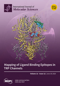
Cover Story (view full-size image): Characterization of the binding epitopes of various endogenous modulators of transient receptor potential (TRP) channels is essential for understanding the necessary cellular signaling pathways as well as novel approaches to rational drug design. TRP channel binding epitopes serve as hotspots for the transmission of environmental signals with specific information to the channel. Deeper structural and functional knowledge of TRP channels allows us to describe the way in which endogenous modulators interact, and how they allosterically affect the communication and function of these multicomplex receptors. View this paper.
  • Issues are regarded as officially published after their release is announced to the table of contents alert mailing list.
  • You may sign up for e-mail alerts to receive table of contents of newly released issues.
  • PDF is the official format for papers published in both, html and pdf forms. To view the papers in pdf format, click on the "PDF Full-text" link, and use the free Adobe Reader to open them.
Order results
Result details
Section
Select all
Export citation of selected articles as:
11 pages, 1704 KiB  
Article
Validation of Breast Cancer Margins by Tissue Spray Mass Spectrometry
by Vitaliy V. Chagovets, Natalia L. Starodubtseva, Alisa O. Tokareva, Vladimir E. Frankevich, Valerii V. Rodionov, Vlada V. Kometova, Konstantin Chingin, Eugene N. Kukaev, Huanwen Chen and Gennady T. Sukhikh
Int. J. Mol. Sci. 2020, 21(12), 4568; https://doi.org/10.3390/ijms21124568 - 26 Jun 2020
Cited by 7 | Viewed by 2891
Abstract
Current methods for the intraoperative determination of breast cancer margins commonly suffer from the insufficient accuracy, specificity and/or low speed of analysis, increasing the time and cost of operation as well the risk of cancer recurrence. The purpose of this study is to [...] Read more.
Current methods for the intraoperative determination of breast cancer margins commonly suffer from the insufficient accuracy, specificity and/or low speed of analysis, increasing the time and cost of operation as well the risk of cancer recurrence. The purpose of this study is to develop a method for the rapid and accurate determination of breast cancer margins using direct molecular profiling by mass spectrometry (MS). Direct molecular fingerprinting of tiny pieces of breast tissue (approximately 1 × 1 × 1 mm) is performed using a home-built tissue spray ionization source installed on a Maxis Impact quadrupole time-of-flight mass spectrometer (qTOF MS) (Bruker Daltonics, Hamburg, Germany). Statistical analysis of MS data from 50 samples of both normal and cancer tissue (from 25 patients) was performed using orthogonal projections onto latent structures discriminant analysis (OPLS-DA). Additionally, the results of OPLS classification of new 19 pieces of two tissue samples were compared with the results of histological analysis performed on the same tissues samples. The average time of analysis for one sample was about 5 min. Positive and negative ionization modes are used to provide complementary information and to find out the most informative method for a breast tissue classification. The analysis provides information on 11 lipid classes. OPLS-DA models are created for the classification of normal and cancer tissue based on the various datasets: All mass spectrometric peaks over 300 counts; peaks with a statistically significant difference of intensity determined by the Mann–Whitney U-test (p < 0.05); peaks identified as lipids; both identified and significantly different peaks. The highest values of Q2 have models built on all MS peaks and on significantly different peaks. While such models are useful for classification itself, they are of less value for building explanatory mechanisms of pathophysiology and providing a pathway analysis. Models based on identified peaks are preferable from this point of view. Results obtained by OPLS-DA classification of the tissue spray MS data of a new sample set (n = 19) revealed 100% sensitivity and specificity when compared to histological analysis, the “gold” standard for tissue classification. “All peaks” and “significantly different peaks” datasets in the positive ion mode were ideal for breast cancer tissue classification. Our results indicate the potential of tissue spray mass spectrometry for rapid, accurate and intraoperative diagnostics of breast cancer tissue as a means to reduce surgical intervention. Full article
(This article belongs to the Special Issue Breast Cancer: From Pathophysiology to Novel Therapeutic Approaches)
Show Figures

Figure 1

16 pages, 2955 KiB  
Article
Binding of CML-Modified as Well as Heat-Glycated β-lactoglobulin to Receptors for AGEs Is Determined by Charge and Hydrophobicity
by Hannah E. Zenker, Malgorzata Teodorowicz, Arifa Ewaz, R.J. Joost van Neerven, Huub F.J. Savelkoul, Nicolette W. De Jong, Harry J. Wichers and Kasper A. Hettinga
Int. J. Mol. Sci. 2020, 21(12), 4567; https://doi.org/10.3390/ijms21124567 - 26 Jun 2020
Cited by 10 | Viewed by 2719
Abstract
Intake of dietary advanced glycation end products (AGEs) is associated with inflammation-related health problems. Nε-carboxymethyl lysine (CML) is one of the best characterised AGEs in processed food. AGEs have been described as ligands for receptors present on antigen presenting cells. However, changes in [...] Read more.
Intake of dietary advanced glycation end products (AGEs) is associated with inflammation-related health problems. Nε-carboxymethyl lysine (CML) is one of the best characterised AGEs in processed food. AGEs have been described as ligands for receptors present on antigen presenting cells. However, changes in protein secondary and tertiary structure also induce binding to AGE receptors. We aimed to discriminate the role of different protein modifications in binding to AGE receptors. Therefore, β-lactoglobulin was chemically modified with glyoxylic acid to produce CML and compared to β-lactoglobulin glycated with lactose. Secondary structure was monitored with circular dichroism, while hydrophobicity and formation of β-sheet structures was measured with ANS-assay and ThT-assay, respectively. Aggregation was monitored using native-PAGE. Binding to sRAGE, CD36, and galectin-3 was measured using inhibition ELISA. Even though no changes in secondary structure were observed in all tested samples, binding to AGE receptors increased with CML concentration of CML-modified β-lactoglobulin. The negative charge of CML was a crucial determinant for the binding of protein bound CML, while binding of glycated BLG was determined by increasing hydrophobicity. This shows that sRAGE, galectin-3, and CD36 bind to protein bound CML and points out the role of negatively charged AGEs in binding to AGE receptors. Full article
(This article belongs to the Special Issue Immunoregulatory Receptor Signaling Networks)
Show Figures

Figure 1

14 pages, 1089 KiB  
Review
Diverse Physiological Functions of Cation Proton Antiporters across Bacteria and Plant Cells
by Masaru Tsujii, Ellen Tanudjaja and Nobuyuki Uozumi
Int. J. Mol. Sci. 2020, 21(12), 4566; https://doi.org/10.3390/ijms21124566 - 26 Jun 2020
Cited by 21 | Viewed by 4809
Abstract
Membrane intrinsic transport systems play an important role in maintaining ion and pH homeostasis and forming the proton motive force in the cytoplasm and cell organelles. In most organisms, cation/proton antiporters (CPAs) mediate the exchange of K+, Na+ and Ca [...] Read more.
Membrane intrinsic transport systems play an important role in maintaining ion and pH homeostasis and forming the proton motive force in the cytoplasm and cell organelles. In most organisms, cation/proton antiporters (CPAs) mediate the exchange of K+, Na+ and Ca2+ for H+ across the membrane in response to a variety of environmental stimuli. The tertiary structure of the ion selective filter and the regulatory domains of Escherichia coli CPAs have been determined and a molecular mechanism of cation exchange has been proposed. Due to symbiogenesis, CPAs localized in mitochondria and chloroplasts of eukaryotic cells resemble prokaryotic CPAs. CPAs primarily contribute to keeping cytoplasmic Na+ concentrations low and controlling pH, which promotes the detoxification of electrophiles and formation of proton motive force across the membrane. CPAs in cyanobacteria and chloroplasts are regulators of photosynthesis and are essential for adaptation to high light or osmotic stress. CPAs in organellar membranes and in the plasma membrane also participate in various intracellular signal transduction pathways. This review discusses recent advances in our understanding of the role of CPAs in cyanobacteria and plant cells. Full article
(This article belongs to the Special Issue Ion Transport and Homeostasis in Plants)
Show Figures

Figure 1

18 pages, 315 KiB  
Review
Newly Identified Regulators of Ovarian Folliculogenesis and Ovulation
by Eran Gershon and Nava Dekel
Int. J. Mol. Sci. 2020, 21(12), 4565; https://doi.org/10.3390/ijms21124565 - 26 Jun 2020
Cited by 78 | Viewed by 6711
Abstract
Each follicle represents the basic functional unit of the ovary. From its very initial stage of development, the follicle consists of an oocyte surrounded by somatic cells. The oocyte grows and matures to become fertilizable and the somatic cells proliferate and differentiate into [...] Read more.
Each follicle represents the basic functional unit of the ovary. From its very initial stage of development, the follicle consists of an oocyte surrounded by somatic cells. The oocyte grows and matures to become fertilizable and the somatic cells proliferate and differentiate into the major suppliers of steroid sex hormones as well as generators of other local regulators. The process by which a follicle forms, proceeds through several growing stages, develops to eventually release the mature oocyte, and turns into a corpus luteum (CL) is known as “folliculogenesis”. The task of this review is to define the different stages of folliculogenesis culminating at ovulation and CL formation, and to summarize the most recent information regarding the newly identified factors that regulate the specific stages of this highly intricated process. This information comprises of either novel regulators involved in ovarian biology, such as Ube2i, Phoenixin/GPR73, C1QTNF, and α-SNAP, or recently identified members of signaling pathways previously reported in this context, namely PKB/Akt, HIPPO, and Notch. Full article
18 pages, 335 KiB  
Review
Neuropathophysiology, Genetic Profile, and Clinical Manifestation of Mucolipidosis IV—A Review and Case Series
by Aleksandra Jezela-Stanek, Elżbieta Ciara and Karolina M. Stepien
Int. J. Mol. Sci. 2020, 21(12), 4564; https://doi.org/10.3390/ijms21124564 - 26 Jun 2020
Cited by 10 | Viewed by 3332
Abstract
Mucolipidosis type IV (MLIV) is an ultra-rare lysosomal storage disorder caused by biallelic mutations in MCOLN1 gene encoding the transient receptor potential channel mucolipin-1. So far, 35 pathogenic or likely pathogenic MLIV-related variants have been described. Clinical manifestations include severe intellectual disability, speech [...] Read more.
Mucolipidosis type IV (MLIV) is an ultra-rare lysosomal storage disorder caused by biallelic mutations in MCOLN1 gene encoding the transient receptor potential channel mucolipin-1. So far, 35 pathogenic or likely pathogenic MLIV-related variants have been described. Clinical manifestations include severe intellectual disability, speech deficit, progressive visual impairment leading to blindness, and myopathy. The severity of the condition may vary, including less severe psychomotor delay and/or ocular findings. As no striking recognizable facial dysmorphism, skeletal anomalies, organomegaly, or lysosomal enzyme abnormalities in serum are common features of MLIV, the clinical diagnosis may be significantly improved because of characteristic ophthalmological anomalies. This review aims to outline the pathophysiology and genetic defects of this condition with a focus on the genotype–phenotype correlation amongst cases published in the literature. The authors will present their own clinical observations and long-term outcomes in adult MLIV cases. Full article
(This article belongs to the Special Issue Genetic and Metabolic Molecular Research of Lysosomal Storage Disease)
16 pages, 3353 KiB  
Article
Growth Hormone Treatment Promotes Remote Hippocampal Plasticity after Experimental Cortical Stroke
by Sonia Sanchez-Bezanilla, N. David Åberg, Patricia Crock, Frederick R. Walker, Michael Nilsson, Jörgen Isgaard and Lin Kooi Ong
Int. J. Mol. Sci. 2020, 21(12), 4563; https://doi.org/10.3390/ijms21124563 - 26 Jun 2020
Cited by 16 | Viewed by 3872
Abstract
Cognitive impairment is common after stroke, and disturbances in hippocampal function are often involved, even in remote non-hippocampal injuries. In terms of hippocampal function, growth hormone (GH) is known to affects plasticity and cognition. We aimed to investigate whether GH treatment after an [...] Read more.
Cognitive impairment is common after stroke, and disturbances in hippocampal function are often involved, even in remote non-hippocampal injuries. In terms of hippocampal function, growth hormone (GH) is known to affects plasticity and cognition. We aimed to investigate whether GH treatment after an experimental cortical stroke could enhance remote hippocampal plasticity and the hippocampal-dependent visual discrimination task. C57BL6 male mice were subjected to cortical photothrombotic stroke. Stroke mice were then treated with either saline or GH at 48 h after occlusion for 28 days. We assessed learning and memory using mouse touchscreen platform for the visual discrimination task. We also evaluated markers of neural progenitor cells, synaptic plasticity and cerebrovascular remodelling in the hippocampal formation. GH treatment significantly improved the performance on visual discrimination task after stroke. We observed a concomitant increased number of bromodeoxyuridine-positive cells in the dentate gyrus of the hippocampus. We also detected increased protein levels and density of doublecortin, a neuronal precursor cells marker, as well as glutamate receptor 1 (GLuR1), a synaptic marker. These findings provide further neurobiological evidence for how GH treatment could be used to promote hippocampal plasticity in a remote region from the initial cortical injury, and thus enhance cognitive recovery after stroke. Full article
(This article belongs to the Special Issue Neuroregeneration and Brain Repair after Stroke)
Show Figures

Figure 1

12 pages, 1700 KiB  
Article
Characterization of FLOWERING LOCUS C Homologs in Apple as a Model for Fruit Trees
by Hidenao Kagaya, Naoko Ito, Tomoki Shibuya, Sadao Komori, Kazuhisa Kato and Yoshinori Kanayama
Int. J. Mol. Sci. 2020, 21(12), 4562; https://doi.org/10.3390/ijms21124562 - 26 Jun 2020
Cited by 12 | Viewed by 2823
Abstract
To elucidate the molecular mechanism of juvenility and annual flowering of fruit trees, FLOWERING LOCUS C (FLC), an integrator of flowering signals, was investigated in apple as a model. We performed sequence and expression analyses and transgenic experiments related to juvenility [...] Read more.
To elucidate the molecular mechanism of juvenility and annual flowering of fruit trees, FLOWERING LOCUS C (FLC), an integrator of flowering signals, was investigated in apple as a model. We performed sequence and expression analyses and transgenic experiments related to juvenility with annual flowering to characterize the apple FLC homologs MdFLC. The phylogenetic tree analysis, which included other MADS-box genes, showed that both MdFLC1 and MdFLC3 belong to the same FLC group. MdFLC1c from one of the MdFLC1 splice variants and MdFLC3 contain the four conserved motives of an MIKC-type MADS protein. The mRNA of variants MdFLC1a and MdFLC1b contain intron sequences, and their deduced amino acid sequences lack K- and C-domains. The expression levels of MdFLC1a, MdFLC1b, and MdFLC1c decreased during the flowering induction period in a seasonal expression pattern in the adult trees, whereas the expression level of MdFLC3 did not decrease during that period. This suggests that MdFLC1 is involved in flowering induction in the annual growth cycle of adult trees. In apple seedlings, because phase change can be observed in individuals, seedlings can be used for analysis of expression during phase transition. The expression levels of MdFLC1b, MdFLC1c, and MdFLC3 were high during the juvenile phase and low during the transitional and adult phases. Because the expression pattern of MdFLC3 suggests that it plays a specific role in juvenility, MdFLC3 was subjected to functional analysis by transformation of Arabidopsis. The results revealed the function of MdFLC3 as a floral repressor. In addition, MdFT had CArG box-like sequences, putative targets for the suppression of flowering by MdFLC binding, in the introns and promoter regions. These results indicate that apple homologs of FLC, which might play a role upstream of the flowering signals, could be involved in juvenility as well as in annual flowering. Apples with sufficient genome-related information are useful as a model for studying phenomena unique to woody plants such as juvenility and annual flowering. Full article
(This article belongs to the Special Issue Cell Signaling in Model Plants)
Show Figures

Figure 1

15 pages, 3614 KiB  
Article
Chitosan-Based Coacervate Polymers for Propolis Encapsulation: Release and Cytotoxicity Studies
by Tabata Sato, Daphne Mello, Luana Vasconcellos, Artur J. M. Valente and Alexandre Borges
Int. J. Mol. Sci. 2020, 21(12), 4561; https://doi.org/10.3390/ijms21124561 - 26 Jun 2020
Cited by 23 | Viewed by 4099
Abstract
Chitosan-DNA (CS-DNA) and Chitosan-Pectin (CS-P) hydrogels were formulated as a sustained drug delivery carrier for drug delivery. For this, hydrogels were prepared by emulsion technique: mixing aqueous phase of the CS and DNA or P solution with benzyl alcohol using a high-performance dispersing [...] Read more.
Chitosan-DNA (CS-DNA) and Chitosan-Pectin (CS-P) hydrogels were formulated as a sustained drug delivery carrier for drug delivery. For this, hydrogels were prepared by emulsion technique: mixing aqueous phase of the CS and DNA or P solution with benzyl alcohol using a high-performance dispersing instrument. Green Propolis (GP) was incorporated by imbibition: hydrogels were placed in GP aqueous solution (70 µg/mL) for 2 h. The specimens were freeze-dried and then characterized using different techniques. In vitro cell viability and morphology were also performed using the MG63 cell line. The presence of P was evidenced by the occurrence of a strong band at 1745 cm−1, also occurring in the blend. DNA and CS-DNA showed a strong band at 1650 cm−1, slightly shifted from the chitosan band. The sorption of GP induced a significant modification of the gel surface morphology and some phase separation occurs between chitosan and DNA. Drug release kinetics in water and in saliva follow a two-step mechanism. Significant biocompatibility revealed that these hydrogels were non-toxic and provided acceptable support for cell survival. Thus, the hydrogel complexation of chitosan with DNA and with Pectin provides favorable micro-environment for cell growth and is a viable alternative drug delivery system for Green Propolis. Full article
Show Figures

Graphical abstract

1 pages, 161 KiB  
Correction
Correction: Elbadawy, M., et al. Novel Functions of Death-Associated Protein Kinases through Mitogen-Activated Protein Kinase-Related Signals. Int. J. Mol. Sci. 2018, 19, 3031
by Mohamed Elbadawy, Tatsuya Usui, Hideyuki Yamawaki and Kazuaki Sasaki
Int. J. Mol. Sci. 2020, 21(12), 4560; https://doi.org/10.3390/ijms21124560 - 26 Jun 2020
Cited by 1 | Viewed by 1621
Abstract
The authors wish to make the following corrections to this paper [...] Full article
(This article belongs to the Section Biochemistry)
18 pages, 1955 KiB  
Article
Familial Infertility (Azoospermia and Cryptozoospermia) in Two Brothers—Carriers of t(1;7) Complex Chromosomal Rearrangement (CCR):  Molecular Cytogenetic Analysis
by Marta Olszewska, Tomasz Stokowy, Nijole Pollock, Nataliya Huleyuk, Andrew Georgiadis, Svetlana Yatsenko, Danuta Zastavna, Alexander N. Yatsenko and Maciej Kurpisz
Int. J. Mol. Sci. 2020, 21(12), 4559; https://doi.org/10.3390/ijms21124559 - 26 Jun 2020
Cited by 6 | Viewed by 3972
Abstract
Structural aberrations involving more than two breakpoints on two or more chromosomes are known as complex chromosomal rearrangements (CCRs). They can reduce fertility through gametogenesis arrest developed due to disrupted chromosomal pairing in the pachytene stage. We present a familial case of two [...] Read more.
Structural aberrations involving more than two breakpoints on two or more chromosomes are known as complex chromosomal rearrangements (CCRs). They can reduce fertility through gametogenesis arrest developed due to disrupted chromosomal pairing in the pachytene stage. We present a familial case of two infertile brothers (with azoospermia and cryptozoospermia) and their mother, carriers of an exceptional type of CCR involving chromosomes 1 and 7 and three breakpoints. The aim was to identify whether meiotic disruption was caused by CCR and/or genomic mutations. Additionally, we performed a literature survey for male CCR carriers with reproductive failures. The characterization of the CCR chromosomes and potential genomic aberrations was performed using: G-banding using trypsin and Giemsa staining (GTG banding), fluorescent in situ hybridization (FISH) (including multicolor FISH (mFISH) and bacterial artificial chromosome (BAC)-FISH), and genome-wide array comparative genomic hybridization (aCGH). The CCR description was established as: der(1)(1qter->1q42.3::1p21->1q42.3::7p14.3->7pter), der(7)(1pter->1p2 1::7p14.3->7qter). aCGH revealed three rare genes variants: ASMT, GARNL3, and SESTD1, which were ruled out due to unlikely biological functions. The aCGH analysis of three breakpoint CCR regions did not reveal copy number variations (CNVs) with biologically plausible genes. Synaptonemal complex evaluation (brother-1; spermatocytes II/oligobiopsy; the silver staining technique) showed incomplete conjugation of the chromosomes. Associations between CCR and the sex chromosomes (by FISH) were not found. A meiotic segregation pattern (brother-2; ejaculated spermatozoa; FISH) revealed 29.21% genetically normal/balanced spermatozoa. The aCGH analysis could not detect smaller intergenic CNVs of few kb or smaller (indels of single exons or few nucleotides). Since chromosomal aberrations frequently do not affect the phenotype of the carrier, in contrast to the negative influence on spermatogenesis, there is an obvious need for genomic sequencing to investigate the point mutations that may be responsible for the differences between the azoospermic and cryptozoospermic phenotypes observed in a family. Progeny from the same parents provide a unique opportunity to discover a novel genomic background of male infertility. Full article
(This article belongs to the Collection Feature Papers in Molecular Genetics and Genomics)
Show Figures

Figure 1

25 pages, 1186 KiB  
Review
Neutrophils: Underestimated Players in the Pathogenesis of Multiple Sclerosis (MS)
by Mirre De Bondt, Niels Hellings, Ghislain Opdenakker and Sofie Struyf
Int. J. Mol. Sci. 2020, 21(12), 4558; https://doi.org/10.3390/ijms21124558 - 26 Jun 2020
Cited by 53 | Viewed by 8000
Abstract
Neutrophils are the most abundant circulating and first-responding innate myeloid cells and have so far been underestimated in the context of multiple sclerosis (MS). MS is the most frequent, immune-mediated, inflammatory disease of the central nervous system. MS is treatable but not curable [...] Read more.
Neutrophils are the most abundant circulating and first-responding innate myeloid cells and have so far been underestimated in the context of multiple sclerosis (MS). MS is the most frequent, immune-mediated, inflammatory disease of the central nervous system. MS is treatable but not curable and its cause(s) and pathogenesis remain elusive. The involvement of neutrophils in MS pathogenesis has been suggested by the use of preclinical animal disease models, as well as on the basis of patient sample analysis. In this review, we provide an overview of the possible mechanisms and functions by which neutrophils may contribute to the development and pathology of MS. Neutrophils display a broad variety of effector functions enabling disease pathogenesis, including (1) the release of inflammatory mediators and enzymes, such as interleukin-1β, myeloperoxidase and various proteinases, (2) destruction and phagocytosis of myelin (as debris), (3) release of neutrophil extracellular traps, (4) production of reactive oxygen species, (5) breakdown of the blood–brain barrier and (6) generation and presentation of autoantigens. An important question relates to the issue of whether neutrophils exhibit a predominantly proinflammatory function or are also implicated in the resolution of chronic inflammatory responses in MS. Full article
(This article belongs to the Special Issue Current Trends of Neutrophil Biology)
Show Figures

Figure 1

14 pages, 2587 KiB  
Article
Naringenin-Functionalized Multi-Walled Carbon Nanotubes: A Potential Approach for Site-Specific Remote-Controlled Anticancer Delivery for the Treatment of Lung Cancer Cells
by Renata P. Morais, Gabrielle B. Novais, Leandro S. Sangenito, André L. S. Santos, Ronny Priefer, Margreet Morsink, Marcelo C. Mendonça, Eliana B. Souto, Patrícia Severino and Juliana C. Cardoso
Int. J. Mol. Sci. 2020, 21(12), 4557; https://doi.org/10.3390/ijms21124557 - 26 Jun 2020
Cited by 38 | Viewed by 3349
Abstract
Multi-walled carbon nanotubes functionalized with naringenin have been developed as new drug carriers to improve the performance of lung cancer treatment. The nanocarrier was characterized by Transmission Electron Microscopy (TEM), Fourier-Transform Infrared Spectroscopy (FTIR), X-ray photoelectron spectroscopy, Raman Spectroscopy, and Differential Scanning Calorimetry [...] Read more.
Multi-walled carbon nanotubes functionalized with naringenin have been developed as new drug carriers to improve the performance of lung cancer treatment. The nanocarrier was characterized by Transmission Electron Microscopy (TEM), Fourier-Transform Infrared Spectroscopy (FTIR), X-ray photoelectron spectroscopy, Raman Spectroscopy, and Differential Scanning Calorimetry (DSC). Drug release rates were determined in vitro by the dialysis method. The cytotoxic profile was evaluated using the MTT assay, against a human skin cell line (hFB) as a model for normal cells, and against an adenocarcinomic human alveolar basal epithelial (A569) cell line as a lung cancer in vitro model. The results demonstrated that the functionalization of carbon nanotubes with naringenin occurred by non-covalent interactions. The release profiles demonstrated a pH-responsive behavior, showing a prolonged release in the tumor pH environment. The naringenin-functionalized carbon nanotubes showed lower cytotoxicity on non-malignant cells (hFB) than free naringenin, with an improved anticancer effect on malignant lung cells (A549) as an in vitro model of lung cancer. Full article
(This article belongs to the Special Issue Remote Controlled Drug Delivery)
Show Figures

Figure 1

13 pages, 2165 KiB  
Article
Anticancer Effects of Cold Atmospheric Plasma in Canine Osteosarcoma Cells
by Jaehak Lee, Hyunjin Moon, Bonghye Ku, Keunho Lee, Cheol-Yong Hwang and Seung Joon Baek
Int. J. Mol. Sci. 2020, 21(12), 4556; https://doi.org/10.3390/ijms21124556 - 26 Jun 2020
Cited by 7 | Viewed by 3740
Abstract
Osteosarcoma is known to be one of the frequently occurring cancers in dogs. Its prognosis is usually very poor, with a high incidence of lung metastasis. Although radiation therapy has become a major therapeutic choice for canine osteosarcoma, the high costs and unexpected [...] Read more.
Osteosarcoma is known to be one of the frequently occurring cancers in dogs. Its prognosis is usually very poor, with a high incidence of lung metastasis. Although radiation therapy has become a major therapeutic choice for canine osteosarcoma, the high costs and unexpected side effects prevent some patients from considering this treatment. Cold atmospheric plasma (CAP) is an ionized gas with high energy at low temperatures, and it produces reactive oxygen species that mediate many signaling pathways. Although many researchers have used CAP as an anticancer therapeutic approach in humans, its importance has been neglected in veterinary medicine. In this study, D-17 and DSN canine osteosarcoma cell lines were treated with CAP to observe its anticancer activity. By high-content screening and flow cytometry, CAP-treated cells showed growth arrest and apoptosis induction. Moreover, the osteosarcoma cells exhibited reduced migration and invasion activity when treated with CAP. Overall, CAP exerted an anticancer effect on canine osteosarcoma cell lines. CAP may have the potential to be used as a novel modality for treating cancer in veterinary medicine. Full article
Show Figures

Figure 1

25 pages, 5518 KiB  
Article
Metabolomic and Gene Expression Studies Reveal the Diversity, Distribution and Spatial Regulation of the Specialized Metabolism of Yacón (Smallanthus sonchifolius, Asteraceae)
by Guillermo F. Padilla-González, Evelyn Amrehn, Maximilian Frey, Javier Gómez-Zeledón, Alevtina Kaa, Fernando B. Da Costa and Otmar Spring
Int. J. Mol. Sci. 2020, 21(12), 4555; https://doi.org/10.3390/ijms21124555 - 26 Jun 2020
Cited by 13 | Viewed by 4574
Abstract
Smallanthus sonchifolius, also known as yacón, is an Andean crop species commercialized for its nutraceutical and medicinal properties. The tuberous roots of yacón accumulate a diverse array of probiotic and bioactive metabolites including fructooligosaccharides and caffeic acid esters. However, the metabolic diversity [...] Read more.
Smallanthus sonchifolius, also known as yacón, is an Andean crop species commercialized for its nutraceutical and medicinal properties. The tuberous roots of yacón accumulate a diverse array of probiotic and bioactive metabolites including fructooligosaccharides and caffeic acid esters. However, the metabolic diversity of yacón remains unexplored, including the site of biosynthesis and accumulation of key metabolite classes. We report herein a multidisciplinary approach involving metabolomics, gene expression and scanning electron microscopy, to provide a comprehensive analysis of the diversity, distribution and spatial regulation of the specialized metabolism in yacón. Our results demonstrate that different metabolic fingerprints and gene expression patterns characterize specific tissues, organs and cultivars of yacón. Manual inspection of mass spectrometry data and molecular networking allowed the tentative identification of 71 metabolites, including undescribed structural analogues of known bioactive compounds. Imaging by scanning electron microscopy revealed the presence of a new type of glandular trichome in yacón bracts, with a distinctive metabolite profile. Furthermore, the high concentration of sesquiterpene lactones in capitate glandular trichomes and the restricted presence of certain flavonoids and caffeic acid esters in underground organs and internal tissues suggests that these metabolites could be involved in protective and ecological functions. This study demonstrates that individual organs and tissues make specific contributions to the highly diverse and specialized metabolome of yacón, which is proving to be a reservoir of previously undescribed molecules of potential significance in human health. Full article
(This article belongs to the Section Molecular Plant Sciences)
Show Figures

Graphical abstract

19 pages, 626 KiB  
Review
A Review on the Environmental Fate Models for Predicting the Distribution of Engineered Nanomaterials in Surface Waters
by Edward Suhendra, Chih-Hua Chang, Wen-Che Hou and Yi-Chin Hsieh
Int. J. Mol. Sci. 2020, 21(12), 4554; https://doi.org/10.3390/ijms21124554 - 26 Jun 2020
Cited by 22 | Viewed by 4370
Abstract
Exposure assessment is a key component in the risk assessment of engineered nanomaterials (ENMs). While direct and quantitative measurements of ENMs in complex environmental matrices remain challenging, environmental fate models (EFMs) can be used alternatively for estimating ENMs’ distributions in the environment. This [...] Read more.
Exposure assessment is a key component in the risk assessment of engineered nanomaterials (ENMs). While direct and quantitative measurements of ENMs in complex environmental matrices remain challenging, environmental fate models (EFMs) can be used alternatively for estimating ENMs’ distributions in the environment. This review describes and assesses the development and capability of EFMs, focusing on surface waters. Our review finds that current engineered nanomaterial (ENM) exposure models can be largely classified into three types: material flow analysis models (MFAMs), multimedia compartmental models (MCMs), and spatial river/watershed models (SRWMs). MFAMs, which is already used to derive predicted environmental concentrations (PECs), can be used to estimate the releases of ENMs as inputs to EFMs. Both MCMs and SRWMs belong to EFMs. MCMs are spatially and/or temporally averaged models, which describe ENM fate processes as intermedia transfer of well-mixed environmental compartments. SRWMs are spatiotemporally resolved models, which consider the variability in watershed and/or stream hydrology, morphology, and sediment transport of river networks. As the foundation of EFMs, we also review the existing and emerging ENM fate processes and their inclusion in recent EFMs. We find that while ENM fate processes, such as heteroaggregation and dissolution, are commonly included in current EFMs, few models consider photoreaction and sulfidation, evaluation of the relative importance of fate processes, and the fate of weathered/transformed ENMs. We conclude the review by identifying the opportunities and challenges in using EFMs for ENMs. Full article
(This article belongs to the Special Issue Nanotoxicology and Nanosafety 2.0)
Show Figures

Figure 1

15 pages, 2909 KiB  
Article
HNG, A Humanin Analogue, Promotes Hair Growth by Inhibiting Anagen-to-Catagen Transition
by Sung Min Kim, Jung-Il Kang, Hoon-Seok Yoon, Youn Kyung Choi, Ji Soo Go, Sun Kyung Oh, Meejung Ahn, Jeongtae Kim, Young Sang Koh, Jin Won Hyun, Eun-Sook Yoo and Hee-Kyoung Kang
Int. J. Mol. Sci. 2020, 21(12), 4553; https://doi.org/10.3390/ijms21124553 - 26 Jun 2020
Cited by 7 | Viewed by 4175
Abstract
The hair follicle goes through repetitive cycles including anagen, catagen, and telogen. The interaction of dermal papilla cells (DPCs) and keratinocytes regulates the hair cycle and hair growth. Humanin was discovered in the surviving brain cells of patients with Alzheimer’s disease. HNG, a [...] Read more.
The hair follicle goes through repetitive cycles including anagen, catagen, and telogen. The interaction of dermal papilla cells (DPCs) and keratinocytes regulates the hair cycle and hair growth. Humanin was discovered in the surviving brain cells of patients with Alzheimer’s disease. HNG, a humanin analogue, activates cell growth, proliferation, and cell cycle progression, and it protects cells from apoptosis. This study was performed to investigate the promoting effect and action mechanisms of HNG on hair growth. HNG significantly increased DPC proliferation. HNG significantly increased hair shaft elongation in vibrissa hair follicle organ culture. In vivo experiment showed that HNG prolonged anagen duration and inhibited hair follicle cell apoptosis, indicating that HNG inhibited the transition from the anagen to catagen phase mice. Furthermore, HNG activated extracellular signal-regulated kinase (Erk)1/2, Akt, and signal transducer and activator of transcription (Stat3) within minutes and up-regulated vascular endothelial growth factor (VEGF) levels on DPCs. This means that HNG could induce the anagen phase longer by up-regulating VEGF, which is a Stat3 target gene and one of the anagen maintenance factors. HNG stimulated the anagen phase longer with VEGF up-regulation, and it prevented apoptosis by activating Erk1/2, Akt, and Stat3 signaling. Full article
(This article belongs to the Section Molecular Biology)
Show Figures

Graphical abstract

16 pages, 2716 KiB  
Article
Maternal Tryptophan Supplementation Protects Adult Rat Offspring against Hypertension Programmed by Maternal Chronic Kidney Disease: Implication of Tryptophan-Metabolizing Microbiome and Aryl Hydrocarbon Receptor
by Chien-Ning Hsu, I-Chun Lin, Hong-Ren Yu, Li-Tung Huang, Mao-Meng Tiao and You-Lin Tain
Int. J. Mol. Sci. 2020, 21(12), 4552; https://doi.org/10.3390/ijms21124552 - 26 Jun 2020
Cited by 20 | Viewed by 2776
Abstract
Hypertension and chronic kidney disease (CKD) can originate during early-life. Tryptophan metabolites generated by different pathways have both detrimental and beneficial effects. In CKD, uremic toxins from the tryptophan-generating metabolites are endogenous ligands of the aryl hydrocarbon receptor (AHR). The interplay between AHR, [...] Read more.
Hypertension and chronic kidney disease (CKD) can originate during early-life. Tryptophan metabolites generated by different pathways have both detrimental and beneficial effects. In CKD, uremic toxins from the tryptophan-generating metabolites are endogenous ligands of the aryl hydrocarbon receptor (AHR). The interplay between AHR, nitric oxide (NO), the renin–angiotensin system (RAS), and gut microbiota is involved in the development of hypertension. We examined whether tryptophan supplementation in pregnancy can prevent hypertension and kidney disease programmed by maternal CKD in adult offspring via the aforementioned mechanisms. Sprague–Dawley (SD) female rats received regular chow or chow supplemented with 0.5% adenine for 3 weeks to induce CKD before pregnancy. Pregnant controls or CKD rats received vehicle or tryptophan 200 mg/kg per day via oral gavage during pregnancy. Male offspring were divided into four groups (n = 8/group): control, CKD, tryptophan supplementation (Trp), and CKD plus tryptophan supplementation (CKDTrp). All rats were sacrificed at the age of 12 weeks. We found maternal CKD induced hypertension in adult offspring, which tryptophan supplementation prevented. Maternal CKD-induced hypertension is related to impaired NO bioavailability and non-classical RAS axis. Maternal CKD and tryptophan supplementation differentially shaped distinct gut microbiota profile in adult offspring. The protective effect of tryptophan supplementation against maternal CKD-induced programmed hypertension is relevant to alterations to several tryptophan-metabolizing microbes and AHR signaling pathway. Our findings support interplay among tryptophan-metabolizing microbiome, AHR, NO, and the RAS in hypertension of developmental origins. Furthermore, tryptophan supplementation in pregnancy could be a potential approach to prevent hypertension programmed by maternal CKD. Full article
(This article belongs to the Special Issue Decoding the Complex Crossroad of Tryptophan Metabolic Pathways)
Show Figures

Figure 1

15 pages, 4030 KiB  
Article
On the Catalytic Activity of the Engineered Coiled-Coil Heptamer Mimicking the Hydrolase Enzymes: Insights from a Computational Study
by Mario Prejanò, Isabella Romeo, Nino Russo and Tiziana Marino
Int. J. Mol. Sci. 2020, 21(12), 4551; https://doi.org/10.3390/ijms21124551 - 26 Jun 2020
Cited by 10 | Viewed by 2754
Abstract
Recently major advances were gained on the designed proteins aimed to generate biomolecular mimics of proteases. Although such enzyme-like catalysts must still suffer refinements for improving the catalytic activity, at the moment, they represent a good example of artificial enzymes to be tested [...] Read more.
Recently major advances were gained on the designed proteins aimed to generate biomolecular mimics of proteases. Although such enzyme-like catalysts must still suffer refinements for improving the catalytic activity, at the moment, they represent a good example of artificial enzymes to be tested in different fields. Herein, a de novo designed homo-heptameric peptide assembly (CC-Hept) where the esterase activity towards p-nitro-phenylacetate was obtained for introduction of the catalytic triad (Cys-His-Glu) into the hydrophobic matrix, is the object of the present combined molecular dynamics and quantum mechanics/molecular mechanics investigation. Constant pH Molecular Dynamics simulations on the apoform of CC-Hept suggested that the Cys residues are present in the protonated form. Molecular dynamics (MD) simulations of the enzyme–substrate complex evidenced the attitude of the enzyme-like system to retain water molecules, necessary in the hydrolytic reaction, in correspondence of the active site, represented by the Cys-His-Glu triad on each of the seven chains, without significant structural perturbations. A detailed reaction mechanism of esterase activity of CC-Hept-Cys-His-Glu was investigated on the basis of the quantum mechanics/molecular mechanics calculations employing a large quantum mechanical (QM) region of the active site. The proposed mechanism is consistent with available esterases kinetics and structural data. The roles of the active site residues were also evaluated. The deacylation phase emerged as the rate-determining step, in agreement with esterase activity of other natural proteases. Full article
(This article belongs to the Special Issue Designer Biopolymers: Self-Assembling Proteins and Nucleic Acids 2020)
Show Figures

Graphical abstract

13 pages, 1809 KiB  
Article
Peptides Derived from (RRWQWRMKKLG)2-K-Ahx Induce Selective Cellular Death in Breast Cancer Cell Lines through Apoptotic Pathway
by Diego Sebastián Insuasty-Cepeda, Andrea Carolina Barragán-Cárdenas, Alejandra Ochoa-Zarzosa, Joel E. López-Meza, Ricardo Fierro-Medina, Javier Eduardo García-Castañeda and Zuly Jenny Rivera-Monroy
Int. J. Mol. Sci. 2020, 21(12), 4550; https://doi.org/10.3390/ijms21124550 - 26 Jun 2020
Cited by 8 | Viewed by 2853
Abstract
The effect on the cytotoxicity against breast cancer cell lines of the substitution of 26Met residue in the sequence of the Bovine Lactoferricin-derived dimeric peptide LfcinB (20-30)2: (20RRWQWRMKKLG30)2-K-Ahx with amino acids of different [...] Read more.
The effect on the cytotoxicity against breast cancer cell lines of the substitution of 26Met residue in the sequence of the Bovine Lactoferricin-derived dimeric peptide LfcinB (20-30)2: (20RRWQWRMKKLG30)2-K-Ahx with amino acids of different polarity was evaluated. The process of the synthesis of the LfcinB (20-30)2 analog peptides was similar to the original peptide. The cytotoxic assays showed that some analog peptides exhibited a significant cytotoxic effect against breast cancer cell lines HTB-132 and MCF-7, suggesting that the substitution of the Met with amino acids of a hydrophobic nature drastically enhances its cytotoxicity against HTB-132 and MCF-7 cells, reaching IC50 values up to 6 µM. In addition, these peptides have a selective effect, since they exhibit a lower cytotoxic effect on the non-tumorigenic cell line MCF-12. Interestingly, the cytotoxic effect is fast (90 min) and is maintained for up to 48 h. Additionally, through flow cytometry, it was found that the obtained dimeric peptides generate cell death through the apoptosis pathway and do not compromise the integrity of the cytoplasmic membrane, and there are intrinsic apoptotic events involved. These results show that the obtained peptides are extremely promising molecules for the future development of drugs for use against breast cancer. Full article
(This article belongs to the Special Issue Peptides for Health Benefits 2020)
Show Figures

Figure 1

34 pages, 3957 KiB  
Review
SARS-CoV-2 Evolutionary Adaptation toward Host Entry and Recognition of Receptor O-Acetyl Sialylation in Virus–Host Interaction
by Cheorl-Ho Kim
Int. J. Mol. Sci. 2020, 21(12), 4549; https://doi.org/10.3390/ijms21124549 - 26 Jun 2020
Cited by 68 | Viewed by 8648
Abstract
The recently emerged SARS-CoV-2 is the cause of the global health crisis of the coronavirus disease 2019 (COVID-19) pandemic. No evidence is yet available for CoV infection into hosts upon zoonotic disease outbreak, although the CoV epidemy resembles influenza viruses, which use sialic [...] Read more.
The recently emerged SARS-CoV-2 is the cause of the global health crisis of the coronavirus disease 2019 (COVID-19) pandemic. No evidence is yet available for CoV infection into hosts upon zoonotic disease outbreak, although the CoV epidemy resembles influenza viruses, which use sialic acid (SA). Currently, information on SARS-CoV-2 and its receptors is limited. O-acetylated SAs interact with the lectin-like spike glycoprotein of SARS CoV-2 for the initial attachment of viruses to enter into the host cells. SARS-CoV-2 hemagglutinin-esterase (HE) acts as the classical glycan-binding lectin and receptor-degrading enzyme. Most β-CoVs recognize 9-O-acetyl-SAs but switched to recognizing the 4-O-acetyl-SA form during evolution of CoVs. Type I HE is specific for the 9-O-Ac-SAs and type II HE is specific for 4-O-Ac-SAs. The SA-binding shift proceeds through quasi-synchronous adaptations of the SA-recognition sites of the lectin and esterase domains. The molecular switching of HE acquisition of 4-O-acetyl binding from 9-O-acetyl SA binding is caused by protein–carbohydrate interaction (PCI) or lectin–carbohydrate interaction (LCI). The HE gene was transmitted to a β-CoV lineage A progenitor by horizontal gene transfer from a 9-O-Ac-SA–specific HEF, as in influenza virus C/D. HE acquisition, and expansion takes place by cross-species transmission over HE evolution. This reflects viral evolutionary adaptation to host SA-containing glycans. Therefore, CoV HE receptor switching precedes virus evolution driven by the SA-glycan diversity of the hosts. The PCI or LCI stereochemistry potentiates the SA–ligand switch by a simple conformational shift of the lectin and esterase domains. Therefore, examination of new emerging viruses can lead to better understanding of virus evolution toward transitional host tropism. A clear example of HE gene transfer is found in the BCoV HE, which prefers 7,9-di-O-Ac-SAs, which is also known to be a target of the bovine torovirus HE. A more exciting case of such a switching event occurs in the murine CoVs, with the example of the β-CoV lineage A type binding with two different subtypes of the typical 9-O-Ac-SA (type I) and the exclusive 4-O-Ac-SA (type II) attachment factors. The protein structure data for type II HE also imply the virus switching to binding 4-O acetyl SA from 9-O acetyl SA. Principles of the protein–glycan interaction and PCI stereochemistry potentiate the SA–ligand switch via simple conformational shifts of the lectin and esterase domains. Thus, our understanding of natural adaptation can be specified to how carbohydrate/glycan-recognizing proteins/molecules contribute to virus evolution toward host tropism. Under the current circumstances where reliable antiviral therapeutics or vaccination tools are lacking, several trials are underway to examine viral agents. As expected, structural and non-structural proteins of SARS-CoV-2 are currently being targeted for viral therapeutic designation and development. However, the modern global society needs SARS-CoV-2 preventive and therapeutic drugs for infected patients. In this review, the structure and sialobiology of SARS-CoV-2 are discussed in order to encourage and activate public research on glycan-specific interaction-based drug creation in the near future. Full article
(This article belongs to the Special Issue Cellular Interaction in Immunity)
Show Figures

Figure 1

17 pages, 2409 KiB  
Review
Roles of Organellar RNA-Binding Proteins in Plant Growth, Development, and Abiotic Stress Responses
by Kwanuk Lee and Hunseung Kang
Int. J. Mol. Sci. 2020, 21(12), 4548; https://doi.org/10.3390/ijms21124548 - 26 Jun 2020
Cited by 28 | Viewed by 4007
Abstract
Organellar gene expression (OGE) in chloroplasts and mitochondria is primarily modulated at post-transcriptional levels, including RNA processing, intron splicing, RNA stability, editing, and translational control. Nucleus-encoded Chloroplast or Mitochondrial RNA-Binding Proteins (nCMRBPs) are key regulatory factors that are crucial for the fine-tuned regulation [...] Read more.
Organellar gene expression (OGE) in chloroplasts and mitochondria is primarily modulated at post-transcriptional levels, including RNA processing, intron splicing, RNA stability, editing, and translational control. Nucleus-encoded Chloroplast or Mitochondrial RNA-Binding Proteins (nCMRBPs) are key regulatory factors that are crucial for the fine-tuned regulation of post-transcriptional RNA metabolism in organelles. Although the functional roles of nCMRBPs have been studied in plants, their cellular and physiological functions remain largely unknown. Nevertheless, existing studies that have characterized the functions of nCMRBP families, such as chloroplast ribosome maturation and splicing domain (CRM) proteins, pentatricopeptide repeat (PPR) proteins, DEAD-Box RNA helicase (DBRH) proteins, and S1-domain containing proteins (SDPs), have begun to shed light on the role of nCMRBPs in plant growth, development, and stress responses. Here, we review the latest research developments regarding the functional roles of organellar RBPs in RNA metabolism during growth, development, and abiotic stress responses in plants. Full article
(This article belongs to the Special Issue Structural/Functional Characterization of Plant Proteins)
Show Figures

Figure 1

14 pages, 2513 KiB  
Article
Functional Expression of Adenosine A3 Receptor in Yeast Utilizing a Chimera with the A2AR C-Terminus
by Abhinav R. Jain and Anne S. Robinson
Int. J. Mol. Sci. 2020, 21(12), 4547; https://doi.org/10.3390/ijms21124547 - 26 Jun 2020
Cited by 3 | Viewed by 3817
Abstract
The adenosine A3 receptor (A3R) is the only adenosine receptor subtype to be overexpressed in inflammatory and cancer cells and therefore is considered a novel and promising therapeutic target for inflammatory diseases and cancer. Heterologous expression of A3R [...] Read more.
The adenosine A3 receptor (A3R) is the only adenosine receptor subtype to be overexpressed in inflammatory and cancer cells and therefore is considered a novel and promising therapeutic target for inflammatory diseases and cancer. Heterologous expression of A3R at levels to allow biophysical characterization is a major bottleneck in structure-guided drug discovery efforts. Here, we apply protein engineering using chimeric receptors to improve expression and activity in yeast. Previously we had reported improved expression and trafficking of the chimeric A1R variant using a similar approach. In this report, we constructed chimeric A3/A2AR comprising the N-terminus and transmembrane domains from A3R (residues 1–284) and the cytoplasmic C-terminus of the A2AR (residues 291–412). The chimeric receptor showed approximately 2-fold improved expression with a 2-fold decreased unfolded protein response when compared to wild type A3R. Moreover, by varying culture conditions such as initial cell density and induction temperature a further 1.7-fold increase in total receptor yields was obtained. We observed native-like coupling of the chimeric receptor to Gai-Gpa1 in engineered yeast strains, activating the downstream, modified MAPK pathway. This strategy of utilizing chimeric receptor variants in yeast thus provides an exciting opportunity to improve expression and activity of “difficult-to-express” receptors, expanding the opportunity for utilizing yeast in drug discovery. Full article
Show Figures

Figure 1

28 pages, 1698 KiB  
Review
Betacoronavirus Genomes: How Genomic Information has been Used to Deal with Past Outbreaks and the COVID-19 Pandemic
by Alejandro Llanes, Carlos M. Restrepo, Zuleima Caballero, Sreekumari Rajeev, Melissa A. Kennedy and Ricardo Lleonart
Int. J. Mol. Sci. 2020, 21(12), 4546; https://doi.org/10.3390/ijms21124546 - 26 Jun 2020
Cited by 32 | Viewed by 6221
Abstract
In the 21st century, three highly pathogenic betacoronaviruses have emerged, with an alarming rate of human morbidity and case fatality. Genomic information has been widely used to understand the pathogenesis, animal origin and mode of transmission of coronaviruses in the aftermath of the [...] Read more.
In the 21st century, three highly pathogenic betacoronaviruses have emerged, with an alarming rate of human morbidity and case fatality. Genomic information has been widely used to understand the pathogenesis, animal origin and mode of transmission of coronaviruses in the aftermath of the 2002–2003 severe acute respiratory syndrome (SARS) and 2012 Middle East respiratory syndrome (MERS) outbreaks. Furthermore, genome sequencing and bioinformatic analysis have had an unprecedented relevance in the battle against the 2019–2020 coronavirus disease 2019 (COVID-19) pandemic, the newest and most devastating outbreak caused by a coronavirus in the history of mankind. Here, we review how genomic information has been used to tackle outbreaks caused by emerging, highly pathogenic, betacoronavirus strains, emphasizing on SARS-CoV, MERS-CoV and SARS-CoV-2. We focus on shared genomic features of the betacoronaviruses and the application of genomic information to phylogenetic analysis, molecular epidemiology and the design of diagnostic systems, potential drugs and vaccine candidates. Full article
(This article belongs to the Special Issue Molecular Research in Emerging Viruses 2020)
Show Figures

Figure 1

14 pages, 1967 KiB  
Article
Genetic Deletion of Vasohibin-2 Exacerbates Ischemia-Reperfusion-Induced Acute Kidney Injury
by Hiromasa Miyake, Katsuyuki Tanabe, Satoshi Tanimura, Yuri Nakashima, Tomoyo Morioka, Kana Masuda, Hitoshi Sugiyama, Yasufumi Sato and Jun Wada
Int. J. Mol. Sci. 2020, 21(12), 4545; https://doi.org/10.3390/ijms21124545 - 26 Jun 2020
Cited by 3 | Viewed by 2467
Abstract
Acute kidney injury (AKI) has been increasingly recognized as a risk factor for transition to chronic kidney disease. Recent evidence suggests that endothelial damage in peritubular capillaries can accelerate the progression of renal injury. Vasohibin-2 (VASH2) is a novel proangiogenic factor that promotes [...] Read more.
Acute kidney injury (AKI) has been increasingly recognized as a risk factor for transition to chronic kidney disease. Recent evidence suggests that endothelial damage in peritubular capillaries can accelerate the progression of renal injury. Vasohibin-2 (VASH2) is a novel proangiogenic factor that promotes tumor angiogenesis. However, the pathophysiological roles of VASH2 in kidney diseases remain unknown. In the present study, we examined the effects of VASH2 deficiency on the progression of ischemia–reperfusion (I/R) injury-induced AKI. I/R injury was induced by bilaterally clamping renal pedicles for 25 min in male wild-type (WT) and Vash2 homozygous knockout mice. Twenty-four hours later, I/R injury-induced renal dysfunction and tubular damage were more severe in VASH2-deficient mice than in WT mice, with more prominent neutrophil infiltration and peritubular capillary loss. After induction of I/R injury, VASH2 expression was markedly increased in injured renal tubules. These results suggest that VASH2 expression in renal tubular epithelial cells might be essential for alleviating I/R injury-induced AKI, probably through protecting peritubular capillaries and preventing inflammatory infiltration. Full article
(This article belongs to the Section Molecular Pathology, Diagnostics, and Therapeutics)
Show Figures

Figure 1

16 pages, 729 KiB  
Review
MicroRNAs as Key Players in Melanoma Cell Resistance to MAPK and Immune Checkpoint Inhibitors
by Maria Letizia Motti, Michele Minopoli, Gioconda Di Carluccio, Paolo Antonio Ascierto and Maria Vincenza Carriero
Int. J. Mol. Sci. 2020, 21(12), 4544; https://doi.org/10.3390/ijms21124544 - 26 Jun 2020
Cited by 21 | Viewed by 3442
Abstract
Advances in the use of targeted and immune therapies have revolutionized the clinical management of melanoma patients, prolonging significantly their overall and progression-free survival. However, both targeted and immune therapies suffer limitations due to genetic mutations and epigenetic modifications, which determine a great [...] Read more.
Advances in the use of targeted and immune therapies have revolutionized the clinical management of melanoma patients, prolonging significantly their overall and progression-free survival. However, both targeted and immune therapies suffer limitations due to genetic mutations and epigenetic modifications, which determine a great heterogeneity and phenotypic plasticity of melanoma cells. Acquired resistance of melanoma patients to inhibitors of BRAF (BRAFi) and MEK (MEKi), which block the mitogen-activated protein kinase (MAPK) pathway, limits their prolonged use. On the other hand, immune checkpoint inhibitors improve the outcomes of patients in only a subset of them and the molecular mechanisms underlying lack of responses are under investigation. There is growing evidence that altered expression levels of microRNAs (miRNA)s induce drug-resistance in tumor cells and that restoring normal expression of dysregulated miRNAs may re-establish drug sensitivity. However, the relationship between specific miRNA signatures and acquired resistance of melanoma to MAPK and immune checkpoint inhibitors is still limited and not fully elucidated. In this review, we provide an updated overview of how miRNAs induce resistance or restore melanoma cell sensitivity to mitogen-activated protein kinase inhibitors (MAPKi) as well as on the relationship existing between miRNAs and immune evasion by melanoma cell resistant to MAPKi. Full article
(This article belongs to the Special Issue New Prognostic and Predictive Markers in Cancer Progression)
Show Figures

Graphical abstract

17 pages, 10092 KiB  
Article
The Influence of Bisphenol a on the Nitrergic Nervous Structures in the Domestic Porcine Uterus
by Liliana Rytel and Slawomir Gonkowski
Int. J. Mol. Sci. 2020, 21(12), 4543; https://doi.org/10.3390/ijms21124543 - 26 Jun 2020
Cited by 4 | Viewed by 2297
Abstract
Bisphenol A (BPA) is one of the most common environmental pollutants among endocrine disruptors. Due to its similarity to estrogen, BPA may affect estrogen receptors and show adverse effects on many internal organs. The reproductive system is particularly vulnerable to the impact of [...] Read more.
Bisphenol A (BPA) is one of the most common environmental pollutants among endocrine disruptors. Due to its similarity to estrogen, BPA may affect estrogen receptors and show adverse effects on many internal organs. The reproductive system is particularly vulnerable to the impact of BPA, but knowledge about BPA-induced changes in the innervation of the uterus is relatively scarce. Therefore, this study aimed to investigate the influence of various doses of BPA on nitrergic nerves supplying the uterus with the double immunofluorescence method. It has been shown that even low doses of BPA caused an increase in the number of nitrergic nerves in the uterine wall and changed their neurochemical characterization. During the present study, changes in the number of nitrergic nerves simultaneously immunoreactive to substance P, vasoactive intestinal polypeptide, pituitary adenylate cyclase-activating peptide, and/or cocaine- and amphetamine-regulated transcript were found under the influence of BPA. The obtained results strongly suggest that nitrergic nerves in the uterine wall participate in adaptive and/or protective processes aimed at homeostasis maintenance in the uterine activity under the impact of BPA. Full article
(This article belongs to the Special Issue Nitric Oxide: Physiology, Pharmacology, and Therapeutic Applications)
Show Figures

Figure 1

18 pages, 2657 KiB  
Article
Rationally Designed Antibodies as Research Tools to Study the Structure–Toxicity Relationship of Amyloid-β Oligomers
by Ryan Limbocker, Benedetta Mannini, Rodrigo Cataldi, Shianne Chhangur, Aidan K. Wright, Ryan P. Kreiser, J. Alex Albright, Sean Chia, Johnny Habchi, Pietro Sormanni, Janet R. Kumita, Francesco S. Ruggeri, Christopher M. Dobson, Fabrizio Chiti, Francesco A. Aprile and Michele Vendruscolo
Int. J. Mol. Sci. 2020, 21(12), 4542; https://doi.org/10.3390/ijms21124542 - 25 Jun 2020
Cited by 12 | Viewed by 4528
Abstract
Alzheimer’s disease is associated with the aggregation of the amyloid-β peptide (Aβ), resulting in the deposition of amyloid plaques in brain tissue. Recent scrutiny of the mechanisms by which Aβ aggregates induce neuronal dysfunction has highlighted the importance of the Aβ oligomers of [...] Read more.
Alzheimer’s disease is associated with the aggregation of the amyloid-β peptide (Aβ), resulting in the deposition of amyloid plaques in brain tissue. Recent scrutiny of the mechanisms by which Aβ aggregates induce neuronal dysfunction has highlighted the importance of the Aβ oligomers of this protein fragment. Because of the transient and heterogeneous nature of these oligomers, however, it has been challenging to investigate the detailed mechanisms by which these species exert cytotoxicity. To address this problem, we demonstrate here the use of rationally designed single-domain antibodies (DesAbs) to characterize the structure–toxicity relationship of Aβ oligomers. For this purpose, we use Zn2+-stabilized oligomers of the 40-residue form of Aβ (Aβ40) as models of brain Aβ oligomers and two single-domain antibodies (DesAb18-24 and DesAb34-40), designed to bind to epitopes at residues 18–24 and 34–40 of Aβ40, respectively. We found that the DesAbs induce a change in structure of the Zn2+-stabilized Aβ40 oligomers, generating a simultaneous increase in their size and solvent-exposed hydrophobicity. We then observed that these increments in both the size and hydrophobicity of the oligomers neutralize each other in terms of their effects on cytotoxicity, as predicted by a recently proposed general structure–toxicity relationship, and observed experimentally. These results illustrate the use of the DesAbs as research tools to investigate the biophysical and cytotoxicity properties of Aβ oligomers. Full article
(This article belongs to the Special Issue Protein Aggregates Toxicity: New Insights into the Mechanisms)
Show Figures

Graphical abstract

44 pages, 2128 KiB  
Review
Platelets in Healthy and Disease States: From Biomarkers Discovery to Drug Targets Identification by Proteomics
by Erica Gianazza, Maura Brioschi, Roberta Baetta, Alice Mallia, Cristina Banfi and Elena Tremoli
Int. J. Mol. Sci. 2020, 21(12), 4541; https://doi.org/10.3390/ijms21124541 - 25 Jun 2020
Cited by 33 | Viewed by 7634
Abstract
Platelets are a heterogeneous small anucleate blood cell population with a central role both in physiological haemostasis and in pathological states, spanning from thrombosis to inflammation, and cancer. Recent advances in proteomic studies provided additional important information concerning the platelet biology and the [...] Read more.
Platelets are a heterogeneous small anucleate blood cell population with a central role both in physiological haemostasis and in pathological states, spanning from thrombosis to inflammation, and cancer. Recent advances in proteomic studies provided additional important information concerning the platelet biology and the response of platelets to several pathophysiological pathways. Platelets circulate systemically and can be easily isolated from human samples, making proteomic application very interesting for characterizing the complexity of platelet functions in health and disease as well as for identifying and quantifying potential platelet proteins as biomarkers and novel antiplatelet therapeutic targets. To date, the highly dynamic protein content of platelets has been studied in resting and activated platelets, and several subproteomes have been characterized including platelet-derived microparticles, platelet granules, platelet releasates, platelet membrane proteins, and specific platelet post-translational modifications. In this review, a critical overview is provided on principal platelet proteomic studies focused on platelet biology from signaling to granules content, platelet proteome changes in several diseases, and the impact of drugs on platelet functions. Moreover, recent advances in quantitative platelet proteomics are discussed, emphasizing the importance of targeted quantification methods for more precise, robust and accurate quantification of selected proteins, which might be used as biomarkers for disease diagnosis, prognosis and therapy, and their strong clinical impact in the near future. Full article
(This article belongs to the Special Issue Proteomics Analysis in Biomarker Discovery)
Show Figures

Graphical abstract

18 pages, 1085 KiB  
Review
Understanding the Biology of Human Interstitial Cells of Cajal in Gastrointestinal Motility
by Daphne Foong, Jerry Zhou, Ali Zarrouk, Vincent Ho and Michael D. O’Connor
Int. J. Mol. Sci. 2020, 21(12), 4540; https://doi.org/10.3390/ijms21124540 - 25 Jun 2020
Cited by 56 | Viewed by 6858
Abstract
Millions of patients worldwide suffer from gastrointestinal (GI) motility disorders such as gastroparesis. These disorders typically include debilitating symptoms, such as chronic nausea and vomiting. As no cures are currently available, clinical care is limited to symptom management, while the underlying causes of [...] Read more.
Millions of patients worldwide suffer from gastrointestinal (GI) motility disorders such as gastroparesis. These disorders typically include debilitating symptoms, such as chronic nausea and vomiting. As no cures are currently available, clinical care is limited to symptom management, while the underlying causes of impaired GI motility remain unaddressed. The efficient movement of contents through the GI tract is facilitated by peristalsis. These rhythmic slow waves of GI muscle contraction are mediated by several cell types, including smooth muscle cells, enteric neurons, telocytes, and specialised gut pacemaker cells called interstitial cells of Cajal (ICC). As ICC dysfunction or loss has been implicated in several GI motility disorders, ICC represent a potentially valuable therapeutic target. Due to their availability, murine ICC have been extensively studied at the molecular level using both normal and diseased GI tissue. In contrast, relatively little is known about the biology of human ICC or their involvement in GI disease pathogenesis. Here, we demonstrate human gastric tissue as a source of primary human cells with ICC phenotype. Further characterisation of these cells will provide new insights into human GI biology, with the potential for developing novel therapies to address the fundamental causes of GI dysmotility. Full article
(This article belongs to the Special Issue Telocytes and Other Interstitial Cells: From Structure to Function)
Show Figures

Figure 1

15 pages, 1727 KiB  
Article
HDAC8 Inhibition Reduces Lesional Iba-1+ Cell Infiltration after Spinal Cord Injury without Effects on Functional Recovery
by Sven Hendrix, Selien Sanchez, Elissia Ventriglia and Stefanie Lemmens
Int. J. Mol. Sci. 2020, 21(12), 4539; https://doi.org/10.3390/ijms21124539 - 25 Jun 2020
Cited by 8 | Viewed by 2082
Abstract
Pan-histone deacetylase (HDAC) inhibition with valproic acid (VPA) has beneficial effects after spinal cord injury (SCI), although with side effects. We focused on specific HDAC8 inhibition, because it is known to reduce anti-inflammatory mediators produced by macrophages (Mφ). We hypothesized that HDAC8 inhibition [...] Read more.
Pan-histone deacetylase (HDAC) inhibition with valproic acid (VPA) has beneficial effects after spinal cord injury (SCI), although with side effects. We focused on specific HDAC8 inhibition, because it is known to reduce anti-inflammatory mediators produced by macrophages (Mφ). We hypothesized that HDAC8 inhibition improves functional recovery after SCI by reducing pro-inflammatory classically activated Mφ. Specific HDAC8 inhibition with PCI-34051 reduced the numbers of perilesional Mφ as measured by histological analyses, but did not improve functional recovery (Basso Mouse Scale). We could not reproduce the published improvement of functional recovery described in contusion SCI models using VPA in our T-cut hemisection SCI model. The presence of spared fibers might be the underlying reason for the conflicting data in different SCI models. Full article
(This article belongs to the Special Issue Molecular Mechanisms Underlying CNS Inflammation)
Show Figures

Graphical abstract

Previous Issue
Next Issue
Back to TopTop