Article

Koninginins X-Z, Three New Polyketides from Trichoderma koningiopsis SC-5

1
Xiangya School of Pharmaceutical Sciences, Central South University, Changsha 410013, China
2
Hunan Key Laboratory of Diagnostic and Therapeutic Drug Research for Chronic Diseases, Changsha 410013, China
3
Key Laboratory of South China Agricultural Plant Molecular Analysis and Genetic Improvement, Guangdong Provincial Key Laboratory of Applied Botany, South China Botanical Garden, Chinese Academy of Sciences, Guangzhou 510520, China
4
Affiliated Haikou Hospital of Xiangya School of Medicine, Central South University, Haikou 570208, China
*
Authors to whom correspondence should be addressed.
These authors contributed equally to this work.
Molecules 2023, 28(23), 7848; https://doi.org/10.3390/molecules28237848
Submission received: 28 October 2023 / Revised: 22 November 2023 / Accepted: 27 November 2023 / Published: 29 November 2023

Abstract

:
Koninginins X-Z (13), three novel polyketides, were isolated from the solid fermentation of the endophytic fungus Trichoderma koningiopsis SC-5. Their structures, including the absolute configurations, were comprehensively characterized by a combination of NMR spectroscopic methods, HRESIMS, 13C NMR, DFT GIAO 13C NMR, and electronic circular dichroism calculations as well as single crystal X-ray diffraction. In addition, all the compounds were evaluated for antifungal activity against Candida albicans.

Graphical Abstract

1. Introduction

Plant pathogens are ubiquitous microorganisms that have profound impacts on the agricultural, industrial, pharmaceutical, and medical domains [1]. Beyond causing crop diseases, they play a dual role by also serving as biological control agents in agriculture, safeguarding crops [2,3]. Their unique characteristics enable applications in biofuel and industrial chemical production, while their secondary metabolites, rich in biological activities, hold promise for new drug developments in medicine [4,5]. The genus Trichoderma, a widely distributed fungal group in nature, mainly exists in the ecological environment of plant inter-root soil, foliage, seeds, and bulbs. It stands as the most extensively researched and utilized phytopathogenic antagonist fungus among biopreventive fungi employed for plant disease control [6,7]. Notably, Trichoderma is one of the best-represented plant antagonistic fungi, contributing to biological control and the discovery of new natural products, such as polyketides, alkaloids, and terpenoids [8,9,10,11,12]. Koninginin derivatives, a kind of typical polyketides produced by the species of the Trichoderma genus, most of which share an intriguing bicyclic pyran skeleton with a characteristic hemiketal or ketal moiety [13,14,15,16,17,18,19,20,21,22], have successfully aroused our keen interest in medicinal discoveries.
During our continuing search for novel microbial natural products [23,24,25,26,27], we have previously reported two novel koninginin derivatives sharing unprecedented nitrogenous fused polycyclic polyketide skeletons from Trichoderma koningiopsis SC-5 [28]. With the examination of other similar structurally unique and biologically meaningful koninginins, the further chemical investigation of Trichoderma koningiopsis SC-5 successfully led to the isolation of three novel koninginin derivatives, koninginins X-Z (13) (Figure 1). Herein, the isolation, structural elucidation, and antifungal activity evaluation of these three isolates are described in detail.

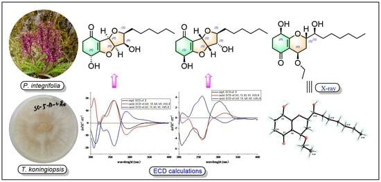
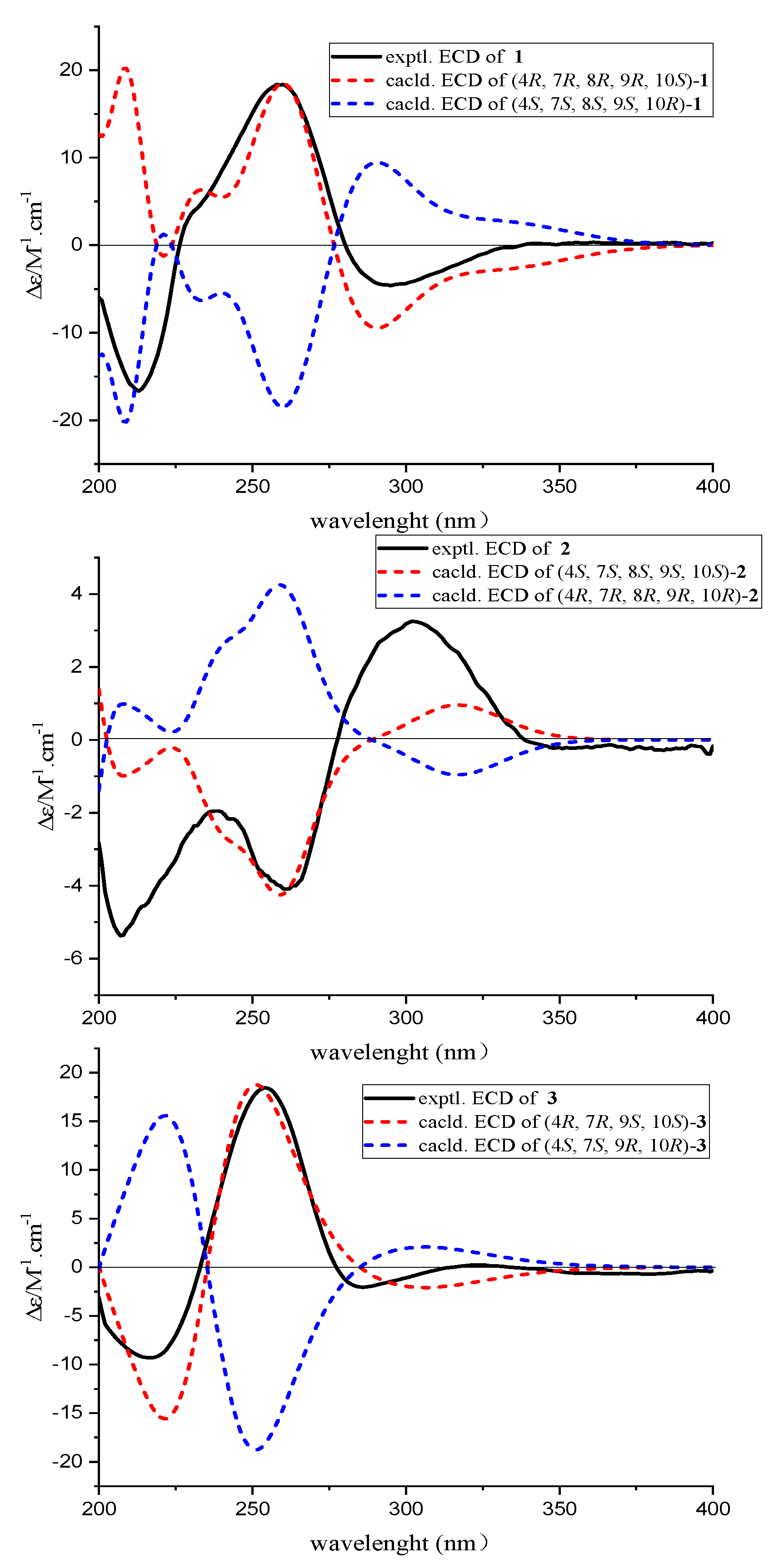
2. Results and Discussion

Koninginin X (1) was isolated as a yellow oil, HRESIMS-(+) analysis of molecular ion peaks at m/z 297.1703 [M + H]+ (calcd. for C16H25O5+, 297.1702) and 319.1523 [M + Na]+ (calcd. for C16H24O5Na+, 319.1521) collectively pointed to the molecular formula C16H24O5, requiring 5 indices of hydrogen deficiency (IHDs). Its emblematic NMR spectroscopic signals (Table 1) of five oxygenated methines at δC/H 64.0/4.75 (C-4), 79.4/5.25 (C-7), 89.5/5.25 (C-8), 72.0/4.35 (C-9), 82.1/3.66 (C-10) and a terminal methyl group at δC/H 14.0/0.87 (C-16) together with a carbonyl functionality at δC 194.0 (C-1) and two olefinic carbons at δC 178.5 (C-5), 115.1 (C-6) were distinctively recognized by holistic analysis of 1H, 13C NMR, DEPT 135, and HSQC spectra. The established functionalities of 1 chemo-logically accounted for two IHDs and strongly suggested the chemical structure of 1 to be a tricyclic skeleton.
The planar structure of 1 was established by a thorough analysis of its 1H-1H COSY and HMBC spectra (Figure 2). Firstly, two spin-spin coupling segments of H2-2/H2-3/H-4 and H-7/H-8/H-9/H-10/H2-11/H2-12/H2-13/H2-14/H2-15/H3-16 were observed in the 1H-1H COSY spectrum. Then, the critical HMBC correlations from H2-2 to C-1/C-3/C-4/C-6, H2-3 to C-1/C-2/C-4/C-5, H-4 to C-2/C-6, and the COSY cross peaks of H2-2/H2-3/H-4 absolutely constructed the ring system of cyclohexanone. In addition, the HMBC correlations from H-7 to C-5/C-9/C-10, H-8 to C-5/C-6/C-10, together with the remaining two IHDs, necessitated C-5/C-6/C-7/C-8/C-9/C-10 to form a double-furan fused ring system. Therefore, the planar structure of 1 was completely illustrated, and it was recommended to share an identical framework with that of trichodermaketone B [9]. Similarly, an obvious negative Cotton effect at 286 nm (∆ε − 3.26) was observed in the CD spectrum of 1, which informatively suggested the absolute configuration to be 4R [9]. In addition, compound 1 possessed the same relative configuration as that of trichodermaketone B, referring to the key NOE correlations (Figure 2), while the absolute configuration has not been completely identified.
The chemical structure of 1 was further affirmed by 13C NMR calculation, and the 13C NMR calculated data for the 4R*,7R*,8R*,9R*,10S*-1 conformer showed a correlation coefficient (R2) of 0.9991 (Figure 3) and a small mean absolute error (MAE) of 1.15. Moreover, a minute root mean square (RMS) of 1.60 and the critical Prel value of 100% were also obtained for the 4R*,7R*,8R*,9R*,10S*-1 conformer, which thus further evidenced the probability of the calculated result (Table 2). Subsequently, we determined the absolute configuration of 1 by theoretical ECD calculation, and its absolute configuration was finally revealed to be 4R,7R,8R,9R,10S, as depicted in Figure 4. Consequently, the absolute configuration of 1 was absolutely elucidated and given a trivial name, (4R,7R,8R,9R,10S)-koninginin X.
Compound 2 was gathered as a stereoisomer of 1 and had the same molecular formula, C16H24O5, determined by HRESIMS-(+) analysis (m/z 297.1710 [M + H]+ and 319.1530 [M + Na]+). The NMR data (Table 1) of 2 and key 1H-1H COSY and HMBC correlations (Figure 2) also demonstrated that both compounds should share the same planar structure. However, according to the 13C NMR spectrum, the chemical shift of C-2 at δC 35.6 in trichodermaketone B was upshifted to δC 22.5 in 2, while the chemical shift of C-4 at δC 63.6 in trichodermaketone B was downshifted to δC 71.7 in 2; the aforementioned informative results tentatively indicated the relative configuration of C-4 of 2 might differ from that of the known trichodermaketone B [9,29,30]. Interestingly, the CD experiment of 2 showed a clear positive Cotton effect for the n → π* transition at 302 nm (∆ε + 3.25), which further suggested the absolute configuration for 2 to be 4S and consolidated the aforementioned deduction. Notably, the 4S configuration of 2 was absolutely opposite to 4R for the known compounds, trichodermaketones A and B [9].
Furthermore, in the NOE difference experiments (Figure 2), the irradiation of H-8 (δH 5.05) led to the enhancement of H-7 (δH 5.65) and H-9 (δH 4.16), the irradiation of H-7 (δH 5.65) led to the enhancement of H-8 (δH 5.05) and H-9 (δH 4.16), and the irradiation of H-10 (δH 3.43) resulted in the enhancement of H-9 (δH 4.16). Based on these NOE signals, the chiral protons of H-7, H-8, and H-9 were then suggested to be on the same side, while H-10 should be on the other orientation. Therefore, the relative configuration of 2 was completely established.
Subsequently, with the aim to verify the relative configuration and determine the absolute configuration of 2, the time-dependent density functional (TDDFT)-ECD calculations in Gaussian 16 were applied. In Figure 4, the calculated ECD curve of 4S,7S,8S,9S,10S-2 was consistent with that of the experimental one by a close comparison, thus ambiguously suggesting that the absolute configuration of 2 was 4S,7S,8S,9S,10S. As a result, the structure of 2 was fully determined and given the trivial name, (4S,7S,8S,9S,10S)-koninginin Y.
Compound 3 was obtained as a yellow oil; its molecular formula was confirmed as C18H30O5 on the basis of the positive HRESIMS ion peak at m/z 349.1989 [M + Na]+ (calcd. for C18H30O5Na+, 349.1991), which required four degrees of unsaturation. Comprehensive analyses of the 1D and 2D NMR spectra for 3 disclosed the presence of two methyl groups [δC/H 14.6/0.91 (C-16) and 16.0/1.15 (C-18)], nine methylenes [δC/H 34.4/2.63, 2.29 (C-2), 30.6/2.18, 1.97 (C-3), 29.3/2.03, 1.55 (C-8), 33.8/1.64 (C-11), 26.8/1.55, 1.41 (C-12), 30.6/1.41, 1.33 (C-13), 33.2/1.33 (C-14), 23.8/1.33 (C-15), and 65.4/3.64, 3.55 (C-17)], four methines [δC/H 67.1/4.38 (C-4), 65.4/4.35 (C-7), 78.2/4.14 (C-9), and 73.6/3.68 (C-10)], and three non-proton carbons with the formation of α,β-unsaturated ketone units [δC 113.2 (C-6), 174.9 (C-5), and 199.4 (C-1)].
Then, a detailed comparison of the NMR data of 3 with those of 7-O-methylkoninginin D [9], a koninginin derivative isolated from the marine fungus Trichoderma koningii, implied that 3 should also be a closely similar derivative of koninginin D. The NMR data for both compounds are very similar, excepting that the methoxy group [δC 59.6 (C-17)] in 7-O-methylkoninginin D was changed to be an ethoxy group [δC 65.4 (C-17) and δC 16.0 (C-18)] in 3. Moreover, the cross peak between H2-17 and H3-18 in the 1H-1H COSY spectrum, together with the critical HMBC correlations from H3-18 to C-17 and H2-17 to C-7, further strengthened the presence of the ethoxy functional group and suggested its location at the C-7 position. Moreover, other observed HMBC correlations and 1H-1H COSY cross peaks (Figure 2) further established the planar structure of 3.
Nevertheless, the relative configuration of compound 3 was unable to be established by the NOESY experiment, because there were no diagnosable correlative signals obviously observed, and the configuration of 7-O-methylkoninginin D was previously determined from an inference of biogenetic perspective [9]. Then, the J values of 1H NMR were used to determine the relative configuration of 3 [29,30,31]. In the 1H NMR spectrum, the coupling constants for H-7/H-8α with J = 2.4 Hz, H-8α/H-8β with J = 14.4 Hz, and H-9/H-8β with J = 2.4 Hz were essentially located H-7 and H-9 in a trans-diequatorial relationship. Additionally, the coupling constant (J = 12.6 Hz) for H-10/H-9 also indicated H-9 and H-10 in a trans-diequatorial relationship. Moreover, a negative Cotton effect at 286 nm (∆ε − 2.05) for the n → π* transition in the CD spectrum of 3 also suggested the absolute configuration of 4R [9,32].
Subsequently, the absolute configuration of 3 was further confirmed by comparison of the experimental and the simulated circular dichroism (CD) spectra. As shown in Figure 4, the calculated ECD curve of 4R,8R,9S,10S-3 was perfectly matched with the experimental one, thus strongly illustrating that the absolute configuration of 3 was 4R,8R,9S,10S. Satisfactorily, the absolute configuration of 3 with 4R,8R,9S,10S was further ambiguously verified by single crystal X-ray diffraction with a Flack parameter of 0.15 (11) (Figure 5), Therefore, the absolute configuration of compound 3 was completely determined and given the trivial name, (4R,8R,9S,10S)-koninginin Z.
Conclusively, the type of chemical structure for the koninginin family was first reported in 1989 [33]; nowadays, it has been increasingly arousing the great interest of many pharmacists with an aim to discover novel lead drugs with structurally fascinating skeletons and biologically significant activities [10,17,21], which thus boosts the reporting of novel compounds in the koninginin family [9,10,11,12,28]. However, the intractable challenge and imperfect issue clouding the koninginin family is that the configurations for many chemical structures had not been thoroughly solved [9,18,21]. In this research, the chemical structures, including the absolute configuration of these novel isolates, were comprehensively characterized with the help of the combination of NMR spectroscopic methods, HRESIMS, electronic circular dichroism calculations, and single crystal X-ray diffraction. We believe that these reliable methodologies towards efficient structure resolution in this study could act as a promisingly applicable strategy to resolve the configuration determination of other koninginin derivatives.
Notably, the genus Trichoderma has been revealed in recent years to be a high producer of natural products [34]. Polyketides are the critical characteristic chemical constituents for the genus Trichoderma and reported extensively. They usually share a variety of intriguing structures and novel carbon-chain skeletons [34]. As exemplified by the sorbicillinoid-based compounds, saturnispols A-H, all of them share a novel cyclic hexaketide nucleus and a typical sorbyl sidechain in their chemical scaffold [35]. Moreover, trichorenins A-C with a unique tetracyclic carbon skeleton were also discovered from the genus Trichoderma [36], and tandyukisins G-I possess an attractive chromone core with a natural rarely occurring 4-oxo-4H-1-benzopyran scaffold representing a class of unprecedented polyketides that are widely distributed in Trichoderma sp. [37]. Although the genus Trichoderma has been widely studied due to the diversity of natural environmental systems, the genus Trichoderma parasitized by different biological groups is likely to produce more natural products with novel structures and significant biological activities under the influence of an environmental host and their own evolution [35,38,39]. Therefore, the genus Trichoderma is a significantly promising strategic bio-resource for the excavation of structurally novel lead natural products, and further extensive research efforts on their chemical constituents are still called upon for their great research value and spacious application prospect.
In the previously reported literature [18,19,40], the natural products derived from the genus Trichoderma usually exhibit considerable antifungal activity against a variety of different pathogenic fungi. For example, trichodermatide B from Emericella nidulans showed significant antifungal activity against Cryptococcus neoformans, with an IC50 value of 4.9 μg/mL [37]. Therefore, compounds 13 were further evaluated for antifungal activity against Candida albicans, where they did not exhibit any notable antifungal effect at the concentration of 100 μg/mL. Notably, we evaluated the antibacterial and cytotoxic activities of the isolated koninginin compounds from Trichoderma koningiopsis SC-5 in our previous experiments [28], and none of them showed any significant activity. In the future, more pathogenic fungi should be considered to evaluate the antifungal effects of these koninginin compounds, and many other biological activities such as antiviral and anti-inflammatory activities also worthy of testing.

3. Materials and Methods

3.1. General Experimental Procedures

The HRESIMS analyses were carried out on an Agilent 6500 series Q-TOF mass spectrometer (Agilent, Singapore) in the positive ion mode. The 1D and 2D NMR data were measured on a Bruker AVIII-600 spectrometer (Bruker, Karlsruhe, Germany) using TMS as an internal standard. Optical rotations were recorded on an Anton Paar MCP-500 spectropolarimeter (Anton Paar, Graz, Austria). Experimental ECD spectra were acquired from Applied PhotoPhysics’ Chiascan circular dichroism spectrometer (APL, Surrey, UK). The single crystal data were collected on an Agilent Xcalibur Novasingle crystal diffractometer equipped with CuKα radiation. The reversed-phase preparative HPLC was conducted using an Agilent 1100 (Agilent Technologies, Santa Clara, CA, USA) instrument connected to an Innoval ODS-2 column (5 μm, 10 mm × 250 mm) with a VWD detector. The UV spectra were recorded with the use of a UV-2600 spectrophotometer (Shimadzu, Kyoto, Japan). Silica gel (Qingdao Marine Chemical Factory, Qingdao, China), macroporous resin (Hebei, China), Sephadex LH-20 (Toyopearl Tosoh, Tokyo, Japan), and octadecylsilane reversed-phase silica gel (50 µm, Fuji, Kasugai, Japan) were utilized for column chromatography. All the solvents were of analytical grade.

3.2. Fungal Material

The strain of Trichoderma koningiopsis SC-5 was isolated from Pedicularis integrifolia in Li County, Aba Tibetan and Qiang Autonomous Prefecture, Sichuan Province. It was collected in July 2021. This endophytic strain was subsequently identified by the sequence analysis of the rDNA ITS region, and the sequence of its rDNA ITS region was submitted to GenBank (Accession: OP646773). This endophytic fungus is preserved in the Department of Medicinal Chemistry, Xiangya School of Pharmaceutical Sciences, Central South University.

3.3. Fermentation, Extraction, and Isolation

The fungus was cultured in autoclaved rice solid medium (120 × 500 mL Erlenmeyer flasks, each containing 100 g of grain and 120 mL of ultrapure water). The fermentation was performed under static conditions at a constant temperature of 28 °C for 4 weeks. The fungal culture (5 kg) was extracted with EtOAc (10 L) three times, and the extract was subsequently decompression-concentrated to provide 110.0 g crude products.
The EtOAc extract was subjected to macroporous resin column chromatography (CC) eluting with EtOH-H2O (v/v, 20:80–0:100) to give three fractions (Fr. 1 to Fr. 3). Fr. 2 (45 g) was further separated on a silica gel CC eluting with CH2Cl2-MeOH (v/v, 100:1–0:100) to obtain 7 fractions (Fr. 2a to Fr. 2g). Subsequently, Fr. 2d (8.5 g) was separated by a reversed-phase ODS column with a gradient of MeOH-H2O (v/v, 5:95–100:0) to provide 11 subfractions (Fr. 2d-1 to Fr. 2d-11); among them, Fr. 2d-4 was separated by a Sephadex LH-20 column with MeOH-H2O (v/v, 80:20) and was further purified by prep-HPLC with CH3CN-H2O (0–50 min, 25–50%, 2 mL/min) to yield compound 3 (15.2 mg, tR = 42.5 min). Fr. 2d-3 was separated by a Sephadex LH-20 column with MeOH-H2O (v/v, 80:20), and was further purified by prep-HPLC with CH3CN-H2O (0–40 min, 25–45%, 2 mL/min) to obtain compound 1 (6.0 mg, tR = 36.2 min) and 2 (2.8 mg, tR = 38.2 min).
Koninginin X (1). Yellow oil; [ α ] D 20 + 9.32 (c 0.30, MeOH); UV (MeOH): λmax (log ε): 257 (2.75) nm; ECD (0.36 mg/mL, MeOH): λmaxε) 233 (+6.30), 260 (+18.48), 291 (−9.45) nm; 1H (CDCl3, 600 MHz) and 13C (CDCl3, 150 MHz) NMR spectral data, see Table 1; HRESIMS: m/z 297.1703 [M + H]+ (calcd. for C16H25O5+, 297.1702), 319.1523 [M + Na]+ (calcd. for C16H24O5Na+, 319.1521).
Koninginin Y (2). Yellow oil; [ α ] D 20 − 55.14 (c 0.13, MeOH); UV (MeOH): λmax (log ε): 257 (3.12) nm; ECD (0.36 mg/mL, MeOH): λmaxε) 207 (−5.37), 239 (−1.95), 261 (−4.10), 302 (+3.25) nm; 1H (CDCl3, 600 MHz) and 13C (CDCl3, 150 MHz) NMR spectral data, see Table 1; HRESIMS: m/z 297.1710 [M + H]+ (calcd. for C16H25O5+, 297.1702), 319.1530 [M + Na]+ (calcd. for C16H24O5Na+, 319.1521).
Koninginin Z (3). Yellow oil; [ α ] D 20 + 164.87 (c 0.36, MeOH); HPLC-UV (CH3CN-H2O) λmax: 258 nm; ECD (0.36 mg/mL, MeOH): λmaxε) 217 (−14.21), 254 (+18.21), 306 (−0.74) nm; 1H (CD3OD, 600 MHz) and 13C (CD3OD, 150 MHz) NMR spectral data, see Table 1; HRESIMS: m/z 349.1989 [M + Na]+ (calcd. for C18H30O5Na+, 349.1991).

3.4. X-ray Crystallographic Analysis

Crystal data for 3 C18H30O5 (M = 326.43 g/mol): monoclinic, space group P21 (no. 4), a = 5.0609(2) Å, b = 39.7106(9) Å, c = 9.8073(3) Å, V = 1905.11(11) Å3, Z = 2, T = 100.15 K, μ(CuKα) = 0.759 mm−1, Dcalc = 1.232 g/cm3, 6178 reflections measured (8.908 ≤ 2Θ ≤ 148.692), 6178 unique (Rint = 0.0298, Rsigma = 0.0394), which were used in all the calculations. The final R1 was 0.0423 (I > 2σ(I)), and wR2 was 0.1121 (all data). Flack parameter = 0.15(11). The crystallographic data for 3 reported in this paper was deposited in the Cambridge Crystallographic Data Centre. (Deposition number: CCDC 2264777). Copies of these data can be obtained free of charge via https://www.ccdc.cam.ac.uk (accessed on 18 June 2023).

3.5. Quantum Chemistry Calculations

A conformational search was performed by Crest [41], followed by optimization on a GFN2-xTB [42] level with a 4 kcal/mol energy window to remove high-energy conformers. The optimization and frequency calculation of each conformer was implemented on a B3LYP-D3(BJ)/TZVP [43,44] level of theory by using a Gaussian 16 software package with the keyword: g09defaults [45]. The DFT GIAO 13C NMR calculation was performed on the ωB97x-D/6-31G* level [46], and the data processing followed the reported STS protocol [47]. The ECD calculations were by TDDFT on the ωB97x-D/TZVP level of theory and were Boltzmann-averaged according to the Gibbs free energy. SpecDis v1.71 [48] was used to simulate the ECD curves of compounds 13, with sigma/gamma values of 0.30 eV, 0.30 eV, and 0.40 eV, respectively. The averaged calculated ECD curves of compounds 12 were adjusted by blue shifting for 15 nm and 10 nm, respectively.

3.6. Biological Activity Assay

The antifungal activity against Candida albicans (ATCC 10231) was evaluated by using a broth microdilution protocol modified from the CLSI (formerly NCCLS) M-38A and M-27A2 methods [9]. The compounds and positive drug vancomycin were prepared at a concentration of 100 µg/mL in the growth medium, and the solvent control used DMSO. The MIC value was regarded as the lowest inhibitory concentration for the visible growth of the tested fungi.

4. Conclusions

In summary, this study describes the whole process of separation and structure identification of three novel compounds, koninginins X-Z (13). Their structures including the absolute configurations are clearly characterized. Koninginin X (1) and koninginin Y (2) share a cyclohexanone concatenate double furan fused-ring system as the critical structure backbone, while koninginin Z (3) possesses a bicyclic pyran skeleton. These findings not only greatly elevate the chemical diversity of secondary metabolites for koninginin derivatives, but also further enrich the chemical compositions of the genus of Trichoderma koningiopsis.

Supplementary Materials

The following supporting information can be downloaded at: https://www.mdpi.com/article/10.3390/molecules28237848/s1, Table S1. X-ray crystallographic data and structure refinement for 3; Figure S1. HRESIMS spectrum of compound 1; Figure S2. UV spectrum of compound 1; Figure S3. 1H NMR spectrum of compound 1; Figure S4. 13C NMR spectrum of compound 1; Figure S5. DEPT 135 spectrum of compound 1 recorded in CDCl3; Figure S6. 1H-1H COSY spectrum of compound 1 recorded in CDCl3; Figure S7. HSQC spectrum of compound 1 recorded in CDCl3; Figure S8. HMBC spectrum of compound 1 recorded in CDCl3; Figure S9. NOE difference spectrum of compound 1 recorded in CDCl3; Figure S10. CD spectrum of compound 1; Figure S11. Regression analyses of experimental and calculated 13C NMR chemical shifts for 1; Figure S12. Experimental and calculated ECD spectra of compound 1; Figure S13. HRESIMS spectrum of compound 2; Figure S14. UV spectrum of compound 2; Figure S15. 1H NMR spectrum of compound 2; Figure S16. 13C NMR spectrum of compound 2; Figure S17. DEPT 135 spectrum of compound 2 recorded in CDCl3; Figure S18. 1H-1H COSY spectrum of compound 2 recorded in CDCl3; Figure S19. HSQC spectrum of compound 2 recorded in CDCl3; Figure S20. HMBC spectrum of compound 2 recorded in CDCl3; Figure S21. NOE difference spectrum of compound 2 recorded in CDCl3; Figure S22. NOE difference spectrum of compound 2 recorded in CDCl3; Figure S23. NOE difference spectrum of compound 2 recorded in CDCl3; Figure S24. CD spectrum of compound 2; Figure S25. Experimental and calculated ECD spectra of compound 2; Figure S26. HRESIMS spectrum of compound 3; Figure S27. UV spectrum of compound 3; Figure S28. 1H NMR spectrum of compound 3; Figure S29. 13C NMR spectrum of compound 3; Figure S30. DEPT 135 spectrum of compound 3 recorded in CD3OD; Figure S31. 1H-1H COSY spectrum of compound 3 recorded in CD3OD; Figure S32. HSQC spectrum of compound 3 recorded in CD3OD; Figure S33. HMBC spectrum of compound 3 recorded in CD3OD; Figure S34. NOESY spectrum of compound 3 recorded in CD3OD; Figure S35. CD spectrum of compound 3; Figure S36. Experimental and calculated ECD spectra of compound 3.

Author Contributions

Conceptualization, Z.Z. and H.T.; methodology, W.P., J.T. and Y.H.; software, Z.S. and L.X.; validation, Z.Z., Y.Z. and S.Q.; formal analysis, W.P. and J.T.; investigation, Y.Z.; resources, J.T.; data curation, W.P., J.T. and S.Q.; writing—original draft preparation, W.P., J.T. and Z.Z.; writing—review and editing, W.P., J.T. and Z.Z.; visualization, Z.S.; supervision, Z.Z. and H.T. All authors have read and agreed to the published version of the manuscript.

Funding

This work was supported by the National Natural Science Foundation of China (82173711), Key Research and Development Project of Hainan Province (No. ZDYF2022SHFZ048), Natural Science Foundation of Hunan Province (No. 2021JJ30917), Youth Innovation Promotion Association of CAS (2020342), the Central South University postgraduate independent exploration and innovation project (Nos. 2022zzts0899; 2021zzts0994), and the Open Sharing Fund for the Large-Scale Instruments and Equipment of Central South University.

Institutional Review Board Statement

Not applicable.

Informed Consent Statement

Not applicable.

Data Availability Statement

All the data including X-ray crystallographic data and UV, HRMS, 1D/2D NMR, and CD spectra are available in this publication and the Supplementary Materials.

Conflicts of Interest

The authors declare no conflict of interest.

References

  1. Wang, Y.; Pruitt, R.N.; Nürnberger, T.; Wang, Y.C. Evasion of plant immunity by microbial pathogens. Nat. Rev. Microbiol. 2022, 20, 449–464. [Google Scholar] [CrossRef] [PubMed]
  2. Dutta, A.; Hartmann, F.E.; Francisco, C.S.; McDonald, B.A.; Croll, D. Mapping the adaptive landscape of a major agricultural pathogen reveals evolutionary constraints across heterogeneous environments. ISME J. 2021, 15, 1402–1419. [Google Scholar] [CrossRef] [PubMed]
  3. Keswani, C.; Singh, S.P.; García-Estrada, C.; Mezaache-Aichour, S.; Glare, T.R.; Borriss, R.; Rajput, V.D.; Minkina, T.M.; Ortiz, A.; Sansinenea, E. Biosynthesis and beneficial effects of microbial gibberellins on crops for sustainable agriculture. J. Appl. Microbiol. 2022, 132, 1597–1615. [Google Scholar] [CrossRef] [PubMed]
  4. Winn, M.; Rowlinson, M.; Wang, F.; Bering, L.; Francis, D.; Levy, C.; Micklefield, J. Discovery, characterization and engineering of ligases for amide synthesis. Nature 2021, 593, 391–398. [Google Scholar] [CrossRef] [PubMed]
  5. Fan, L.G.; Wang, T.T.; Hua, C.F.; Sun, W.J.; Li, X.Y.; Grunwald, L.; Liu, J.G.; Wu, N.; Shao, X.L.; Yin, Y.M.; et al. A compendium of DNA-binding specificities of transcription factors in Pseudomonas syringae. Nat. Commun. 2020, 11, 4947. [Google Scholar] [CrossRef] [PubMed]
  6. Yao, X.; Guo, H.L.; Zhang, K.X.; Zhao, M.Y.; Ruan, J.J.; Chen, J. Trichoderma and its role in biological control of plant fungal and nematode disease. Front. Microbiol. 2023, 14, 1160551. [Google Scholar] [CrossRef]
  7. Tyśkiewicz, R.; Nowak, A.; Ozimek, E.; Jaroszuk-Ściseł, J. Trichoderma: The current status of its application in agriculture for the biocontrol of fungal phytopathogens and stimulation of plant growth. Int. J. Mol. Sci. 2022, 23, 2329. [Google Scholar] [CrossRef] [PubMed]
  8. Miao, F.P.; Liang, X.R.; Yin, X.L.; Wang, G.; Ji, N.Y. Absolute configurations of unique harziane diterpenes from Trichoderma species. Org. Lett. 2012, 14, 3815–3817. [Google Scholar] [CrossRef]
  9. Song, F.H.; Dai, H.Q.; Tong, Y.J.; Ren, B.; Chen, C.X.; Sun, N.; Liu, X.Y.; Bian, J.; Liu, M.; Gao, H.; et al. Trichodermaketones A-D and 7-O-methylkoninginin D from the marine fungus Trichoderma koningii. J. Nat. Prod. 2010, 73, 806–810. [Google Scholar] [CrossRef]
  10. Sun, Y.; Tian, L.; Huang, J.; Ma, H.Y.; Zheng, Z.; Lv, A.L.; Yasukawa, K.; Pei, Y.H. Trichodermatides A-D, novel polyketides from the marine-derived fungus Trichoderma reesei. Org. Lett. 2008, 10, 393–396. [Google Scholar] [CrossRef]
  11. Chen, S.C.; Li, H.H.; Chen, Y.C.; Li, S.N.; Xu, J.L.; Guo, H.; Liu, Z.M.; Zhu, S.; Liu, H.X.; Zhang, W.M. Three new diterpenes and two new sesquiterpenoids from the endophytic fungus Trichoderma koningiopsis A729. Bioorg. Chem. 2019, 86, 368–374. [Google Scholar] [CrossRef] [PubMed]
  12. Shi, X.S.; Meng, L.H.; Li, X.; Wang, D.J.; Zhou, X.W.; Du, F.Y.; Wang, B.G.; Li, X.M. Polyketides and terpenoids with potent antibacterial activities from the Artemisia argyi-derived fungus Trichoderma koningiopsis QA-3. Chem. Biodivers. 2020, 17, e2000566. [Google Scholar] [CrossRef] [PubMed]
  13. Liu, K.; Yang, Y.; Miao, C.P.; Zheng, Y.K.; Chen, J.L.; Chen, Y.W.; Xu, L.H.; Guang, H.L.; Ding, Z.T.; Zhao, L.X. Koningiopisins A-H, polyketides with synergistic antifungal activities from the endophytic fungus Trichoderma koningiopsis. Planta Med. 2016, 82, 371–376. [Google Scholar] [CrossRef] [PubMed]
  14. Chen, L.; Wu, G.W.; Liu, D.; Zhuang, W.Y.; Yin, W.B. Trichodermatides E and F from fungus Trichoderma applanatum. J. Asian Nat. Prod. Res. 2019, 21, 659–665. [Google Scholar] [CrossRef] [PubMed]
  15. Wang, Y.L.; Hu, B.Y.; Qian, M.A.; Wang, Z.H.; Zou, J.M.; Sang, X.Y.; Li, L.; Luo, X.D.; Zhao, L.X. Koninginin W, a new polyketide from the endophytic fungus Trichoderma koningiopsis YIM PH30002. Chem. Biodivers. 2021, 18, e2100460. [Google Scholar] [CrossRef]
  16. Cutler, H.G.; Cutler, S.J.; Ross, S.A.; Sayed, K.E.; Dugan, F.M.; Bartlett, M.G.; Hill, A.A.; Hill, R.A.; Parker, S.R. Koninginin G, a new metabolite from Trichoderma aureoviride. J. Nat. Prod. 1999, 62, 137–139. [Google Scholar] [CrossRef]
  17. Shi, X.S.; Li, H.L.; Li, X.M.; Wang, D.J.; Li, X.; Meng, L.H.; Zhou, X.W.; Wang, B.G. Highly oxygenated polyketides produced by Trichoderma koningiopsis QA-3, an endophytic fungus obtained from the fresh roots of the medicinal plant Artemisia argyi. Bioorg. Chem. 2020, 94, 103448. [Google Scholar] [CrossRef]
  18. Hu, M.; Li, Q.L.; Yang, Y.B.; Liu, K.; Miao, C.P.; Zhao, L.X.; Ding, Z.T. Koninginins R-S from the endophytic fungus Trichoderma koningiopsis. Nat. Prod. Res. 2017, 31, 835–839. [Google Scholar] [CrossRef]
  19. Liu, K.; Yang, Y.B.; Chen, J.L.; Miao, C.P.; Wang, Q.; Zhou, H.; Chen, Y.W.; Li, Y.Q.; Ding, Z.T.; Zhao, L.X. Koninginins N-Q, polyketides from the endophytic fungus Trichoderma koningiopsis Harbored in Panax notoginseng. Nat. Prod. Bioprospect. 2016, 6, 49–55. [Google Scholar] [CrossRef]
  20. Lang, B.Y.; Li, J.; Zhou, X.X.; Chen, Y.H.; Yang, Y.H.; Li, X.N.; Zeng, Y.; Zhao, P.J. Koninginins L and M, two polyketides from Trichoderma koningii 8662. Phytochem. Lett. 2015, 11, 1–4. [Google Scholar] [CrossRef]
  21. Zhou, X.X.; Li, J.; Yang, Y.H.; Zeng, Y.; Zhao, P.J. Three new koninginins from Trichoderma neokongii 8722. Phytochem. Lett. 2014, 8, 137–140. [Google Scholar] [CrossRef]
  22. Shi, X.S.; Wang, D.J.; Li, X.M.; Li, H.L.; Meng, L.H.; Li, X.; Pi, Y.; Zhou, X.W.; Wang, B.G. Antimicrobial polyketides from Trichoderma koningiopsis QA-3, an endophytic fungus obtained from the medicinal plant Artemisia argyi. RSC Adv. 2017, 7, 51335–51342. [Google Scholar] [CrossRef]
  23. Zhang, S.; Chen, D.K.; Kuang, M.; Peng, W.W.; Chen, Y.; Tan, J.B.; Kang, F.H.; Xu, K.P.; Zou, Z.X. Rhytidhylides A and B, two new phthalide derivatives from the endophytic fungus Rhytidhysteron sp. BZM-9. Molecules 2021, 26, 6092. [Google Scholar] [CrossRef] [PubMed]
  24. Zhang, W.; Lu, X.X.; Huo, L.Q.; Zhang, S.; Chen, Y.; Zou, Z.X.; Tan, H.B. Sesquiterpenes and steroids from an endophytic Eutypella scoparia. J. Nat. Prod. 2021, 84, 1715–1724. [Google Scholar] [CrossRef] [PubMed]
  25. Peng, W.W.; Kuang, M.; Huang, Y.T.; Li, M.F.; Zheng, Y.T.; Xu, L.; Tan, J.B.; Kang, F.H.; Tan, H.B.; Zou, Z.X. Pseudocercones A-C, three new polyketide derivatives from the endophytic fungus Pseudocercospora sp. TSS-1. Nat. Prod. Res. 2022, 1–8. [Google Scholar] [CrossRef]
  26. Kuang, M.; Peng, W.W.; Huang, Y.T.; Li, M.F.; Qin, S.Y.; Zheng, Y.T.; Xu, L.; Huang, Q.; Zou, Z.X. Two new chromone derivatives from the rhizosphere soil fungus Ilyonectria robusta. Nat. Prod. Res. 2022, 1–8. [Google Scholar] [CrossRef]
  27. Chen, Y.; Wang, H.; Ke, X.; Sang, Z.H.; Kuang, M.; Peng, W.W.; Tan, J.B.; Zheng, Y.T.; Zou, Z.X.; Tan, H.B. Five new secondary metabolites from an endophytic fungus Phomopsis sp. SZSJ-7B. Front. Plant Sci. 2022, 13, 1049015. [Google Scholar] [CrossRef]
  28. Peng, W.W.; Huang, Q.; Ke, X.; Wang, W.X.; Chen, Y.; Sang, Z.H.; Chen, C.; Qin, S.Y.; Zheng, Y.T.; Tan, H.B.; et al. Koningipyridines A and B, two nitrogen-containing polyketides from fungus Trichoderma koningiopsis SC-5. Front. Plant Sci. 2023, 14, 1161420. [Google Scholar]
  29. Cutler, H.G.; Himmelsbach, D.S.; Yagen, B.; Arrendale, R.F.; Cox, R.H. Koninginin B: A biologically active congener of koninginin A from Trichoderma koningii. J. Agric. Food Chem. 1991, 39, 977–980. [Google Scholar] [CrossRef]
  30. Ghisalberti, E.L.; Rowland, C.Y. Antifungal metabolites from Trichoderma harzianum. J. Nat. Prod. 1993, 56, 1799–1804. [Google Scholar] [CrossRef]
  31. Liu, Z.; Yashiroda, Y.; Sun, P.; Ma, H.; Wang, Y.; Li, L.; Yan, F.; Sun, Y. Argenteolides A and B, glycosylated polyketide-peptide hybrid macrolides from an Actinomycete Streptomyces argenteolus. Org. Lett. 2023, 25, 571–575. [Google Scholar] [CrossRef] [PubMed]
  32. He, Y.C.; Wang, R.L.; Huang, B.; Dai, Q.; Lin, J. Pholiotone A, a new polyketide derivative from Pholiota sp. Nat. Prod. Res. 2020, 34, 1957–1961. [Google Scholar] [CrossRef] [PubMed]
  33. Cutler, H.G.; Himmelsbach, D.S.; Arrendale, R.F.; Cole, P.D.; Cox, R.H. Koninginin A: A novel plant growth regulator from Trichoderma koningii (Microbiology & Fermentation Industry). Agric. Biol. Chem. 1989, 53, 2605–26011. [Google Scholar]
  34. Li, M.F.; Li, G.H.; Zhang, K.Q. Non-Volatile Metabolites from Trichoderma spp. Metabolites 2019, 9, 58. [Google Scholar] [CrossRef] [PubMed]
  35. Meng, J.J.; Cheng, W.; Heydari, H.; Wang, B.; Zhu, K.; Konuklugil, B.; Lin, W.H. Sorbicillinoid-based metabolites from a sponge-derived fungus Trichoderma saturnisporum. Mar. Drugs 2018, 16, 226. [Google Scholar] [CrossRef] [PubMed]
  36. Shi, Z.Z.; Miao, F.P.; Fang, S.T.; Yin, X.L.; Ji, N.Y. Trichorenins A-C, algicidal tetracyclic metabolites from the marine-alga-epiphytic fungus Trichoderma virens Y13-3. J. Nat. Prod. 2018, 81, 1121–1124. [Google Scholar] [CrossRef]
  37. Lai, C.; Chen, J.; Liu, J.; Tian, D.; Lan, D.; Liu, T.; Wu, B.; Bi, H.; Tang, J. New polyketides from a hydrothermal vent sediment fungus Trichoderma sp. JWM29-10-1 and their antimicrobial effects. Mar. Drugs 2022, 20, 720. [Google Scholar] [CrossRef]
  38. Hashem, A.H.; Attia, M.S.; Kandil, E.K.; Fawzi, M.M.; Abdelrahman, A.S.; Khader, M.S.; Khodaira, M.A.; Emam, A.E.; Goma, M.A.; Abdelaziz, A.M. Bioactive compounds and biomedical applications of endophytic fungi: A recent review. Microb. Cell Fact. 2023, 22, 107. [Google Scholar] [CrossRef]
  39. Toppo, P.; Kagatay, L.L.; Gurung, A.; Singla, P.; Chakraborty, R.; Roy, S.; Mathur, P. Endophytic fungi mediates production of bioactive secondary metabolites via modulation of genes involved in key metabolic pathways and their contribution in different biotechnological sector. 3 Biotech. 2023, 13, 191. [Google Scholar] [CrossRef]
  40. Tarawneh, A.H.; León, F.; Radwan, M.M.; Rosa, L.H.; Cutler, S.J. Secondary metabolites from the fungus Emericella nidulans. Nat. Prod. Commun. 2013, 8, 1285–1288. [Google Scholar] [CrossRef]
  41. Pracht, P.; Bohle, F.; Grimme, S. Automated exploration of the low-energy chemical space with fast quantum chemical methods. Phys. Chem. Chem. Phys. 2020, 22, 7169–7192. [Google Scholar] [CrossRef] [PubMed]
  42. Bannwarth, C.; Ehlert, S.; Grimme, S. GFN2-xTB-An accurate and broadly parametrized self-consistent tight-binding quantum chemical method with multipole electrostatics and density-dependent dispersion contributions. J. Chem. Theory Comput. 2019, 15, 1652–1671. [Google Scholar] [CrossRef] [PubMed]
  43. Tsuzuki, S.; Uchimaru, T. Accuracy of intermolecular interaction energies, particularly those of hetero-atom containing molecules obtained by DFT calculations with Grimme’s D2, D3 and D3BJ dispersion corrections. Phys. Chem. Chem. Phys. 2020, 22, 22508–22519. [Google Scholar] [CrossRef] [PubMed]
  44. Grimme, S.; Ehrlich, S.; Goerigk, L. Effect of the damping function in dispersion corrected density functional theory. J. Comput. Chem. 2011, 32, 1456–1465. [Google Scholar] [CrossRef]
  45. Frisch, M.J.; Trucks, G.W.; Schlegel, H.B.; Scuseria, G.E.; Robb, M.A.; Cheeseman, J.R.; Scalmani, G.; Barone, V.; Petersson, G.A.; Nakatsuji, H.; et al. Gaussian 16. Revision C.01; Gaussian, Inc.: Wallingford, CT, USA, 2016. [Google Scholar]
  46. Chai, J.D.; Head-Gordon, M. Long-range corrected hybrid density functionals with damped atom-atom dispersion corrections. Phys. Chem. Chem. Phys. 2008, 10, 6615–6620. [Google Scholar] [CrossRef]
  47. Li, J.; Liu, J.K.; Wang, W.X. GIAO 13C NMR calculation with sorted training sets improves accuracy and reliability for structural assignation. J. Org. Chem. 2020, 85, 11350–11358. [Google Scholar] [CrossRef]
  48. Bruhn, T.; Schaumlöffel, A.; Hemberger, Y.; Bringmann, G. SpecDis: Quantifying the comparison of calculated and experimental electronic circular dichroism spectra. Chirality 2013, 25, 243–249. [Google Scholar] [CrossRef]
Figure 1. Structures of compounds 13.
Molecules 28 07848 g001
Figure 2. 1H-1H COSY, key HMBC, and NOESY correlations of 13.
Molecules 28 07848 g002
Figure 3. Regression analyses of experimental and calculated 13C NMR chemical shifts of 1.
Molecules 28 07848 g003
Figure 4. Experimental and calculated ECD spectra of 13 (in MeOH).
Molecules 28 07848 g004
Figure 5. ORTEP drawing of the X-ray structure of 3.
Molecules 28 07848 g005
Table 1. 1H and 13C NMR (δC) data for 13.
No.1 a2 a3 b
δH (J in Hz)δCδH (J in Hz)δCδH (J in Hz)δC
1 194.0 194.6 199.4
22.42 (1H, m);
2.58 (1H, m)
35.32.71 (1H, m);
2.56 (1H, m)
22.52.63 (1H, m);
2.29 (1H, m)
34.4
32.43 (1H, m);
2.36 (1H, m)
31.12.46 (1H, m);
1.90 (1H, m)
29.72.18 (1H, m);
1.97 (1H, m)
30.6
44.75 (1H, dd, 9.0, 5.4)64.04.14 (1H, d, 4.8)71.74.38 (1H, td, 5.4, 2.4)67.1
5 178.5 180.6 174.9
6 115.1 111.3 113.2
75.25 (1H, dt, 6.0)79.45.65 (1H, d, 6.0)78.34.35 (1H, t, 2.4)65.4
85.25 (1H, dt, 6.0)89.55.05 (1H, d, 6.0)93.52.03 (1H, dt 14.4, 2.4);
1.55 (1H, m)
29.3
94.35 (1H, d, 4.2)72.04.16 (1H, d, 4.8)74.64.14 (1H, td, 12.6, 2.4)78.2
103.66 (1H, m)82.13.43 (1H, td, 7.2)78.63.68 (1H, td, 12.6, 6.0)73.6
111.67 (2H, m)27.71.70 (1H, m);
1.64 (1H, m)
27.11.64 (2H, m)33.8
121.29 (2H, m)26.11.31 (2H, m)26.01.55 (1H, m);
1.41 (1H, m)
26.8
131.29 (2H, m)29.31.31 (2H, m)29.41.41 (1H, m);
1.33 (1H, m)
30.6
141.29 (2H, m)31.71.31 (2H, m)31.61.33 (2H, m)33.2
151.29 (2H, m)22.61.31 (2H, m)22.71.33 (2H, m)23.8
160.87 (3H, t, 6.6)14.00.87 (3H, t, 7.2)14.00.91 (3H, t, 6.6)14.6
17 3.64 (1H, m);
3.55 (1H, m)
65.4
18 1.15 (3H, t, 6.6)16.0
a Record in CDCl3, 600 MHz for 1H, and 150 MHz for 13C, δ in ppm. b Record in CD3OD, 600 MHz for 1H, and 150 MHz for 13C, δ in ppm.
Table 2. Calculated 13C chemical shifts (CDCl3) of structure 1 fitting to the experimental data of compound 1.
No.Exptl. δ1
1abs dev c
1194.0192.841.16
235.334.530.77
331.129.981.12
464.064.250.25
5178.5177.161.34
6115.1119.534.43
779.479.340.06
889.589.960.46
972.074.362.36
1182.179.272.83
1227.728.771.07
1326.125.420.68
1429.328.920.38
1531.730.870.83
1622.622.830.23
MAE a1.15
RMS b1.60
Pmean30.45%
Prel100%
a Mean absolute error. b Root mean square. c Absolute deviation of calcd. δC.
Disclaimer/Publisher’s Note: The statements, opinions and data contained in all publications are solely those of the individual author(s) and contributor(s) and not of MDPI and/or the editor(s). MDPI and/or the editor(s) disclaim responsibility for any injury to people or property resulting from any ideas, methods, instructions or products referred to in the content.

