Article

Six New Phenolic Glycosides from the Seeds of Moringa oleifera Lam. and Their α-Glucosidase Inhibitory Activity

1
School of Pharmacy, Guizhou Medical University, Guiyang 550004, China
2
Key Laboratory of Basic Pharmacology of Ministry of Education, Zunyi Medical University, Zunyi 563000, China
3
State Key Laboratory of Functions and Applications of Medicinal Plants, Guizhou Medical University, Guiyang 550004, China
4
Engineering Research Center for the Development and Application of Ethnic Medicine and TCM, Ministry of Education, Guizhou Medical University, Guiyang 550004, China
*
Authors to whom correspondence should be addressed.
These authors contributed equally to this work.
Molecules 2023, 28(17), 6426; https://doi.org/10.3390/molecules28176426
Submission received: 15 July 2023 / Revised: 26 August 2023 / Accepted: 1 September 2023 / Published: 4 September 2023

Abstract

:
Plant-derived phytochemicals have recently drawn interest in the prevention and treatment of diabetes mellitus (DM). The seeds of Moringa oleifera Lam. are widely used in food and herbal medicine for their health-promoting properties against various diseases, including DM, but many of their effective constituents are still unknown. In this study, 6 new phenolic glycosides, moringaside B–G (16), together with 10 known phenolic glycosides (716) were isolated from M. oleifera seeds. The structures were elucidated by 1D and 2D NMR spectroscopy and high-resolution electrospray ionization mass spectrometry (HR-ESI-MS) data analysis. The absolute configurations of compounds 2 and 3 were determined by electronic circular dichroism (ECD) calculations. Compounds 2 and 3 especially are combined with a 1,3-dioxocyclopentane moiety at the rhamnose group, which are rarely reported in phenolic glycoside backbones. A biosynthetic pathway of 2 and 3 was assumed. Moreover, all the isolated compounds were evaluated for their inhibitory activities against α-glucosidase. Compounds 4 and 16 exhibited marked activities with IC50 values of 382.8 ± 1.42 and 301.4 ± 6.22 μM, and the acarbose was the positive control with an IC50 value of 324.1 ± 4.99 μM. Compound 16 revealed better activity than acarbose.

1. Introduction

Diabetes mellitus (DM) is a metabolic disorder characterized by persistent hyperglycemia due to insufficient insulin secretion or impairment of islet function, which is now one of the major threats to human health in the 21st century [1]. Type II DM (T2DM, i.e., non-insulin-dependent DM) accounts for about 90% of the total DM patients in the current world [2]. Glycemic control is considered an effective therapy for the treatment of T2DM [3]. As is well-known, α-glucosidase is a carbohydrate hydrolase that acts on the terminal α (1→4) bonds of starch and disaccharides to release α-glucose in the brush border of the small intestine [4]. Through inhibiting the activities of α-glucosidase, the absorption of glucose in the intestine is slowed down, and the blood sugar level can be well managed [5]. Therefore, α-glucosidase inhibitors have become the focus of hypoglycemic drug research in recent years. At present, the most common α-glycosidase inhibitors are acarbose, voglibose, and miglitol. However, these inhibitors all have serious side effects, such as flatulence, abdominal cramping, and diarrhea [6]. Natural products are a rich source of safe and highly effective α-glucosidase inhibitors. Most of these natural bioactive compounds not only reduce hyperglycemia but are also associated with fewer side effects than currently applied α-glycosidase inhibitors and offer nutritional benefits for DM patients [7]. In recent years, a large number of studies have shown that compounds with α-glucosidase inhibitory activity have been screened from natural products [8].
Moringa oleifera Lam. belongs to the genus Moringa (family Moringaceae), native to the dry tropical forests of northwestern India [9]. M. oleifera is referred to as a “miracle tree” because of its rich nutritional and pharmacological properties [10]. It has high nutritional value, including protein, fiber, and a variety of vitamins, especially in seeds [11]. The seeds are rich in oils and unsaturated fatty acids, which can be used as a potential source of edible oil [12]. The seeds have many benefits for humans. These have aroused the interest of researchers. At present, there are few studies on the chemical constituents of the seeds; the biological activities are mainly directed to the crude extracts, and the pharmacodynamic material basis is not clear. To date, only several flavonoids, phenolic glycosides, and sterols [13,14,15] have been reported, which exhibit significant properties such as anti-hyperglycaemic, anti-inflammatory, anti-oxidation, and so on [16,17,18].
Herein, in the current study, we have studied the chemical constituents from an 85% ethanol extract of M. oleifera seeds and six new phenolic glycosides (16); ten known phenolic glycosides (716) (Figure 1) have been isolated and identified. All the secondary metabolites were evaluated for their inhibitory activities against α-glucosidase.

2. Results and Discussion

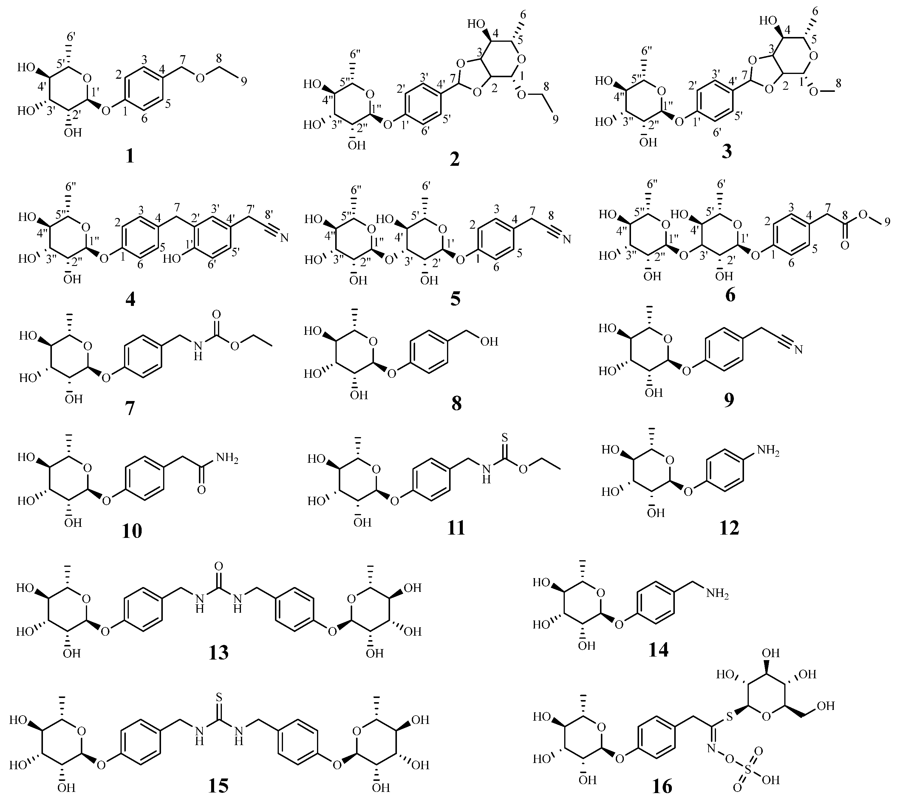
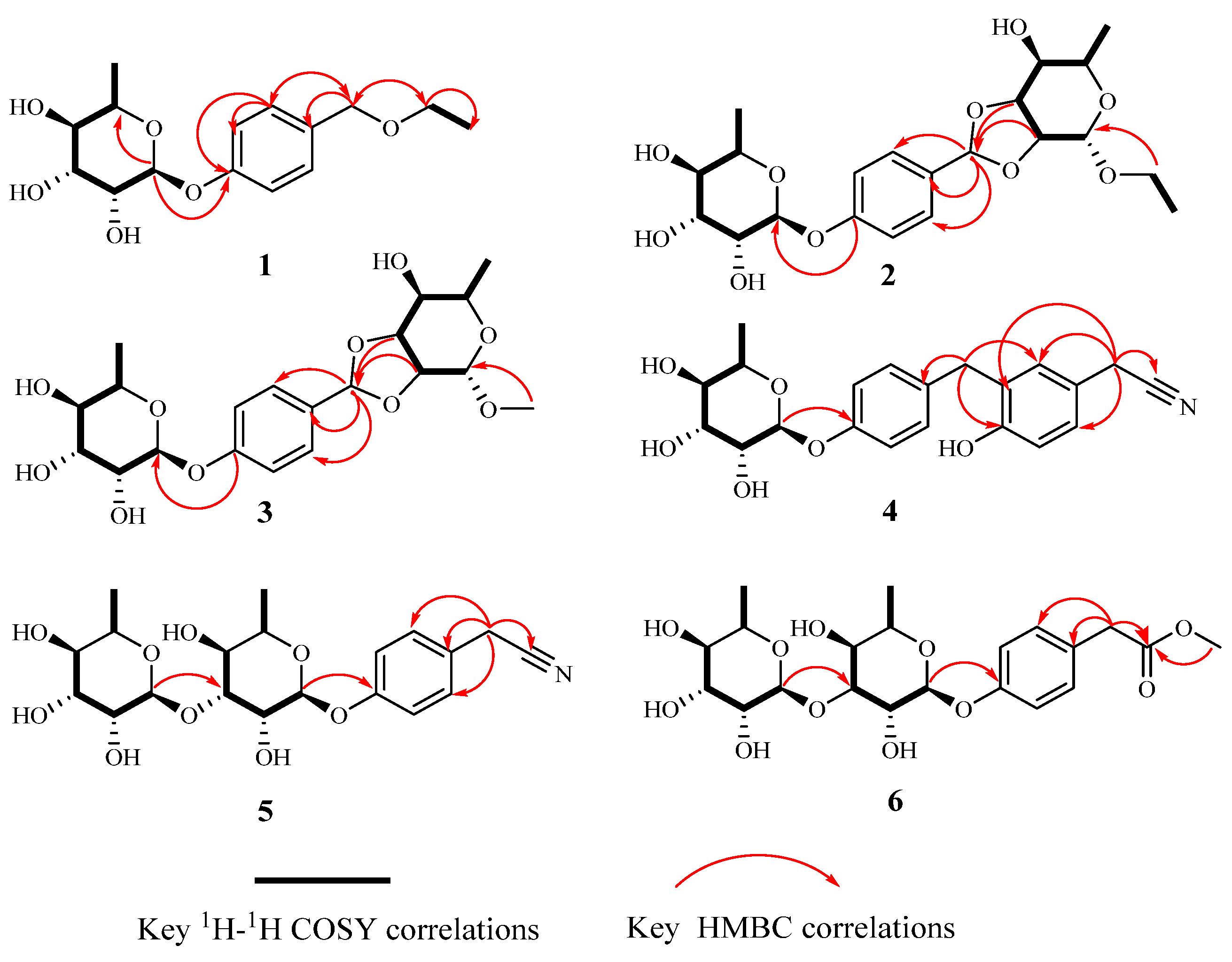
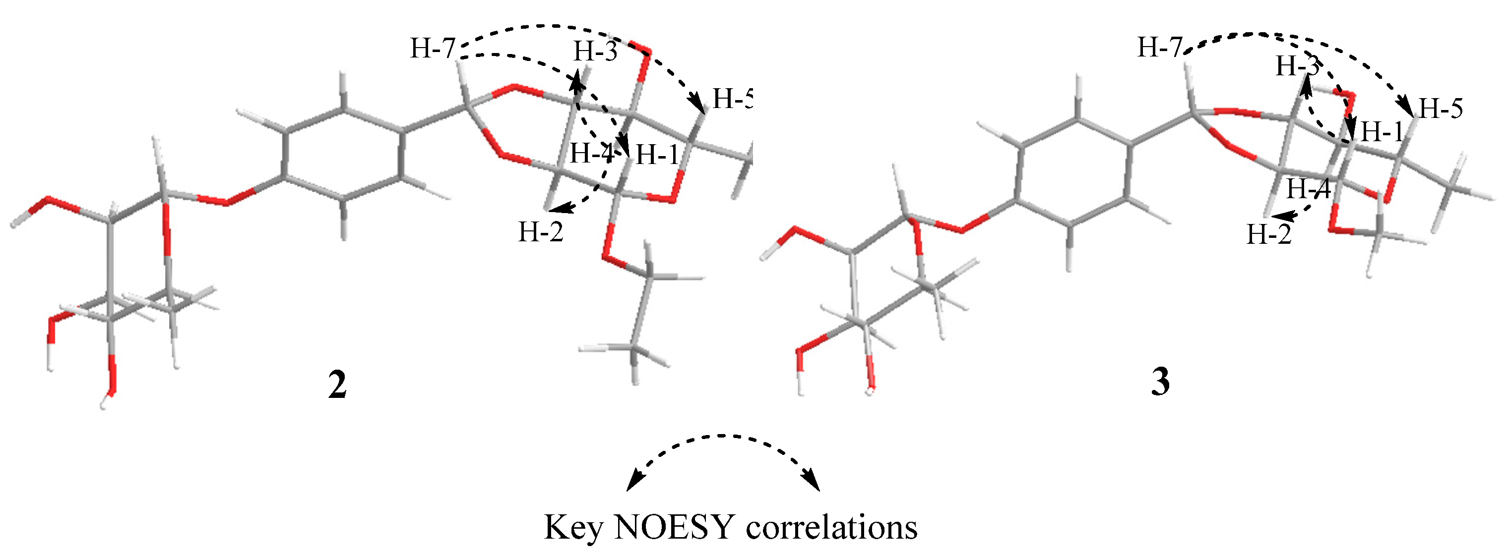
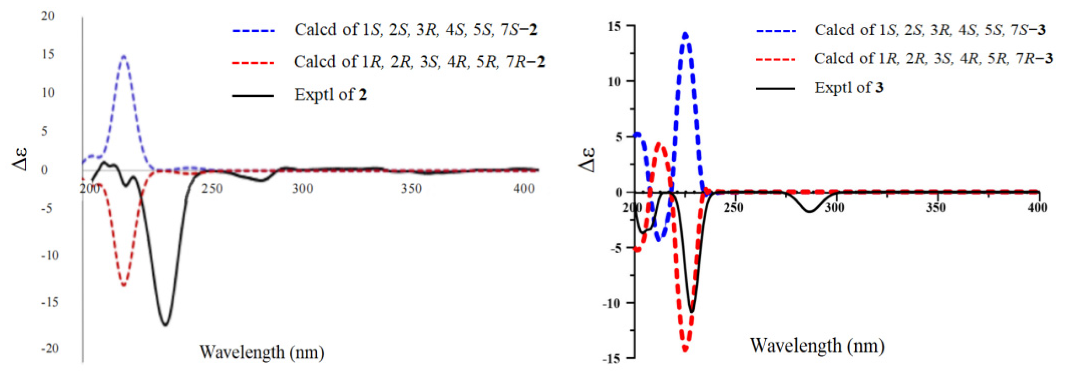
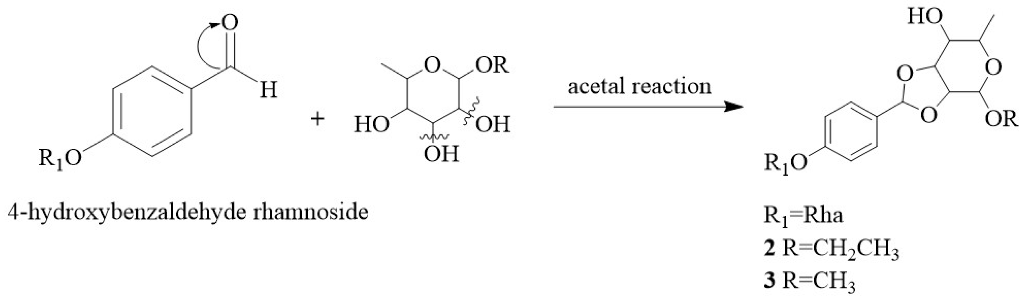
2.1. Structural Elucidation of the Isolated Compounds

The 85% EtOH extract from M. oleifera seeds was subjected to repeated column chromatography (CC) such as silica gel, Sephadex LH-20, Toyopearl HW-40F, and ODS, afforded six new phenolic glycosides (16) and ten known phenolic glycosides (716). Compounds 716 were identified as O-ethyl-4-[(4-α-L-rhamnosyl)-benzyl] carbamate (7) [19], 1-O-(4-hydroxymethylphenyl), α-L-rhamnopyranoside (8) [20], niazirin (9) [21], marumoside A (10) [22], niazimicin (11) [22], 4-aminophenol-α-L-rhamnopyranose (12) [23], moringa A (13) [24], 4-(α-L-rhamnosyloxy)benzylamine) (14) [25], N,N′-bis (4-[(α-L-rhamnosyloxy)benzyl]) thiourea (15) [26], and glucomoringin (16) [27] (Figure 1) by comparison of their spectroscopic data with those previously reported in the literature.
Compound 1 was obtained as a colorless viscous oil. Its molecular formula was determined as C15H22O6 by a positive high-resolution electrospray ionization mass spectrometry (HR-ESI-MS) ion at m/z 321.1302 [M + Na]+ (calculated for C15H22O6Na, 321.1309), indicating 5 degrees of unsaturation. The infrared (IR) spectrum showed absorption of hydroxyl (3392 cm−1), a methylene group (2915 cm−1), a benzene ring (1613, 1511 cm−1), and an ether bond (1231 cm−1). The 1H-nuclear magnetic resonance (NMR) spectrum of 1 revealed the presence of a 1,4-disubstituted benzene ring at δH 7.27 (2H, d, J = 8.5 Hz) and 7.04 (2H, d, J = 8.5 Hz), one ethoxy group hydrogen signal at δH 3.53 (2H, q) and 1.32–1.21 (3H, overlapped), one singlet methylene hydrogen signal at δH 4.43 (2H, s), and a rhamnose anomeric proton at δH 5.42 (1H, d, J = 1.6 Hz). The 13C NMR data showed 15 resonances, including 6 aromatic carbon signals at δC 157.4 (C-1), 133.4 (C-4), 130.4 (C-3, C-5), and 117.4 (C-2, C-6), 2 ethoxy carbon signals at δC 66.6 (C-8) and 15.4 (C-9), 1 methylene carbon signal at δC 73.2 (C-7), and 6 rhamnosyl carbon signals at δC 99.8 (C-1′), 73.8 (C-4′), 72.2 (C-3′), 72.0 (C-2′), 70.6 (C-5′), and 18.0 (C-6′). Furthermore, the location of the rhamnose at δC 157.4 (C-1) was verified by the 1H-detected heteronuclear multiple bond correlation (HMBC) spectrum correlation from δH 5.42 (1H, d, J = 1.6 Hz, H-1′) to C-1 (Figure 2). The chemical shifts and coupling constants of H-1′ indicated that the sugar is linked to the aglycone with α-glycosidic linkage. The singlet methylene signal at C-4 was confirmed by the HMBC correlation from δH 4.43 to C-4 and C-3. The HMBC correlations from δH 3.53 to C-7 indicated that the ethoxyl group was attached to C-7 (Figure 2). The gas chromatography (GC) analysis showed that the derivative of acid hydrolysis from 1 had the same retention time (tR = 26.24 min) as the derivative of authentic L-rhamnose. Thus, compound 1 was identified as a phenolic glycoside derivative and named 4-(α-L-rhamnosyl) benzyl ethyl ester, which we trivially named moringaside B. All the 1H and 13C-NMR data of compound 1 were assigned (Table 1).
Compound 2 was obtained as a yellow oil. Its molecular formula was determined as C21H30O10 by positive HR-ESI-MS ion at m/z 443.1904 [M + H]+ (calculated for C21H31O10, 443.1912), indicating 7 degrees of unsaturation. The presence of a hydroxyl (3413 cm−1), a methylene group (2918 cm−1), a benzene ring (1613, 1514 cm−1), and an aromatic ether bond (1233 cm−1) was confirmed by its IR spectrum. The 1H-NMR spectrum showed an AA’BB’ coupling system aromatic ring at δH 7.39 (2H, d, J = 8.7 Hz) and 7.06 (2H, d, J = 8.7 Hz), an ethoxy group at δH 3.69 (1H, overlapped), 3.50 (1H, dq, J = 9.8, 7.1 Hz), and 1.19–1.22 (3H, overlapped), one methylene group at δH 5.83 (1H, s), and two sugar anomeric protons at δH 5.11 (1H, d, J = 1.5 Hz) and 5.45 (1H, d, J = 1.8 Hz). The 13C NMR spectrum showed that 2 had six aromatic carbon signals at δC 157.4 (C-1′), 133.4 (C-4′), 130.4 (C-3′, C-5′), and 117.4 (C-2′, C-6′), two ethoxy carbon signals at δC 63.7 (C-8) and 15.4 (C-9), one methylene carbon signal at δC 106.5 (C-7), and two groups of sugar carbon signals at δC 107.5 (C-1), 86.0 (C-2), 80.9 (C-3), 85.6 (C-4), 66.0 (C-5), 21.1 (C-6), and δC 99.7 (C-1″), 72.0 (C-2″), 72.2 (C-3″), 73.8 (C-4″), 70.7 (C-5″), and 18.0 (C-6″). Moreover, the HMBC correlation from δH 5.45 to C-1′ indicated that one sugar fragment group was attached to C-1′, and the α-configuration of anomeric carbon of the sugar fragment was deduced based on the coupling constant of the anomeric proton δH 5.45 (1H, d, J = 1.8 Hz). The location of the singlet methylene signal at C-4′ was confirmed by the HMBC correlation from δH 5.83 to C-4′, C-3′, and C-5′. Other sugars, C-2 and C-3, were attached to C-7 by oxygen atoms, respectively, which were confirmed by the HMBC correlation from δH 4.68 to C-7 and δH 4.89 to C-7 (Figure 2). The 1H-1H homonuclear chemical-shift correlated spectroscopy (COSY) spectrum correlations (Figure 2) of H-1/H-2, H-2/H-3, H-3/H-4, H-4/H-5, and H-5/H-6 showed the assignment in the protons of the sugar moiety. And this sugar was confirmed to α-configuration by the chemical shift and coupling constant of δH 5.11 (1H, d, J = 1.5 Hz). The GC analysis spectrum showed that the acid hydrolysate of 2 had the same retention time (tR = 26.24 min) with the derivative of authentic sample L-rhamnose. The HMBC correlation from δH 3.70 (1H, m) and 3.50 (1H, m) to C-1, as well as the 1H-1H COSY correlation of H-8/H-9 was observed, suggesting that the ethoxyl group was located at C-1. The nuclear overhauser effect spectroscopy (NOESY) spectrum correlations of H-7 with H-1/H-5 and H-1 with H-3 revealed their co-facial relationship, and they were assigned arbitrarily as α-oriented, while the correlations of H-4 with H-6/H-2 indicated that these protons were β-oriented (Figure 3). The absolute configuration of C-1/C-2/C-3/C-4/C-5/C-7 was assigned as 1R/2R/3S/4R/5R/7R by comparing the calculated ECD data (Figure 4) with the experimental data. Consequently, compound 2 was identified as (1R, 2R, 3S, 4R, 5R, 7R)-O-ethly-2,3-di-O-(1′-O-α-L-Rha-phenylmethylene)-α-L-rhamnopyranoside and trivially named moringaside C. All the 1H and 13C-NMR data of compound 2 were assigned (Table 1).
Compound 3 was obtained as a yellow oil. Its molecular formula was determined as C20H18O10 by a positive HR-ESI-MS ion at m/z 451.1567 [M + Na]+ (calculated for C20H28O10Na, 451.1575), indicating 7 degrees of unsaturation. Its IR spectrum exhibited the presence of a hydroxyl (3413 cm−1), a methylene group (2918 cm−1), a benzene ring (1613, 1514 cm−1), and aromatic ether bond (1233 cm−1) functional groups. Analysis of the 1D and 2D NMR data of 3 were similar to those of compound 2, except that the ethoxyl group attached to C-1 was replaced by methoxyl. The GC analysis showed that the acid hydrolysate of 3 had the same retention time as the standard L-rhamnose derivative (tR = 26.24 min). Meanwhile, the chemical shift and coupling constant of the two sugar anomeric protons δH 5.45 (J = 1.8 Hz) and 5.00 (J = 1.5 Hz) proves that the two sugar segment groups are α-configuration. The NOESY spectrum correlation from H-7 to H-1/H-5 and H-1 to H-3 indicated their cofacial orientation (α-oriented), while the correlations from H-4 to H-6/H-2 suggested that these protons were β-oriented (Figure 3). The absolute configuration of C-1/C-2/C-3/C-4/C-5/C-7 was assigned as 1R/2R/3S/4R/5R/7R by comparing ECD spectra (Figure 4). Consequently, compound 3 was determined to be (1R, 2R, 3S, 4R, 5R, 7R)-O-methyl-2,3-di-O-(1′-O-α-L-Rha-phenylmethylene)-α-L-rhamnopyranoside, trivially named moringaside D. All the 1H and 13C-NMR data of compound 3 were assigned (Table 1).
Compound 4 was obtained as a yellow oil. Its molecular formula C21H23NO6 (11 degrees of unsaturation) was deduced by the ion peak of HR-ESI-MS m/z 408.1413 [M + Na]+ (calculated for C21H23NO6Na, 408.1423). The IR spectrum showed characteristic absorption peaks at 3383 cm−1, 2933 cm−1, 2256 cm−1, 1610 cm−1, 1508 cm−1, 1114 cm−1, 1062 cm−1, and 1026 cm−1, which were in agreement with a hydroxyl, a methylene group, a cyanogen group, a benzene ring, and an aromatic ether bond. The 1H NMR spectrum revealed AA’BB’coupling on a benzene ring at δH 7.14 (2H, d, J = 8.4 Hz, H-2, H-6) and 6.95 (2H, d, J = 8.4 Hz, H-3, H-5). Signals at δH 7.00 (1H, dd, J = 8.4, 2.4 Hz, H-5′), 6.97 (1H, d, J = 1.8 Hz, H-3′), and 6.78 (1H, d, J = 7.8 Hz, H-6′) showed that an ABX coupling system in an aromatic ring. Two methylene hydrogen signals were assigned at δH 3.87 (2H, s, H-7) and 3.69 (2H, s, H-7′). One anomeric doublet proton resonance at δH 5.36 (1H, d, J = 1.8 Hz, H-1″) showed the presence of one sugar unit as a glycoside. The 13C NMR spectrum of 4 showed 21 carbon signals including 12 aromatic carbon signals [δC 156.1 (C-1), 156.0 (C-1′), 136.2 (C-4), 131.2 (C-3′), 130.9 (C-3, C-5), 130.2 (C-2′), 127.9 (C-5′), 122.6 (C-4′), 117.5 (C-2, C-6), and 116.5 (C-6′)], 6 sugar carbon signals (δC 100.0 (C-1″), 73.9 (C-4″), 72.2 (C-3″), 72.1 (C-2″), 70.5 (C-5″) and 18.0 (C-6″)), 2 methylene carbon signals (δC 35.7 (C-7) and 22.7 (C-7′)), and 1 CN moiety signal at δC 120.2 (C-8′) [28]. The HMBC correlation from δH 5.36 to C-1 indicated that one sugar segment was attached to C-1, and according to the coupling constant of the anomeric proton δH 5.36 (J = 1.8 Hz), the α-configuration of the anomeric carbon of a sugar fragment was deduced. Furthermore, the GC analysis showed that the retention time of acid hydrolysate 4 was the same as the standard L-rhamnose derivative (tR = 26.32 min), which indicated that 4 had α-L-rhamnose moiety. The methylene (C-7) linked to two benzene rings was confirmed by the HMBC correlation (Figure 2) of H-7 to C-4, C-1′, C-3′, and C-6′, and the downfield resonance of δC 156.0 (C-1′) suggested substitution of a hydroxyl residue. The location of the acetonitrile signal at the ABX coupling aromatic spin systems (C-4′) was confirmed by the key HMBC correlations from H-7′ to C-3′, C-4′, C-5′, and C-8′. Therefore, the structure of 4 was confirmed as 4-(α-L-rhamnopyranosyl) benzyl-1′-hydroxy-4′-phenylacetonitrile and trivially named moringaside E. All the 1H and 13C-NMR data of compound 4 were assigned (Table 2).
Compound 5 was obtained as yellow oil. Its negative HR-ESI-MS showed an [M - H] ion at m/z 424.1600, which is in accordance with the molecular formula C20H27O9N (calculated for C20H26O9N, 424.1602), indicating 8 degrees of unsaturation. The IR spectrum of 5 shows frequencies at 3394, 2933, 2252, 1612, 1510, 1236, 1064, and 1022 cm−1 and was assigned to a hydroxyl, a methylene group, a cyanogen group, a benzene ring, and an aromatic ether bond. The 1D and 2D NMR data of compound 5 showed a high degree of similarity to the compound 4-[(β-D-glucopyranosyl)-(1→3)-(α-L-rhamnopyranosyl)]phenylacetonitrile [29]. The only difference was that the C-3′ of compound 5 was attached to rhamnose by oxygen atoms, not glucose. Two sugar segment groups part of 5 were identified and characterized by the anomeric proton doublet at δH 5.49 (J = 1.8 Hz) and δH 4.75 (J = 1.2 Hz); these data suggested that sugars’ moieties were α-configuration. Additionally, acid hydrolysis of 5 obtained L-rhamnose, which was identified by the GC analysis comparison with authentic samples, which proved that 5 had α-L-rhamnose moieties. Consequently, the structure of compound 5 was confirmed as 4-[(α-L-rhamnopyranosyl)-(1→3)-(α-L-rhamnopyranosyl)]phenylacetonitrile and trivially named moringaside F. All the 1H and 13C-NMR data of compound 5 were assigned (Table 2).
Compound 6 was isolated as a yellow oil, and its molecular formula was assigned as C21H30O11 based on positive HR-ESI-MS data of the protonated species [M + Na]+ at m/z 481.1680 (calculated for 481.1685), indicating 7 degrees of unsaturation. Its IR spectrum exhibited the presence of hydroxyl (3385 cm−1), methylene group (2933 cm−1), carbonyl (1732 cm−1), and fatty ether bond (1064 cm−1, 1022 cm−1), with a comparison of the NMR data of 6 with the known compound methyl 2-[4-(α-L-rhamnopyranosyl)phenyl]acetate [21]. The differences were the addition of a group of sugar carbon signals (99.0, 72.8, 74.8, 73.7, 73.9, and 18.0) at C-3′ in 6. This sugar fragment is attached at the C-3′ position, confirmed by δH 4.76, and has a correlation signal with C-3′ in the HMBC spectrum (Figure 2). The GC analysis showed that the acid hydrolysate of 6 was L-rhamnose; meanwhile, these two sugar moieties’ were α-configuration corroborated by the anomeric protons at δH 5.46 (J = 1.8 Hz) and 4.76 (J = 1.2 Hz). Eventually, compound 6 was elucidated as methyl 2-[4-(α-L-rhamnopyranosyl)-(1→3)-(α-L-rhamnopyranosyl)phenyl]acetate, which we trivially named moringaside G. All the 1H and 13C-NMR data of 6 were assigned (Table 2).
Compounds 16 are without precedent in the natural products literature, especially compounds 23, which possess a rare 1, 3-dioxocyclopentane at the rhamnose group. A putative biosynthetic pathway for their scaffold is proposed in Figure 5. The 4-hydroxybenzaldehyde rhamnoside, a secondary metabolite of the seeds from M. oleifera [24], reacts with ethyl (or methyl) α-L rhamnoside in acidic conditions by acetal reaction to yield ethyl (or methyl) 2,3-O-benzylidene-α-L-rhamnopyranoside, which is synthesized in a pathway similar to that of methyl 2,3-O-benzylidene-α-D-mannopyranoside [30].

2.2. α-Glucosidase Inhibitory Activity Evaluation

α-glucosidase is a key catalytic enzyme for carbohydrate digestion and glucose release. Inhibition of α-glucosidase can delay glucose uptake and reduce postprandial blood glucose levels, which may inhibit the progression of DM [31]. Thus, all the isolated phenolic glycosides were evaluated for their inhibitory activities of α-glucosidase. As shown in Table 3, compared to the positive drug acarbose with an IC50 value of 324.1 ± 4.99 μM, compound 16 revealed excellent inhibitory activity of α-glucosidase with an IC50 value of 301.4 ± 6.22 μM, while compound 4 showed moderate activity with an IC50 value of 382.8 ± 1.42 μM. Other compounds had low inhibitory activity against α-glucosidase and are not listed in Table 3.

3. Material and Methods

3.1. General Experimental Procedure

The 1D and 2D NMR were recorded on a BRUKER 600 NEO NMR spectrometer (Bruker Co., Ltd., Karlsruhe, Germany) and a JEOL ECS 400 NMR spectrometer (Jeol, Tokyo, Japan). HR-ESI-MS data were measured using Thermo Fisher Q Exactive-Plus mass spectroscopy (Thermo Fisher Scientific, Waltham, MA, USA). GC Analysis was carried out on a Shimadzu-2010 Plus gas chromatograph (Shimadzu Co., Ltd., Kyoto, Japan). A JASCO J-715 spectrometer (Jasco, Tokyo, Japan) was used to record the ECD. UV spectra were acquired on a UV-2700 spectrometer (Shimadzu Co., Ltd., Kyoto, Japan). IR spectra were obtained using an IR Tracer-100 spectrometer (Shimadzu Co., Ltd., Kyoto, Japan). AMR-100 enzyme-linked immunosorbent assay (Hangzhou Aosen Instrument Co., Ltd., Hangzhou, China). Silica gel (200–300 mesh and 300–400 mesh, Qingdao Haiyang chemical Co., Ltd., Qingdao, China), Toyopearl HW-40F (Tosoh corporation, Tokyo, Japan), and Sephadex LH-20 (GE Healthcare Bio-Sciences AB, Uppsala, Sweden). The TLC were silica gel GF254 plates (Qingdao Haiyang Chemical Co., Ltd., Qingdao, China).

3.2. Plant Material

The seeds of M. oleifera were collected from Yunnan Province, People’s Republic of China, and identified by Shao-Huan Liu, a senior experimentalist at Guizhou Medical University. The voucher specimens were stored in the Engineering Research Center for the Development and Application of Ethnic Medicine and TCM (Ministry of Education), Guizhou Medical University, Guiyang, China.

3.3. Extraction and Isolation

The seeds of M. oleifera (10.5 kg) were slightly smashed and extracted with 85% ethanol under reflux, then concentrated the extract (2.5 kg). The extract was separated through a D101 macroporous resin, following elution in proper order by water, ethanol-water (30%, 60%, 95%, v/v). Finally, four parts were obtained: water part (1.8 kg), 30% ethanol part (276.7 g), 60% ethanol part (69.2 g), and 95% ethanol part (249.4 g).
The 60% ethanol extract was separated into 10 fractions (Fr. 1–10) through a silica gel column chromatography eluting with CH2Cl2–MeOH (100:0 to 1:1). Fr. 5 was separated by a silica gel CC eluting with CH2Cl2–MeOH (30:1 to 12:1) and HW-40F CC (MeOH) to obtain compounds 1 (23 mg) and 7 (90 mg). Fr. 6 was purified over Sephadex LH-20 (MeOH) and Toyopearl HW-40F (MeOH) to give compounds 8 (10.5 mg), 9 (9.8 mg), 10 (23 mg), and 11 (68.9 mg). Fr. 7 was chromatographed over silica gel (CH2Cl2–MeOH, 8:1 to 2:1) to get nine subfractions (Fr. 7.1–7.9). Fr. 7.6 was subjected to a Sephadex LH-20 (MeOH), Toyopearl HW-40F (MeOH), and ODS column to yield compounds 12 (34.2 mg), 13 (26.1 mg), 14 (26.7 mg), and 15 (470 mg). Fr. 10 was subjected to a silica gel CC eluting with EtOAc–MeOH (5:1 to 1:1) and an ODS column (MeOH/H2O, 1:9 to 7:3) to obtain compound 16 (79.0 mg). The 30% ethanol extract was separated into 11 fractions (Fr. 1–11) through a silica gel CC eluting with CH2Cl2–MeOH (100:0 to 1:1). Fr. 6 was separated by a silica gel CC eluting with EtOAc–MeOH (20:1 to 10:1) to get four subfractions (Fr. 6.1–6.7), and Fr. 6.3 was separated by Sephadex LH-20 (MeOH–Water, 1:1) to obtain four subfractions (Fr. 6.3.1–6.3.4). Fr. 6.3.2 was further separated by an ODS column to gain compounds 2 (5 mg) and 3 (7 mg). Fr. 6.3.3 was also chromatographed on an ODS column to yield compound 4 (8 mg). Fr. 8 was subjected to silica gel CC (EtOAc–MeOH, 20:0 to 8:1) to give seven sub-fractions (Fr. 8.1–8.7). Fr. 8.4 was applied to Sephadex LH-20 (MeOH–Water, 1:1) and an ODS column to get compound 5 (40 mg). Compound 6 (5.9 mg) was isolated from Fr. 8.5 by HW-40F CC (MeOH).
  • Moringaside B (1): colorless viscous oil; α D 25 -90.8 (c 0.10, MeOH); UV (MeOH) λmax (log ε) 206 (3.07), 224 (2.17) nm; IR (KBr) νmax: 3392, 2915, 1613, 1511, and 1231 cm−1; 1H and 13C NMR data, see Table 1; HR-ESI-MS: m/z 321.1302 [M + Na]+ (calculated for C15H22O6Na, 321.1309).
  • Moringaside C (2): yellow oil; α D 25 -111.3 (c 0.1, MeOH); UV (MeOH) λmax (log ε) 206 (3.06), 224 (2.20) nm; IR (KBr) νmax: 3413, 2918, 1613, 1514, and 1233 cm−1; ECD (c 0.10, MeOH) Δε 214 (+0.63), 237 (-18.53) nm; 1H and 13C NMR data, see Table 1; HR-ESI-MS: m/z 443.1904 [M + H]+ (calculated for C21H31O10, 443.1912).
  • Moringaside D (3): yellow oil; α D 25 -109.5 (c 0.1, MeOH); UV (MeOH) λmax (log ε) 206 (3.10), 224 (2.25) nm; IR (KBr) νmax: 3396, 2913, 1612, 1512, and 1232 cm−1; ECD (c 0.08, MeOH) Δε 204 (-3.65), 228 (-10.90) nm; 1H and 13C NMR data, see Table 1; HR-ESI-MS m/z 451.1567 [M + Na]+ (calculated for C20H28O10Na, 451.1575).
  • Moringaside E (4): yellow oil; α D 25 -103.5 (c 0.1, MeOH); UV (MeOH) λmax (log ε) 208 (3.28), 224 (2.21), and 280 nm (2.03) nm; IR (KBr) νmax: 3383, 2933, 2256, 1610, 1508, 1114, 1062, and 1026 cm−1; 1H and 13C NMR data, see Table 2; HR-ESI-MS m/z 408.1413 [M + Na]+ (calculated for C21H23NO6Na, 408.1423).
  • Moringaside F (5): yellow oil; α D 25 -146.2 (c 0.1, MeOH); UV (MeOH) λmax (log ε) 222 (2.64), 272 (2.26) nm; IR (KBr) νmax: 3394, 2933, 2253, 1612, 1510, 1236, 1064, and 1022 cm−1; 1H and 13C NMR data, see Table 2; HR-ESI-MS m/z 424.1600 [M - H] (calculated for C20H26O9N, 424.1602).
  • Moringaside G (6): yellow oil; α D 25 -51.1 (c 0.1, MeOH); UV (MeOH) λmax (log ε) 208 (3.18), 222 (2.44), and 272 (2.16) nm; IR (KBr) νmax: 3385, 2933, 1732, 1512, 1230, 1064, and 1022 cm−1;1H and 13C NMR data, see Table 2; HR-ESI-MS m/z 481.1680 [M + Na]+ (calculated for C21H30O11Na 481.1685).

3.4. Acid Hydrosis and Sugar Identification

The sugar was identified according to the established method [32]. Compounds 16 (each 0.3 mg), respectively, were dissolved with 2 mol/mL Hcl solution (2 mL) at 95 °C for 3 h. After cooling to room temperature, the ethyl acetate was added to the reaction solution and extracted three times. The water-soluble layer was dried to obtain the sugar residual. The sugar residuals, D-rhamnose (0.5 mL) and L-rhamnose (0.5 mL), separately, were added pyridine (0.4 mL) and L-cysteine methyl ester hydrochloride (1.0 mg), then heated at 60 °C for 1 h. N-trimethylsilyllimidazole (0.15 mL) was added to the mixture and reacted at 60 °C for 1 h again. Next, the reaction solution was dried, then dissolved in water (1.0 mL) and extracted with n-hexane (0.5 mL) three times. The organic layer was directly analyzed by GC analysis. The peaks of the acid-hydrolyzed derivatives of compounds 16 coincide with the derivatives of the authentic sample L-rhamnose.

3.5. Electronic Circular Dichroism Calculation of Compounds 23

The theoretical calculations were carried out using Gaussian 09 [33]. At first, all conformers were optimized at PM6. Room-temperature equilibrium populations were calculated according to the Boltzmann distribution law, based on which dominative conformers of population over 1% were kept. The chosen conformers were further optimized at B3LYP/6-31G(d) in the gas phase. Vibrational frequency analysis confirmed the stable structures. ECD calculations [34] were conducted at the B3LYP/6-311G(d,p) level in methanol with the IEFPCM model using the time-dependent density functional theory (TD-DFT). Rotatory strengths for a total of 10 excited states were calculated. The ECD spectrum was simulated in SpecDis by overlapping Gaussian functions for each transition according to Equation (1).
Δ ε ( E ) = 1 2.297 × 10 39 × 1 2 π σ i A Δ E i R i e E E i 2 σ 2

3.6. Inhibitory Activities against α-Glucosidase

The α-glucosidase enzyme inhibition assay was performed according to the previously described method [35]. Compounds 1–16 were screened for α-glucosidase inhibitory activity with acarbose as a positive control (10 μg/mL) and DMSO as a blank control. In sequence, 10 μL of the sample, 100 μL of phosphate buffer (pH = 6.8), and 50 μL of α-glucosidase (0.5 U/mL) were added to 96-well plates and incubated for 15 min in a 37 °C incubator. Then, a further 40 μL of substrate (p-nitrophenyl-β-D-glucopyranoside, 1.25 mmol/L) was added and incubated in a 37 °C incubator for 25 min. After the reaction, the absorbance was measured at 405 nm. The results were obtained from at least three independent experiments.

4. Conclusions

In summary, we have conducted the successful isolation of 16 compounds from M. oleifera seeds, including 6 new and 10 known phenolic glycosides. Among them, compounds 2 and 3 especially are combined with a rare 1,3-dioxocyclopentane moiety at the rhamnose group. The putative biosynthetic pathway for their scaffold is proposed. All of the isolated compounds were evaluated for their inhibitory activities against α-glucosidase, and two compounds (4 and 16) showed comparable inhibitory activity to the positive control in vitro (Table 3). This study enriches the chemical basis of M. oleifera and elucidated the pharmacological basis of the hypoglycemic activity of the seeds. The results not only broaden the horizon of the structural diversity of phenolic glycosides of M. oleifera but also provide new evidence for the clinical applications of herbal medicine. Folk and ethnic medicines are of great importance and are valuable reservoirs for lead compounds in the field of drug research and development. From these results, further in-depth study may be done to discover the lead compounds.

Supplementary Materials

The following supporting information can be downloaded at https://www.mdpi.com/article/10.3390/molecules28176426/s1; Figures S1–S51: HR-ESI-MS, UV, IR, 1D, and 2D NMR spectra of compounds 16, and GC analysis of D-rhamnose, L-rhamnose, and compounds 16.

Author Contributions

L.-Z.L. and J.-S.S. designed the experiment; L.C. performed the isolation and identification of all the compounds and wrote the manuscript; Y.-L.T., X.-J.D. and S.-J.X. contributed to the isolation of the compounds; Y.-J.L. and X.-S.Y. helped to buy the plant material and provided suggestions on the isolation; L.-Z.L. reviewed the manuscript; Y.-J.L. and X.-S.Y. provided comments and suggestions on structure elucidation and reviewed the manuscript. All authors have read and agreed to the published version of the manuscript.

Funding

This work was supported by the National Natural Science Foundation of China (Nos. 82260752 and 81860689) and the project of the Key Laboratory of Basic Pharmacology of the Ministry of Education, Zunyi Medical University (No. (2022)392).

Institutional Review Board Statement

Not applicable.

Informed Consent Statement

Not applicable.

Data Availability Statement

All the data in this research were presented in the manuscript and Supplementary Material.

Conflicts of Interest

The authors declare no conflict of interest.

Sample Availability

Samples of the compounds are available from the authors upon reasonable request.

References

  1. Chiranjeev, S.; Youllee, K.; Dohee, A.; Sang, J.C. Protein tyrosine phosphatases (PTPs) in diabetes: Causes and therapeutic opportunities. Arch. Pharm. Res. 2021, 44, 310–321. [Google Scholar]
  2. Chen, G.L.; Xu, Y.B.; Wu, J.L.; Li, N.A.; Guo, M.Q. Hypoglycemic and hypolipidemic effects of Moringa oleifera leaves and their functional chemical constituents. Food Chem. 2020, 333, 127478. [Google Scholar] [CrossRef] [PubMed]
  3. Dehghan, H.; Salehi, P.; Amiri, M.S. Bioassay-guided purification of α-amylase, α-glucosidase inhibitors and DPPH radical scavengers from roots of Rheum turkestanicum. Ind. Crops Prod. 2018, 117, 303–309. [Google Scholar] [CrossRef]
  4. Watanabe, S.; Okoshi, H.; Yamabe, S.; Shimada, M. Moringa oleifera Lam. in Diabetes Mellitus: A Systematic Review and Meta-Analysis. Molecules 2021, 26, 3513. [Google Scholar] [CrossRef]
  5. Khoo, C.M. Diabetes Mellitus Treatment. Int. Encycl. Public Health 2017, 2, 288–293. [Google Scholar]
  6. Tuo, L.; Kenneth, T.; Dan, S.K. Identification of α-glucosidase Inhibitors in Machilus litseifolia by combined use of High-Resolution α-glucosidase inhibition profilingand HPLCPDA- HRMS-SPE-NMR. J. Nat. Prod. 2019, 82, 249–258. [Google Scholar]
  7. Şöhretoğlu, D.; Sari, S.; Barut, B.; Özel, A. Discovery of potent α-glucosidase inhibitor flavonols: Insights into mechanism of action through inhibition kinetics and docking simulations. Bioorg. Chem. 2018, 79, 257–264. [Google Scholar] [CrossRef]
  8. Chen, H.; Xiong, L.; Wang, W.J. Reviews on α-glucosidase inhibitor from plant secondary metabolites. China J. Chin. Mater. Med. 2017, 42, 2915–2924. [Google Scholar]
  9. Dhakad, A.K.; Ikram, M.; Sharma, S.; Pandey, V.V.; Singh, A. Biological, nutritional, and therapeutic significance of Moringa oleifera Lam. Phytother. Res. 2019, 33, 2870–2903. [Google Scholar] [CrossRef]
  10. Tshabalala, T.; Ncube, B.; Madala, N.E.; Nyakudya, T.T.; Ndhlala, A.R. Scribbling the Cat: A Case of the “Miracle” Plant, Moringa oleifera. Plants 2019, 8, 510. [Google Scholar] [CrossRef]
  11. Verma, K.S.; Nigam, R. Nutritional assessment of different parts of moringa oleifera lammcollected from central india. J. Nat. Prod. Plant Resour. 2014, 4, 81–86. [Google Scholar]
  12. Zhao, B.; Hua, L.; Tao, L.; Di, W.; Chen, Z. Characterization of the Chemical Composition of Chinese Moringa oleifera Seed Oil. J. Am. Oil Chem. Soc. 2019, 96, 523–533. [Google Scholar] [CrossRef]
  13. Renata, A.C.; De, S.; Ernesto, H.; Jair, M.; Geraldo, B.F.; Fernandes, V. Thiocarbamates from moringa oleifera seeds bioactive against virulent and multidrug-resistant vibrio species. BioMed. Res. Int. 2017, 2017, 7963747. [Google Scholar] [CrossRef]
  14. Singh, R.; Negi, P.S.; Radha, C. Phenolic composition, antioxidant and antimicrobial activities of free and bound phenolic extracts of Moringa oleifera seed flour. J. Funct. Foods 2013, 5, 1883–1891. [Google Scholar] [CrossRef]
  15. Mahajan, S.G.; Mali, R.G.; Mehta, A.A. Effect of Moringa oleifera lam. seed extract on toluene diisocyanate-induced immune-mediated inflammatory responses in rats. J. Immunotoxicol. 2007, 4, 85–96. [Google Scholar] [CrossRef] [PubMed]
  16. Al Malki, A.L.; El Rabey, H.A. The antidiabetic effect of low doses of Moringa oleifera lam. seeds on streptozotocin induced diabetes and diabetic nephropathy in male rats. Biomed. Res. Int. 2015, 2015, 381040. [Google Scholar] [CrossRef]
  17. Minaiyan, M.; Asghari, G.; Taheri, D.; Saeidi, M.; Nasr-Esfahani, S. Anti-inflammatory effect of Moringa oleifera Lam. seeds on acetic acid-induced acute colitis in rats. Avicenna J. Phytomedicine 2014, 4, 127–136. [Google Scholar]
  18. Lalas, S.; Tsaknis, J. Extraction and identification of natural antioxidant from the seeds of the Moringa oleifera tree variety of malawi. J. Am. Oil Chem. Soc. 2002, 79, 677–683. [Google Scholar] [CrossRef]
  19. Tang, M.M.; Chen, G.Y.; Jiang, K.C.; Luo, M.Y.; Wu, H.; Hu, B.; Zhou, X.M. A New Phenolic Glycoside from the Seeds of Moringa oleifera. Chem. Nat. Compd. 2020, 56, 642–644. [Google Scholar] [CrossRef]
  20. Grond, S.; Papastavrou, I.; Zeeck, A. Novel α-L-Rhamnopyranosides from a Single Strain of Streptomyces by Supplement-Induced Biosynthetic Steps. Eur. J. Org. Chem. 2002, 2002, 3237–3242. [Google Scholar] [CrossRef]
  21. Ma, N.; Tang, Q.; Wu, W.T.; Huang, X.A.; Xu, Q.; Rong, G.L.; Song, J.P. Three Constituents of Moringa oleifera Seeds Regulate Expression of Th17-Relevant Cytokines and Ameliorate TPA-Induced Psoriasis-Like Skin Lesions in Mice. Molecules 2018, 23, 3256. [Google Scholar] [CrossRef] [PubMed]
  22. Faizi, S.; Siddiqui, B.S.; Saleem, R.; Siddiqui, S.; Aftab, K.; Gilani, A.H. Isolation and structure elucidation of novel hypotensive agents, niazinin a, niazinin b, niazimicin and niaziminin a+b from Moringa oleifera: The first naturally occurring thiocarbamates. Chem. Inform. 1993, 24, 1256–1261. [Google Scholar] [CrossRef]
  23. Zhang, J.; Zhang, Q.S.; Tian, G.Y. Study on synthesis of multivalent neoglycoproteins and their binding properties to hepatic stellate cells. Chin. J. Chem. 2003, 21, 843–846. [Google Scholar] [CrossRef]
  24. Xiong, Y.; Riaz Rajoka, M.S.; Zhang, M.; He, Z. Isolation and identification of two new compounds from the seeds of Moringa oleifera and their antiviral and anti-inflammatory activities. Nat. Prod. Res. 2020, 36, 974–983. [Google Scholar] [CrossRef] [PubMed]
  25. Saleem, R.; Meinwald, J. Synthesis of novel hypotensive aromatic thiocarbamate glycosides. J. Chem. Soc. Perkin Trans. 1 2000, 31, 391–394. [Google Scholar] [CrossRef]
  26. Yun, Y.S.; Satake, M.; Katsuki, S.; Kunugi, A. Phenylpropanoid derivatives from edible canna, Canna edulis. Phytochemistry 2004, 65, 2167–2171. [Google Scholar] [PubMed]
  27. Gueyrard, D.; Iori, R.; Tatibou, T.A.; Rollin, P. Glucosinolate chemistry: Synthesis of O-Glycosylated derivatives of glucosinalbin. Eur. J. Org. Chem. 2010, 2010, 3657–3664. [Google Scholar] [CrossRef]
  28. Faizi, S.; Siddiqui, B.S.; Saleem, R.; Siddiqui, S.; Aftab, K.; Gilani, A. Isolation and structure elucidation of new nitrile and mustard oil glycosides from moringa oleifera and their effect on blood pressure. J. Nat. Prod. 1994, 57, 1256–1261. [Google Scholar] [CrossRef]
  29. Francis, J.; Jayaprakasam, B.; Olson, L.; Nair, M. Insulin secretagogues from moringa oleifera with cyclooxygenase enzyme and lipid peroxidation inhibitory activities. Helv. Chim. Acta 2004, 87, 317–326. [Google Scholar] [CrossRef]
  30. Liotta, L.J.; Chalmers, J.F.; Falco Marshall, J.N.; Ferreira, T.E.; Mullen, H.E.; Pace, N.J. Selective 4,6-O-benzylidene formation of methyl α-D-mannopyranoside using 2,6-dimethylbenzaldehyde. Carbohydr. Res. 2014, 391, 31–36. [Google Scholar] [CrossRef]
  31. Zeng, L.; Ding, H.F.; Hu, X.; Zhang, G.W.; Gong, D.M. Galangin inhibits α-glucosidase activity and formation of non-enzymatic glycation products. Food Chem. 2019, 271, 70–79. [Google Scholar] [CrossRef]
  32. Li, L.Z.; Zhang, Y.; Chen, L.; Cen, Y.Z.; Tu, Y.L.; Yang, X.S.; Li, Y.J. Two New Abietane Diterpenes From the Stems of Clerodendrum trichotomum Thunb. Nat. Prod. Com. 2022, 17, 1–8. [Google Scholar] [CrossRef]
  33. Frisch, M.J.; Trucks, G.W.; Schlegel, J.; Scuseria, G.E.; Robb, M.A.; Cheeseman, J.R. Gaussian 09, Revision C.01; ScienceOpen, Inc.: Burlington, MA, USA, 2010. [Google Scholar]
  34. Bruhn, T.; Schaumlöffel, A.; Hemberger, Y.; Bringmann, G. SpecDis: Quantifying the Comparison of Calculated and Experimental Electronic Circular Dichroism Spectra. Chirality 2013, 25, 243–249. [Google Scholar] [CrossRef] [PubMed]
  35. Tian, J.L.; Si, X.; Wang, Y.H.; Gong, E.S.; Xie, X.; Zhang, Y.; Li, B.; Shu, C. Bioactive flavonoids from rubus corchorifolius inhibit α-glucosidase and α-amylase to improve postprandial hyperglycemia. Food Chem. 2020, 341, 128149–128179. [Google Scholar] [CrossRef] [PubMed]
Figure 1. Chemical structures of compounds 116 isolated from M. oleifera seeds.
Molecules 28 06426 g001
Figure 2. Key 1H-1H COSY and HMBC correlations of compounds 16.
Molecules 28 06426 g002
Figure 3. Key NOESY correlations for compounds 23.
Molecules 28 06426 g003
Figure 4. Calculated and experimental ECD spectra of compounds 23.
Molecules 28 06426 g004
Figure 5. A putative biosynthetic pathway for compounds 2 and 3.
Molecules 28 06426 g005
Table 1. The 1H and 13C NMR spectroscopic data of compounds 13 in CD3OD.
NO.1 aNO.2 b3 b
δH (J in Hz)δCδH (J in Hz)δCδH (J in Hz)δC
1-157.415.11 d (1.5)107.55.00 d (1.5)108.6
2, 67.27 d (8.5)117.424.68 d (5.5)86.04.68 d (5.4)85.8
3, 57.04 d (8.5)130.434.89 ov80.94.89 ov80.8
4-133.443.69 m85.63.65 dd (9.0, 5.4)85.6
74.43 s73.254.07 dq (8.7, 6.3)66.04.08 m65.9
83.53 q (14.0, 7.0)66.661.32 d (6.3)21.11.33 d (6.0)21.1
91.21–1.19 ov15.475.83 s106.55.83 s106.5
1′5.42 d (1.6)99.883.70 m
3.50 dq (9.8, 7.1)
63.73.33 s54.6
2′4.00 dd (3.3, 1.9)72.091.19–1.22 ov15.4--
3′3.85 dd (9.5, 3.4)72.21′-158.7-158.7
4′3.46 t (9.5)73.82′, 6′7.06 d (8.7)117.17.06 d (9.0)117.1
5′3.64 m70.63′, 5′7.39 d (8.7)129.47.39 d (9.0)129.4
6′1.23–1.21 ov18.04′-132.0-132.0
1″5.45 d (1.8)99.75.45 d (1.8)99.7
2″3.99 dd (3.5, 1.8)72.03.99 dd (3.6, 1.8)72.0
3″3.84 dd (9.5, 3.5)72.23.84 dd (9.0, 3.0)72.2
4″3.45 t (9.5)73.83.45 t (9.6)73.8
5″3.60 m70.73.60 m70.7
6″1.19–1.22 ov18.01.21 d (6.0)18.0
a: NMR data (δ) were measured at 400 MHz for 1H and 100 MHz for 13C; b: NMR data (δ) were measured at 600 MHz for 1H and 150 MHz for 13C; ov: overlapping signals within the same column.
Table 2. The 1H (600 MHz) and 13C NMR (150 MHz) data of compounds 46 in CD3OD.
NO.456
δH (J in Hz)δCδH (J in Hz)δCδH (J in Hz)δC
1-156.1-157.2-156.8
2, 66.95 d (8.4)117.57.09 d (8.4)118.17.04 d (8.4)117.6
3, 57.14 d (8.4)130.97.30 d (8.4)130.47.22 d (8.4)131.5
4-136.2-126.0-129.5
73.87 s35.73.83 s22.73.61 s40.9
8---119.8-174.2
9----3.68 s52.5
1′-156.05.49 d (1.8)99.65.46 d (1.8)99.7
2′-130.24.19 dd (1.8)69.24.18 dd (3.2, 2.0)63.8
3′6.97 d (1.8)131.24.04 dd (3.0)79.74.05 dd (9.2, 3.6)79.7
4′-122.63.60 t (9.0)71.93.60 t (7.2)72.0
5′7.00 dd (8.4, 2.4)127.93.67 m70.33.70 m70.2
6′6.78 d (7.8)116.51.24 d (6.0)18.11.25 d (6.0)18.1
7′3.69 s22.7----
8′-120.2----
1″5.36 d (1.8)100.04.75 d (1.2)99.04.76 d (1.2)99.0
2″3.98 dd (3.6, 2.4)72.14.00 dd (3.0)72.74.00 d (3.6)72.8
3″3.83 dd (9.6, 3.6)72.23.48 dd (3.0)74.83.48 dd (6.0, 3.0)74.8
4″3.45 m73.93.39 t (9.0)73.63.40 t (9.0)73.7
5″3.66 t (9.6)70.53.35 m73.83.35 m73.9
6″1.22 d (6.6)18.01.35 d (6.0)18.01.36 d (6.0)18.0
Table 3. α-glucosidase inhibitory activity of compounds 116 (n = 3).
Compound aIC50 (μM)
4382.8 ± 1.42
16301.4 ± 6.22
Acarbose b324.1 ± 4.99
a Data of inactive compounds are not listed. b Positive control.
Disclaimer/Publisher’s Note: The statements, opinions and data contained in all publications are solely those of the individual author(s) and contributor(s) and not of MDPI and/or the editor(s). MDPI and/or the editor(s) disclaim responsibility for any injury to people or property resulting from any ideas, methods, instructions or products referred to in the content.

