
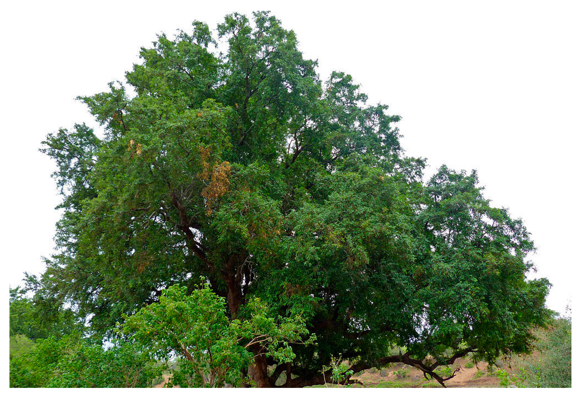
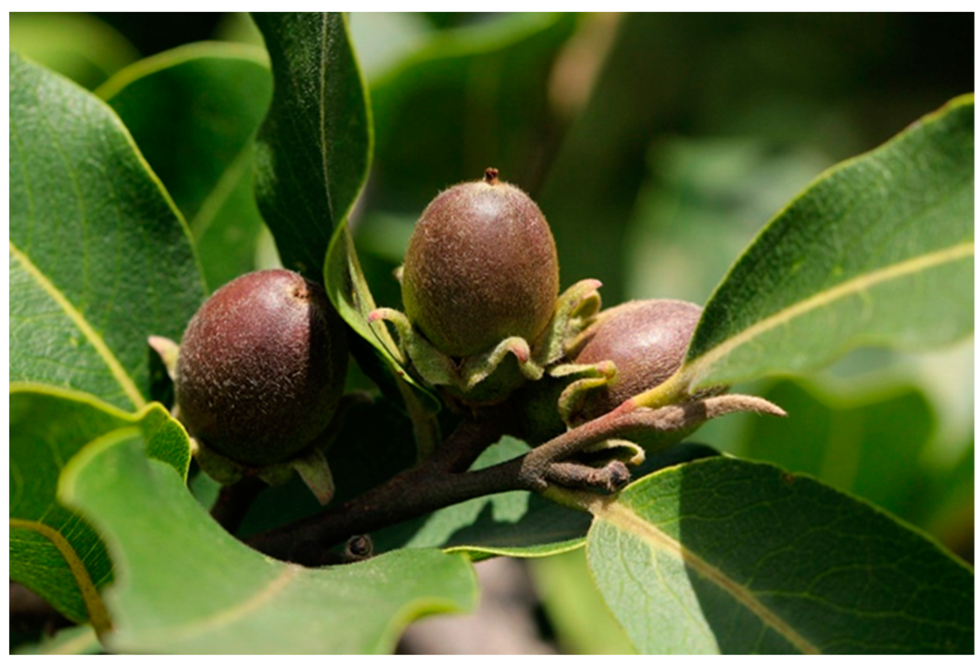
Traditional Uses, Pharmacological Activities, and Phytochemical Analysis of Diospyros mespiliformis Hochst. ex. A. DC (Ebenaceae): A Review

Abstract
:1. Introduction
2. Results and Discussion
2.1. Traditional Uses
2.2. Phytochemical Analysis
2.3. Secondary Metabolites
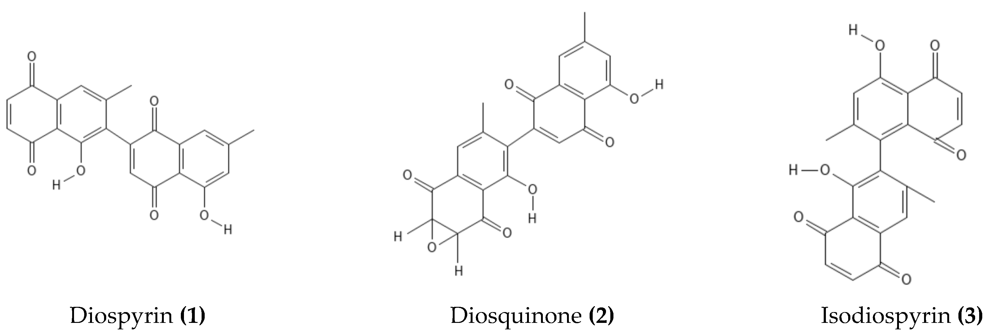
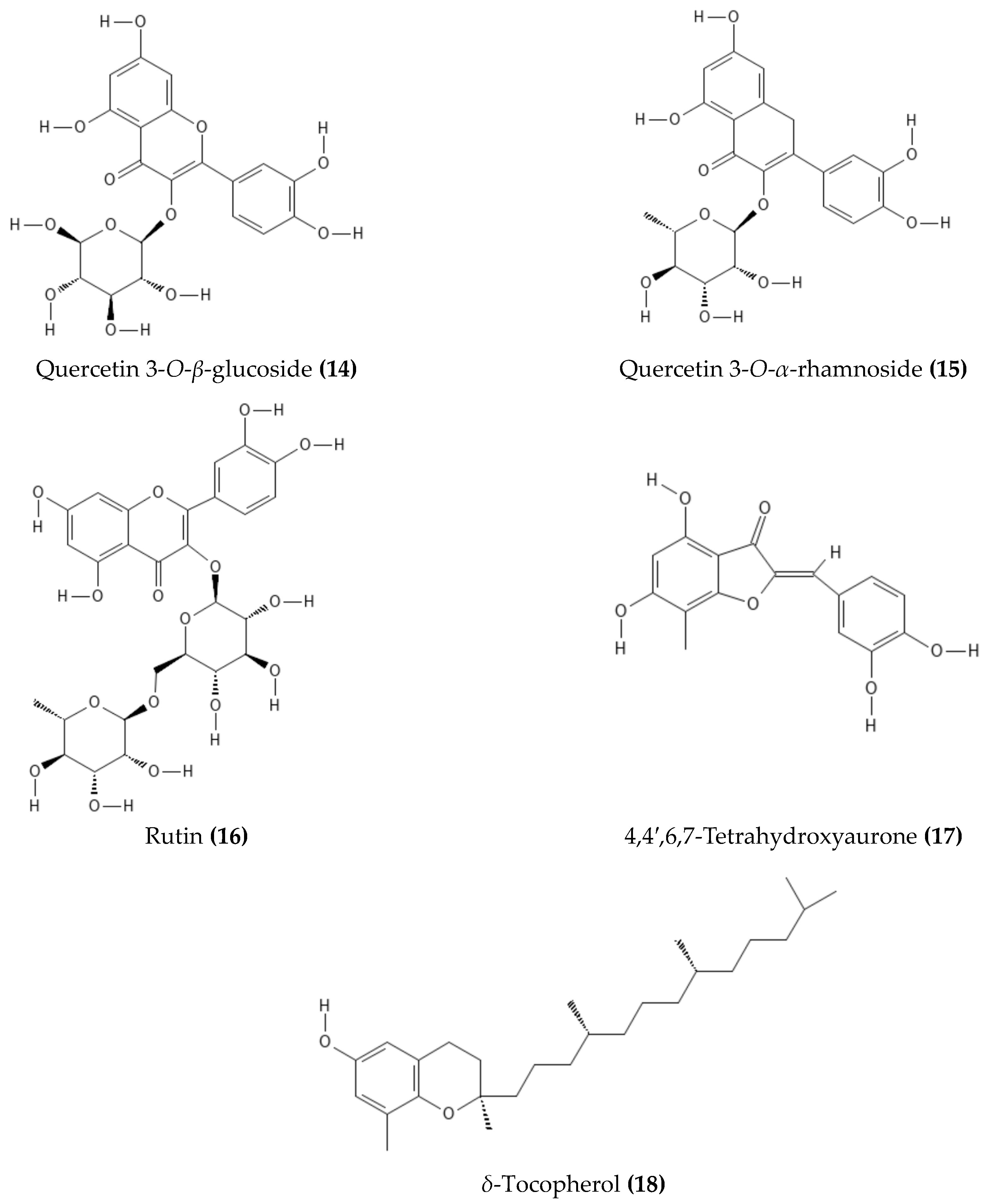
2.4. Isolated or Tentatively Identified Compounds from D. mespiliformis

| No. | Compounds | Plant Part | Detection/Isolation Method | Reference |
|---|---|---|---|---|
| 1 | Diospyrin | Stem bark or wood | Isolated | [61,62] |
| 2 | Diosquinone | Stem bark, wood, roots | Isolated | [60,61,62] |
| 3 | Isodiospyrin | Stem bark or wood | Isolated | [60,61] |
| 4 | Isoscutellarein 7-O-(4′′′-O-acetyl)-β-allopyranosyl (1′′′→2″)-β-glucopyranoside | Leaves | Isolated | [66] |
| 5 | Kaempferol | Stem bark | UPLC-ESI-MS | [12] |
| 6 | Luteolin | Leaves | Isolated | [66] |
| 7 | Luteolin 7-O-β-glucoside | Leaves | Isolated | [66] |
| 8 | Luteolin 4′-O-β-neohesperidoside | Leaves | Isolated | [66] |
| 9 | Luteolin 3′,4′,6,8-tetramethyl ether | Leaves | Isolated | [66] |
| 10 | 8-methoxy-3-methyl-1,2-naphthoquinone | Stem bark | UPLC-ESI-MS | [12] |
| 11 | Myricetin | Stem bark | UPLC-ESI-MS | [12] |
| 12 | Plumbagin | Stem bark, wood, roots | Isolated | [60,61,65] |
| 13 | Quercetin | Stem bark, leaves | Isolated, UPLC-ESI-MS | [12,66] |
| 14 | Quercetin 3-O-β-glucoside | Leaves | Isolated | [66] |
| 15 | Quercetin 3-O-α-rhamnoside | Leaves | Isolated | [66] |
| 16 | Rutin | Leaves | Isolated | [66] |
| 17 | 4,4′,6,7-Tetrahydroxyaurone | Stem bark | UPLC-ESI-MS | [12] |
| 18 | δ-Tocopherol | Stem bark | UPLC-ESI-MS | [12] |
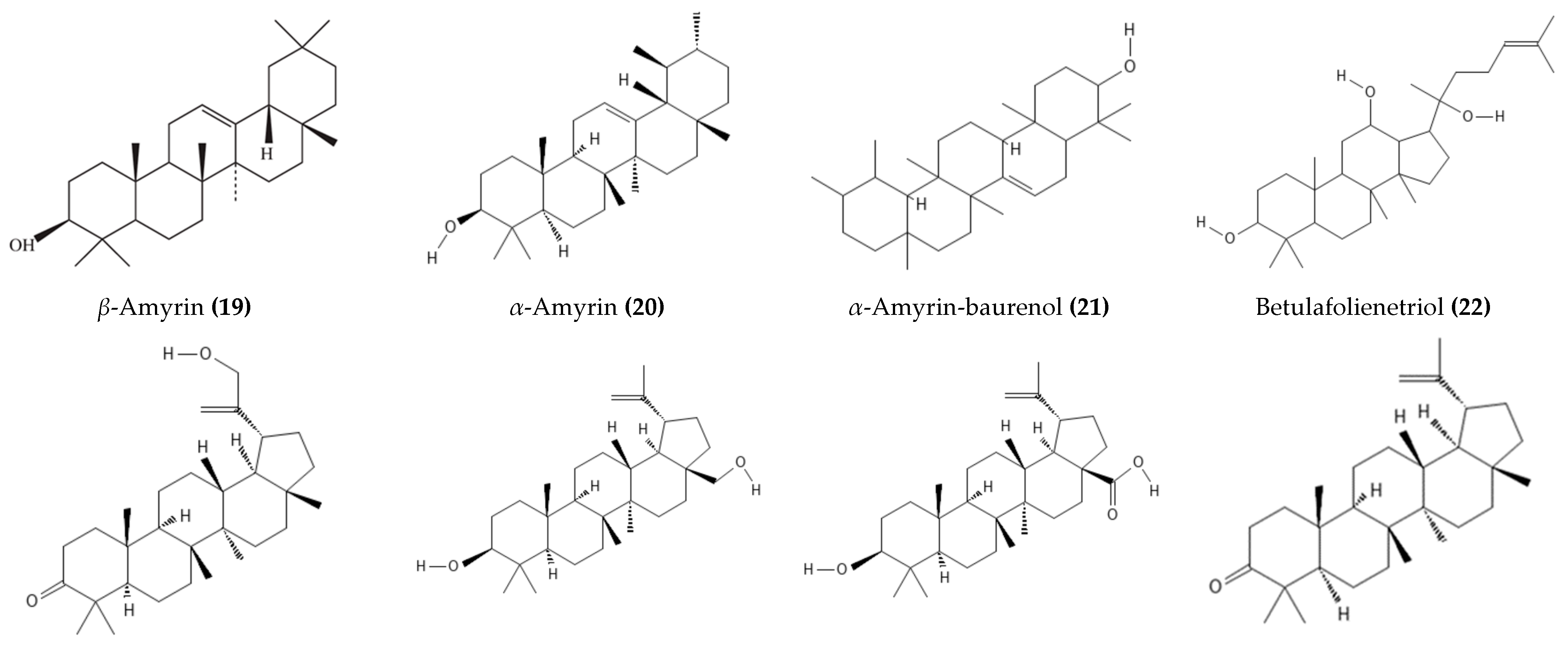
| 19 | β-Amyrin | Seeds | GC-MS | [54] |
| 20 | α-Amyrin | Stem bark or wood | Isolated | [41,60,61,63] |
| 21 | α-Amyrin-baurenol | Stem bark or wood | Isolated | [41,60,61,63] |
| 22 | Betulafolienetriol | Stem bark | UPLC-ESI-MS | [12] |
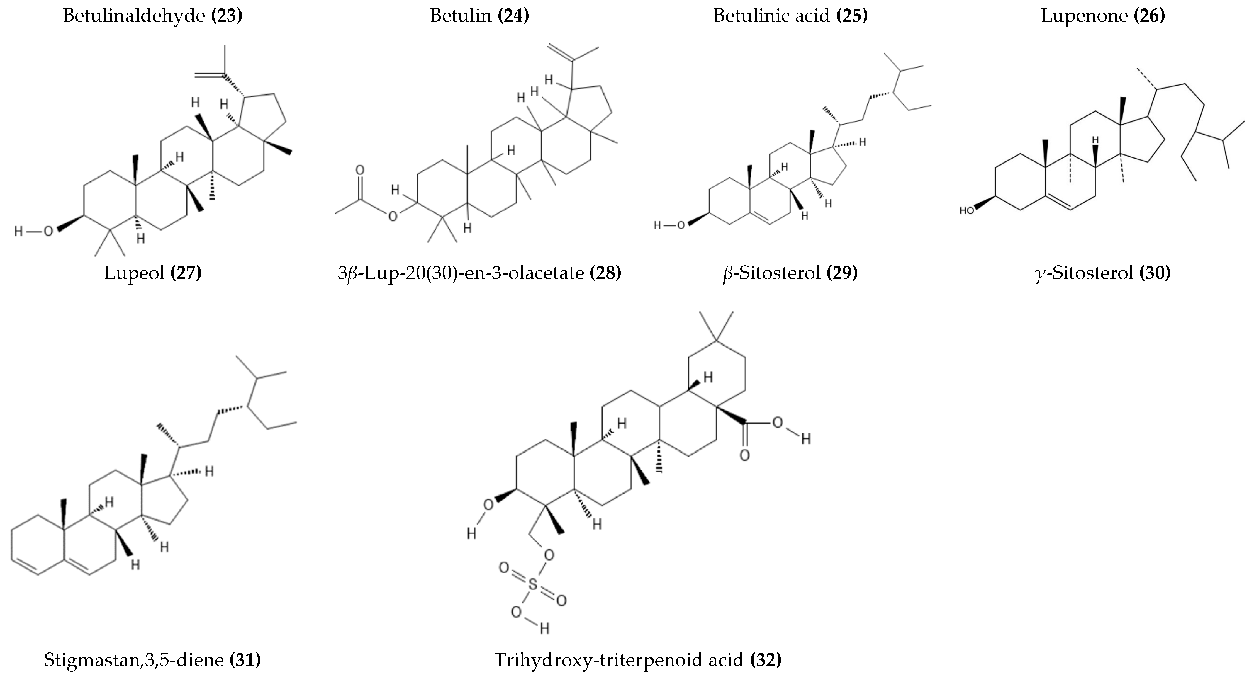
| 23 | 30-Hydroxylup-20(29)-en-3β-ol, betulinaldehyde | Stem bark | UPLC-ESI-MS | [12] |
| 24 | Betulin | Stem bark or wood | Isolated, GC-MS | [14,41,60,61,63] |
| 25 | Betulinic acid | Stem bark or wood | Isolated, UPLC-ESI-MS | [12,14,41,60,61,63] |
| 26 | Lupenone | Stem bark | Isolated | [14] |
| 27 | Lupeol | Stem bark or wood | Isolated, GC-MS | [11,13,14,41,60,61,63,64] |
| 28 | 3β-Lup-20(30)-en-3-olacetate | Wood stem | GC-MS | [11] |
| 29 | β-Sitosterol | Stem bark or wood | Isolated | [11,41,60,61,63] |
| 30 | γ-Sitosterol | Seeds | GC-MS | [54] |
| 31 | Stigmastan,3,5-diene | Wood stem | GC-MS | [11] |
| 32 | Trihydroxy-triterpenoid acid | Stem bark or wood | Isolated | [41,60,61,63] |
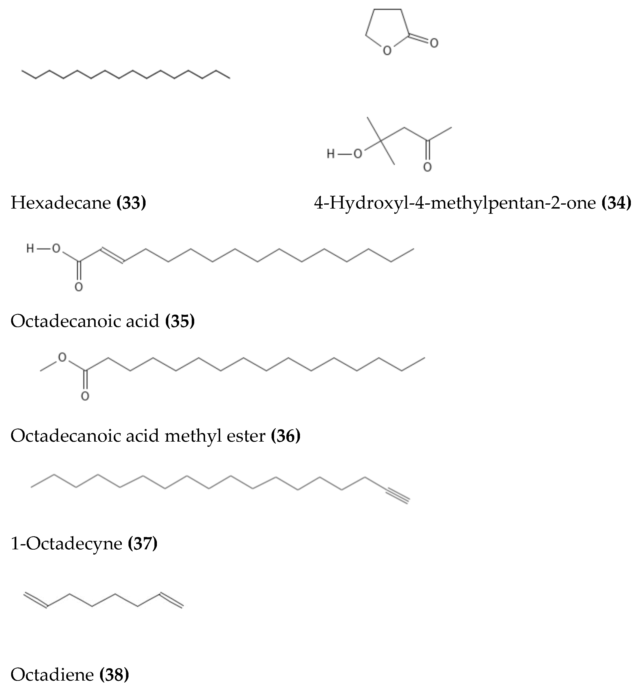
| 33 | Hexadecane | Seeds | GC-MS | [54] |
| 34 | 4-Hydroxyl-4-methylpentan-2-one | Leaves | GC-MS | [4] |
| 35 | Octadecanoic acid | Leaves, wood stem | GC-MS | [4,11] |
| 36 | Octadecanoic acid methyl ester | Wood stem | GC-MS | [11] |
| 37 | 1-Octadecyne | Leaves | GC-MS | [4] |
| 38 | Octadiene | Seeds | GC-MS | [54] |
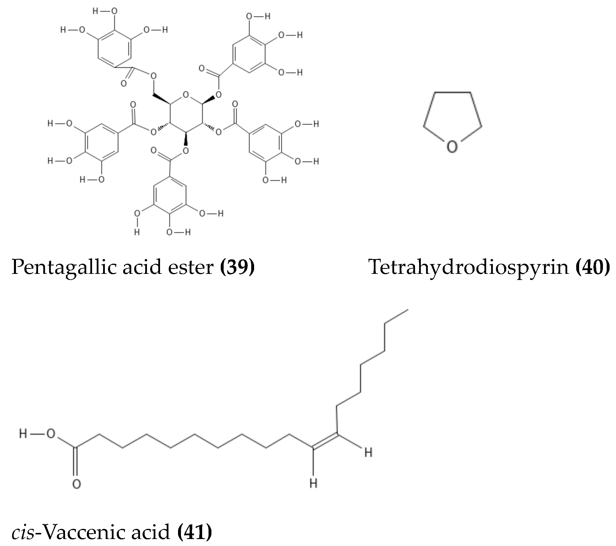
| 39 | Pentagallic acid ester of glucose | Stem bark | UPLC-ESI-MS | [12] |
| 40 | Tetrahydrodiospyrin | Stem bark | UPLC-ESI-MS | [12] |
| 41 | cis-Vaccenic acid | Wood stem | GC-MS | [11] |
2.5. Pharmacological Activity
2.5.1. Antimicrobial Activity
2.5.2. Anti-Inflammatory Activity
2.5.3. Antiparasitic Activity
2.5.4. Antidiabetic Activity
2.5.5. Antiviral Activity
2.5.6. Anti-Hypersensitivity
2.5.7. Antioxidant Activity
2.5.8. Antiproliferative Activity
2.5.9. In Vivo Studies
3. Materials and Methods
4. Conclusions and Future Perspectives
Author Contributions
Funding
Institutional Review Board Statement
Informed Consent Statement
Data Availability Statement
Conflicts of Interest
References
- Atta-U-Rahman, M.; Igbal, C.; Thomson, W.J. Bioassay Techniques for Drug Development, 2nd ed.; Taylor and Francis: Singapore, 2005. [Google Scholar]
- Gerard, J.; Louppe, D. Afzelia africana Sm. ex pers. (Internet) Record from PROTA4U; PROTA (Plant Resources of Tropical Africal/Ressources Vegetales de l’Afrique Tropicale): Wageningen, The Netherlands, 2011. [Google Scholar]
- Orwa, C.; Mutua, A.; Kindt, R.; Jamnadass, R.; Simons, A. Agroforestree Database: A Tree Reference and Selection Guide; Version 4; World Agroforestry Centre: Nairobi, Kenya, 2009. [Google Scholar]
- Dangoggo, S.M.; Hassan, L.G.; Sadiq, I.S.; Manga, S.B. Phytochemical analysis and antibacterial screening of leaves of Diospyros mespiliformis and Ziziphus spina-christi. J. Chem. Eng. 2012, 1, 31–37. [Google Scholar]
- Esimone, C.O.; Nworu, C.S.; Onuigbo, E.B.; Omeje, J.U.; Nsirim, K.L.; Ogbu, J.C.; Ngwu, M.I.; Chah, K.F. Anti-mycobacterial activity of root and leaf extracts of Anthocleista djalonensis (Loganiaceae) and Diospyros mespiliformis (Ebenaceae). Int. J. Green Pharm. 2009, 3, 201–205. [Google Scholar]
- Adeniyi, B.A.; Odelola, H.A.; Oso, B.A. Antimicrobial potentials of Diospyros mespiliformis (Ebenaceae). Afr. J. Med. Med. Sci. 1996, 25, 221–224. [Google Scholar]
- Mudau, T.E.; Olowoyo, J.O.; Amoo, S.O. Ethnobotanical assessment of medicinal plants used traditionally for treating diabetes in Vhembe district, Limpopo Province, South Africa. S. Afr. J. Bot. 2022, 146, 304–324. [Google Scholar] [CrossRef]
- Vandi, V.L.; Amang, A.P.; Mezui, C.; Siwe, G.T.; Ndji, G.L.; Mbida, H.; Baponwa, O.; Tan, P.V. Antihistaminergic and anticholinergic properties of the root bark aqueous extract of Diospyros mespiliformis (Ebenaceae) on hypersecretion of gastric acid induced in wistar rats. eCAM 2022, 2022, 5190499. [Google Scholar] [CrossRef] [PubMed]
- Mustapha, M.D.; Hassan, L.G.; Umar, K.J.; Abubakar, K.; Yusuf, H.M.A. Phytochemical screening and cytotoxic effects of Diospyros mespiliformis Hochst (Ebenaceae) root bark extract using Brine Shrimp (Artemia salina) Test. AJBAR 2022, 1, 1–7. [Google Scholar] [CrossRef]
- Maitera, O.N.; Louis, H.; Oyebanji, O.O.; Anumah, A.O. Investigation of tannin content in Diospyros mespiliformis extract using various extraction solvents. J. Anal. Pharm. Res. 2018, 7, 55–59. [Google Scholar]
- David, O.M.; Olanlokun, J.O.; Owoniyi, B.E.; Ayeni, M.; Ebenezer, O.; Koorbanally, N.A. Studies on the mitochondrial, immunological and inflammatory effects of solvent fractions of Diospyros mespiliformis Hochst in Plasmodium berghei-infected mice. Sci. Rep. 2021, 11, 6941. [Google Scholar] [CrossRef]
- Olanlokun, J.O.; Bodede, O.; Prinsloo, G.; Olorunsogo, O.O. Comparative antimalarial, toxicity and mito-protective effects of Diospyros mespiliformis Hochst. ex A. DC. and Mondia whitei (Hook. f.) Skeels on Plasmodium berghei infection in mice. J. Ethnopharmacol. 2021, 268, 113585. [Google Scholar] [CrossRef]
- Anas, A.; Ahmed, A.; Umar, S.; Jajere, U.M.; Mshelia, E.H.; Natasha, O. Inhibitory effect of isolated lupeol from stem bark of Diospyros mespiliformis Horsch (Ebenaceae) against some microbial pathogens. Bayero J. Pure Appl. Sci. 2017, 10, 293–299. [Google Scholar] [CrossRef]
- Mohamed, I.E.; El Bushra, E.; Choudhary, M.I.; Khan, S.N. Bioactive natural products from two Sudanese medicinal plants Diospyros mespiliformis and Croton zambesicus. Rec. Nat. Prod. 2009, 3, 198. [Google Scholar]
- Mabona, U.; Viljoen, A.; Shikanga, E.; Marston, A.; Van Vuuren, S. Antimicrobial activity of southern African medicinal plants with dermatological relevance: From an ethnopharmacological screening approach, to combination studies and the isolation of a bioactive compound. J. Ethnopharmacol. 2013, 148, 45–55. [Google Scholar] [CrossRef]
- Koenen, E.V. Medicinal, Poisonous and Edible Plants in Namibia; Klaus Hess Verlag: Göttingen, Germany, 1996. [Google Scholar]
- Watt, J.M.; Breyer-Brandwijk, M.G. The Medicinal and Poisonous Plants of Southern and Eastern Africa, 2nd ed.; Livingstone: Edinburg, UK; London, UK, 1962. [Google Scholar]
- Luseba, D.; Van der Merwe, D. Ethnoveterinary medicine practices among Tsonga speaking people of South Africa. Onderstepoort J. Vet. Res. 2006, 73, 115–122. [Google Scholar] [CrossRef] [PubMed]
- Mahwasane, S.T.; Middleton, L.; Boaduo, N. An ethnobotanical survey of indigenous knowledge on medicinal plants used by the traditional healers of the Lwamondo area, Limpopo province, South Africa. S. Afr. J. Bot. 2013, 88, 69–75. [Google Scholar] [CrossRef]
- Bapela, M.J.; Meyer, J.M.; Kaiser, M. In vitro antiplasmodial screening of ethnopharmacologically selected South African plant species used for the treatment of malaria. J. Ethnopharmacol. 2014, 156, 370–373. [Google Scholar] [CrossRef] [PubMed]
- Masevhe, N.A.; McGaw, L.J.; Eloff, J.N. The traditional use of plants to manage candidiasis and related infections in Venda, South Africa. J. Ethnopharmacol. 2015, 168, 364–372. [Google Scholar] [CrossRef]
- Tshikalange, T.E.; Mophuting, B.C.; Mahore, J.; Winterboer, S.; Lall, N. An ethnobotanical study of medicinal plants used in villages under Jongilanga tribal council, Mpumalanga, South Africa. Afr. J. Tradit. Complement. Altern. Med. 2016, 13, 83–89. [Google Scholar] [CrossRef] [PubMed]
- Tapsoba, H.; Deschamps, J.P. Use of medicinal plants for the treatment of oral diseases in Burkina Faso. J. Ethnopharmacol. 2006, 104, 68–78. [Google Scholar] [CrossRef] [PubMed]
- Maroyi, A. An ethnobotanical survey of medicinal plants used by the people in Nhema communal area, Zimbabwe. J. Ethnopharmacol. 2011, 136, 347–354. [Google Scholar] [CrossRef]
- Luka, J.; Badau, S.J.; Mbaya, A.W.; Gadzama, J.J.; Kumshe, H.A. Acute toxicity study and effect of prolonged administration (28 days) of crude ethanolic root extract of Diospyros mespiliformis Hochst (Ebenaceae) on clinical, haematological and biochemical parameters of albino rats. J. Ethnopharmacol. 2014, 153, 268–273. [Google Scholar] [CrossRef]
- Moyo, B.; Ndlovu, S.L.; Moyo, S.; Masika, P.J.; Muchenje, V.; Ndhlovu, D.N.; Maphosa, V. Alternative remedies and approaches used by resources-challenged farmers in the management of cattle black-leg disease in Umzingwane district, Matabeleland South, Zimbabwe. Int. J. Livest. Prod. 2014, 6, 97–102. [Google Scholar]
- Dossou-Yovo, H.O.; Vodouhe, F.G.; Sinsin, B. Assessment of the medicinal uses of plant species found on termitaria in the Pendjari biosphere reserve in Benin. J. Med. Plant Res. 2014, 8, 368–377. [Google Scholar]
- Koné, W.M.; Atindehou, K.K. Ethnobotanical inventory of medicinal plants used in traditional veterinary medicine in Northern Côte d’Ivoire (West Africa). S. Afr. J. Bot. 2008, 74, 76–84. [Google Scholar] [CrossRef]
- Chinsembu, K.C.; Hijarunguru, A.; Mbangu, A. Ethnomedicinal plants used by traditional healers in the management of HIV/AIDS opportunistic diseases in Rundu, Kavango East Region, Namibia. S. Afr. J. Bot. 2015, 100, 33–42. [Google Scholar] [CrossRef]
- Sulaiman, A.N.; Arzai, A.H.; Taura, D.W. Ethnobotanical survey: A comprehensive review of medicinal plants used in treatment of gastrointestinal diseases in Kano state, Nigeria. Phytomed. Plus. 2022, 2, 100180. [Google Scholar] [CrossRef]
- Setshego, M.V.; Aremu, A.O.; Mooki, O.; Otang-Mbeng, W. Natural resources used as folk cosmeceuticals among rural communities in Vhembe district municipality, Limpopo province, South Africa. BMC Complement. Med. 2020, 20, 81. [Google Scholar] [CrossRef]
- Chinsembu, K.C. Ethnobotanical study of medicinal flora utilised by traditional healers in the management of sexually transmitted infections in Sesheke District, Western Province, Zambia. Rev. Bras. Farmacogn. 2016, 26, 268–274. [Google Scholar] [CrossRef]
- Mamba, P.; Adebayo, S.A.; Tshikalange, T.E. Anti-microbial, Anti-inflammatory and HIV-1 reverse transcriptase activity of selected South African plants used to treat sexually transmitted diseases. Int. J. Pharmacogn. Phytochem. Res. 2016, 8, 1870–1876. [Google Scholar]
- Nadembega, P.; Boussim, J.I.; Nikiema, J.B.; Poli, F.; Antognoni, F. Medicinal plants in Baskoure, Kourittenga province, Burkina Faso: An ethnobotanical study. J. Ethnopharmacol. 2011, 133, 378–395. [Google Scholar] [CrossRef]
- Van Wyk, B.; Van Wyk, P.; Van Wyk, B.E. Photo Guide to Trees of Southern Africa; Briza: Pretoria, South Africa, 2008. [Google Scholar]
- Adeniyi, B.A.; Robert, M.F.; Chai, H.; Fong, H.H.S. In vitro cytotoxicity activity of diosquinone, a naphthoquinone epoxide. Phytother. Res. 2003, 17, 282–284. [Google Scholar] [CrossRef] [PubMed]
- Atawodi, S.E.; Bulus, T.; Ibrahim, S.; Ameh, D.A.; Nok, A.J.; Mamman, M.; Galadima, M. In vitro trypanocidal effect of methanolic extract of some Nigerian savannah plants. Afr. J. Biotechnol. 2003, 2, 317–321. [Google Scholar]
- Von Koenen, E. Medicinal, Poisonous and Edible Plants of Namibia; Klaus Hess Publishers: Windhoek, Namibia; Gottingen, Germany, 2001. [Google Scholar]
- Etkin, N.L. Antimalarial plants used by Hausa in Northern Nigeria. Trop. Dr. 1997, 27, 12–16. [Google Scholar] [CrossRef] [PubMed]
- Mabogo, D.E.N. The Ethnobotany of the Vhavenda. Master’s Thesis, University of Pretoria, Pretoria, South Africa, 1990. [Google Scholar]
- Khan, M.R.; Nkunya, M.H.H.; Wevers, H. Triterpenoids from leaves of Diospyros species. Planta Med. 1980, 38, 380–381. [Google Scholar] [CrossRef]
- Arnold, H.; Gulumian, M. Pharmacopoeia of traditional medicine in Venda. J. Ethnopharmacol. 1984, 12, 35–74. [Google Scholar] [CrossRef] [PubMed]
- Kerharo, J. Historic and ethnopharmacognostic review on the belief and traditional practices in the treatment of sleeping sickness in West Africa. Bull. Soc. Med. Afr. Black Lang. Fr. 1974, 19, 400. [Google Scholar]
- Kantati, Y.T.; Kodjo, K.M.; Dogbeavou, K.S.; Vaudry, D.; Leprince, J.; Gbeassor, M. Ethnopharmacological survey of plant species used in folk medicine against central nervous system disorders in Togo. J. Ethnopharmacol. 2016, 181, 214–220. [Google Scholar] [CrossRef] [PubMed]
- Ziblim, I.A.; Timothy, K.A.; Deo-Anyi, E.J. Exploitation and use of medicinal plants, Northern Region, Ghana. J. Med. Plant Res. 2013, 7, 1984–1993. [Google Scholar]
- Klotoé, J.R.; Dougnon, T.V.; Koudouvo, K.; Atègbo, J.M.; Loko, F.; Akoègninou, A.; Aklikokou, K.; Dramane, K.; Gbeassor, M. Ethnopharmacological survey on antihemorrhagic medicinal plants in South of Benin. Eur. J. Med. Plants 2012, 3, 40–51. [Google Scholar] [CrossRef]
- Nfi, A.N.; Mbanya, J.N.; Ndi, C.; Kameni, A.; Vabi, M.; Pingpoh, D.; Yonkeu, S.; Moussa, C. Ethnoveterinary medicine in the Northern Provinces of Cameroon. Vet. Res. Commun. 2001, 25, 71–76. [Google Scholar] [CrossRef]
- Semenya, S.S.; Maroyi, A. Ethnobotanical survey of plants used by Bapedi traditional healers to treat tuberculosis and its opportunistic infections in the Limpopo Province, South Africa. S. Afr. J. Bot. 2019, 122, 401–421. [Google Scholar] [CrossRef]
- Cheikhyoussef, A.; Shapi, M.; Matengu, K.; Mu Ashekele, H. Ethnobotanical study of indigenous knowledge on medicinal plant use by traditional healers in Oshikoto region, Namibia. J. Ethnobiol. Ethnomedicine 2011, 7, 10. [Google Scholar] [CrossRef]
- Green, E.; Samie, A.; Obi, C.L.; Bessong, P.O.; Ndip, R.N. Inhibitory properties of selected South African plants against Mycobacterium tuberculosis. J. Ethnopharmacol. 2010, 130, 151–157. [Google Scholar] [CrossRef]
- Kokwaro, J.O. Medicinal Plants of East Africa; University of Nairobi Press: Nairobi, Kenya, 2009. [Google Scholar]
- Stafford, G.I.; Pedersen, M.E.; van Staden, J.; Jager, A.K. Review on plants with CNS-effects used in traditional South African medicine against mental diseases. J. Ethnopharmacol. 2008, 119, 513–537. [Google Scholar] [CrossRef] [PubMed]
- Chivandi, E.; Erlwanger, K.H.; Davidson, B.C. Lipid content and fatty acid profile of the fruit seeds of Diospyros mespiliformis. Int. J. Integr. Biol. 2009, 5, 121–124. [Google Scholar]
- Hegazy, A.K.; Mohamed, A.A.; Ali, S.I.; Alghamdi, N.M.; Abdel-Rahman, A.M.; Al-Sobeai, S. Chemical ingredients and antioxidant activities of underutilized wild fruits. Heliyon 2019, 5, e01874. [Google Scholar] [CrossRef] [PubMed]
- Ebbo, A.A.; Sani, D.; Suleiman, M.M.; Ahmad, A.; Hassan, A.Z. Assessment of antioxidant and wound healing activity of the crude methanolic extract of Diospyros mespiliformis Hochst ex a. Dc (Ebenaceae) and its fractions in Wistar rats. S. Afr. J. Bot. 2022, 150, 305–312. [Google Scholar] [CrossRef]
- Adewuyi, A.; Oderinde, R.A. Fatty acid composition and lipid profile of Diospyros mespiliformis, Albizia lebbeck, and Caesalpinia pulcherrima seed oils from Nigeria. Int. J. Food Sci. 2014, 2014, 283614. [Google Scholar] [CrossRef]
- Petzke, K.J.; Ezeagu, I.E.; Proll, J.; Akinsoyinu, A.O.; Metges, C.C. Amino acid composition, available lysine content and in vitro protein digestibility of selected tropical crop seeds. Foods Hum. Nutr. 1997, 50, 151–162. [Google Scholar] [CrossRef]
- Glew, R.S.; Vanderjagt, D.J.; Chuang, L.T.; Huang, Y.S.; Millson, M.; Glew, R.H. Nutrient content of four edible wild plants from West Africa. Foods Hum. Nutr. 2005, 60, 187–193. [Google Scholar] [CrossRef]
- Achaglinkame, M.A.; Aderibigbe, R.O.; Hensel, O.; Sturm, B.; Korese, J.K. Nutritional characteristics of four underutilized edible wild fruits of dietary interest in Ghana. Foods 2019, 8, 104. [Google Scholar] [CrossRef]
- Zhong, S.M.; Waterman, P.G.; Jeffreys, J.A.D. Naphthoquinones and triterpenes from African Diospyros species. Phytochemistry 1984, 23, 1067–1072. [Google Scholar] [CrossRef]
- Adzu, B.; Amos, S.; Dzarma, S.; Muazzam, I.; Gamaniel, K.S. Pharmacological evidence favouring the folkloric use of Diospyros mespiliformis Hochst in the relief of pain and fever. J. Ethnopharmacol. 2002, 82, 191–195. [Google Scholar] [CrossRef]
- Adeniyi, B.A.; Fong, H.H.S.; Pezzuto, J.M.; Luyengi, L.; Odelola, H.A. Antibacterial activity of diospyrin, isodiospyrin and bisisodiospyrin from the root of Diospyros piscatoria (Gurke)(Ebenaceae). Phytother. Res. 2000, 14, 112–117. [Google Scholar] [CrossRef]
- Fallas, A.L.; Thomson, R.H. Ebenaceae extractives. Part III. Binaphthaquinones from Diospyros species. J. Chem. Society C Org. 1968, 2279–2282. [Google Scholar] [CrossRef]
- Lajubutu, B.A.; Pinney, R.J.; Roberts, M.F.; Odelola, H.A.; Oso, B.A. Antibacterial activity of diosquinone and plumbagin from the root of Diospyros mespiliformis (Hostch)(Ebenaceae). Phytother. Res. 1995, 9, 346–350. [Google Scholar] [CrossRef]
- Hawas, U.W.; El-Ansari, M.A.; El-Hagrassi, A.M. A new acylated flavone glycoside, in vitro antioxidant and antimicrobial activities from Saudi Diospyros mespiliformis Hochst. ex A. DC (Ebenaceae) leaves. ZNC 2022, 77, 387–393. [Google Scholar] [CrossRef] [PubMed]
- Shai, L.J.; Chauke, M.A.; Magano, S.R.; Mogale, A.M.; Eloff, J.N. Antibacterial activity of sixteen plant species from Phalaborwa, Limpopo Province, South Africa. J. Med. Plants Res. 2013, 7, 899–906. [Google Scholar]
- Aderbauer, B.; Clausen, P.H.; Kershaw, O.; Melzig, M. In vitro and in vivo trypanocidal effect of lipophilic extracts of medicinal plants from Mali and Burkina Faso. J. Ethnopharmacol. 2008, 119, 225–231. [Google Scholar] [CrossRef]
- Hedimbi, M. Evaluation of Selected Namibian Ethno-Medicinal Plants for Anti-HIV Properties. Ph.D. Thesis, University of Namibia, Windhoek, Namibia, 2015. [Google Scholar]
- Traore, M.S.; Diane, S.; Diallo, M.S.T.; Balde, E.S.; Balde, M.A.; Camara, A.; Diallo, A.; Keita, A.; Cos, P.; Maes, L.; et al. In vitro antiprotozoal and cytotoxic activity of ethnopharmacologically selected Guinean plants. Planta Med. 2014, 80, 1340–1344. [Google Scholar] [CrossRef]
- Chukwuma, O.J.T. Antiviral activities of the aqueous, ethanolic and methanolic extracts of Diospyros mespiliformis leaf on some pathogenic Avian viruses. IDOSR J. Exp. Sci. 2017, 2, 35–49. [Google Scholar]
- Belemtougri, R.G.; Constantin, B.; Cognard, C.; Raymond, G.; Sawadogo, L. Effects of two medicinal plants Psidium guajava L. (Myrtaceae) and Diospyros mespiliformis L. (Ebenaceae) leaf extracts on rat skeletal muscle cells in primary culture. J. Zhejiang Univ. Sci. B 2006, 7, 56–63. [Google Scholar] [CrossRef]
- Lawal, F.; Bapela, M.J.; Adebayo, S.A.; Nkadimeng, S.M.; Yusuf, A.A.; Malterud, K.E.; McGaw, L.J.; Tshikalange, T.E. Anti-inflammatory potential of South African medicinal plants used for the treatment of sexually transmitted infections. S. Afr. J. Bot. 2019, 125, 62–71. [Google Scholar] [CrossRef]
- Nafuka, S.N. In Vitro Antiplasmodial Activity and Phytochemicals Screening of Ethnomedicinal Plants Used to Treat Malaria Associated Symptoms. Ph.D. Thesis, University of Namibia, Windhoek, Namibia, 2014. [Google Scholar]
- Adoum, O.A. Screening of medicinal plants native to Kano and Jigawa states of northern Nigeria, using Artemia cysts (brine shrimp test). Am. J. Pharmacol. Sci. 2016, 4, 7–10. [Google Scholar]
- Amang, A.P.; Bouvourne, P.; Mezui, C.; Siwe, G.T.; Kuissu, M.T.; Vernyuytan, P. Gastro-protective activity of the leaves aqueous extract of Diospyros mespiliformis on gastric ulcers in Swiss mice. Int. J. Pharmacogn 2020, 7, 44–51. [Google Scholar]
- Bapela, M.J.; Kaiser, M.; Meyer, J.J.M. Antileishmanial activity of selected South African plant species. S. Afr. J. Bot. 2017, 108, 342–345. [Google Scholar] [CrossRef]
- Agbadoronye, P.; Abolarinwa, S.; Lawal, B.; Odeyemi, S.; Irhue, A.; Achagwa, S.; Ngamdu, S. Alkaloidal fraction of Diospyros mespiliformis protect against Trypanosoma evansi-mediated haematological and hepatic impairment in infected rats. J. Appl. Nat. Sci. 2021, 1, 66–78. [Google Scholar]
- Olanlokun, J.O.; Adetutu, J.A.; Olorunsogo, O.O. ln vitro inhibition of beta-hematin formation and in vivo effects of Diospyros mespiliformis and Mondia whitei methanol extracts on chloroquine-susceptible Plasmodium berghei-induced malaria in mice. Interv. Med. Appl. Sci. 2021, 11, 197–206. [Google Scholar] [CrossRef] [PubMed]
- Jigam, A.A.; Abdulrazaq, U.T.; Suleiman, R.S.; Kali, P.S. Effects of sub-chronic administration of Diospyros mespiliformis Hochst (Ebenaceae) root extracts on some biochemical parameters in mice. J. Appl. Pharm. Sci. 2012, 2, 60–64. [Google Scholar] [CrossRef]
- Adzu, B.; Chindo, B.A.; Tarfa, F.D.; Salawu, O.A.; Igoli, O.J. Isolation and analgesic property of lupeol from Diospyros mespiliformis stem bark. J. Med. Plant Res. 2015, 9, 813–819. [Google Scholar]
- Chinwe, U.J.; Adedotun, A.A.; Mudi, S.Y.; Musa, H.; Oluwagbemiga, A.O.; Babatunde, A. In-vivo therapeutic efficacy and phytochemical investigation of three commonly used plants for malaria treatment by the Hausa community in Kano, Nigeria. J. Pharmacogn. Phytochem. 2021, 10, 111–117. [Google Scholar]
- Nwaogu, J.; Fakai, I.M.; Yahaya, I. Hepatoprotective effect of methanol stem bark extract of Diospyros mespiliformis against carbon tetrachloride induced liver damage. Asian J. Biochem. 2022, 12, 18–25. [Google Scholar] [CrossRef]
- Mallavadhani, U.V.; Panda, A.K.; Rao, Y.R. Pharmacology and chemotaxonomy of Diospyros. Phytochemistry 2007, 49, 901–951. [Google Scholar] [CrossRef] [PubMed]
- Chantal, M.M.B.; Josiane, N.M.T.; Baudelaire, N.E.; Nicolas, N.Y. Antioxidant properties of granulometric classes and solvent extraction of Diospyros mespiliformis Hochst. ex A. fruits powder. Int. J. Mod. Biol. Res. 2021, 9, 1–16. [Google Scholar]
- Ndhlala, A.R.; Chitindingu, K.; Mupure, C.; Murenje, T.; Ndhlala, F.; Benhura, M.A.; Muchuweti, M. Antioxidant properties of methanolic extracts from Diospyros mespiliformis (jackal berry), Flacourtia indica (Batoka plum), Uapaca kirkiana (wild loquat) and Ziziphus mauritiana (yellow berry) fruits. Int. J. Food Sci. Technol. 2008, 43, 284–288. [Google Scholar] [CrossRef]
- Shikwambana, N.; Mahlo, S.M. A survey of antifungal activity of selected south African plant species used for the treatment of skin infections. Nat. Prod. Commun. 2020, 15, 1934578X20923181. [Google Scholar] [CrossRef]
- Sombie, E.N.; Tibiri, A.; N’do, J.Y.P.; Traore, T.K.; Ouedraogo, N.; Hilou, A.; Guissou, P.I.; Nacoulma, O.G. Ethnobotanical study and antioxidant activity of anti-hepatitis plants extracts of the COMOE province, Burkina Faso. Int. J. Biol. Chem. Sci. 2018, 12, 1308–1319. [Google Scholar] [CrossRef]
- Ebbo, A.A.; Sani, D.; Suleiman, M.M.; Ahmad, A.; Hassan, A.Z. Acute and sub-chronic toxicity evaluation of the crude methanolic extract of Diospyros mespiliformis hochst ex a. Dc (ebenaceae) and its fractions. Toxicol. Rep. 2020, 7, 1138–1144. [Google Scholar] [CrossRef]


| Part Used | Traditional Uses | Country | References |
|---|---|---|---|
| Leaves | Ringworm, urinary, and sexually transmitted infections, sleeping sickness, malaria, headaches, anthelmintic, wounds, dysentery, fever, leprosy, scars, skin rashes, bruises, styptic to staunch bleeding, diarrhoea, tonic, febrifuge, stomach aches, and coughs | South Africa, Ivory Coast, Nigeria, Zambia, Burkina Faso, Namibia | [15,16,17,28,30,31,32,33,34,35,36,37,38,39,40,41,42,43] |
| Stem | Blackleg disease in cattle, diabetes mellitus, stroke, traumatic brain injury, and malaria | South Africa, Zimbabwe, Burkina Faso, Togo | [7,26,34,44] |
| Bark | Oral diseases, stomach problems, diarrhoea, coughs, leprosy, STIs and urinary tract infections, dysentery, fever, vomiting, pneumonia, syphilis, and hemorrhages. Ethnoveterinary: Helminthiasis, milk production in animals, mental illness, headaches, epilepsy, and convulsions | South Africa, Burkina Faso, Nigeria, Tanzania, Senegal, Ivory Coast, Ghana, Benin, Cameroon | [18,19,23,39,40,41,42,43,45,46,47] |
| Roots | Ringworm, urinary, and sexually transmitted infections, abdominal pains, stomach aches, tuberculosis, male sexual dysfunction, scars, skin rashes, bruises, wounds, ringworm, dysentery, fever, coughs, epilepsy, pneumonia, syphilis, mental illness, headaches, epilepsy, convulsions, and worm expellant | South Africa, Namibia, Zimbabwe, Nigeria, Ghana, Kenya | [15,16,17,24,33,35,36,38,40,45,48,49,50,51,52] |
| Fruits | Dysentery, fungal infections, diarrhoea, tonic, febrifuge, skin diseases, menstrual pain, and ringworms | South Africa, Benin, Burkina Faso | [21,27,28,31,34,40] |
| Twigs | Teeth cleaning | South Africa | [31] |
| Plant Part/Compounds | Solvents Used | Pharmacological Activity | Bioassay Model | Results | References |
|---|---|---|---|---|---|
| Leaves | Acetone | Antioxidant | DPPH | IC50 = 25 ± 2 μg/mL | [55] |
| Antibacterial | MIC (B. stearothermophilus) | 80 µg/mL | [65] | ||
| Antifungal | MIC (C. albicans and M. canis and T. rubrum) | 80 µg/mL for C. albicans, 20 µg/mL for M. canis and 20 µg/mL for T. rubrum | [66] | ||
| DCM | Antiproliferative | In vitro cytotoxicity | MTC > 500 µg/mL on fibroblast-like mammalian cells | [67] | |
| DCM: MeOH | Antibacterial | MIC (P. acnes ATCC 11827 and T. mentagrophytes) | 50 µg/mL for P. acnes and 100 µg/mL for T. mentagrophytes. | [15] | |
| Antiparasitic | Long-term viability assay (T. brucei) | MIC = 500 µg/mL | [67] | ||
| 70% Ethanol | Antimicrobial | MIC (C. albicans ATCC 10231, G. vaginalis ATCC 14018, N. gonorrhoeae ATCC 19424 and O. ureolytica ATCC 43534) | 3.1–6.3 mg/mL | [33] | |
| Antiviral | HIV-1 RT colorimetric ELISA kit (Roche) | 78.7% at 0.1 mg/mL had | [68] | ||
| Antiparasitic | In vitro antiplasmodial activity | IC50 = 25.8 µg/mL for Trypanosoma cruzi, IC50 = >64 µg/mL for Leishmania infantum | [69] | ||
| Antiproliferative | In vitro cytotoxicity | IC50 = >64 µg/mL for MRC-5 fibroblasts | [69] | ||
| Ethanol | Antiviral | In vitro allantoic sac routes of developing chick embryos | 95.0%, 90.5%, and 89.0% at 400 mg/mL, 200 mg/mL, and 100 mg/mL respectively, for Newcastle disease virus | [70] | |
| Anti-hypersensitivity | Intracellular free calcium measurements | Reduced amplitude of Ca2+ release from SR at 10 mg/mL. IC50 = 9.23 mg/mL and 54% inhibited calcium release. | [71] | ||
| MeOH | Antioxidant | DPPH | IC50 = 6.94 ± 0.49 µg/mL | [72] | |
| Antimycobacterial | MIC (M. smegmatis) | 167 µg/mL | [5] | ||
| Antiparasitic | In vitro antiplasmodial bioassay | IC50 = 1.51 µg/mL for P. falciparum 3D7A | [73] | ||
| Antiviral | In vitro allantoic sac routes of developing chick embryos | 100.0%, 92.8%, and 90.5% at 400 mg/mL for Newcastle disease virus | [70] | ||
| Toxicity | Acute and subchronic toxicity in rats | LD50 of >5g/kg. No notable adverse effects seen on parameters studied | [72] | ||
| Water | Antioxidant | ABTS, FRAP | 1.17 ± 0.00 TEAC in mM (ABTS). 70.77 ± 0.4 M ET/g | [74] | |
| Antifungal | MIC (C. albicans, and T. rubrum) | 20 µg/mL for C. albicans, 40 µg/mL for T. rubrum. | [66] | ||
| Antiparasitic | In vitro antiplasmodial bioassay (P. falciparum 3D7A) | IC50 = 3.01 µg/mL | [73] | ||
| Antiviral | In vitro allantoic sac routes of developing chick embryos | 91.0%, 86.0%, and 85.0% at 400 mg/mL, 200 mg/mL, and 100 mg/mL, respectively, for Newcastle disease virus | [70] | ||
| Anti-hypersensitivity | Intracellular free calcium measurements | IC50 = 8.84 mg/mL at 10 mg/mL. 29% inhibited calcium release. | [71] | ||
| Toxicity | Gastroprotective efficacy: stomach ulcer | 200 mg/kg had the highest level of ulcer inhibition (88.13%) | [75] | ||
| Leaf fractions | Butanol | Antioxidant | DPPH | IC50 = 1.44 ± 0.01 µg/mL | [55] |
| Hexane | Antioxidant | DPPH | IC50 = 28.03 ± 2.57 µg/mL | [55] | |
| Ethyl acetate | Antioxidant | DPPH | IC50 = 1.08 ± 0.04 µg/mL | [55] | |
| Water | Antioxidant | DPPH | IC50 = 4.73 ± 0.23 µg/mL | [55] | |
| Roots | DCM: 50% MeOH | Antiparasitic | In vitro hypoxanthine incorporation assay (P. falciparum NF54) | IC50 = 4.40/28.4 µg/mL | [20] |
| Antileishmanial, resazurin assay (L. donovani MHOM-ET-67/L82) | IC50 = 7.7 µg/mL for DCM and IC50 = 54 µg/mL for 50% MeOH. | [76] | |||
| Antiproliferative | In vitro inhibition of mammalian cell proliferation | IC50 = 24.3 µg/mL for DCM and 60.4 µg/mL for MeOH | [77] | ||
| Ethanol | Antiparasitic | Acute toxicity and prolonged administration in rats | Intraperitoneal LD50 of 570 mg/kg | [25] | |
| 70% Ethanol | Anti-inflammatory | 15-LOX | IC50 = 188.1 µg/mL | [33] | |
| Antiviral | HIV-1 RT colorimetric ELISA kit (Roche) | 17.4% inhibition | [33] | ||
| MeOH | Antioxidant | DPPH | IC50 = 3.47 ± 0.05 µg/mL | [55] | |
| Antiparasitic | In vitro antiplasmodial bioassay (P. falciparum 3D7A) | IC50 = 2.12 µg/mL | [73] | ||
| In vivo antiplasmodial activity in mice (Plasmodium berghei) | High rate of parasite clearance (84.7%) and lower parasitemia (0.67%) | [78] | |||
| Toxicity | Subchronic in vivo studies | Safe dose of 400mg/Kg bw and LD50 of 620mg/kg bw of mice. | [79] | ||
| Water | Antibacterial | Disc diffusion (S. aureus, P. aeruginosa, E. coli and Shigella spp). | 10–13 mm on S. aureus, 11–13 mm on P. aeruginosa, 11–14 mm on E. coli and 10–11 mm on Shigella spp. | [4] | |
| Antiparasitic | In vitro antiplasmodial bioassay (P. falciparum 3D7A) | IC50 = 2.91 µg/mL | [73] | ||
| Root bark | Acetone | Anti-inflammatory | XO, NO | IC50 = 142 8 µg/mL (XO) and IC50 = 79.8 ± 2.7 µg/mL (NO) | [80] |
| Hexane | Antiproliferative | Brine shrimp (Artemia salina) cytotoxicity | 8203.52 μg/mL lethal dose | [9] | |
| Water | Antiproliferative | Brine shrimp (Artemia salina) cytotoxicity | 100% safe at 10–1000 μg/mL | [9] | |
| Antioxidant | ABTS, DPPH and FRAP | IC50 = 220 µg/mL for ABTS, 494 µg/mL DPPH and 543 µg/mL | [8] | ||
| Antisecretory mechanism | Pyloric ligation, pyloric ligation plus histamine, and carbachol pretreatments | Increased mucus mass and stomach ulcer inhibition ranging from 9.50% to 59.52% | [8] | ||
| Bark | 95% Ethanol | Antiparasitic | In vivo antitrypanosomal activity of Trypanosoma evansi-infected rats | Increased red blood cells and elevated bilirubin. Reduced total proteins | [77] |
| Hexane | Antimycobacterial | MIC (M. tuberculosis H37Ra) | 100 µg/mL | [50] | |
| MeOH | Antioxidant | DPPH | IC50 = 7.82 ± 0.76 µg/mL | [55] | |
| Antiparasitic | In vivo antiplasmodial activity in mice (Plasmodium berghei NK65) | 53% at 800 mg/kg dosage | [81] | ||
| Bark fractions | Ethyl acetate and hexane | Anti-inflammatory | Wound healing | Fully healed | [55] |
| Stem | Ethanol | Antiproliferative | Brine shrimp (Artemia cysts) lethality test (BST) | LC50 >100 μg/mL | [79] |
| Stem bark | MeOH | Antipyretic | In vivo studies | LD50 = 513.80 ± 33.92 mg/kg i.p. in mice. | [63] |
| Antiparasitic | In vivo antiplasmodial activity against P. berghei ANKA in mice | Parasitemia (5 ± 1), increased packed cell volume (36% ± 1.4), increased platelets (2 ± 1.4 105 mm3), decreased alkaline phosphatase (56 ± 0.7 U/L), alanine aminotransferases (6.2 ± 0.8 U/L), and alanine aminotransferases (8 ± 3.8 U/L). | [12] | ||
| Toxicity | Acute toxicity and hepatoprotective effects | LD50 > 5000 mg/kg bw. Possess hepatoprotective property by inhibiting lipid peroxidation. | [82] | ||
| Water | Neuropharmacological | In vivo studies in mice | Increased pentobarbital-induced sleep, decreased exploratory and spontaneous motor behavior | [83] | |
| Stem fractions | DCM | Antiparasitic | In vivo antiplasmodial activity against P. berghei NK 65 in mice | High parasite clearance | [11] |
| Fruits | Ethanol | Antioxidant | DPPH. In vivo antioxidants in rats. | IC50 = 1.037 ± 0.204 mg/mL. Increased the levels of the enzymes SOD, catalase, peroxidase, alanine transaminase, and aspartate aminotransferase | [84] |
| Hydroethanolic | Antioxidant | DPPH | IC50 = 1.111 ± 0.135 mg/mL | [84] | |
| MeOH | Antioxidant | DPPH radical scavenging, reducing power effects, and superoxide-anion-radical scavenging. | Increase radical-scavenging effect, reducing power and superoxide-anion-radical-scavenging. | [85] | |
| Antioxidant | DPPH, H2O2 scavenging | 87.36% at 1 mg/mL for DPPH. >85% at 1 mg/m for H2O2. | [58] | ||
| Isoscutellarein 7-O-(4′′′-O-acetyl)-β-allopyranosyl (1′′′ → 2″)-β-glucopyranoside (8) | Antioxidant | DPPH | IC50 = 15.46 μg/mL | [65] | |
| Luteolin 3′,4′,6,8-tetramethyl ether (9) | Antimycobacterial | MIC (M. smegmatis) | 250 µg/mL | [5] | |
| Antibacterial | Disc diffusion (E. coli) | 34 mm | [65] | ||
| Antibacterial | MIC (S. aureus) | 9.77 μg/mL | [65] | ||
| Antioxidant | DPPH | IC50 = 15.46 μg/mL | [65] | ||
| Quercetin 3-O-α-rhamnoside (15) | Antibacterial | MIC (S. aureus NCTC 6571, S. aureus E3T, E. coli KL16 and P. aeruginosa NCTC 6750) | 3 to 30 μg/mL for S. aureus, 15 for E. coli and 16 μg/mL for P. aeruginosa | [64] | |
| Antioxidant | DPPH | IC50 = 12.32 μg/mL | [65] | ||
| Diosquinone (2) | Antiproliferative | In vitro cytotoxicity | ED50 = 0.18 μg/mL for U373 cells, ED50 = 0.2 µg/mL for BC-1, HT-1080, Lu-1, KB, and SKNSH cells, ED50 = 1–1.7 µg/mL for KB-V(V-VLB) cells | [36] | |
| Plumbagin (12) | Antidiabetic | α- Glucosidase enzyme inhibition assay | IC50 = 0.002 ± 0.004 mM | [14] | |
| Lupeol (27) | Antidiabetic | α- Glucosidase enzyme inhibition assay | IC50 = 0.46 ± 0.002 mM | [14] | |
| Betulin (24) | Antidiabetic | α- Glucosidase enzyme inhibition assay | IC50 = 0.0624 ± 0.002 mM | [14] |
Disclaimer/Publisher’s Note: The statements, opinions and data contained in all publications are solely those of the individual author(s) and contributor(s) and not of MDPI and/or the editor(s). MDPI and/or the editor(s) disclaim responsibility for any injury to people or property resulting from any ideas, methods, instructions or products referred to in the content. |
© 2023 by the authors. Licensee MDPI, Basel, Switzerland. This article is an open access article distributed under the terms and conditions of the Creative Commons Attribution (CC BY) license (https://creativecommons.org/licenses/by/4.0/).
Share and Cite
Ramadwa, T.E.; Meddows-Taylor, S. Traditional Uses, Pharmacological Activities, and Phytochemical Analysis of Diospyros mespiliformis Hochst. ex. A. DC (Ebenaceae): A Review. Molecules 2023, 28, 7759. https://doi.org/10.3390/molecules28237759
Ramadwa TE, Meddows-Taylor S. Traditional Uses, Pharmacological Activities, and Phytochemical Analysis of Diospyros mespiliformis Hochst. ex. A. DC (Ebenaceae): A Review. Molecules. 2023; 28(23):7759. https://doi.org/10.3390/molecules28237759
Chicago/Turabian StyleRamadwa, Thanyani Emelton, and Stephen Meddows-Taylor. 2023. "Traditional Uses, Pharmacological Activities, and Phytochemical Analysis of Diospyros mespiliformis Hochst. ex. A. DC (Ebenaceae): A Review" Molecules 28, no. 23: 7759. https://doi.org/10.3390/molecules28237759