

Molecular Biomarkers and Recent Liquid Biopsy Testing Progress: A Review of the Application of Biosensors for the Diagnosis of Gliomas

Abstract
:1. Introduction
2. Glioma Biomarkers
2.1. Major Molecular Biomarkers and Recent Testing Progress in 5 Years
2.1.1. IDH
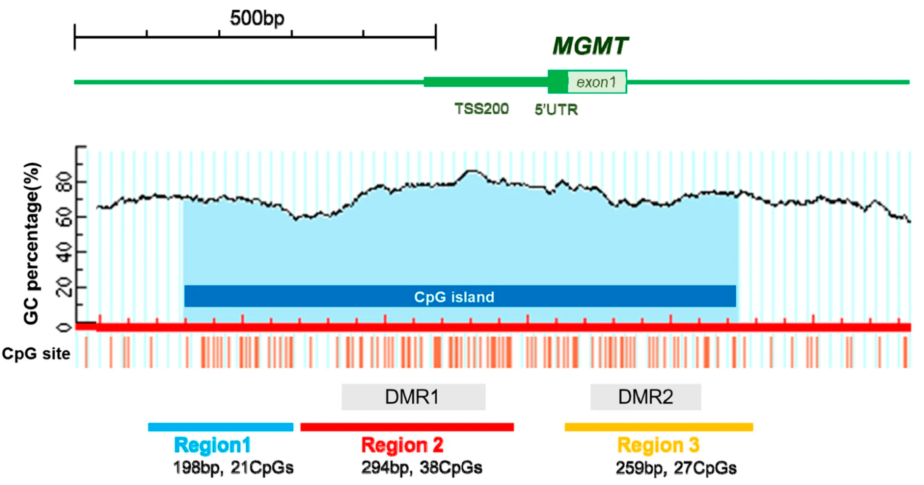
2.1.2. MGMT
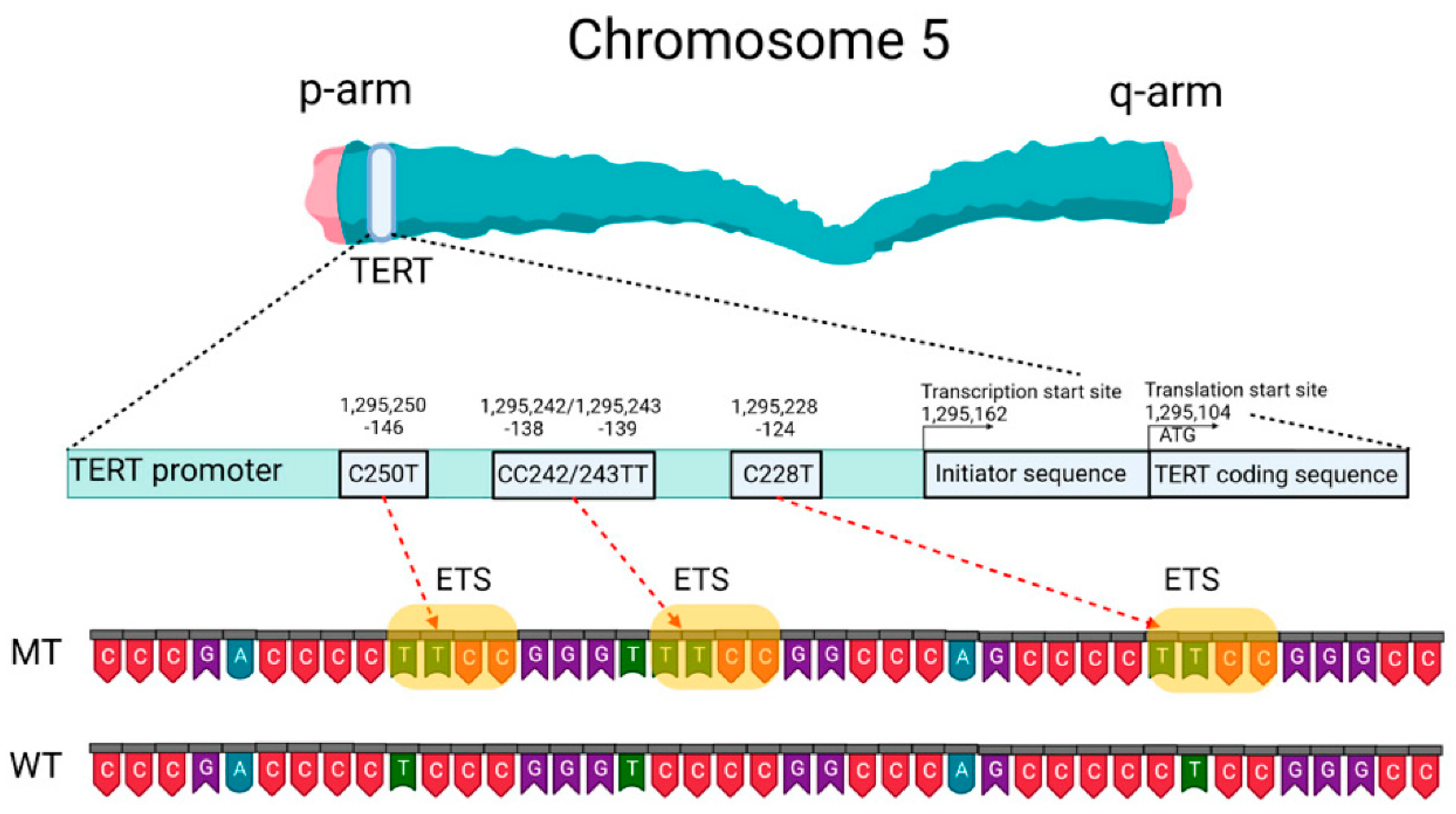
2.1.3. TERT
2.2. Other Promising Molecular Biomarkers
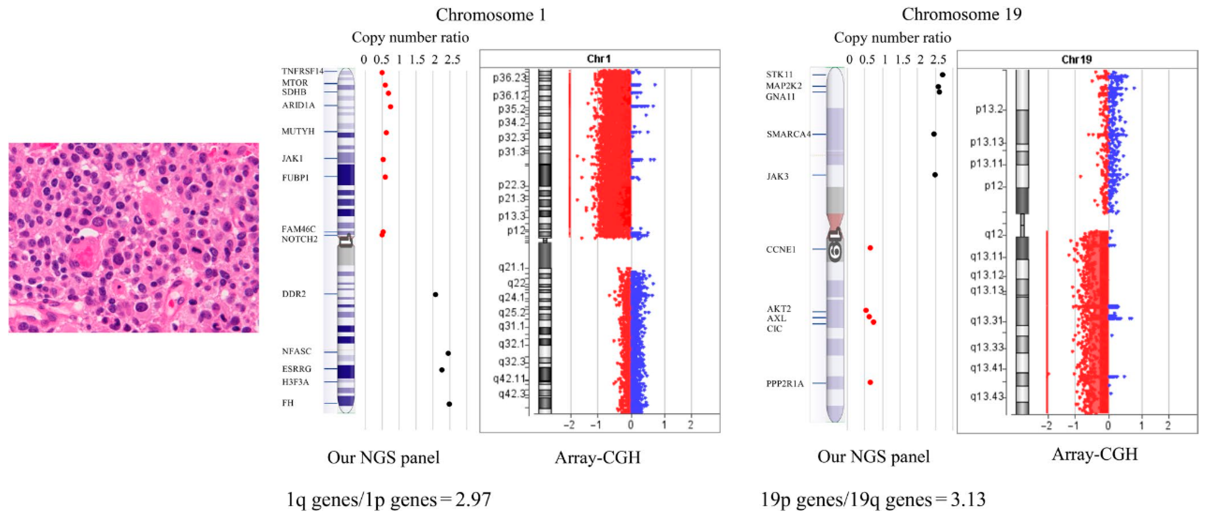
2.2.1. 1p/19q Co-Deletion
2.2.2. ATRX
2.2.3. EGFR
2.2.4. CDKN2A
2.2.5. Exosomes
2.2.6. cfDNA
2.2.7. ctDNA
2.2.8. CTCs
3. Available Liquid Biopsy and Their Prospects on Gliomas
3.1. Electrochemical Biosensor
3.1.1. Electrochemical Protein Sensor
3.1.2. Electrochemical Immunosensor
3.1.3. Electrochemical Nucleic Acid Aptamer Sensor
3.1.4. Electrochemical Microbial Sensor
3.2. Optical or Colorimetric Biosensor
3.2.1. Resonance-Based Optical Sensors
3.2.2. SERS Based Sensors
3.2.3. Electrochemiluminescence, Chemiluminescent Biosensor
3.2.4. Terahertz Based Biosensors
3.2.5. Fluorescence Based Biosensors
3.2.6. Colorimetric Biosensors
3.2.7. Near-Infrared Spectroscopy
3.2.8. Fiber-Based Biosensors
4. Perspectives
Author Contributions
Funding
Institutional Review Board Statement
Informed Consent Statement
Conflicts of Interest
References
- Preusser, M.; Lim, M.; Hafler, D.A.; Reardon, D.A.; Sampson, J.H. Prospects of Immune Checkpoint Modulators in the Treatment of Glioblastoma. Nat. Rev. Neurol. 2015, 11, 504–514. [Google Scholar] [CrossRef] [PubMed] [Green Version]
- Stupp, R.; Mason, W.P.; van den Bent, M.J.; Weller, M.; Fisher, B.; Taphoorn, M.J.B.; Belanger, K.; Brandes, A.A.; Marosi, C.; Bogdahn, U.; et al. Radiotherapy plus Concomitant and Adjuvant Temozolomide for Glioblastoma. N. Engl. J. Med. 2005, 352, 987–996. [Google Scholar] [CrossRef] [PubMed] [Green Version]
- Tsougos, I.; Svolos, P.; Kousi, E.; Fountas, K.; Theodorou, K.; Fezoulidis, I.; Kapsalaki, E. Differentiation of Glioblastoma Multiforme from Metastatic Brain Tumor Using Proton Magnetic Resonance Spectroscopy, Diffusion and Perfusion Metrics at 3 T. Cancer Imaging 2012, 12, 423–436. [Google Scholar] [CrossRef] [PubMed]
- Zhang, B.-H.; Yang, B.-H.; Tang, Z.-Y. Randomized Controlled Trial of Screening for Hepatocellular Carcinoma. J. Cancer Res. Clin. Oncol. 2004, 130, 417–422. [Google Scholar] [CrossRef]
- Schröder, F.H.; Hugosson, J.; Roobol, M.J.; Tammela, T.L.J.; Ciatto, S.; Nelen, V.; Kwiatkowski, M.; Lujan, M.; Lilja, H.; Zappa, M.; et al. Prostate-Cancer Mortality at 11 Years of Follow-Up. N. Engl. J. Med. 2012, 366, 981–990. [Google Scholar] [CrossRef]
- McNamara, C.; Mankad, K.; Thust, S.; Dixon, L.; Limback-Stanic, C.; D’Arco, F.; Jacques, T.S.; Löbel, U. 2021 WHO Classification of Tumours of the Central Nervous System: A Review for the Neuroradiologist. Neuroradiology 2022, 64, 1919–1950. [Google Scholar] [CrossRef]
- Higa, N.; Akahane, T.; Yokoyama, S.; Yonezawa, H.; Uchida, H.; Takajo, T.; Kirishima, M.; Hamada, T.; Matsuo, K.; Fujio, S.; et al. A Tailored Next-Generation Sequencing Panel Identified Distinct Subtypes of Wildtype IDH and TERT Promoter Glioblastomas. Cancer Sci. 2020, 111, 3902–3911. [Google Scholar] [CrossRef]
- Xue, H.; Han, Z.; Li, H.; Li, X.; Jia, D.; Qi, M.; Zhang, H.; Zhang, K.; Gong, J.; Wang, H.; et al. Application of Intraoperative Rapid Molecular Diagnosis in Precision Surgery for Glioma: Mimic the World Health Organization CNS5 Integrated Diagnosis. Neurosurgery 2022, 92, 762–771. [Google Scholar] [CrossRef]
- Sciortino, T.; Secoli, R.; d’Amico, E.; Moccia, S.; Conti Nibali, M.; Gay, L.; Rossi, M.; Pecco, N.; Castellano, A.; De Momi, E.; et al. Raman Spectroscopy and Machine Learning for IDH Genotyping of Unprocessed Glioma Biopsies. Cancers 2021, 13, 4196. [Google Scholar] [CrossRef]
- Haapala, I.; Kondratev, A.; Roine, A.; Mäkelä, M.; Kontunen, A.; Karjalainen, M.; Laakso, A.; Koroknay-Pál, P.; Nordfors, K.; Haapasalo, H.; et al. Method for the Intraoperative Detection of IDH Mutation in Gliomas with Differential Mobility Spectrometry. Curr. Oncol. 2022, 29, 3252–3258. [Google Scholar] [CrossRef]
- Alfaro, C.M.; Pirro, V.; Keating, M.F.; Hattab, E.M.; Cooks, R.G.; Cohen-Gadol, A.A. Intraoperative Assessment of Isocitrate Dehydrogenase Mutation Status in Human Gliomas Using Desorption Electrospray Ionization-Mass Spectrometry. J. Neurosurg. 2019, 132, 180–187. [Google Scholar] [CrossRef]
- Brown, H.M.; Alfaro, C.M.; Pirro, V.; Dey, M.; Hattab, E.M.; Cohen-Gadol, A.A.; Cooks, R.G. Intraoperative Mass Spectrometry Platform for IDH Mutation Status Prediction, Glioma Diagnosis, and Estimation of Tumor Cell Infiltration. J. Appl. Lab. Med. 2021, 6, 902–916. [Google Scholar] [CrossRef] [PubMed]
- Mimosa, M.L.; Al-Ameri, W.; Simpson, J.T.; Nakhla, M.; Boissinot, K.; Munoz, D.G.; Das, S.; Feilotter, H.; Fattouh, R.; Saleeb, R.M. A Novel Approach to Detect IDH Point Mutations in Gliomas Using Nanopore Sequencing: Test Validation for the Clinical Laboratory. J. Mol. Diagn. 2022, 25, 133–142. [Google Scholar] [CrossRef] [PubMed]
- Wu, D.; Chi, J.; Zhang, M.; Cheng, L.; Wang, X.; Fan, J.; Huang, Z.; Wang, H.; Xie, H.; Pan, Q.; et al. Water-Dispersing Perovskite Probes for the Rapid Imaging of Glioma Cells. Adv. Opt. Mater. 2022, 10, 2101835. [Google Scholar] [CrossRef]
- Reifenberger, G.; Wirsching, H.-G.; Knobbe-Thomsen, C.B.; Weller, M. Advances in the Molecular Genetics of Gliomas–Implications for Classification and Therapy. Nat. Rev. Clin. Oncol. 2017, 14, 434–452. [Google Scholar] [CrossRef] [PubMed]
- Thakkar, J.P.; Dolecek, T.A.; Horbinski, C.; Ostrom, Q.T.; Lightner, D.D.; Barnholtz-Sloan, J.S.; Villano, J.L. Epidemiologic and Molecular Prognostic Review of Glioblastoma. Cancer Epidemiol. Biomark. Prev. 2014, 23, 1985–1996. [Google Scholar] [CrossRef] [Green Version]
- Liu, L.; Gerson, S.L. Targeted Modulation of MGMT: Clinical Implications. Clin. Cancer Res. 2006, 12, 328–331. [Google Scholar] [CrossRef] [Green Version]
- Esteller, M.; Garcia-Foncillas, J.; Andion, E.; Goodman, S.N.; Hidalgo, O.F.; Vanaclocha, V.; Baylin, S.B.; Herman, J.G. Inactivation of the DNA-Repair Gene MGMT and the Clinical Response of Gliomas to Alkylating Agents. N. Engl. J. Med. 2000, 343, 1350–1354. [Google Scholar] [CrossRef] [PubMed]
- Silantyev, A.S.; Falzone, L.; Libra, M.; Gurina, O.I.; Kardashova, K.S.; Nikolouzakis, T.K.; Nosyrev, A.E.; Sutton, C.W.; Mitsias, P.D.; Tsatsakis, A. Current and Future Trends on Diagnosis and Prognosis of Glioblastoma: From Molecular Biology to Proteomics. Cells 2019, 8, 863. [Google Scholar] [CrossRef] [PubMed] [Green Version]
- Hegi, M.E.; Genbrugge, E.; Gorlia, T.; Stupp, R.; Gilbert, M.R.; Chinot, O.L.; Nabors, L.B.; Jones, G.; Van Criekinge, W.; Straub, J.; et al. MGMT Promoter Methylation Cutoff with Safety Margin for Selecting Glioblastoma Patients into Trials Omitting Temozolomide: A Pooled Analysis of Four Clinical Trials. Clin. Cancer Res. 2019, 25, 1809–1816. [Google Scholar] [CrossRef] [Green Version]
- Nguyen, N.; Redfield, J.; Ballo, M.; Michael, M.; Sorenson, J.; Dibaba, D.; Wan, J.; Ramos, G.D.; Pandey, M. Identifying the Optimal Cutoff Point for MGMT Promoter Methylation Status in Glioblastoma. CNS Oncol. 2021, 10, CNS74. [Google Scholar] [CrossRef] [PubMed]
- Schulze Heuling, E.; Knab, F.; Radke, J.; Eskilsson, E.; Martinez-Ledesma, E.; Koch, A.; Czabanka, M.; Dieterich, C.; Verhaak, R.G.; Harms, C.; et al. Prognostic Relevance of Tumor Purity and Interaction with MGMT Methylation in Glioblastoma. Mol. Cancer Res. 2017, 15, 532–540. [Google Scholar] [CrossRef] [PubMed] [Green Version]
- Lipp, E.S.; Healy, P.; Austin, A.; Clark, A.; Dalton, T.; Perkinson, K.; Herndon, J.E.; Friedman, H.S.; Friedman, A.H.; Bigner, D.D.; et al. MGMT: Immunohistochemical Detection in High-Grade Astrocytomas. J. Neuropathol. Exp. Neurol. 2019, 78, 57–64. [Google Scholar] [CrossRef] [Green Version]
- Sahara, N.; Hartanto, R.A.; Yoshuantari, N.; Dananjoyo, K.; Widodo, I.; Malueka, R.G.; Dwianingsih, E.K. Diagnostic Accuracy of Immunohistochemistry in Detecting MGMT Methylation Status in Patients with Glioma. Asian Pac. J. Cancer Prev. 2021, 22, 3803–3808. [Google Scholar] [CrossRef] [PubMed]
- Preusser, M.; Charles Janzer, R.; Felsberg, J.; Reifenberger, G.; Hamou, M.-F.; Diserens, A.-C.; Stupp, R.; Gorlia, T.; Marosi, C.; Heinzl, H.; et al. Anti-O6-Methylguanine-Methyltransferase (MGMT) Immunohistochemistry in Glioblastoma Multiforme: Observer Variability and Lack of Association with Patient Survival Impede Its Use as Clinical Biomarker. Brain Pathol. 2008, 18, 520–532. [Google Scholar] [CrossRef]
- Li, M.; Dong, G.; Zhang, W.; Ren, X.; Jiang, H.; Yang, C.; Zhao, X.; Zhu, Q.; Li, M.; Chen, H.; et al. Combining MGMT Promoter Pyrosequencing and Protein Expression to Optimize Prognosis Stratification in Glioblastoma. Cancer Sci. 2021, 112, 3699–3710. [Google Scholar] [CrossRef]
- Preusser, M.; Berghoff, A.S.; Manzl, C.; Filipits, M.; Weinhäusel, A.; Pulverer, W.; Dieckmann, K.; Widhalm, G.; Wöhrer, A.; Knosp, E.; et al. Clinical Neuropathology Practice News 1-2014: Pyrosequencing Meets Clinical and Analytical Performance Criteria for Routine Testing of MGMT Promoter Methylation Status in Glioblastoma. Clin. Neuropathol. 2014, 33, 6–14. [Google Scholar] [CrossRef] [Green Version]
- Christians, A.; Hartmann, C.; Benner, A.; Meyer, J.; von Deimling, A.; Weller, M.; Wick, W.; Weiler, M. Prognostic Value of Three Different Methods of MGMT Promoter Methylation Analysis in a Prospective Trial on Newly Diagnosed Glioblastoma. PLoS ONE 2012, 7, e33449. [Google Scholar] [CrossRef]
- Grasbon-Frodl, E.M.; Kreth, F.W.; Ruiter, M.; Schnell, O.; Bise, K.; Felsberg, J.; Reifenberger, G.; Tonn, J.-C.; Kretzschmar, H.A. Intratumoral Homogeneity of MGMT Promoter Hypermethylation as Demonstrated in Serial Stereotactic Specimens from Anaplastic Astrocytomas and Glioblastomas. Int. J. Cancer 2007, 121, 2458–2464. [Google Scholar] [CrossRef]
- Tetzner, R. Prevention of PCR Cross-Contamination by UNG Treatment of Bisulfite-Treated DNA. In DNA Methylation; Methods in Molecular Biology; Humana Press: Totowa, NJ, USA, 2009; Volume 507, pp. 357–370. [Google Scholar] [CrossRef]
- Wojdacz, T.K.; Dobrovic, A. Methylation-Sensitive High Resolution Melting (MS-HRM): A New Approach for Sensitive and High-Throughput Assessment of Methylation. Nucleic Acids Res. 2007, 35, e41. [Google Scholar] [CrossRef] [Green Version]
- Rosas-Alonso, R.; Colmenarejo-Fernandez, J.; Pernia, O.; Rodriguez-Antolín, C.; Esteban, I.; Ghanem, I.; Sanchez-Cabrero, D.; Losantos-Garcia, I.; Palacios-Zambrano, S.; Moreno-Bueno, G.; et al. Clinical Validation of a Novel Quantitative Assay for the Detection of MGMT Methylation in Glioblastoma Patients. Clin. Epigenetics 2021, 13, 52. [Google Scholar] [CrossRef]
- Hanihara, M.; Miyake, K.; Watanabe, A.; Yamada, Y.; Oishi, N.; Kawataki, T.; Inukai, T.; Kondo, T.; Kinouchi, H. Assessment of MGMT Methylation Status Using High-Performance Liquid Chromatography in Newly Diagnosed Glioblastoma. Clin. Epigenetics 2020, 12, 174. [Google Scholar] [CrossRef] [PubMed]
- Braczynski, A.K.; Capper, D.; Jones, D.T.; Schittenhelm, J.; Stichel, D.; von Deimling, A.; Harter, P.N.; Mittelbronn, M. High Density DNA Methylation Array Is a Reliable Alternative for PCR-Based Analysis of the MGMT Promoter Methylation Status in Glioblastoma. Pathol. Res. Pract. 2020, 216, 152728. [Google Scholar] [CrossRef]
- Wick, W.; Weller, M.; van den Bent, M.; Sanson, M.; Weiler, M.; von Deimling, A.; Plass, C.; Hegi, M.; Platten, M.; Reifenberger, G. MGMT Testing—The Challenges for Biomarker-Based Glioma Treatment. Nat. Rev. Neurol. 2014, 10, 372–385. [Google Scholar] [CrossRef] [PubMed] [Green Version]
- Bady, P.; Sciuscio, D.; Diserens, A.-C.; Bloch, J.; van den Bent, M.J.; Marosi, C.; Dietrich, P.-Y.; Weller, M.; Mariani, L.; Heppner, F.L.; et al. MGMT Methylation Analysis of Glioblastoma on the Infinium Methylation BeadChip Identifies Two Distinct CpG Regions Associated with Gene Silencing and Outcome, Yielding a Prediction Model for Comparisons across Datasets, Tumor Grades, and CIMP-Status. Acta Neuropathol. 2012, 124, 547–560. [Google Scholar] [CrossRef] [Green Version]
- Estival, A.; Sanz, C.; Ramirez, J.-L.; Velarde, J.M.; Domenech, M.; Carrato, C.; de Las Peñas, R.; Gil-Gil, M.; Sepúlveda, J.; Armengol, R.; et al. Pyrosequencing versus Methylation-Specific PCR for Assessment of MGMT Methylation in Tumor and Blood Samples of Glioblastoma Patients. Sci. Rep. 2019, 9, 11125. [Google Scholar] [CrossRef] [PubMed] [Green Version]
- Chen, X.; Zeng, M.; Tong, Y.; Zhang, T.; Fu, Y.; Li, H.; Zhang, Z.; Cheng, Z.; Xu, X.; Yang, R.; et al. Automatic Prediction of MGMT Status in Glioblastoma via Deep Learning-Based MR Image Analysis. BioMed Res. Int. 2020, 2020, 9258649. [Google Scholar] [CrossRef]
- Yogananda, C.G.B.; Shah, B.R.; Nalawade, S.S.; Murugesan, G.K.; Yu, F.F.; Pinho, M.C.; Wagner, B.C.; Mickey, B.; Patel, T.R.; Fei, B.; et al. MRI-Based Deep-Learning Method for Determining Glioma MGMT Promoter Methylation Status. AJNR Am. J. Neuroradiol. 2021, 42, 845–852. [Google Scholar] [CrossRef]
- Hasanau, T.; Pisarev, E.; Kisil, O.; Nonoguchi, N.; Le Calvez-Kelm, F.; Zvereva, M. Detection of TERT Promoter Mutations as a Prognostic Biomarker in Gliomas: Methodology, Prospects, and Advances. Biomedicines 2022, 10, 728. [Google Scholar] [CrossRef]
- Horn, S.; Figl, A.; Rachakonda, P.S.; Fischer, C.; Sucker, A.; Gast, A.; Kadel, S.; Moll, I.; Nagore, E.; Hemminki, K.; et al. TERT Promoter Mutations in Familial and Sporadic Melanoma. Science 2013, 339, 959–961. [Google Scholar] [CrossRef] [Green Version]
- Killela, P.J.; Reitman, Z.J.; Jiao, Y.; Bettegowda, C.; Agrawal, N.; Diaz, L.A.; Friedman, A.H.; Friedman, H.; Gallia, G.L.; Giovanella, B.C.; et al. TERT Promoter Mutations Occur Frequently in Gliomas and a Subset of Tumors Derived from Cells with Low Rates of Self-Renewal. Proc. Natl. Acad. Sci. USA 2013, 110, 6021–6026. [Google Scholar] [CrossRef] [PubMed]
- Arita, H.; Narita, Y.; Fukushima, S.; Tateishi, K.; Matsushita, Y.; Yoshida, A.; Miyakita, Y.; Ohno, M.; Collins, V.P.; Kawahara, N.; et al. Upregulating Mutations in the TERT Promoter Commonly Occur in Adult Malignant Gliomas and Are Strongly Associated with Total 1p19q Loss. Acta Neuropathol. 2013, 126, 267–276. [Google Scholar] [CrossRef]
- Diplas, B.H.; Liu, H.; Yang, R.; Hansen, L.J.; Zachem, A.L.; Zhao, F.; Bigner, D.D.; McLendon, R.E.; Jiao, Y.; He, Y.; et al. Sensitive and Rapid Detection of TERT Promoter and IDH Mutations in Diffuse Gliomas. Neuro-Oncology 2019, 21, 440–450. [Google Scholar] [CrossRef]
- Lee, H.; Lee, B.; Kim, D.G.; Cho, Y.A.; Kim, J.-S.; Suh, Y.-L. Detection of TERT Promoter Mutations Using Targeted Next-Generation Sequencing: Overcoming GC Bias through Trial and Error. Cancer Res. Treat. 2022, 54, 75–83. [Google Scholar] [CrossRef]
- Kang, S.Y.; Kim, D.G.; Kim, H.; Cho, Y.A.; Ha, S.Y.; Kwon, G.Y.; Jang, K.-T.; Kim, K.-M. Direct Comparison of the Next-Generation Sequencing and ITERT PCR Methods for the Diagnosis of TERT Hotspot Mutations in Advanced Solid Cancers. BMC Med. Genom. 2022, 15, 25. [Google Scholar] [CrossRef] [PubMed]
- Yu, P.-C.; Tan, L.-C.; Zhu, X.-L.; Shi, X.; Chernikov, R.; Semenov, A.; Zhang, L.; Ma, B.; Wang, Y.; Zhou, X.-Y.; et al. Arms-QPCR Improves Detection Sensitivity of Earlier Diagnosis of Papillary Thyroid Cancers with Worse Prognosis Determined by Coexisting BRAF V600E and Tert Promoter Mutations. Endocr. Pract. 2021, 27, 698–705. [Google Scholar] [CrossRef] [PubMed]
- Muralidharan, K.; Yekula, A.; Small, J.L.; Rosh, Z.S.; Kang, K.M.; Wang, L.; Lau, S.; Zhang, H.; Lee, H.; Bettegowda, C.; et al. TERT Promoter Mutation Analysis for Blood-Based Diagnosis and Monitoring of Gliomas. Clin. Cancer Res. 2021, 27, 169–178. [Google Scholar] [CrossRef]
- Fontanilles, M.; Marguet, F.; Beaussire, L.; Magne, N.; Pépin, L.-F.; Alexandru, C.; Tennevet, I.; Hanzen, C.; Langlois, O.; Jardin, F.; et al. Cell-Free DNA and Circulating TERT Promoter Mutation for Disease Monitoring in Newly-Diagnosed Glioblastoma. Acta Neuropathol. Commun. 2020, 8, 179. [Google Scholar] [CrossRef]
- Stasik, S.; Salomo, K.; Heberling, U.; Froehner, M.; Sommer, U.; Baretton, G.B.; Ehninger, G.; Wirth, M.P.; Thiede, C.; Fuessel, S. Evaluation of TERT Promoter Mutations in Urinary Cell-Free DNA and Sediment DNA for Detection of Bladder Cancer. Clin. Biochem. 2019, 64, 60–63. [Google Scholar] [CrossRef]
- Akuta, N.; Kawamura, Y.; Kobayashi, M.; Arase, Y.; Saitoh, S.; Fujiyama, S.; Sezaki, H.; Hosaka, T.; Kobayashi, M.; Suzuki, Y.; et al. TERT Promoter Mutation in Serum Cell-Free DNA Is a Diagnostic Marker of Primary Hepatocellular Carcinoma in Patients with Nonalcoholic Fatty Liver Disease. Oncology 2021, 99, 114–123. [Google Scholar] [CrossRef]
- Akuta, N.; Suzuki, F.; Kobayashi, M.; Fujiyama, S.; Kawamura, Y.; Sezaki, H.; Hosaka, T.; Kobayashi, M.; Saitoh, S.; Arase, Y.; et al. Detection of TERT Promoter Mutation in Serum Cell-Free DNA Using Wild-Type Blocking PCR Combined with Sanger Sequencing in Hepatocellular Carcinoma. J. Med. Virol. 2020, 92, 3604–3608. [Google Scholar] [CrossRef] [PubMed]
- Momeni-Boroujeni, A.; Yousefi, E.; Gupta, S.; Benayed, R.; Berger, M.F.; Ladanyi, M.; Monroe, R.; Kim, J.; Jungbluth, A.; Weigelt, B.; et al. Evaluation of TERT MRNA Expression Using RNAscope®: A Potential Histopathologic Diagnostic and Prognostic Tool. Pathol. Res. Pract. 2022, 233, 153892. [Google Scholar] [CrossRef]
- Shin, I.; Park, Y.W.; Ahn, S.S.; Kang, S.-G.; Chang, J.H.; Kim, S.H.; Lee, S.-K. Clinical and Diffusion Parameters May Noninvasively Predict TERT Promoter Mutation Status in Grade II Meningiomas. J. Neuroradiol. 2022, 49, 59–65. [Google Scholar] [CrossRef] [PubMed]
- Ivanidze, J.; Lum, M.; Pisapia, D.; Magge, R.; Ramakrishna, R.; Kovanlikaya, I.; Fine, H.A.; Chiang, G.C. MRI Features Associated with TERT Promoter Mutation Status in Glioblastoma. J. Neuroimaging 2019, 29, 357–363. [Google Scholar] [CrossRef]
- Kong, Z.; Zhang, Y.; Liu, D.; Liu, P.; Shi, Y.; Wang, Y.; Zhao, D.; Cheng, X.; Wang, Y.; Ma, W. Role of Traditional CHO PET Parameters in Distinguishing IDH, TERT and MGMT Alterations in Primary Diffuse Gliomas. Ann. Nucl. Med. 2021, 35, 493–503. [Google Scholar] [CrossRef] [PubMed]
- Li, C.; Liu, Z.; Zhang, X.; Wang, H.; Friedman, G.K.; Ding, Q.; Zhao, X.; Li, H.; Kim, K.; Yu, X.; et al. Generation of Chromosome 1p/19q Co-Deletion by CRISPR/Cas9-Guided Genomic Editing. Neurooncol. Adv. 2022, 4, vdac131. [Google Scholar] [CrossRef]
- Hu, N.; Richards, R.; Jensen, R. Role of Chromosomal 1p/19q Co-Deletion on the Prognosis of Oligodendrogliomas: A Systematic Review and Meta-Analysis. Interdiscip. Neurosurg. 2016, 5, 58–63. [Google Scholar] [CrossRef] [Green Version]
- Chen, R.; Smith-Cohn, M.; Cohen, A.L.; Colman, H. Glioma Subclassifications and Their Clinical Significance. Neurotherapeutics 2017, 14, 284–297. [Google Scholar] [CrossRef] [Green Version]
- Qin, T.; Mullan, B.; Ravindran, R.; Messinger, D.; Siada, R.; Cummings, J.R.; Harris, M.; Muruganand, A.; Pyaram, K.; Miklja, Z.; et al. ATRX Loss in Glioma Results in Dysregulation of Cell-Cycle Phase Transition and ATM Inhibitor Radio-Sensitization. Cell Rep. 2022, 38, 110216. [Google Scholar] [CrossRef]
- Sampson, J.H.; Gunn, M.D.; Fecci, P.E.; Ashley, D.M. Brain Immunology and Immunotherapy in Brain Tumours. Nat. Rev. Cancer 2020, 20, 12–25. [Google Scholar] [CrossRef]
- Sun, J.; Li, B.; Shu, C.; Ma, Q.; Wang, J. Functions and Clinical Significance of Circular RNAs in Glioma. Mol. Cancer 2020, 19, 34. [Google Scholar] [CrossRef] [Green Version]
- Li, D.; Patel, C.B.; Xu, G.; Iagaru, A.; Zhu, Z.; Zhang, L.; Cheng, Z. Visualization of Diagnostic and Therapeutic Targets in Glioma With Molecular Imaging. Front. Immunol. 2020, 11, 592389. [Google Scholar] [CrossRef] [PubMed]
- Richard, S.A.; Eugene, K.D. The Pivotal Immunomodulatory and Anti-Inflammatory Effect of Histone-Lysine N-Methyltransferase in the Glioma Microenvironment: Its Biomarker and Therapy Potentials. Anal. Cell. Pathol. 2021, 2021, 4907167. [Google Scholar] [CrossRef] [PubMed]
- Yang, K.; Wu, Z.; Zhang, H.; Zhang, N.; Wu, W.; Wang, Z.; Dai, Z.; Zhang, X.; Zhang, L.; Peng, Y.; et al. Glioma Targeted Therapy: Insight into Future of Molecular Approaches. Mol. Cancer 2022, 21, 39. [Google Scholar] [CrossRef]
- Lah, T.T.; Novak, M.; Breznik, B. Brain Malignancies: Glioblastoma and Brain Metastases. Semin. Cancer Biol. 2020, 60, 262–273. [Google Scholar] [CrossRef] [PubMed]
- Mehrjardi, N.Z.; Hänggi, D.; Kahlert, U.D. Current Biomarker-Associated Procedures of Cancer Modeling-a Reference in the Context of IDH1 Mutant Glioma. Cell Death Dis. 2020, 11, 998. [Google Scholar] [CrossRef] [PubMed]
- Śledzińska, P.; Bebyn, M.G.; Furtak, J.; Kowalewski, J.; Lewandowska, M.A. Prognostic and Predictive Biomarkers in Gliomas. Int. J. Mol. Sci. 2021, 22, 10373. [Google Scholar] [CrossRef] [PubMed]
- Kalluri, R.; LeBleu, V.S. The Biology, Function, and Biomedical Applications of Exosomes. Science 2020, 367, eaau6977. [Google Scholar] [CrossRef]
- Chen, G.; Huang, A.C.; Zhang, W.; Zhang, G.; Wu, M.; Xu, W.; Yu, Z.; Yang, J.; Wang, B.; Sun, H.; et al. Exosomal PD-L1 Contributes to Immunosuppression and Is Associated with Anti-PD-1 Response. Nature 2018, 560, 382–386. [Google Scholar] [CrossRef]
- Poggio, M.; Hu, T.; Pai, C.-C.; Chu, B.; Belair, C.D.; Chang, A.; Montabana, E.; Lang, U.E.; Fu, Q.; Fong, L.; et al. Suppression of Exosomal PD-L1 Induces Systemic Anti-Tumor Immunity and Memory. Cell 2019, 177, 414–427.e13. [Google Scholar] [CrossRef] [Green Version]
- Lin, H.; Yu, J.; Gu, X.; Ge, S.; Fan, X. Novel Insights into Exosomal Circular RNAs: Redefining Intercellular Communication in Cancer Biology. Clin. Transl. Med. 2021, 11, e636. [Google Scholar] [CrossRef] [PubMed]
- Liu, J.; Ren, L.; Li, S.; Li, W.; Zheng, X.; Yang, Y.; Fu, W.; Yi, J.; Wang, J.; Du, G. The Biology, Function, and Applications of Exosomes in Cancer. Acta Pharm. Sin. B 2021, 11, 2783–2797. [Google Scholar] [CrossRef] [PubMed]
- Al-Nedawi, K.; Meehan, B.; Micallef, J.; Lhotak, V.; May, L.; Guha, A.; Rak, J. Intercellular Transfer of the Oncogenic Receptor EGFRvIII by Microvesicles Derived from Tumour Cells. Nat. Cell Biol. 2008, 10, 619–624. [Google Scholar] [CrossRef] [PubMed]
- Skog, J.; Würdinger, T.; van Rijn, S.; Meijer, D.H.; Gainche, L.; Sena-Esteves, M.; Curry, W.T.; Carter, B.S.; Krichevsky, A.M.; Breakefield, X.O. Glioblastoma Microvesicles Transport RNA and Proteins That Promote Tumour Growth and Provide Diagnostic Biomarkers. Nat. Cell Biol. 2008, 10, 1470–1476. [Google Scholar] [CrossRef] [PubMed]
- Penders, J.; Nagelkerke, A.; Cunnane, E.M.; Pedersen, S.V.; Pence, I.J.; Coombes, R.C.; Stevens, M.M. Single Particle Automated Raman Trapping Analysis of Breast Cancer Cell-Derived Extracellular Vesicles as Cancer Biomarkers. ACS Nano 2021, 15, 18192–18205. [Google Scholar] [CrossRef] [PubMed]
- Hanjani, N.A.; Esmaelizad, N.; Zanganeh, S.; Gharavi, A.T.; Heidarizadeh, P.; Radfar, M.; Omidi, F.; MacLoughlin, R.; Doroudian, M. Emerging Role of Exosomes as Biomarkers in Cancer Treatment and Diagnosis. Crit. Rev. Oncol. Hematol. 2021, 169, 103565. [Google Scholar] [CrossRef] [PubMed]
- Vaidya, M.; Sugaya, K. DNA Associated with Circulating Exosomes as a Biomarker for Glioma. Genes 2020, 11, 1276. [Google Scholar] [CrossRef]
- Eibl, R.H.; Schneemann, M. Liquid Biopsy and Primary Brain Tumors. Cancers 2021, 13, 5429. [Google Scholar] [CrossRef]
- Kan, L.K.; Drummond, K.; Hunn, M.; Williams, D.; O’Brien, T.J.; Monif, M. Potential Biomarkers and Challenges in Glioma Diagnosis, Therapy and Prognosis. BMJ Neurol. Open 2020, 2, e000069. [Google Scholar] [CrossRef]
- Birkó, Z.; Nagy, B.; Klekner, Á.; Virga, J. Novel Molecular Markers in Glioblastoma-Benefits of Liquid Biopsy. Int. J. Mol. Sci. 2020, 21, 7522. [Google Scholar] [CrossRef]
- Gatto, L.; Franceschi, E.; Di Nunno, V.; Tosoni, A.; Lodi, R.; Brandes, A.A. Liquid Biopsy in Glioblastoma Management: From Current Research to Future Perspectives. Oncologist 2021, 26, 865–878. [Google Scholar] [CrossRef] [PubMed]
- Diaz, L.A.; Bardelli, A. Liquid Biopsies: Genotyping Circulating Tumor DNA. J. Clin. Oncol. 2014, 32, 579–586. [Google Scholar] [CrossRef] [PubMed]
- Bagley, S.J.; Nabavizadeh, S.A.; Mays, J.J.; Till, J.E.; Ware, J.B.; Levy, S.; Sarchiapone, W.; Hussain, J.; Prior, T.; Guiry, S.; et al. Clinical Utility of Plasma Cell-Free DNA in Adult Patients with Newly Diagnosed Glioblastoma: A Pilot Prospective Study. Clin. Cancer Res. 2020, 26, 397–407. [Google Scholar] [CrossRef]
- Krol, I.; Castro-Giner, F.; Maurer, M.; Gkountela, S.; Szczerba, B.M.; Scherrer, R.; Coleman, N.; Carreira, S.; Bachmann, F.; Anderson, S.; et al. Detection of Circulating Tumour Cell Clusters in Human Glioblastoma. Br. J. Cancer 2018, 119, 487–491. [Google Scholar] [CrossRef] [Green Version]
- De Wit, M.C.Y.; de Bruin, H.G.; Eijkenboom, W.; Sillevis Smitt, P.a.E.; van den Bent, M.J. Immediate Post-Radiotherapy Changes in Malignant Glioma Can Mimic Tumor Progression. Neurology 2004, 63, 535–537. [Google Scholar] [CrossRef]
- Biela, A.; Watkinson, M.; Meier, U.C.; Baker, D.; Giovannoni, G.; Becer, C.R.; Krause, S. Disposable MMP-9 Sensor Based on the Degradation of Peptide Cross-Linked Hydrogel Films Using Electrochemical Impedance Spectroscopy. Biosens. Bioelectron. 2015, 68, 660–667. [Google Scholar] [CrossRef] [Green Version]
- Sun, B.; Ni, X.; Cao, Y.; Cao, G. Electrochemical Sensor Based on Magnetic Molecularly Imprinted Nanoparticles Modified Magnetic Electrode for Determination of Hb. Biosens. Bioelectron. 2017, 91, 354–358. [Google Scholar] [CrossRef]
- Zhang, Z.; Cong, Y.; Huang, Y.; Du, X. Nanomaterials-Based Electrochemical Immunosensors. Micromachines 2019, 10, 397. [Google Scholar] [CrossRef] [PubMed] [Green Version]
- Police Patil, A.V.; Chuang, Y.-S.; Li, C.; Wu, C.-C. Recent Advances in Electrochemical Immunosensors with Nanomaterial Assistance for Signal Amplification. Biosensors 2023, 13, 125. [Google Scholar] [CrossRef]
- Asai, K.; Yamamoto, T.; Nagashima, S.; Ogata, G.; Hibino, H.; Einaga, Y. An Electrochemical Aptamer-Based Sensor Prepared by Utilizing the Strong Interaction between a DNA Aptamer and Diamond. Analyst 2020, 145, 544–549. [Google Scholar] [CrossRef]
- Zhang, X.; Feng, Y.; Duan, S.; Su, L.; Zhang, J.; He, F. Mycobacterium Tuberculosis Strain H37Rv Electrochemical Sensor Mediated by Aptamer and AuNPs-DNA. ACS Sens. 2019, 4, 849–855. [Google Scholar] [CrossRef] [PubMed]
- Chu, N.; Liang, Q.; Hao, W.; Jiang, Y.; Zeng, R.J. Micro-Microbial Electrochemical Sensor Equipped with Combined Bioanode and Biocathode for Water Biotoxicity Monitoring. Bioresour. Technol. 2021, 326, 124743. [Google Scholar] [CrossRef] [PubMed]
- Hassan, R.Y.A.; Febbraio, F.; Andreescu, S. Microbial Electrochemical Systems: Principles, Construction and Biosensing Applications. Sensors 2021, 21, 1279. [Google Scholar] [CrossRef] [PubMed]
- Nguyen, H.H.; Park, J.; Kang, S.; Kim, M. Surface Plasmon Resonance: A Versatile Technique for Biosensor Applications. Sensors 2015, 15, 10481–10510. [Google Scholar] [CrossRef] [Green Version]
- Li, L.; Zhang, Y.-N.; Zheng, W.; Li, X.; Zhao, Y. Optical Fiber SPR Biosensor Based on Gold Nanoparticle Amplification for DNA Hybridization Detection. Talanta 2022, 247, 123599. [Google Scholar] [CrossRef]
- Xu, K.; Zhou, R.; Takei, K.; Hong, M. Toward Flexible Surface-Enhanced Raman Scattering (SERS) Sensors for Point-of-Care Diagnostics. Adv. Sci. 2019, 6, 1900925. [Google Scholar] [CrossRef]
- Si, Y.; Xu, L.; Deng, T.; Zheng, J.; Li, J. Catalytic Hairpin Self-Assembly-Based SERS Sensor Array for the Simultaneous Measurement of Multiple Cancer-Associated MiRNAs. ACS Sens. 2020, 5, 4009–4016. [Google Scholar] [CrossRef]
- Fan, X.; Wang, S.; Liu, H.; Li, Z.; Sun, Q.; Wang, Y.; Fan, X. A Sensitive Electrochemiluminescence Biosensor for Assay of Cancer Biomarker (MMP-2) Based on NGQDs-Ru@SiO2 Luminophore. Talanta 2022, 236, 122830. [Google Scholar] [CrossRef]
- Zhuo, Y.; Gui, G.; Chai, Y.; Liao, N.; Xiao, K.; Yuan, R. Sandwich-Format Electrochemiluminescence Assays for Tumor Marker Based on PAMAM Dendrimer-L-Cysteine-Hollow Gold Nanosphere Nanocomposites. Biosens. Bioelectron. 2014, 53, 459–464. [Google Scholar] [CrossRef]
- Zhou, J.; Zhao, X.; Huang, G.; Yang, X.; Zhang, Y.; Zhan, X.; Tian, H.; Xiong, Y.; Wang, Y.; Fu, W. Molecule-Specific Terahertz Biosensors Based on an Aptamer Hydrogel-Functionalized Metamaterial for Sensitive Assays in Aqueous Environments. ACS Sens. 2021, 6, 1884–1890. [Google Scholar] [CrossRef]
- Tan, C.; Wang, S.; Li, S.; Liu, X.; Wei, J.; Zhang, G.; Ye, H. Cancer Diagnosis Using Terahertz-Graphene-Metasurface-Based Biosensor with Dual-Resonance Response. Nanomaterials 2022, 12, 3889. [Google Scholar] [CrossRef] [PubMed]
- Zhu, D.; Liu, B.; Wei, G. Two-Dimensional Material-Based Colorimetric Biosensors: A Review. Biosensors 2021, 11, 259. [Google Scholar] [CrossRef] [PubMed]
- Materón, E.M.; Gómez, F.R.; Almeida, M.B.; Shimizu, F.M.; Wong, A.; Teodoro, K.B.R.; Silva, F.S.R.; Lima, M.J.A.; Angelim, M.K.S.C.; Melendez, M.E.; et al. Colorimetric Detection of SARS-CoV-2 Using Plasmonic Biosensors and Smartphones. ACS Appl. Mater. Interfaces 2022, 14, 54527–54538. [Google Scholar] [CrossRef] [PubMed]
- Beć, K.B.; Grabska, J.; Huck, C.W. Near-Infrared Spectroscopy in Bio-Applications. Molecules 2020, 25, 2948. [Google Scholar] [CrossRef] [PubMed]
- Zhang, M.; Zhou, Q.; Huang, C.; Chan, C.T.; Wu, W.; Li, G.; Lim, M.; Gambhir, S.S.; Daldrup-Link, H.E. In Vivo Evaluation of Near-Infrared Fluorescent Probe for TIM3 Targeting in Mouse Glioma. Mol. Imaging Biol. 2022, 24, 280–287. [Google Scholar] [CrossRef]
- Hands, J.R.; Abel, P.; Ashton, K.; Dawson, T.; Davis, C.; Lea, R.W.; McIntosh, A.J.S.; Baker, M.J. Investigating the Rapid Diagnosis of Gliomas from Serum Samples Using Infrared Spectroscopy and Cytokine and Angiogenesis Factors. Anal. Bioanal. Chem. 2013, 405, 7347–7355. [Google Scholar] [CrossRef] [PubMed]
- Wen, X.; Chang, X.; Li, A.; Yang, X.; Tian, F.; Liu, Z.; Copner, N.; Teng, P.; Yuan, L. ZnO/Cu2O Heterojunction Integrated Fiber-Optic Biosensor for Remote Detection of Cysteine. Biosens. Bioelectron. 2023, 223, 115021. [Google Scholar] [CrossRef]
- Paltusheva, Z.U.; Ashikbayeva, Z.; Tosi, D.; Gritsenko, L.V. Highly Sensitive Zinc Oxide Fiber-Optic Biosensor for the Detection of CD44 Protein. Biosensors 2022, 12, 1015. [Google Scholar] [CrossRef]
- Marazuela, D.; Moreno-Bondi, M.C. Fiber-Optic Biosensors—An Overview. Anal. Bioanal. Chem. 2002, 372, 664–682. [Google Scholar] [CrossRef]


Disclaimer/Publisher’s Note: The statements, opinions and data contained in all publications are solely those of the individual author(s) and contributor(s) and not of MDPI and/or the editor(s). MDPI and/or the editor(s) disclaim responsibility for any injury to people or property resulting from any ideas, methods, instructions or products referred to in the content. |
© 2023 by the authors. Licensee MDPI, Basel, Switzerland. This article is an open access article distributed under the terms and conditions of the Creative Commons Attribution (CC BY) license (https://creativecommons.org/licenses/by/4.0/).
Share and Cite
Wu, Y.; Wang, X.; Zhang, M.; Wu, D. Molecular Biomarkers and Recent Liquid Biopsy Testing Progress: A Review of the Application of Biosensors for the Diagnosis of Gliomas. Molecules 2023, 28, 5660. https://doi.org/10.3390/molecules28155660
Wu Y, Wang X, Zhang M, Wu D. Molecular Biomarkers and Recent Liquid Biopsy Testing Progress: A Review of the Application of Biosensors for the Diagnosis of Gliomas. Molecules. 2023; 28(15):5660. https://doi.org/10.3390/molecules28155660
Chicago/Turabian StyleWu, Yuanbin, Xuning Wang, Meng Zhang, and Dongdong Wu. 2023. "Molecular Biomarkers and Recent Liquid Biopsy Testing Progress: A Review of the Application of Biosensors for the Diagnosis of Gliomas" Molecules 28, no. 15: 5660. https://doi.org/10.3390/molecules28155660