Article

Chemical Compositions of Eupatorium heterophyllum Leaf Samples from Yunnan and Sichuan Provinces of China—Isolation of 13 New Sesquiterpene Lactones

1
Graduate School of Biomedical Sciences, Nagasaki University, 1-14 Bunkyo-Machi, Nagasaki 852-8521, Japan
2
Faculty of Pharmaceutical Sciences, Tokushima Bunri University, Yamashiro-cho, Tokushima 770-8514, Japan
3
Kunming Institute of Botany, Chinese Academy of Sciences, Kunming 650201, China
*
Authors to whom correspondence should be addressed.
Molecules 2023, 28(13), 5107; https://doi.org/10.3390/molecules28135107
Submission received: 29 April 2023 / Revised: 24 June 2023 / Accepted: 26 June 2023 / Published: 29 June 2023
(This article belongs to the Special Issue Natural Secondary Metabolites III)

Abstract

:
Eight samples of Eupatorium heterophyllum leaves were collected at different locations in Yunnan and Sichuan provinces in China, and their chemical constituents were investigated. Thirteen previously undescribed sesquiterpene lactones—seven germacranolides, three eudesmanolides, two guaianolides, and a 2-norelemanolide—were isolated, and their structures were elucidated based on extensive spectroscopic analyses. The major constituents in the six samples from northwestern Yunnan and Sichuan are hiyodorilactones A and B, whereas that in the two samples from the region near Kunming, Yunnan is eupatoriopicrin. These results and previously reported results suggest the presence of locality-dependent intra-specific diversity in the chemical constituents of E. heterophyllum leaves.

Graphical Abstract

1. Introduction

The Hengduan Mountains and surrounding areas consist of many mountain ranges and deep river valleys with the elevation range from approximately 1500 to 6000 m, which separate these areas into various subdivisions. These areas are also climatically diverse and rich in plant resources, providing us with good plant materials for the study of diversity in secondary metabolites.
Eupatorium heterophyllum DC. (Asteraceae) is an endemic species in China. It is widely distributed in grasslands and forest areas at altitudes of 1700–3000 m in Southwest China (Sichuan, Yunnan, and Guizhou provinces and Xizang Autonomous Region) and has not been artificially cultivated [1]. We have been studying the intra-specific diversity in leaf and root chemicals of E. heterophyllum native to Yunnan and Sichuan provinces [2,3,4,5,6] as part of our continuing research on the chemical diversity of Asteraceae plants for obtaining unique secondary metabolites produced by limited populations within a species and for getting an insight into a chemical aspect of adaptation/differentiation. To date, diversity was observed in the minor constituents of the root chemicals of E. heterophyllum samples taken from different locations, affording various heterocyclic aromatic compounds, such as benzofuran/dihydrobenzofuran derivatives and propynyl thiophenes, in addition to some common major constituents [2,3,4]. In particular, seven oligomeric benzofurans were recently isolated for the first time from the sample collected in Lijiang City of Yunnan Province, which suggest an ongoing diversification of secondary metabolites in this species [4,5]. The chemical composition of the leaves, which is mainly composed of sesquiterpene lactones, is significantly different from that of the roots. Our previous study has suggested the presence of a higher degree of chemical diversity in the leaves than that in the roots: hiyodorilactones A (1) and B (3), which were marked major constituents, and several germacranolides with a hydroperoxy group at C-1β were found in the leaf samples from northwestern Yunnan and southwestern Sichuan (this chemotype is referred to as hiyodorilactone-type), whereas almost no hiyodorilactones and a small amount of eupatoriopicrin (26) were detected in the sample from a region near Kunming [6]. However, the overall chemical composition of the samples from the latter region has not been sufficiently clarified due to the presence of many unstable or conformationally flexible constituents. Therefore, further phytochemical studies using newly collected samples are required for understanding the chemical diversity of E. heterophyllum leaves. In this study, eight additional samples of E. heterophyllum leaves were collected in Yunnan (samples 1, 2, 7, and 8) and Sichuan (samples 3–6) provinces of China, and a detailed phytochemical study for each sample was performed. Herein, we report the isolation and structure elucidation of thirteen new sesquiterpene lactones from MeOH extracts of the collected samples as well as the differences in chemical compositions of them.

2. Results and Discussion

2.1. LC-MS Analysis

For an initial assessment of chemical diversity, a portion of each fresh leaf sample collected at the locations shown in Figure 1 was immediately extracted with EtOH, and the chemical composition of the extract was analyzed using LC-MS. The base peak ion (BPI) chromatograms of the samples are shown in Figure 2. Major peaks were identified using isolated compounds (Figure 3). The samples from northwestern Yunnan (samples 1 and 2) and Sichuan (samples 3–6) showed major peaks corresponding to hiyodorilactone D (38) [7], hiyodorilactone A (1) [8], eupaformosanin (2) [9], hiyodorilactone B (3) [8], and 20-desoxyeupaformosanin (4) [10] at tR = 7.95, 8.58, 9.17, 9.68, and 10.05 min, respectively, indicating that these chemical compositions are of hiyodorilactone-type samples. In contrast, the BPI chromatograms of the samples taken from a region near Kunming (samples 7 and 8) showed a significant peak of eupatoriopicrin (26) [11] at tR = 9.54 min, suggesting the presence of two chemotypes in the samples. There were some differences in the minor peaks of samples 7 and 8. Several peaks derived from (E,E)-germacranolides (25 [12], 28 [13], 27 [14], and 30 [15]) were detected at tR = 10.66, 10.77, 10.90, and 12.59 min, respectively, for sample 7. Weak peaks characteristic of hyodorilactone-type samples were observed for sample 8 but not for sample 7.

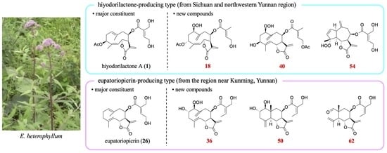
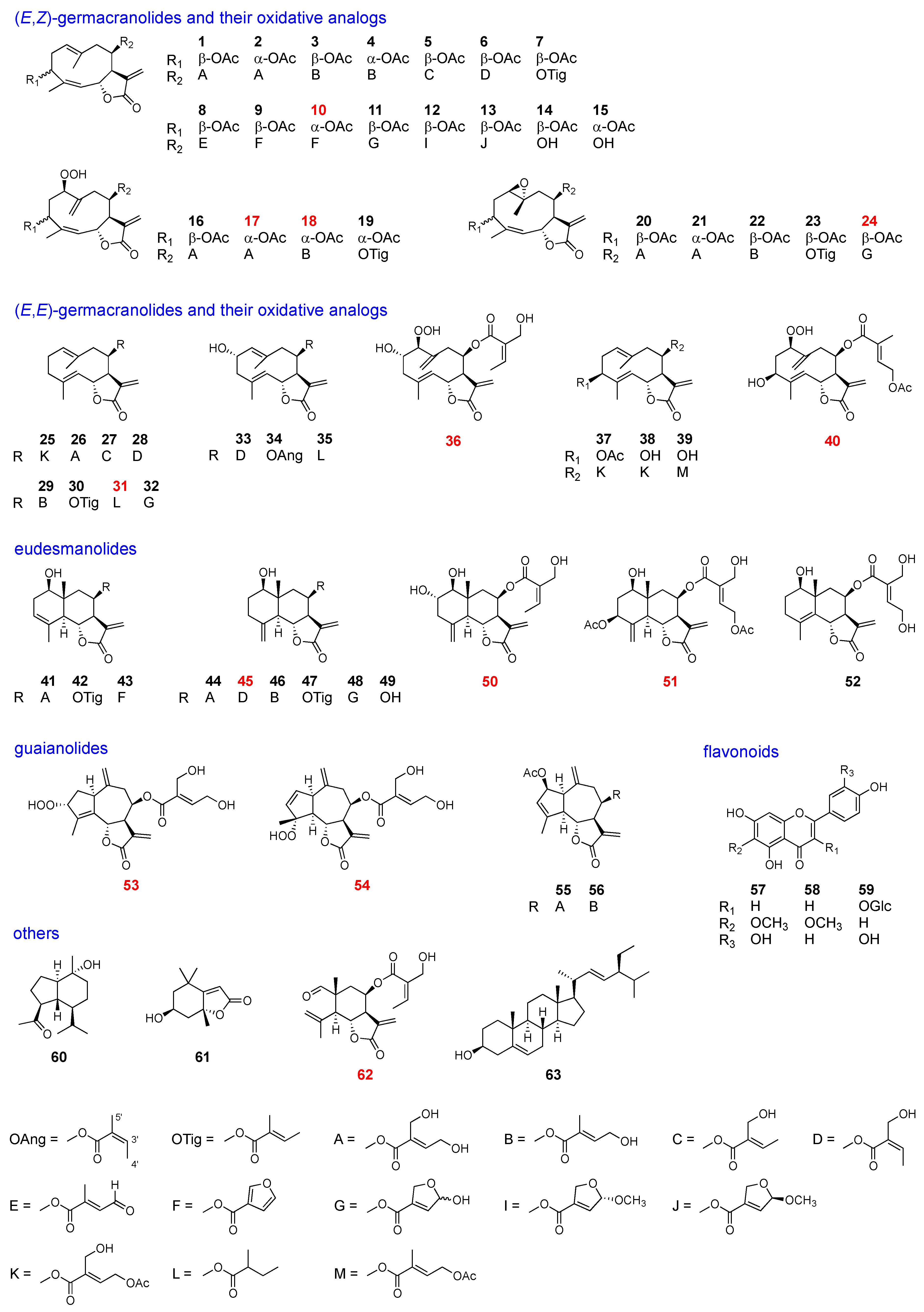
2.2. Isolation and Structural Elucidation of Leaf Chemicals

Dried leaves of each sample were extracted with MeOH, and the compounds were separated using silica-gel column chromatography and normal phase HPLC to yield 63 compounds, 13 of which were previously unreported. The isolated compounds were categolized into six types: (E,Z)-germacranolides and their oxidative analogs, (E,E)-germacranolides and their oxidative analogs, eudesmanolides, guaianolides, flavonoids, and others, as listed in Figure 3 and Table 1.
Among the isolated compounds, the following were known: hiyodorilactone A (1) [8], eupaformosanin (2) [9], hiyodorilactone B (3) [8], 20-desoxyeupaformasanin (4) [10], eupasimplicins B (5) [16,17], eupachinsin B (6) [18], 3β-acetoxy-8β-tigloyloxyheliangolide (7) [19], 4′-dehydrochromolaenide (8) [20], santhemoidin A (9) [21], 20-dehydroeucannabinolide-semi acetal (11) [10], santhemoidin B (12) [21], 4′-epi-santhemoidin B (13) [19], hiyodorilactone C (14) [8], eupaformonin (15) [22], hydroperoxyheterophyllin A (16) [6], hydroperoxyheterophyllin H (19) [6], epoxyeucannabinolid (20) [23], 1β,10α-epoxyeupaformosanin (21) [9], eupalinin B (22) [24], heliangin-3-O-acetate (23) [25], 8β-(4′-acetoxy-5′-hydroxytigloyloxy)-costunolide (25) [12], eupatoriopicrin (26) [11], 8β-(5′-hydroxytigloyloxy)-costunolide (27) [14], eupaglehnin C (28) [13], 20-desoxyeupatoriopicrin (29) [10], 8β-tigloyloxycostunolide (30) [15], 20-dehydroeupatoriopicrin-semi acetal (32) [10], deacetyleupaserrin (33) [26], 2α-hydroxyeupatolide-8-O-angelate (34) [27], 2α-hydroxy-8β-(2-methylbutyryloxy)-germacra-1(10)E,4E,11(13)-trien-12,6α-olide (35) [28], 8β-(4′-acetoxy-5′-hydroxytigloyloxy)-novanin (37) [19], hiyodorilactone D (38) [7], 4E-deacetyl chromolaenide-4′-O-acetate (39) [29], 1-hydroxy-8-(4′,5′-dihydroxytigloyloxy)-3,11(13)-eudesmadien-6,12-olide (41) [12], 1β-hydroxy-8β-tiglinoyloxyarbusculin B (42) [25], 1-hydroxy-8-furoyloxy-eudesma-3,11(13)-dien-6,12-olide (43) [30], 1-hydroxy-8-(4,5-dihydroxytiglyloxy)-eudesma-4(15),11(13)-dien-6,12-olide (44) [30], l-hydroxy-8-sarracenyloxyeudesma-4(15),11(13)-dien-6,12-olide (46) [31], 8β-tiglinoyloxyreynosin (47) [25], 1-hydroxy-8-(3-[2,5-dihydro-5-hydroxy]-furoyloxy)-eudesma-4(15),11(13)-dien-6,12-olide (48) [30], 8β-hydroxyreynosin (49) [32], 1-hydroxy-8-(4′,5′-dihydroxytigloyloxy)-4,11(13)-eudesmadien-6,12-olide (52) [12], eupahakonesin (55) [33], eupachifolin C (56) [34], eupafolin (57) [35], hispidulin (58) [36], quercetin-3-glucoside (59) [37], oplopanone (60) [38], loliolide (61) [39], and stigmasterol (63) [40]. The structures of the new compounds (10, 17, 18, 24, 31, 36, 40, 45, 50, 51, 53, 54, and 62) were elucidated as follows.
Compound 10 was obtained as a colorless oil. Its HREIMS spectrum showed the molecular ion peak at m/z 400.1518 to establish the molecular formula of C22H24O7 with 11 degrees of unsaturation. The IR spectrum of 10 exhibited absorptions at 1765 and 1743 cm−1, suggesting the presence of a γ-lactone and an ester group. The 1H and 13C NMR spectra of 10 (Table 2 and Table 3) were similar to those of santhemoidin A (9) [
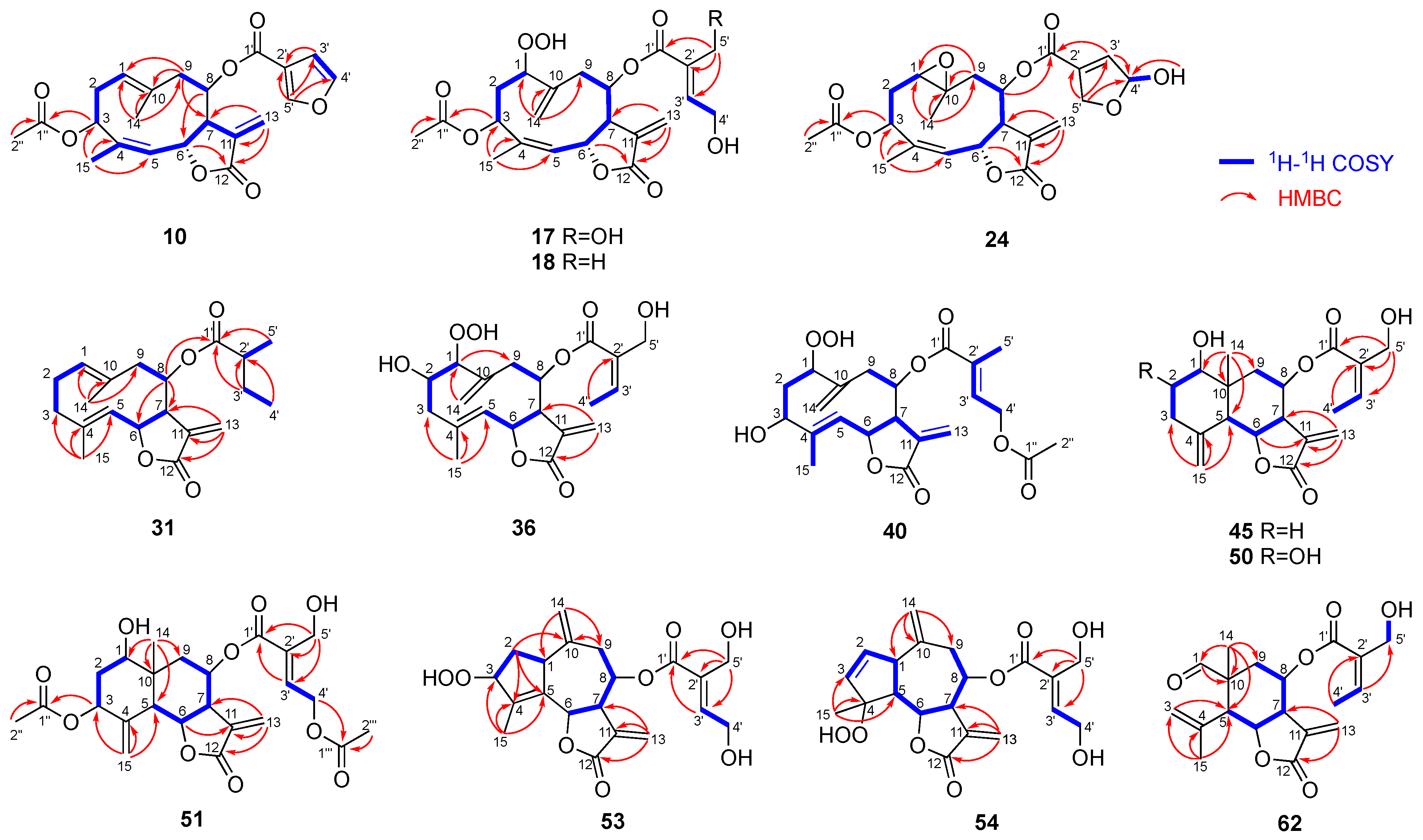
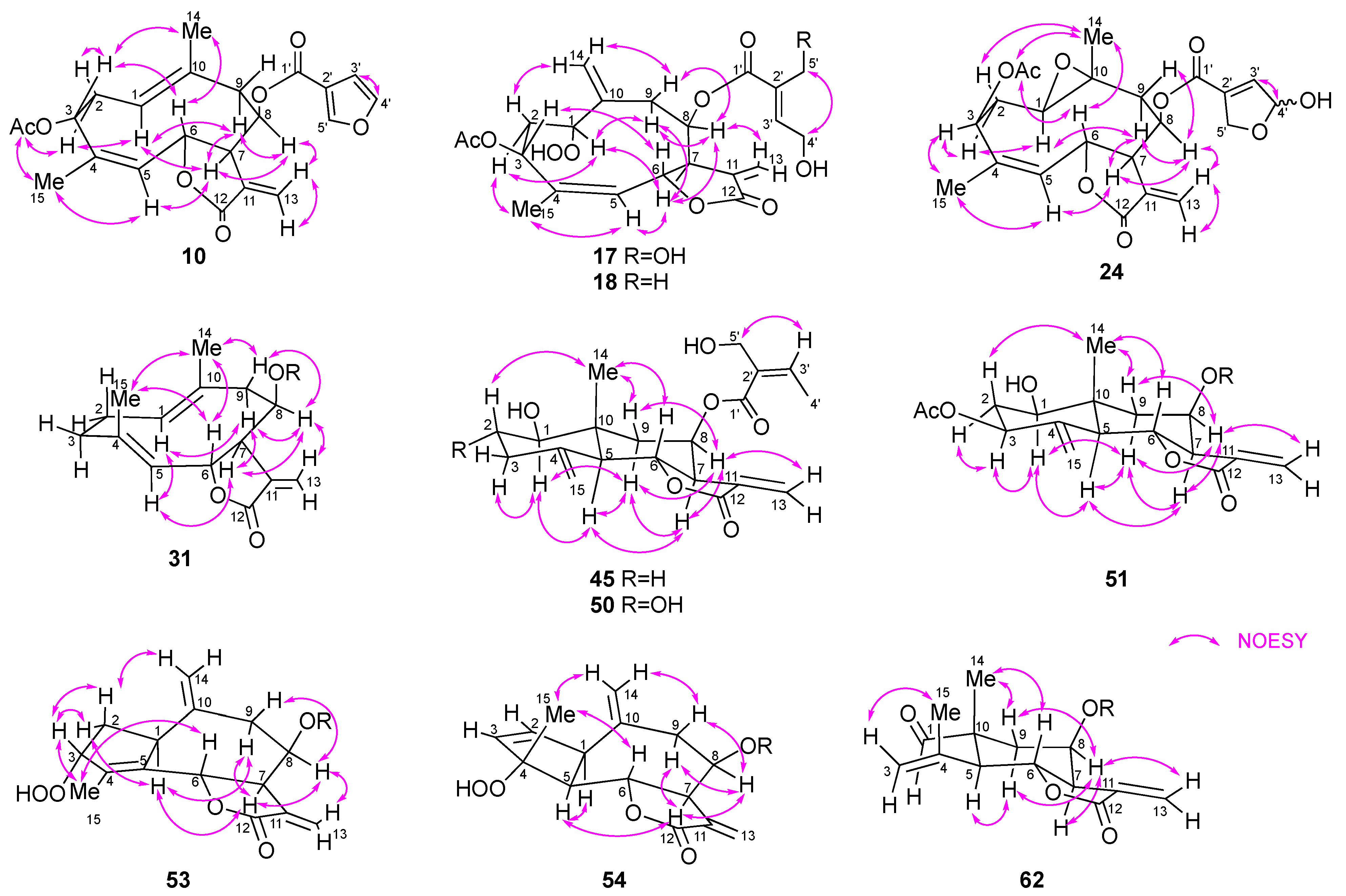
21]. In addition, HMBC correlations from H-3 to C-1″ and from H-6 to C-12 and a NOESY correlation between H-8 and H-13b (δH 5.80) (Figure 4 and Figure 5) indicated that 10 was a (4Z)-germacranolide with an acetoxy group at C-3, a 3-furoyloxy group at C-8, and a γ-lactone between C-12 and C-6, respectively. Thus, 10 has the same planar structure as 9. However, the signals attributable to H-3 and H-6 were observed at δH 5.63 and 5.31 in the 1H NMR spectrum of 10, whereas the corresponding signals of 9 were at δH 5.25 and 5.92, respectively. This characteristic is found between the C-3 epimers of (4Z)-germacranolide, hiyodorilactone A (1) [8], and eupaformosanin (2) [9], which suggests that 10 is the C-3 epimer of 9. This was confirmed by the NOESY correlations among H-3, H-6, and H3-14 (Figure 5). Thus, the structure of 10 was identified as (4Z)-3α-acetoxy-8β-(3-furoyloxy)germacra-1(10),4,11(13)-trien-(12,6α)-olide. The absolute configuration was determined to be (3R,6R,7R,8R)-10 because the experimental ECD spectrum of 10 was in good agreement with the theoretical ECD spectrum (Figure S79).
Compound 17 showed a quasimolecular ion [M + H]+ at m/z 453.1764 in its HRFABMS, which suggests a molecular formula of C22H28O10. The 1H and 13C NMR spectra of 17 (Table 2 and Table 3) resembled those of hydroperoxyheterophyllin A (16) [6], suggesting that 17 was a 1β-hydroperoxyheliangolide related to 16. The major differences between their 1H NMR spectra were observed in the chemical shifts of H-3 (17: δH 5.75; 16: δH 5.41) and H-6 (17: δH 5.69; 16: δH 6.15). These observations were similar to the above-mentioned case of 9 and 10, indicating an α-orientation of the acetoxy group at C-3 in 17. This conclusion was supported by the NOE between H-3 and H-6 (Figure 5). Thus, 17 was identified as (4Z)-3α-acetoxy-8β-(4′,5′-dihydroxytigloyloxy)-1β-hydroperoxygermacra-4,10(14),11(13)-trien-(12,6α)-olide. In a similar manner, 18 was determined to be a 5′-deoxy derivative of 17. Its molecular formula C22H28O9 with one less oxygen atom than that of 17, and the HMBC correlations from H3-5′ (δH 1.82) to C-1′/C-2′/C-3′ support this inference (Figure 4).
The molecular formula of 24 was determined to be C22H26O9 via HRFABMS. Its 1H NMR spectrum is similar to that of 1β,10α-epoxyeucannabinolide (20) [23] (Table 2), differing only in the signals attributable to the ester group at C-8. The signals δH 6.66 (m, H-3′), 6.18 (m, H-4′), 4.89 (m, H-5′a), and 4.69 (m, H-5′b) suggested the presence of a 4′,5′-epoxy-4′-hydroxytigloyl group in 24 [10,30]. Moreover, a pair of 4′-OH signals at δH 3.03/2.94 (each 0.5H, d, J = 8.4 Hz) indicated that 24 was a mixture of hemiacetal isomers (ca. 1:1). The NOESY correlations shown in Figure 5 suggest that the stereochemistry of the heliangolide core is the same as that of 20. Thus, 24 was characterized as a C-4′ epimer of (4Z)-3β-acetoxy-1β,10α-epoxy-8β-(4′,5′-epoxy-4′-hydroxytigloyloxy)germacra-4,11(13)-dien-(12,6α)-olide.
Compound 31 had the molecular formula of C20H28O4 as indicated by the quasimolecular ion peak at m/z 333.2059 [M + H]+ in its HRCIMS. The 1H and 13C NMR spectra showed signals corresponding to a 2-methylbutanoyloxy group (Table 2 and Table 3). The remaining signals of 31 were nearly identical to those of the terpene scaffold of 8β-tigloyloxycostunolide (30) [15]. The NOESY correlations of H-1/H-5, H-5/H-7, H-7/H-8, H-6/H3-14, and H-6/H3-15 established the relative configuration of the germacranolide moiety as illustrated in Figure 5. Based on these observations, 31 was identified as 8β-(2′-methylbutanoyloxy)germacra-1(10),4,11(13)-trien-(12,6α)-olide.
Compound 36 showed a [M + K]+ peak at m/z 433.1241 in HRFABMS, confirming its molecular formula as C20H26O8. The IR absorptions at 3380, 1745, and 1715 cm−1 suggested the presence of a hydroxy group, γ-lactone, and ester group, respectively. The 1D and 2D NMR spectra of 36 were recorded at 233 K because its 1H NMR spectrum exhibited broad signals at room temperature, suggesting conformational flexibility. The 1H and 13C NMR spectra of 36 (Table 4) were similar to those of deacetyleupaserrin (33) [26] except that the signals corresponding to an olefinic methine and a methyl group in 33 were replaced with those of an oxygen-bearing methine [δH 4.13 (H-1); δC 97.9 (C-1)] and an exomethylene group [δH 5.44 and 5.12 (H2-14); δC 120.0 (C-14)], respectively, implying that 36 was a C-1 hydroperoxy analog of deacetyleupaserrin (33). This was confirmed by the COSY correlations of H-1/H-2/H2-3, H-5/H-6/H-7, H-8/H2-9, and H-3′/H-4′, along with the HMBC correlations from H3-15 to C-3, C-4, and C-5; from H2-14 to C-1; from H-1 to C-9; from H2-13 to C-7 and C-12; and from H3-4′ to C-2′ (Figure 4). Therefore, the planar structure of 36 was determined as shown in Figure 4. Unfortunately, the NOE correlations required for determining the relative configurations of 36 were not observed; nevertheless, considering the stereochemistry of 33 and other 1-hydroperoxy germacranolides found in this plant, 36 was concluded as 1β-hydroperoxy-2α-hydroxy-8β-(5′-hydroxyangeloyloxy)germacra-4,10(14),11(13)-trien-(12,6α)-olide.
The HRESIMS spectrum of 40 showed a [M + Na]+ peak at m/z 459.1632 to establish a molecular formula of C22H28O9 with nine degrees of unsaturation. The IR absorptions at 3400 cm−1 corresponded to a hydroxy group and those at 1743, 1735, and 1715 cm−1 are attributed to carbonyl groups. Similar to the case of 36, the 1H NMR spectrum of 40 also afforded broad signals at room temperature. Even at 233 K, the quality of the 13C NMR spectrum remained insufficient owing to the small amount of 40 obtained; however, the 1H NMR spectrum clearly showed pairs of signals (in a ratio of 2:3 based on the integration), suggesting the coexistence of two conformers. A careful analysis of the 1H NMR (Table 4) and the 1H-1H COSY spectra (Figure 4) of both conformers suggested a structural similarity of 40 with 4E-deacetyl chromolaenide-4′-O-acetate (39) [29] as well as the presence of a hydroperoxy group [δH 8.56 (major) and 8.43 (minor)] and an additional exomethylene [δH 5.44/5.13 (major) and 5.35/5.02 (minor)]. The differences in the 1H NMR spectrum of 40 with that of 39 were attributable to a 1β-hydroperoxy-10(14)-ene structure, as is the case with 36 and 33. Therefore, 40 was identified as 8β-(4′-acetoxytigloyloxy)-1β-hydroperoxy-3β-hydroxygermacra-4,10(14),11(13)-trien-(12,6α)-olide.
Compound 45 showed a quasimolecular ion [M + H]+ at m/z 363.1808 in its HRFABMS, which suggests a molecular formula of C20H26O6. The 1H NMR data suggested a structural similarity of 45 to that of the known eudesmanolide 44 [30] (Table 5); however, the COSY correlation between H-3′ [δH 6.40 (q, J = 7.3 Hz)] and H3-4′ [δH 2.04 (d, J = 7.3 Hz)] and the NOESY correlation between H-3′ and H2-5′ indicated that the 4′,5′-dihydroxytigloyl group in 44 was replaced with a 5′-hydroxyangeloyl group in 45 (Figure 4 and Figure 5). Therefore, 45 was identified as 1β-hydroxy-8β-(5′-hydroxyangeloyloxy)eudesma-4(15),11(13)-dien-(12,6α)-olide.
The 1H and 13C NMR spectra of 50 revealed that it is also an eudesmanolide similar to 45. Its molecular formula was determined to be C20H26O7, one more oxygen atom than 45, using HRFABMS. In addition, the COSY correlations between H-1 (δH 3.19)/H-2 (δH 3.62)/H2-3 (δH 2.66 and 2.11) and the NOE correlation between H-2β and H3-14 suggested the presence of another hydroxy group at C-2α in 50 compared to that in 45 (Figure 4 and Figure 5). Thus, 50 was identified as 1β,2α-dihydroxy-8β-(5′-hydroxyangeloyloxy)eudesma-4(15),11(13)-dien-(12,6α)-olide.
The molecular formula of 51 was determined to be C24H30O10 using HRFABMS. Its 1H and 13C NMR data (Table 5) were closely related to those of eupakirunsin H [41], suggesting that 51 was also a eudesmanolide. The downfield shift of H-3 (δH 5.20) and H-8 (δH 5.83) in the 1H NMR spectrum as well as the COSY and HMBC correlations shown in Figure 4 indicated that the hydroxy group at C-3 and tigloyloxy group at C-8 in eupakirunsin H were replaced by acetoxy and 4′-acetoxy-5′-hydroxytigloyl groups, respectively, in 51.
The HRESIMS spectrum of compound 53 showed a [M + Na]+ peak at m/z 415.1368, which suggests the molecular formula C20H24O8 with nine degrees of unsaturation. The 1H and 13C NMR spectra revealed the presence of one methyl, two oxymethylenes, three oxymethines, two exocyclic double bonds, one trisubstituted double bond, one tetrasubstituted double bond, and two carbonyls (Table 6). The above spectroscopic data accounted for six degrees of unsaturation, and therefore, 53 should be tricyclic. Compound 53 was deduced to be a guaianolide with oxygen-functionalities at C-3, C-6, and C-8, one of which is a 4′,5′-dihydroxytigloyloxy group, as evidenced by the COSY and HMBC correlations shown in Figure 4. A significant downfield shift of H-8 (δH 5.72) as well as the NOESY correlation between H-13b and H-8 suggested the presence of a 4′,5′-dihydroxytigloyloxy moiety at C-8 and a γ-lactone between C-12 and C-6. Moreover, the molecular formula of 53 and the chemical shift of C-3 (δ 94.2) suggested the presence of a hydroperoxy group at this position. The elucidated planar structure of 53 is shown in Figure 4. The NOESY spectrum showed a cross-peak between H-7 and H-1, H-8, and H-9α, indicating that these hydrogens were in the same orientation (Figure 5). H-3 showed NOE correlations with H-2a and H-2b, but not with H-1, indicating the α-orientation of hydroperoxy group. Finally, H-6 was assigned a β-orientation owing to its coupling constant (J6,7 = 10.5 Hz). Therefore, 53 was identified as 8β-(4′,5′-dihydroxytigloyloxy)-3α-hydroperoxyguaia-4,10(14),11(13)-trien-(12,6α)-olide.
HRFABMS and 1D/2D NMR spectra of 54 revealed that it is also a guaianolide with the same molecular formula as that of 53 (Table 6 and Figure 4 and Figure 5). A COSY correlation between two olefinic protons at C-2 (δC 133.0; δH 5.77) and C-3 (δC 137.2; δH 6.01) indicated a disubstituted double bond. The chemical shift of C-4 observed at δC 95.2 in the 13C NMR spectrum and NOE correlation between H3-15 and H-6 indicated the presence of a hydroperoxy group at C-4α. Thus, 54 was concluded to be a 4α-hydroperoxy-2-ene isomer of 53.
Compound 62 was isolated as a colorless oil. A [M + Na]+ peak was observed at m/z 371.1472 in its HRESIMS, corresponding to the molecular formula of C19H24O6 with eight degrees of unsaturation. The IR spectrum suggested the presence of hydroxy (3501 cm−1), γ-lactone (1769 cm−1), aldehyde (1726 cm−1), and α,β-unsaturated carbonyl (1715 cm−1) groups. The 1H and 13C NMR spectra showed the characteristic signals for a 5′-hydroxyangeloyl moiety (Table 6), which implied that 62 was a norsesquiterpenoid. The 1H-1H COSY spectrum exhibited a spin system from H-5 to H2-9 (Figure 4). Furthermore, in the HMBC spectrum, H3-14 was correlated with C-1/C-5/C-9/C-10 and H3-15 with C-3/C-4/C-5, indicating that 62 was a 2-norelemanolide. A downfield shift of H-8 (δH 5.87) as well as the NOE between H-8 and H-13b (δH 5.57) as shown in Figure 5 confirmed the position of an α-methylene-γ-lactone and 5′-hydroxyangeloyl group. The relative stereochemistry of 62 is similar to that of 45 based on NOE correlations and coupling constants. Thus, 62 was identified as 8β-(5′-hydroxyangeloyloxy)-1-oxo-2-norelema-3,11(13)-dien-(12,6α)-olide.
The experimental ECD spectra of 17, 18, 24, 45, 50, 51, 54, and 62 showed a similar trend to that of 10, especially the negative Cotton effect around 210 nm mainly owing to the α-methylene-γ-lactone moiety. This indicated that the absolute configurations at C-6 and C-7 of these compounds are the same as those of 10 while the other chromophore might have a weaker contribution to their experimental ECD spectra [18]. In addition, considering the biosynthesis of sesquiterpenoids in higher plants, the other new compounds, 31, 36, 40, and 53, would have the same stereochemistry.

2.3. Discussion

In this study, 63 compounds, including 13 new compounds, were isolated from 8 leaf samples of E. heterophyllum collected in Yunnan and Sichuan provinces (Figure 3). Among the isolated compounds, 57 compounds were sesquiterpene lactones, including germacranolides (140), eudesmanolides (4152), guaianolides (5356), and elemanolides (62). Most of the new compounds would be produced from major constituents vir oxidative metabolism, which often involve the introduction of a hydroperoxy group. The chemical composition of each sample is summarized in Table 1. The major peaks in the BPI chromatograms (Figure 2) confirm the major constituents in each sample. Hiyodorilactones A (1) and B (3) are the major constituents in samples 1–6, confirming that hiyodorilactone-type samples are predominant in Sichuan and northwestern Yunnan regions. In contrast, eupatoriopicrin (26), which is not detected in samples 1–6, is the major constituent in samples 7 and 8 from the region near Kunming. A variety of (E,E)-germacranolides and eudesmanolides are also contained in these samples. Therefore, samples 7 and 8 should be classified as another chemotype, eupatoriopicrin-type. Considering these results and previously obtained results [6], we conclude the presence of locality-dependent intra-specific diversity in the leaf chemicals of E. heterophyllum.
Eupatoriopicrin (26) showed higher cytotoxicity against HL-60 cells than hyodorilactones A (1) and B (3) [42]. Moreover, Pan et al. recently reported genetic diversity in E. heterophyllum [43]. Notably, the geographical distribution of the different genotypes is in good agreement with the chemotypes observed in this study, indicating that the intra-specific diversity in the leaf chemicals of E. heterophyllum is related to its genetic background. These observations can provide us a new insight into a chemical aspect of adaptation and differentiation of Eupatorium plants. We plan to conduct further chemical studies on E. heterophyllum sampled from other regions to obtain secondary metabolites produced by limited populations within a species and to understand the relationship between chemical diversity, genetic diversity, and geographical distribution.

3. Materials and Methods

3.1. General Experimental Procedures

Optical rotations were measured with a JASCO P1020NK digital polarimeter. IR spectra were recorded using a JASCO FT/IR-410 spectrophotometer with the diffuse reflectance method. The 1D and 2D NMR spectra were measured using a Varian Unity plus 500 spectrometer (1H: 500 MHz; 13C: 126 MHz) or JEOL JNM-AL 400 spectrometer (1H: 400 MHz; 13C: 100 MHz). The coupling constants (J) are expressed in Hertz, and the chemical shifts (δ) are reported in ppm with the residual solvent signal used as a reference (CDCl3: TMS). Mass spectra, including high-resolution spectra, were recorded on a JEOL JMS-700 MStation. Column chromatography was performed on silica gel 60 (100–210 mesh, Kanto Chemical Co., Inc., Tokyo, Japan). TLC was with silica gel 60 F254 plates (Merck, Rahway, NJ, USA). Preparative HPLC was performed on a JASCO chromatograph (n-hexane–EtOAc, CHCl3–EtOAc, CHCl3–MeOH) equipped with a JASCO PU-2086 pump, a JASCO UV-970 detector, a JASCO RI-2031 detector, and various columns: COSMOSIL 5SL-II (20 × 250 mm, Nacalai Tesque Inc., Kyoto, Japan), COSMOSIL 5SL-II (10 × 250 mm, Nacalai Tesque Inc., Kyoto, Japan), COSMOSIL 5SL-II (4.6 × 250 mm, Nacalai Tesque Inc., Kyoto, Japan), YMC-Pack Diol-120-NP (4.6 × 250 mm, YMC Co., Ltd., Kyoto, Japan), Inertsil CN-3 (4.6 × 250 mm, GL Sciences, Tokyo, Japan), Inertsil Diol (10 × 250 mm, GL science, Tokyo, Japan), TSK gel silica 60 (4.6 × 250 mm, Tosoh, Tokyo, Japan), and TSK gel G1000HHR (7.8 × 300 mm, Tosoh, Tokyo, Japan).

3.2. Plant Materials

Samples were collected in August of 2014 (samples 1 and 2) and 2015 (samples 3–8) at the several geographically isolated locations shown in Figure 1 and Table 7. Each sample was authenticated by Dr. Takayuki Kawahara, Japan Forest Technology Association, General Incorporated Association, Japan. The voucher specimens (No. 2014-10, 2014-48, 2015-14, 2015-25, 2015-27, 2015-65, 2015-70, and 2015-71 for samples 1–8, respectively) are deposited in Kunming Institute of Botany, Kunming, China.

3.3. LC-MS Analysis

Parts of the fresh leaves (a few grams) of each sample were extracted with ethanol immediately after harvesting, and the extracted ethanol solutions were filtered and then separated using a Waters Acquity™ UPLC I-Class system coupled with a Waters ACQUITY UPLC BEH C18 column (2.1 × 100 mm, 1.7 μm). The temperature of the column was held at 45 °C. The mobile phases, which consisted of two solvent systems (eluent A, 0.1% formic acid in the water, v/v; eluent B (0.1% formic acid in methanol, v/v)), were delivered at a flow rate of 0.25 mL/min using a linear gradient program. The linear elution gradient program was set as follows: 0 min (95:5)–14.75 min (2:98)–17.00 min (2:98)–17.20 min (95:5)–20.00 min (95:5).
A Waters SYNAPT G2-Si HDMS mass spectrometer was connected to the UPLC system via an ESI interface. The conditions of analysis were as follows: capillary voltage was set at 1.5 kV under positive ion and 2.0 kV under negative mode, sampling cone voltage at 40.0 V, source temperature at 120 °C, desolvation gas temperature at 500 °C. The cone gas flow was at 500 L/h, desolvation gas at 1200 L/h, and the nebulizer gas at 6.5 bar.

3.4. Extraction and Isolation

The dried leaves of sample 1 (39.1 g) were cut into small pieces and extracted with MeOH two times at room temperature. After removal of the solvent under reduced pressure not exceeding 30 °C, a concentrated and combined MeOH extract (7.5 g) was obtained, which was then separated using a silica gel column (40 × 160 mm, n-hexane–EtOAc, 9:1, 8:2, 7:3, 6:4, 1:1, 2:8; EtOAc–MeOH, 1:0, 95:5, 9:1, 7:3, 0:1) to afford ten subfractions (Fr. 1–10). Compound 1 (573.7 mg) was obtained as Fr. 7. Fr. 4 (165.1 mg) was purified with semipreparative HPLC (COSMOSIL 5SL-II, 10 × 250 mm, n-hexane–EtOAc, 8:2) to give 7 (11.0 mg) and 9 (36.4 mg). Fr. 5 (141.7 mg) was suspended in n-hexane–EtOAc (7:3) and then filtered to obtain 58 (6.3 mg) as a precipitate. The filtrate was separated by COSMOSIL 5SL-II (10 × 250 mm, n-hexane–EtOAc, 7:3) to afford 58 (7.8 mg), Fr. 5-4 (9.5 mg), and 14 (9.5 mg). Fr. 5-4 (9.5 mg) was purified on YMC-Pack Diol-120-NP (4.6 × 250 mm, n-hexane:EtOAc, 7:3) to yield 60 (0.7 mg), a mixture (4:3) of 12 and 13 (2.1 mg), and 15 (0.4 mg). Fr. 6 (670.1 mg) was separated by COSMOSIL 5SL-II (20 × 250 mm, n-hexane–EtOAc, 4:6) to give five fractions: Fr. 6-0–6-4. Fr. 6-4 (47.6 mg) was identified as 38. Fr. 6-2 (251.5 mg) was purified on COSMOSIL 5SL-II (20 × 250 mm, CHCl3–EtOAc, 7:3) and gave 11 (83.9 mg) and 3 (112.5 mg). Fr. 6-3 (90.3 mg) was purified on YMC-Actus Triart Diol-HILIC (20 × 250 mm, CHCl3–EtOAc, 6:4) and Inertsil CN-3 (4.6 × 250 mm, n-hexane–EtOAc, 1:1) to obtain 22 (2.1 mg) and 24 (1.9 mg).
The extract of sample 2 (5.4 g from 31.2 g of leaves) was similarly treated to afford 1 (187.7 mg), 2 (64.3 mg), 3 (85.4 mg), 4 (18.8 mg), 7 (9.6 mg), 8 (3.2 mg), 9 (38.1 mg), 10 (17.9 mg), 11 (20.0 mg), 14 (7.9 mg), 15 (10.8 mg), 17 (11.5 mg), 20 (16.6 mg), 21 (21.0 mg), 22 (4.2 mg), 24 (2.8 mg), 37 (28.1 mg), 51 (7.4 mg), 57 (26.5 mg), and 58 (4.3 mg).
The extract of sample 3 (2.3 g from 14.7 g of leaves) was similarly treated to afford 1 (11.2 mg), 3 (68.6 mg), 4 (2.4 mg), 18 (1.5 mg), and 60 (1.7 mg).
The extract of sample 4 (7.8 g from 46.2 g of leaves) was similarly treated to afford 1 (305.1 mg), 2 (129.6 mg), 3 (161.0 mg), 4 (15.5 mg), 5 (0.5 mg), 7 (6.9 mg), 16 (20.4 mg), 17 (6.4 mg), 19 (0.8 mg), 20 (1.9 mg), 40 (0.9 mg), 53 (0.5 mg), 54 (6.8 mg), and 61 (2.1 mg).
The extract of sample 5 (1.0 g from 5.2 g of leaves) was similarly treated to give 1 (101.7 mg), 2 (37.6 mg), 3 (12.9 mg), 4 (2.2 mg), 7 (0.9 mg), 9 (0.9 mg), 11 (4.8 mg), 14 (0.4 mg), 16 (4.8 mg), 17 (2.2 mg), 38 (6.8 mg), 57 (3.5 mg), and 60 (0.5 mg).
The extract of sample 6 (3.1 g from 24.8 g of leaves) was similarly treated to afford 1 (303.8 mg), 3 (34.6 mg), 4 (6.1 mg), 5 (0.6 mg), 6 (5.2 mg), 7 (1.4 mg), 9 (1.2 mg), 11 (8.1 mg), 12 (0.5 mg), 13 (0.3 mg), 14 (3.0 mg), 15 (0.5 mg), 37 (1.6 mg), 38 (16.1 mg), 39 (0.9 mg), 56 (0.7 mg), 57 (5.4 mg), 58 (1.7 mg), and 61 (1.3 mg).
The extract of sample 7 (4.0 g from 25.9 g of leaves) was similarly treated to afford 3 (2.1 mg), 23 (0.6 mg), 25 (22.5 mg), 26 (514.5 mg), 27 (27.1 mg), 28 (73.0 mg), 29 (11.0 mg), 30 (70.4 mg), 31 (23.6 mg), 32 (1.0 mg), 34 (3.1 mg), 35 (4.7 mg), 42 (4.3 mg), 47 (2.6 mg), 57 (16.5 mg), 61 (1.8 mg), and 63 (21.3 mg).
The extract of sample 8 (3.3 g from 26.2 g of leaves) was similarly treated to afford 1 (7.8 mg), 2 (16.3 mg), 3 (8.8 mg), 4 (1.1 mg), 11 (1.0 mg), 16 (0.2 mg), 26 (50.3 mg), 28 (10.0 mg), 29 (1.2 mg), 32 (7.9 mg), 33 (5.6 mg), 36 (2.0 mg), 38 (8.4 mg), 39 (0.4 mg), 41 (6.9 mg), 43 (1.9 mg), 44 (20.0 mg), 45 (4.6 mg), 46 (1.0 mg), 48 (1.9 mg), 49 (0.2 mg), 50 (1.4 mg), 52 (2.3 mg), 55 (0.2 mg), 57 (23.7 mg), 58 (3.4 mg), 59 (68.1 mg), 60 (7.2 mg), 61 (1.0 mg), and 62 (0.4 mg).

3.5. Calculation of ECD Spectra

A conformational search was performed using the Monte Carlo method and the MMFF94 force field with Spartan ′20 (Wavefunction, Irvine, CA, USA). The obtained low-energy conformers within 6 kcal/mol were optimized at the B3LYP/6-31G(d,p) level in MeOH (PCM). The vibrational frequencies were also calculated at the same level to confirm their stability, and no imaginary frequencies were found. The energies, oscillator strengths, and rotational strengths of the low-energy conformers were calculated using TDDFT at the CAM-B3LYP/6-31G(d,p) in MeOH (PCM) level and weight-averaged. The ECD spectra were simulated using GaussView [44] with the overlapping Gaussian function with 0.35 eV exponential half-width, and UV correction was performed (redshifted by 10 nm). All DFT calculations were performed using Gaussian 09 [45].

3.6. Compound Data

  • Compound 10, Colorless oil; [α ] D 20 : −76.6 (c = 0.10, MeOH); FT-IR cm−1: 3448, 1765, 1743; MS (EI) m/z: 400 (M)+; HRMS (EI) m/z: 400.1518 (calcd for C22H24O7: 400.1522). UV (CH3OH) λmax (log ε) 204 (0.84) nm; ECD (CH3OH, c = 5.1 × 10⁻⁵ mol/L) λmax (Δε): 208 (−22.74) nm. 1H and 13C NMR: see Table 2 and Table 3.
  • Compound 17, Colorless oil; [α ] D 30 : +19.0 (c = 0.59, MeOH); FT-IR cm−1: 3411, 1740, 1715; MS (FAB) m/z: 453 (M + H)+; HRMS (FAB) m/z: 453.1764 (calcd for C22H29O10: 453.1761). UV (CH3OH) λmax (log ε) 204 (1.03) nm; ECD (CH3OH, c = 4.6 × 10⁻⁵ mol/L) λmax (Δε): 233 (+1.39), 215 (−6.48) nm. 1H and 13C NMR: see Table 2 and Table 3.
  • Compound 18, Colorless oil; [α ] D 30 : +9.9 (c = 0.12, MeOH); FT-IR cm−1: 3390, 1739, 1714; MS (FAB) m/z: 475 (M + K)+; HRMS (FAB) m/z: 475.1383 (calcd for C22H28O9K: 475.1370). UV (CH3OH) λmax (log ε) 203 (0.63) nm; ECD (CH3OH, c = 4.7 × 10⁻⁵ mol/L) λmax (Δε): 302 (+0.32), 255 (−0.25), 236 (+0.25), 214 (−4.86) nm. 1H and 13C NMR: see Table 2 and Table 3.
  • Compound 24, Colorless oil; [α ] D 17 : −59.8 (c = 0.20, MeOH); FT-IR cm−1: 3419, 1760, 1730; MS (FAB) m/z: 457 (M + Na)+; HRMS (FAB) m/z: 475.1475 (calcd for C22H26O9Na: 475.1475). UV (CH3OH) λmax (log ε) 203 (0.87) nm; ECD (CH3OH, c = 4.3 × 10⁻⁵ mol/L) λmax (Δε): 296 (+0.23), 263 (−0.16), 238 (+0.67), 214 (−10.21) nm. 1H and 13C NMR: see Table 2 and Table 3.
  • Compound 31, Colorless oil; [α ] D 15 : +39.2 (c = 0.54, CHCl3); FT-IR cm−1: 1769, 1731; MS (CI) m/z: 333 (M + H)+; HRMS (CI) m/z: 333.2059 (calcd for C20H29O4: 333.2066). 1H and 13C NMR: see Table 2 and Table 3.
  • Compound 36, Colorless oil; [α ] D 20 : +105.1 (c = 0.15, MeOH); FT-IR cm−1: 3380, 1745, 1715; MS (FAB) m/z: 433 (M + K)+; HRMS (FAB) m/z: 433.1241 (calcd for C20H26O8K: 433.1265). 1H and 13C NMR: see Table 4.
  • Compound 40, Colorless oil; [α ] D 30 : +9.9 (c = 0.12, MeOH); FT-IR cm−1: 3400, 1743, 1735, 1715; MS (ESI) m/z: 459 (M + Na)+; HRMS (ESI) m/z: 459.1632 (calcd for C22H28O9Na: 459.1631). 1H and 13C NMR: see Table 4.
  • Compound 45, Colorless oil; [α ] D 29 : +31.6 (c = 0.33, CHCl3); FT-IR cm−1: 3444, 1769, 1715; MS (FAB) m/z: 363 (M + H)+; HRMS (FAB) m/z: 363.1808 (calcd for C20H27O6: 363.1808). UV (CH3OH) λmax (log ε) 208 (1.25) nm; ECD (CH3OH, c = 7.6 × 10⁻⁵ mol/L) λmax (Δε): 258 (−0.79), 238 (+0.02), 216 (−3.22) nm. 1H and 13C NMR: see Table 5.
  • Compound 50, Colorless oil; [α ] D 28 : +9.3 (c = 0.16, MeOH); FT-IR cm−1: 3390, 1767, 1714; MS (FAB) m/z: 379 (M + H)+; HRMS (FAB) m/z: 379.1749 (calcd for C20H27O7: 379.1757). UV (CH3OH) λmax (log ε) 203 (1.02) nm; ECD (CH3OH, c = 6.8 × 10⁻⁵ mol/L) λmax (Δε): 251 (−0.95), 212 (−3.26) nm. 1H and 13C NMR: see Table 5.
  • Compound 51, Colorless oil; [α ] D 17 : +7.7 (c = 0.75, MeOH); FT-IR cm−1: 3459, 1765, 1745, 1721; MS (FAB) m/z: 501 (M + Na)+; HRMS (FAB) m/z: 501.1737 (calcd for C24H30O10Na: 501.1737). UV (CH3OH) λmax (log ε) 204 (1.02) nm; ECD (CH3OH, c = 4.5 × 10⁻⁵ mol/L) λmax (Δε): 255 (−1.31), 216 (−5.00) nm. 1H and 13C NMR: see Table 5.
  • Compound 53, Colorless oil; [α ] D 16 : +32.2 (c = 0.07, MeOH); FT-IR cm−1: 3384, 1759, 1714; MS (ESI) m/z: 415 (M + Na)+; HRMS (ESI) m/z: 415.1368 (calcd for C20H24O8Na: 415.1369). 1H and 13C NMR: see Table 6.
  • Compound 54, Colorless oil; [α ] D 28 : −62.9 (c = 0.63, CHCl3 + MeOH); FT-IR cm−1: 3398, 1748, 1714; MS (FAB) m/z: 415 (M + Na)+; HRMS (FAB) m/z: 415.1369 (calcd for C20H24O8Na: 415.1369). UV (CH3OH) λmax (log ε) 203 (1.12) nm; ECD (CH3OH, c = 5.7 × 10⁻⁵ mol/L) λmax (Δε): 258 (−1.18), 216 (−1.42) nm. 1H and 13C NMR: see Table 6.
  • Compound 62, Colorless oil; [α ] D 29 : +7.9 (c = 0.03, CHCl3); FT-IR cm−1: 3501, 1769, 1715; MS (ESI) m/z: 371 (M + Na) +; HRMS (ESI) m/z: 371.1472 (calcd for C19H24O6Na: 371.1471). UV (CH3OH) λmax (log ε) 203 (0.62) nm; ECD (CH3OH, c = 6.3 × 10⁻⁵ mol/L) λmax (Δε): 255 (−1.00), 210 (−1.09) nm. 1H and 13C NMR: see Table 6.

Supplementary Materials

The following supporting information can be downloaded at: https://www.mdpi.com/article/10.3390/molecules28135107/s1, Figures S1 and S2: LC-MS chromatograms of samples 7 and 8 (under negative mode); Figures S3–S78: 1D and 2D NMR spectra of compounds 10, 17, 18, 24, 31, 36, 40, 45, 50, 51, 53, 54, and 62; Figure S79: Experimental and calculated ECD spectra of 10. Table S1: LC-HR-MS data for the characteristic compounds.

Author Contributions

Y.S., X.G. and T.T. conceived and designed the study; Y.H. isolated the chemicals; Y.H., Y.S., Y.O. and Y.M. analyzed the spectroscopic data; Y.H., Y.S. and T.T. discussed the conclusion and wrote the paper. All authors have read and agreed to the published version of the manuscript.

Funding

This work was partly supported by a Grant-in-Aid for Scientific Research from JSPS (grant numbers 16K18897, 25303010, and 20K07102) and the Japan–China Scientific Cooperation Program from JSPS and NSFC.

Institutional Review Board Statement

Not applicable.

Informed Consent Statement

Not applicable.

Data Availability Statement

Not applicable.

Acknowledgments

The authors thank Chiaki Kuroda (Rikkyo University), Ryo Hanai (Rikkyo University), Katsuyuki Nakashima (Tokushima Bunri University), and Takayuki Kawahara (Forestry and Forest Products Research Institute; Japan Forest Technology Association in present affiliation) for their help with sample collection and plant identification; Nobuaki Tsuda (Nagasaki University) for collecting the MS data. This work was the result of using research equipment shared in the MEXT Project for promoting public utilization of advanced research infrastructure (Program for supporting introduction of the new sharing system) Grant Number JPMXS0422500320.

Conflicts of Interest

The authors declare no conflict of interest. The foundation sponsors had no role in the design of the study; in the collection, analyses, or interpretation of data; in writing of the manuscript, and in the decision to publish the results.

Sample Availability

Samples of the compounds are not available from the authors.

References

  1. Chen, Y.; Kawahara, T.; Hind, D.J.N. Tribe Eupatorieae. In Flora of China; Wu, Z.Y., Raven, P.H., Hong, D.Y., Eds.; Science Press: Beijing, China; Missouri Botanical Garden Press: St. Louis, MO, USA, 2011; Volume 20–21, pp. 879–891. [Google Scholar]
  2. Saito, Y.; Takiguchi, K.; Gong, X.; Kuroda, C.; Tori, M. Thiophene, Furans, and Related Aromatic Compounds from Eupatorium heterophyllum. Nat. Prod. Commun. 2011, 6, 361–366. [Google Scholar] [CrossRef] [PubMed] [Green Version]
  3. Hu, Y.; Saito, Y.; Gong, X.; Matsuo, Y.; Tanaka, T. Dihydrobenzofurans and Propynylthiophenes from the Roots of Eupatorium heterophyllum. Nat. Prod. Commun. 2022, 17, 1–9. [Google Scholar] [CrossRef]
  4. Hu, Y.; Saito, Y.; Matsuo, Y.; Gong, X.; Tanaka, T. New Benzofuran Oligomers from the Roots of Eupatorium heterophyllum Collected in China. Molecules 2022, 27, 8856. [Google Scholar] [CrossRef] [PubMed]
  5. Hu, Y.; Saito, Y.; Matsuo, Y.; Gong, X.; Tanaka, T. Two new dimeric benzofuran diastereomers from the roots of Eupatorium heterophyllum. Tetrahedron Lett. 2022, 102, 153924. [Google Scholar] [CrossRef]
  6. Saito, Y.; Mukai, T.; Iwamoto, Y.; Baba, M.; Takiguchi, K.; Okamoto, Y.; Gong, X.; Kawahara, T.; Kuroda, C.; Tori, M. Germacranolides and Their Diversity of Eupatorium heterophyllum Collected in P. R. China. Chem. Pharm. Bull. 2014, 62, 1092–1099. [Google Scholar] [CrossRef] [Green Version]
  7. Takahashi, T.; Ichimura, T.; Murae, T. Hiyodorilactones D, E, and F, New Cytotoxic Sesquiterpene Lactones from Eupatorium sachalinense Makino. Chem. Pharm. Bull. 1979, 27, 2539–2543. [Google Scholar] [CrossRef] [Green Version]
  8. Takahashi, T.; Eto, H.; Ichimura, T.; Murae, T. Hiyodorilactones A and B, new tumor inhibitory germacradienolides from Eupatorium sachalinense Makino. Chem. Lett. 1978, 7, 1345–1348. [Google Scholar] [CrossRef]
  9. Lee, K.-H.; Kimura, T.; Haruna, M.; McPhail, A.T.; Onan, K.D.; Huang, H.-C. Structure and stereochemistry of eupaformosanin, a new antileukemic and antisarcoma germacranolide from Eupatorium formosanum. Phytochemistry 1977, 16, 1068–1070. [Google Scholar] [CrossRef]
  10. Bohlmann, F.; Schmeda-Hirschmann, G.; Jakupovic, J. Heliangolides and germacrolides from Disynaphia multicrenulata. Phytochemistry 1984, 23, 1435–1437. [Google Scholar] [CrossRef]
  11. Drożdż, B.; Grabarczyk, H.; Samek, Z.; Holub, M.; Herout, V.; Šorm, F. On terpenes. CCXVI. Sesquiterpenic lactones from Eupatorium cannabinum L. Revision of the structure of eupatoriopicrin. Collect. Czech. Chem. Commun. 1972, 37, 1546–1554. [Google Scholar] [CrossRef]
  12. De Hernández, Z.N.J.; Catalán, C.A.N.; Hernández, L.R.; Guerra-Ramírez, D.; Joseph-Nathan, P. Sesquiterpene lactones from Stevia alpina var. glutinosa. Phytochemistry 1999, 51, 79–82. [Google Scholar] [CrossRef]
  13. Tori, M.; Takeichi, Y.; Kuga, H.; Nakashima, K.; Sono, M. Seven Germacranolides, Eupaglehnins A, B, C, D, E, and F, and 2α-Acetoxyepitulipinolide from Eupatorium glehni. Chem. Pharm. Bull. 2002, 50, 1250–1254. [Google Scholar] [CrossRef] [PubMed] [Green Version]
  14. Herz, W.; De Groote, R.; Murari, R.; Kumar, N.; Blount, J.F. Sesquiterpene lactones of Eupatorium serotinum. J. Org. Chem. 1979, 44, 2784–2788. [Google Scholar] [CrossRef]
  15. Bohlmann, F.; Zdero, C.; King, R.M.; Robinson, H. Further Germacranolides from Eupatorium serotinum. Planta Med. 1985, 51, 76–77. [Google Scholar] [CrossRef]
  16. Takahashi, T.; Utagawa, S.; Murae, T. Symposium on the Chemistry of Terpenes, Essential Oils, and Aromatics, 23rd ed.; Chemical Society of Japan: Tottori, Japan, 1979; pp. 276–278. [Google Scholar]
  17. Shen, Y.-C.; Jang, J.-Y.; Khalil, A.T.; Chiang, L.C. New Gemacranolides from Eupatorium hualienense. Chem. Biodivers. 2005, 2, 244–252. [Google Scholar] [CrossRef]
  18. Yu, X.; Zhang, Q.; Tian, L.; Guo, Z.; Liu, C.; Chen, J.; Ebrahim, W.; Liu, Z.; Proksch, P.; Zou, K. Germacrane-Type Sesquiterpenoids with Antiproliferative Activities from Eupatorium chinense. J. Nat. Prod. 2018, 81, 85–91. [Google Scholar] [CrossRef] [PubMed]
  19. Jakupovic, J.; Pathak, V.P.; Bohlmann, F.; Gage, D.; Dillon, M.O. Sesquiterpene lactones from Helogyne hutchisonii. Phytochemistry 1986, 25, 2563–2565. [Google Scholar] [CrossRef]
  20. Ahmed, A.A.; Whittemore, A.T.; Mabry, T.J. A heliangolide from Chromolaena glaberrima. Phytochemistry 1985, 24, 605–606. [Google Scholar] [CrossRef]
  21. Pérez, A.L.; Mendoza, J.S.; Romo de Vivar, A. Germacranolides from Schkuhria anthemoidea. Phytochemistry 1984, 23, 2911–2913. [Google Scholar] [CrossRef]
  22. McPhail, A.T.; Onan, K.D.; Lee, K.-H.; Ibuka, T.; Huang, H.-C. Structure and stereochemistry of eupaformonin, a novel cytotoxic sesquiterpene lactone from Eupatorium formosanum Hay. Tetrahedron Lett. 1974, 36, 3203–3206. [Google Scholar] [CrossRef]
  23. Bohlmann, F.; Zdero, C.; King, R.M.; Robinson, H. Epoxycannabinolid und Diterpene mit neuem Kohlenstoffgerüst aus Villanova titicaensis. Liebigs Ann. Chem. 1984, 1984, 250–258. [Google Scholar] [CrossRef]
  24. Ito, K.; Sakakibara, Y.; Haruna, M.; Lee, K.-H. Four new germacranolides from Eupatorium lindleyanum DC. Chem. Lett. 1979, 8, 1469–1472. [Google Scholar] [CrossRef] [Green Version]
  25. Bohlmann, F.; Gupta, R.K.; Jakupovic, J.; King, R.M.; Robinson, H. Eudesmanolides and heliangolides from Calea rotundifolia. Phytochemistry 1981, 20, 1635–1637. [Google Scholar] [CrossRef]
  26. Kupchan, S.M.; Fujita, T.; Maruyama, M.; Britton, R.W. Tumor inhibitors. LXXXIV. Isolation and structural elucidation of eupaserrin and deacetyleupaserrin, new antileukemic sesquiterpene lactones from Eupatorium semiserratum. J. Org. Chem. 1973, 38, 1260–1264. [Google Scholar] [CrossRef]
  27. Bohlmann, F.; Ziesche, J.; Robinson, H.; King, R.M. Seven germacranolides and four eudesmanolides from Tithonia rotundifolia. Phytochemistry 1981, 20, 267–270. [Google Scholar] [CrossRef]
  28. Zdero, C.; Bohlmann, F.; Niemeyer, H.M. Sesquiterpene lactones from Perityle emoryi. Phytochemistry 1990, 29, 891–894. [Google Scholar] [CrossRef]
  29. Boeker, R.; Jakupovic, J.; Bohlmann, F.; King, R.M.; Robinson, H. Further heliangolides and guaianolides from Eupatorium altissimum. Phytochemistry 1986, 25, 1669–1672. [Google Scholar] [CrossRef]
  30. De Gutierrez, A.N.; Bardón, A.; Catalán, C.A.N.; Gedris, T.B.; Herz, W. Sesquiterpene lactones and other constituents of Disynaphia multicrenulata from Argentina. Biochem. Syst. Ecol. 2001, 29, 633–647. [Google Scholar] [CrossRef]
  31. Hernández, L.R.; De Riscala, E.C.; Catalán, C.A.N.; Díaz, J.G.; Herz, W. Sesquiterpene lactones and other constituents of Stevia maimarensis and Synedrellopsis grisebachii. Phytochemistry 1996, 42, 681–684. [Google Scholar] [CrossRef]
  32. Zdero, C.; Bohlmann, F.; Scott, R. Germacranolides, guaianolides and eudesmanolides from Greenmaniella resinosa. Phytochemistry 1987, 26, 1999–2006. [Google Scholar]
  33. Ito, K.; Sakakibara, Y.; Haruna, M. Seven guaianolides from Eupatorium chinense. Phytochemistry 1982, 21, 715–720. [Google Scholar] [CrossRef]
  34. Ito, K.; Sakakibara, Y.; Haruna, M. New sesquiterpene lactones from Eupatorium chinense var. simplicifolium (Makino) Kitam. Chem. Lett. 1979, 8, 1473–1476. [Google Scholar] [CrossRef]
  35. Wei, X.; Huang, H.; Wu, P.; Cao, H.; Ye, W. Phenolic constituents from Mikania micrantha. Biochem. Syst. Ecol. 2004, 32, 1091–1096. [Google Scholar] [CrossRef]
  36. Akkal, S.; Benayache, F.; Bentamene, A.; Medjroubi, K.; Seguin, E.; Tillequin, F. Flavonoid Aglycones from Centaurea napifolia. Chem. Nat. Compd. 2003, 39, 219–220. [Google Scholar] [CrossRef]
  37. Kazuma, K.; Noda, N.; Suzuki, M. Malonylated flavonol glycosides from the petals of Clitoria ternatea. Phytochemistry 2003, 62, 229–237. [Google Scholar] [CrossRef]
  38. Takeda, K.; Minato, H.; Ishikawa, M. Structure and absolute configuration of oplopanone, from Oplopanax japonicus (Nakai) Nakai. Chem. Commun. 1965, 5, 79–81. [Google Scholar] [CrossRef]
  39. Hodges, R.; Porte, A.L. The structure of loliolide: A terpene from Lolium perenne. Tetrabedron 1964, 20, 1463–1467. [Google Scholar] [CrossRef]
  40. Kojima, H.; Sato, N.; Hatano, A.; Ogura, H. Sterol glucosides from Prunella vulgaris. Phytochemistry 1990, 29, 2351–2355. [Google Scholar] [CrossRef]
  41. Shen, Y.-C.; Lo, K.-L.; Kang, Y.H.; Khalil, A.T. Bioactive Sesquiterpene Lactones from Eupatorium kiirunense. Nat. Prod. Commun. 2006, 1, 531–536. [Google Scholar] [CrossRef]
  42. Tori, M.; Morishita, N.; Hirota, N.; Saito, Y.; Nakashima, K.; Sono, M.; Tanaka, M.; Utagawa, A.; Hirota, H. Sesquiterpenoids Isolated from Eupatorium glehnii. Isolation of Guaiaglehnin A, Structure Revision of Hiyodorilactone B, and Genetic Comparison. Chem. Pharm. Bull. 2008, 56, 677–681. [Google Scholar] [CrossRef] [Green Version]
  43. Pan, Y.Z.; Zhao, Y.J.; Gong, X. Genetic diversity and population demography of Eupatorium heterophyllum (Asteraceae). Guihaia 2021, 41, 340–350. [Google Scholar]
  44. Dennington, R.; Keith, T.A.; Millam, J.M. GaussView 6.1.1; Semichem Inc.: Shawnee Mission, KS, USA, 2016. [Google Scholar]
  45. Frisch, M.J.; Trucks, G.W.; Schlegel, H.B.; Scuseria, G.E.; Robb, M.A.; Cheeseman, J.R.; Scalmani, G.; Barone, V.; Mennucci, B.; Petersson, G.A.; et al. Gaussian 09, Revision D.01; Gaussian, Inc.: Wallingford, CT, USA, 2013. [Google Scholar]
Figure 1. Locations of the collected samples of E. heterophyllum (green and purple squares). Samples 9–16 are samples 1–8 from our previous report [6]. Solid and dotted lines indicate rivers and boundaries of provinces, respectively.
Molecules 28 05107 g001
Figure 2. Base peak ion chromatograms of E. heterophyllum leaf samples (ESI, positive ion mode). Samples 1–6 were from northwestern Yunnan and Sichuan, and samples 7 and 8 from a region near Kunming of Yunnan.
Molecules 28 05107 g002
Figure 3. Compounds isolated from the leaves of E. heterophyllum (new compounds shown in red).
Molecules 28 05107 g003
Figure 4. Key 1H 1H COSY and HMBC correlations for new compounds.
Molecules 28 05107 g004
Figure 5. Key NOESY correlations for new compounds.
Molecules 28 05107 g005
Table 1. Chemical composition of samples 1–8 a.
Sample No. b(E,Z)-Germacranolides and Their Oxidative Analogs(E,E)-Germacranolides and Their Oxidative AnalogsEudesmanolidesGuaianolidesFlavonoidsOthersChemotype
11, 3, 7, 9, 11, 12, 13, 14, 15, 22, 2438 5860Hiyodorilactone
21, 2, 3, 4, 7, 8, 9, 10, 11, 14, 15, 17, 20, 21, 22, 243751 57, 58 Hiyodorilactone
31, 3, 4, 18 60Hiyodorilactone
41, 2, 3, 4, 5, 7, 16, 17, 19, 2040 53, 54 61Hiyodorilactone
51, 2, 3, 4, 7, 9, 11, 14, 16, 1738 5760Hiyodorilactone
61, 3, 4, 5, 6, 7, 9, 11, 12, 13, 14, 1537, 38, 39 5657, 5861Hiyodorilactone
73, 2325, 26, 27, 28, 29, 30, 31, 32, 34, 3542, 47 5761, 63Eupatoriopicrin
81, 2, 3, 4, 11, 1626, 28, 29, 32, 33, 36, 38, 3941, 43, 44, 45, 46, 48, 49, 50, 525557, 58, 5960, 61, 62Eupatoriopicrin
a Red and underlines denote new compounds and major constituents, respectively. b Samples 1, 2, 7, and 8 were collected in Yunnan Province. Samples 3–6 were collected in Sichuan Province. (See Table 7 and Figure 1).
Table 2. 1H NMR data of compounds 10, 17, 18, 24, and 31 (measured at 500 MHz in CDCl3).
Position1017182431
δHmult. (J in Hz)δHmult. (J in Hz)δHmult. (J in Hz)δHmult. (J in Hz)δHmult. (J in Hz)
15.10(1H, t, 7.8)4.00(1H, dd, 11.7, 3.4)4.05(1H, dd, 12.5, 3.5)2.878(1H, dd, 10.0, 4.2)4.88(1H, m)
22.75(1H, m)2.45(1H, td, 12.7, 4.4)2.50(1H, td, 12.5, 4.5)2.589(1H, dt, 15.5, 4.6)2.36(1H, m)
2.11(1H, overlapped)2.31(1H, td, 12.7, 3.4)2.29(1H, td, 12.5, 3.5)1.746(1H, ddd, 15.5, 10.0, 2.4)2.25(1H, m)
35.63(1H, dd, 11.7, 5.1)5.75(1H, dd, 12.7, 4.4)5.76(1H, dd, 12.5, 4.5)5.262(1H, m)2.25(1H, m)
2.10(1H, m)
55.24(1H, d, 10.8)5.34(1H, d, 11.0)5.35(1H, d, 11.0)5.289(1H, dq, 11.3, 1.5)4.76(1H, d, 9.8)
65.31(1H, dd, 10.8, 1.7)5.69(1H, dd, 11.0, 2.2)5.66(1H, dd, 11.0, 2.4)6.137/6.118(1H, dd, 11.3, 1.9)5.12(1H, dd, 9.8, 8.5)
73.00(1H, m)3.05(1H, br s)3.01(1H, br s)2.909(1H, quint, 1.5)2.92(1H, dq, 8.5, 3.4, 3.1)
85.34(1H, t, 3.0)5.19(1H, m)5.19(1H, m)5.262/5.225(1H, br s)5.76(1H, m)
92.78(1H, dd, 14.2, 3.0)2.98(1H, dd, 15.1, 4.1)2.98(1H, dd, 15.2, 4.4)2.824(1H, dd, 15.0, 4.4)2.79(1H, dd, 14.4, 4.6)
2.44(1H, dd, 14.2, 3.0)2.50(1H, dd, 15.1, 2.7)2.48(1H, dd, 15.2, 2.9)1.410(1H, br d, 15.0)2.36(1H, m)
136.38(1H, d, 2.4)6.38(1H, d, 2.2)6.38(1H, d, 2.2)6.400(1H, t, 2.0)6.30(1H, d, 3.4)
5.80(1H, d, 1.9)5.83(1H, d, 2.0)5.79(1H, d, 2.2)5.813(1H, d, 1.9)5.59(1H, d, 3.1)
141.90(3H, s)5.74(1H, s)5.80(1H, s)1.492(3H, s)1.50(1H, s)
5.42(1H, s)5.42(1H, s)
151.81(3H, d, 1.2)1.85(3H, d, 1.2)1.86(3H, d, 1.4)1.921(3H, d, 1.5)1.76(3H, d, 1.2)
3′6.70(1H, dd, 2.0, 0.7)6.95(1H, t, 5.9)6.83(1H, td, 6.0, 1.2)6.655(1H, m)1.66(1H, m)
1.44(1H, m)
4′7.43(1H, t, 1.7)4.40(2H, d, 5.9)4.36(2H, dd, 6.0, 1.0)6.177(1H, m)0.89(3H, t, 7.3)
5′8.01(1H, dd, 1.5, 0.7)4.35(2H, br s)1.82(3H, d, 1.2)4.885(1H, m)1.11(1H, d, 7.1)
4.690(1H, m)
2″2.11(3H, s)2.12(3H, s)2.11(3H, s)2.163/2.160(3H, s)
4′-OH 3.031/2.942(1H, d, 8.4)
Table 3. 13C NMR data of compounds 10, 17, 18, 24, and 31 (measured at 126 MHz in CDCl3).
Position1017182431
1124.583.483.760.13130.5
230.631.531.430.5226.2
370.669.469.372.8139.5
4135.9137.3137.0138.92142.5
5125.4126.3126.6125.56127.4
674.272.672.274.48/74.4675.6
748.748.448.548.2152.8
879.176.776.276.9071.2
943.438.138.343.52/43.4344.3
10135.9136.4136.158.00134.7
11137.2136.9137.0136.46/136.38136.7
12169.3169.7169.1169.21169.6
13124.8125.5125.1125.68/125.56121.1
1418.6122.5122.719.5519.2
1518.117.617.623.0217.5
1′162.1166.0166.4160.83/160.81175.6
2′118.5131.3127.7136.21/136.1041.6
3′109.6145.4142.2137.5226.5
4′144.258.959.8103.4011.8
5′148.256.712.972.40/72.3517.1
1″170.2170.6170.2169.17/169.12
2″21.221.121.121.20/21.18
Table 4. 1H (400 MHz) and 13C (100 MHz) NMR data of compounds 36 and 40 (in CDCl3, at 233 K).
Position3640 (Major Conformer)40 (Minor Conformer)
δHmult. (J in Hz)δCδHmult. (J in Hz)δHmult. (J in Hz)
14.13(1H, m)97.94.60(1H, m)4.45(1H, m)
23.79(1H, m)66.02.13(1H, m)2.47(1H, m)
2.06(1H, m)2.13(1H, m)
32.50(1H, d, 11.2)44.24.39(1H, d, 8.3)4.10(1H, d, 4.4)
2.42(1H, d, 10.7)
4 138.7
55.37(1H, d, 10.2)127.55.41(1H, d, 11.2)5.44overlaped
64.99(1H, t, 10.2)75.25.10(1H, t, 10.2)5.02(1H, t, 12.7)
73.62(1H, m)45.73.55(1H, m)2.96(1H, m)
86.04(1H, m)66.65.92(1H, m)5.72(1H, d, 6.3)
92.77(1H, m)31.42.60(1H, m)3.23(1H, m)
1.74(1H, dd, 11.2, 17.1) 1.81(1H, overlaped)2.20(1H, m)
10 140.6
11 134.5
12 170.0
136.29(1H, d, 2.9)121.26.30(1H, d, 2.9)6.33(1H, d, 2.9)
5.52(1H, d, 2.9) 5.49(1H, d, 2.9)5.66(1H, d, 2.9)
145.44(1H, s)120.05.44(1H, s)5.35(1H, s)
5.12(1H, s) 5.13(1H, s)5.02(1H, s)
151.97(3H, s)17.42.00(3H, br s)1.69(3H, br s)
1′ 166.3
2′ 131.0
3′6.42(1H, q, 7.3)141.16.67(1H, t, 5.9)6.76(1H, t, 5.9)
4′1.99(3H, d, 7.3)16.14.77(2H, d, 5.9)4.79(2H, d, 5.9)
5′4.20(1H, br s)64.11.86(3H, br s)1.89(1H, br s)
1″
2″ 2.18(3H, s)2.16(3H, s)
1-OOH 8.56(1H, br s)8.43(1H, br s)
Table 5. 1H (500 MHz) and 13C (126 MHz) NMR data of compounds 45, 50, and 51 in CDCl3.
Position455051
δHmult. (J in Hz)δCδHmult. (J in Hz)δCδHmult. (J in Hz)δC
13.53(1H, dd, 11.4, 4.9)78.53.19(1H, d, 9.0)83.03.63(1H, dd, 11.6, 4.5)76.1
21.84(1H, m)30.83.62(1H, ddd, 10.9, 9.1, 5.7)70.62.24(1H, m)36.6
1.59(1H, m) 1.64(1H, m)
32.36(1H, ddd, 14.0, 5.2, 1.9)33.32.66(1H, dd, 13.2, 5.7)41.85.20(1H, dd, 11.7, 5.6)70.1
2.14(1H, td, 13.5, 5.2) 2.11(1H, dd, 13.2, 10.9)
4 141.7 140.1 139.5
52.28(1H, d, 11.0)53.42.37(1H, d, 11.0)53.52.24(1H, m)50.6
64.52(1H, t, 11.0)75.24.52(1H, t, 11.0)75.34.59(1H, t, 11.0)74.3
72.88(1H, dq, 11.0, 3.2, 2.9)51.92.90(1H, dq, 11.0, 3.2, 2.9)51.92.90(1H, dq 11.0, 3.4, 2.9)51.9
85.85(1H, ddd, 3.6, 2.7, 2.5)66.45.84(1H, ddd, 3.7, 2.9, 2.2)66.35.83(1H, dd, 5.6, 2.6)67.0
92.47(1H, dd, 15.4, 2.5)40.22.46(1H, dd, 15.4, 2.2)40.02.45(1H, dd, 15.4, 2.6)40.2
1.65(1H, dd, 15.4, 3.6) 1.63(1H, dd, 15.4, 3.7) 1.64(1H, m)
10 42.6 41.9 42.4
11 169.6 134.3 133.9
12 134.5 169.9 169.4
136.20(1H, d, 3.2)119.76.20(1H, d, 3.2)119.96.19(1H, d, 3.4)120.0
5.50(1H, d, 2.9) 5.52(1H, d, 2.9) 5.49(1H, d, 2.9)
140.96(3H, s)13.50.97(3H, s)14.51.00(1H, s)13.6
155.04(1H, br s)111.05.11(1H, br s)112.65.24(1H, s)108.5
4.95(1H, br s) 5.03(1H, br s) 5.13(1H, s)
1′ 166.0 166.2 165.7
2′ 131.4 131.5 133.4
3′6.40(1H, q, 7.3)141.46.38(1H, q, 7.3)140.96.73(1H, t, 6.6)138.5
4′2.04(3H, d, 7.3)15.92.02(3H, d, 7.3)15.94.86(2H, d, 6.6)60.2
5′4.26(1H, d, 12.8)64.74.22(1H, d, 2.7)64.24.38(3H, s)57.3
4.20(1H, d, 12.8) 4.17(1H, d, 2.7)
1″ 169.8
2″ 2.15(3H, s)21.0
1‴ 170.8
2‴ 2.11(3H, s)20.8
Table 6. 1H (500 MHz) and 13C (126 MHz) NMR data of compounds 53, 54, and 62 in CDCl3.
Position535462
δHmult. (J in Hz)δCδHmult. (J in Hz)δCδHmult. (J in Hz)δC
13.74(1H, m)50.83.58(1H, br d, 9.8)52.09.50(1H, s)202.8
22.40(1H, dd, 14.2, 6.9)34.75.77(1H, dd, 5.8, 2.4)133.0
2.11(1H, m)
34.87(1H, d, 7.4)94.26.01(1H, dd, 5.8, 1.5)137.25.10(1H, br s)117.3
4.84(1H, br s)
4 134.7 a 95.2 139.3
5 140.5 a2.96(1H, dd, 11.3, 9.8)49.32.92(1H, d, 11.3)51.7
65.25(1H, dd, 10.5, 1.7)75.64.72(1H, dd, 11.3, 9.0)76.84.62(1H, t, 11.3)75.4
73.40(1H, ddd, 10.5, 5.0, 3.0)47.33.23(1H, dq, 9.0, 3.5, 2.9)48.52.91(1H, dd, 11.3, 2.7)51.1
85.72(1H, m)67.95.71(1H, ddd, 3.4, 3.0, 2.0)67.95.87(1H, ddd, 3.4, 2.7, 2.4)65.8
92.60(1H, dd, 13.7, 4.4)43.22.81(1H, dd, 14.5, 3.0)43.62.10(1H, dd, 15.2, 3.4)36.4
2.56(1H, dd, 13.7, 4.4) 2.41(1H, d, 14.5, 3.4) 1.88(1H, dd, 15.2, 2.4)
10 143.3 141.8 51.0
11 134.3 134.6 134.0
12 168.8 169.7 169.1
136.31(1H, d, 3.4)122.16.32(1H, d, 3.5)122.96.24(1H, d, 3.2)120.5
5.62(1H, d, 3.0) 5.66(1H, d, 2.9) 5.57(1H, d, 3.0)
145.05(1H, s)114.94.93(1H, br s)117.31.29(3H, s)17.7
4.99(1H, s) 4.91(1H, br s)
152.00(3H, s)13.81.41(3H, s)20.41.81(3H, s)22.5
1′ 165.8 166.0 168.9
2′ 131.7 131.6 131.2
3′6.86(1H, t, 5.9)143.96.84(1H, t, 5.9)144.36.43(1H, q, 7.1)141.9
4′3.89(2H, d, 5.9)59.34.40(2H, br d, 5.9)59.02.05(3H, d, 7.1)15.9
5′4.33(2H, br s)57.54.33(1H, br s)57.14.26(1H, dd, 12.4, 4.9)64.7
4.23(1H, dd, 12.4, 4.9)
5′-OH 1.73(1H, t, 4.9)
a Interchangeable.
Table 7. Collection localities of the eight E. heterophyllum samples.
Sample No.Specimen No.Location (County, Province)Longitude/LatitudeAltitude (m)
12014-10Tongdian (Lanpin, Yunnan)99.51° E/26.72° N2400
22014-48Jinan (Lijiang, Yunnan)100.41° E/26.85° N2400
32015-14Wolong (Wenchuan, Sichuan)103.14° E/31.00° N2100
42015-25Jiaochang (Mao, Sichuan)103.67° E/31.95° N1900
52015-27Hongyan (Heishui, Sichuan)103.09° E/32.08° N2200
62015-65Xiameng (Li, Sichuan)103.18° E/31.64° N2000
72015-70Liangwangshan (Songming, Yunnan)102.74° E/25.26° N2100
82015-71Maanshan (Kunming, Yunnan)102.62° E/25.09° N2200
Disclaimer/Publisher’s Note: The statements, opinions and data contained in all publications are solely those of the individual author(s) and contributor(s) and not of MDPI and/or the editor(s). MDPI and/or the editor(s) disclaim responsibility for any injury to people or property resulting from any ideas, methods, instructions or products referred to in the content.

