Article

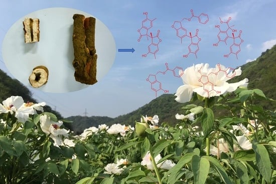
New Phenolic Dimers from Plant Paeonia suffruticosa and Their Cytotoxicity and NO Production Inhibition

1
School of Pharmaceutical Sciences, South-Central Minzu University, Wuhan 430074, China
2
School of Pharmacy, Anhui University of Chinese Medicine, Hefei 230012, China
3
Medical School, Fuyang Normal University, Fuyang 236037, China
4
State Key Laboratory of Phytochemistry and Plant Resources in West China, Kunming Institute of Botany, Chinese Academy of Sciences, Kunming 650201, China
*
Authors to whom correspondence should be addressed.
Molecules 2023, 28(12), 4590; https://doi.org/10.3390/molecules28124590
Submission received: 15 May 2023 / Revised: 3 June 2023 / Accepted: 5 June 2023 / Published: 6 June 2023

Abstract

:
The Paeonia suffruticosa, known as ‘Feng Dan’, has been used for thousands of years in traditional Chinese medicine. In our chemical investigation on the root bark of the plant, five new phenolic dimers, namely, paeobenzofuranones A–E (15), were characterized. Their structures were determined using spectroscopic analysis including 1D and 2D NMR, HRESIMS, UV, and IR, as well as ECD calculations. Compounds 2, 4, and 5 showed cytotoxicity against three human cancer cell lines, with IC50 values ranging from 6.7 to 25.1 μM. Compounds 1 and 2 showed certain inhibitory activity on NO production. To the best of our knowledge, the benzofuranone dimers and their cytotoxicity of P. suffruticosa are reported for the first time in this paper.

Graphical Abstract

1. Introduction

Paeonia suffruticosa is a perennial deciduous shrub belonging to the family Paeoniaceae. The dried root bark of P. suffruticosa is called ‘Feng Dan’ or ‘Mudanpi’ in China, which is used as a traditional Chinese medicine to clear pathogenic heat from the blood and promote blood circulation to remove blood stasis, as recorded in the Chinese Pharmacopoeia (2020 edition). Currently, pharmacological studies on P. suffruticosa demonstrate anti-inflammatory, antioxidant, and anti-tumor activities, as well as central nervous system and cardiovascular system protective activities [1,2,3]. In order to understand the bioactive metabolites, P. suffruticosa was chemically investigated, and more than 190 constituents were reported in the past ten years including phenolics, monoterpenes and glycosides, flavonoids, triterpenes, sesquiterpenes, and lignans. A pharmaceutical investigation on these metabolites demonstrated promising anti-inflammatory and anti-tumor properties; the representatives are paeonisides A and B, mudanpiosides C and F, and suffruticosol A [4,5,6,7,8].
In order to further clarify the chemical constituents and biological activities of genuine medicinal materials of ‘Feng Dan’ from Tongling (China), as well as part of our ongoing work on bioactive natural products from natural sources [9,10,11,12,13,14], the study on the chemical constituents of the root bark of P. suffruticosa was carried out. As a result, five new phenolics were isolated, namely, paeobenzofuranones A–E (15). Their structures (Figure 1) were determined using extensive spectroscopic methods. All compounds were evaluated for their cytotoxicity against three human cancer cell lines including breast cancer MDA-MB-231, human myeloid leukemia HL-60, and colon cancer SW480. In addition, their anti-inflammatory activity by inhibiting NO production was also evaluated. Herein, the isolation, structural elucidation, and bioactivities of the compounds from P. suffruticosa are reported.

2. Results and Discussion

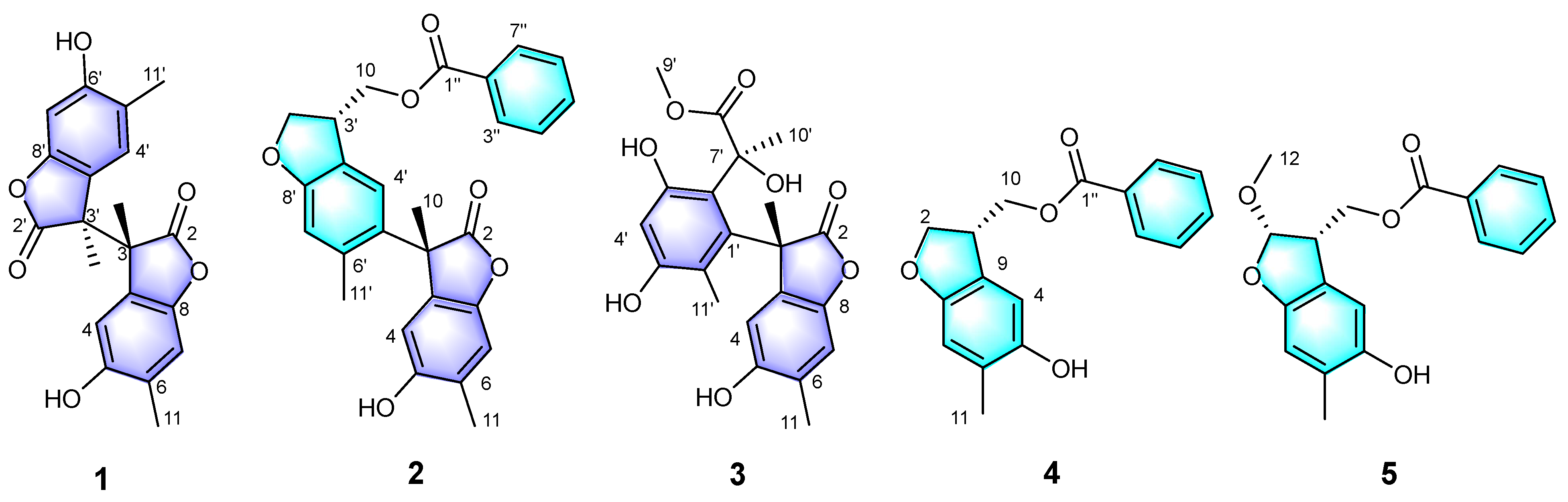
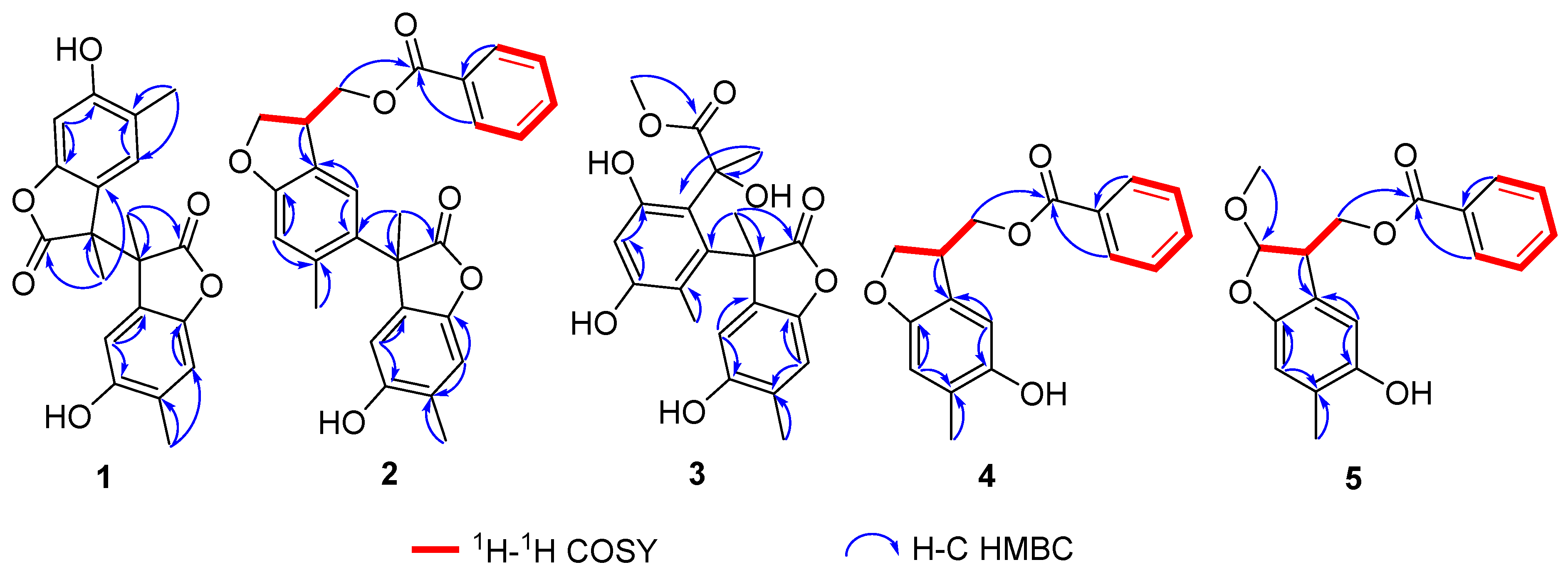
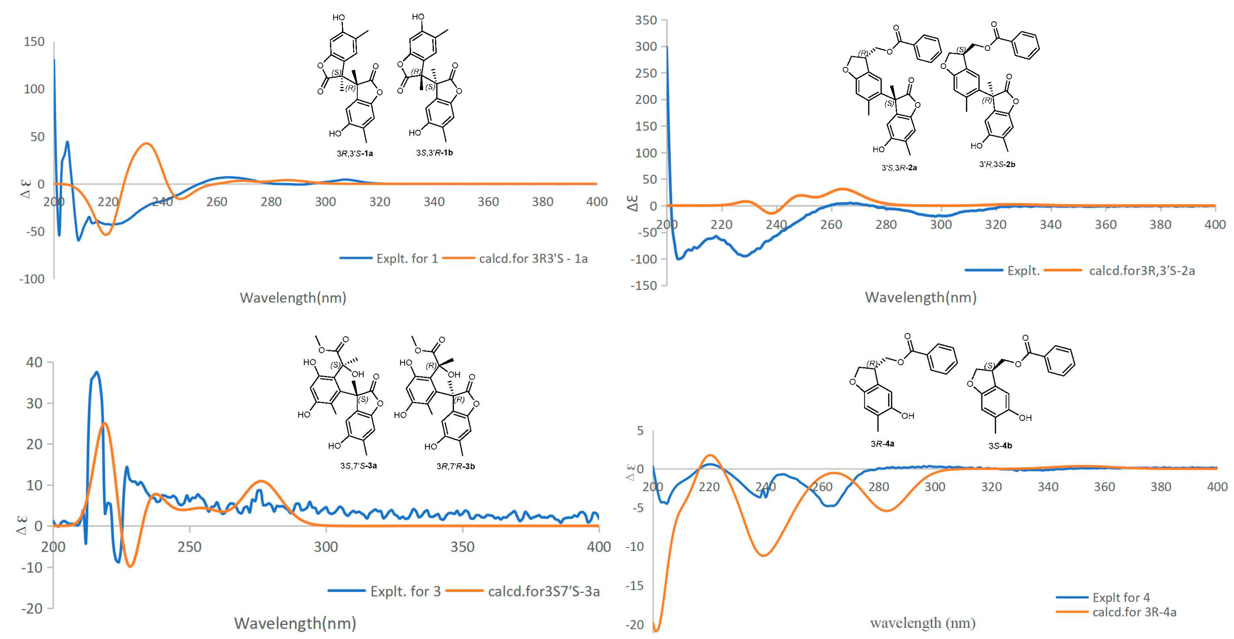
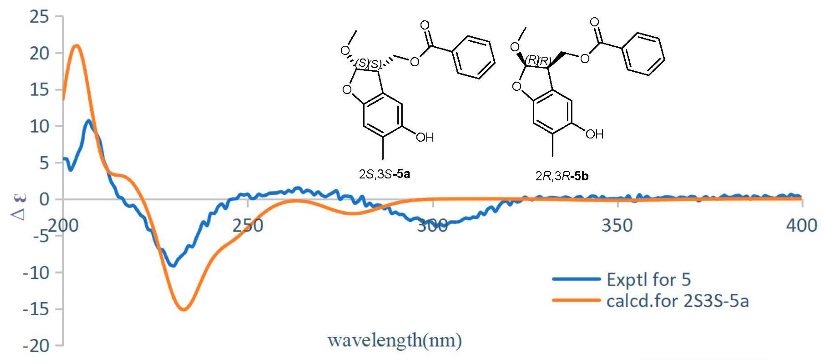
2.1. Structural Elucidation of Compounds 15

Compound 1 was obtained as a colorless oily liquid. Its molecular formula was determined as C20H18O6 by the HR-ESI-MS with a molecular ion peak at m/z 377.09952 ([M + Na]+, calcd. 377.10011). The UV absorption peaks at λmax 290 and 230 nm indicated the presence of a conjugated system. The IR spectrum indicated that compound 1 possessed hydroxyl (3406 cm−1) and lactone (1716 cm−1) groups. The 1H NMR spectroscopic data (Table 1) of compound 1 showed two methyl signals at δH 1.67 (s, 3H) and 2.15 (s, 3H), and two singlets for aromatic protons at δH 6.61 (1H, s, H-7) and 7.18 (1H, s, H-4). The interpretation of the 13C NMR (Table 2) and DEPT spectra data displayed 10 carbon signals, which indicated six non-protonated carbons (C-2: δC 179.4, C-3: δC 52.6, C-5: δC 113.3, C-6: δC 153.7, C-8: δC 146.2, C-9: δC 127.4), two CH (C-4: δC 128.1, C-7: δC 110.3), and two CH3 (C-10: δC 18.7, C-11: δC 16.6). A preliminary analysis of these data suggested that compound 1 should be a benzofuranone derivative with a structure similar to that of 4,6-dihydroxy-3,5-dimethylcoumaran-2-one [15]. The presence of a methyl at δC 18.7 and a carbonyl carbon at δC 179.4 indicated the differences. In the HMBC spectrum (Figure 2), correlations from H3-11 (δH 2.11) to C-4 (δC 128.1), C-5 (δC 113.3), and C-6 (δC 153.7), and from H3-10 (δH 1.67) to C-3 (δC 52.6), C-2 (δC 179.4), and C-9 (δC 127.4) enabled the assignment of the methyl and the carbonyl carbon. A further analysis of the 13C NMR data for a quaternary carbon at C-3 (δC 52.6) revealed that compound 1 should be a symmetric dimer with a linkage by the bond of C-3/C-3′. It was also supported by the MS data analysis. The absolute configurations of C-3 and C-3′ were identified as 3R and 3′S by the ECD calculations (Figure 3). Finally, the structure of compound 1 was identified and trivially named as paeobenzofuranone A.
Compound 2 was obtained as a white powder. Its molecular formula was determined as C27H24O6 by HR-ESI-MS (measured at m/z 445.16489 [M + Na]+; calcd. 445.16511). The UV spectrum revealed the conjugated system by peaks at λmax 295 and 230 nm. The 1D spectra data of compound 2 (Table 1 and Table 2) are partially identical to those of compound 1. The interpretation of the 1H and 13C spectroscopic data of compound 2 showed two benzofuran parts and an additional benzoyl moiety. The locations of the benzofuran parts were assigned by the HMBC correlations from H-10 (δH 1.75) to C-3 (δC 52.6), C-6 (δC 126.3), and C-2 (δC 179.3), as well as from H-4 (δH 6.61) to C-5 (δC 126.3) and C-3 (δC 52.6) (Figure 2). Furthermore, the location of the benzoyl was assigned by the key HMBC peaks from H-3′ (δH 3.81) to C-10′ (δC 66.9); from H-2′ (δH 4.43) to C-3′ (δC 43.5) and C-2′ (δC 73.1); and from H-10′ (δH 4.44) to C-3′ (δC 43.5) and C-10′ (δC 66.9). The absolute configurations of C-3 and C-3′ were established as 3S and 3′R by the ECD calculations (Figure 3). Then, the structure of compound 2 was established and named as paeobenzofuranone B.
Compound 3 was obtained as a white powder. The IR spectrum indicated that compound 3 possessed hydroxyl (3394 cm−1) and lactone (1712 cm−1) groups. Its molecular formula was determined as C21H22O8 by the HR-ESI-MS data analysis (m/z 425.12030 ([M + Na]+, calcd. 425.12124). The 1H and 13C NMR spectra data of compound 3 (Table 1 and Table 2) are partially the same as those of compound 1. The interpretation of the 1H and 13C spectroscopic data of compound 3 revealed one benzofuran part and one benzene ring. The benzofuran part was assigned by the HMBC correlations from H-10 (δH 1.75) to C-3 (δC 50.2), C-1′ (δC 133.7), and C-9 (δC 126.4); from H-11 (δH 2.02) to C-7 (δC 118.9), C-9 (δC 126.4), and C-5 (δC 149.2); and from H-4′ (δH 6.88) to C-2′ (δC 153.2) and C-5′ (δC 144.6) (Figure 2). The 1H-1H COSY cross peaks from δH 2.02 (3H, s, H-11) to δH 6.33 (1H, s, H-7), and from δH 1.77 (3H, s, H-11′) to δH 6.88 (1H, s, H-4′) verified the location of 10-CH3 and 11-CH3. The HMBC correlations verified the benzofuran group attached to C-8 (δC 144.3). Furthermore, from the HMBC correlations, the signal of another ester carbonyl group was connected to the benzene ring through the C-7′ (δC 75.1), as evidenced from δH 1.63 (3H, s, H-10′) to δC 75.1 (CH, C-7′), and from δH 3.73 (OCH3, s, H-9′) to δC 176.8 (C, C-8′). The absolute configurations of C-3 and C-7′ were established as 3S and 7′S by the ECD calculations (Figure 3). Eventually, the structure of compound 3 was elucidated as paeobenzofuranone C.
Compound 4 was obtained as a white powder. The IR spectrum indicated that compound 4 possessed hydroxyl (3383 cm−1) and lactone (1708 cm−1) groups. Its molecular formula was determined as C17H16O4 by the HR-ESI-MS data analysis (m/z 285.11215 ([M + H]+, calcd. 285.11268). The 1H and 13C spectra data of compound 4 (Table 1 and Table 2, Supplementary data) are partially the same as those of compound 1, except for the benzoyl and hydroxymethyl groups in compound 4. The interpretation of the 1H and 13C spectroscopic data of compound 4 implied one benzofuran part and one benzoyl. The locations of the benzofuran parts were assigned by the correlations revealed in the HMBC experiment (Figure 2) between the 11-CH3 (δH 2.14) and C-5 (δC 148.9), C-7 (δC 110.6), and C-8 (δC 153.4); as well as from H-4 (δH 6.74) to C-5, C-8, and C-9 (δC 125.6); from H-3 (δH 3.81) to C-10 (δC 66.4); and from H-7 (δH 4.37) to C-1′′ (δC 166.5). The 1H-1H COSY correlations from δH 3.81 (1H, s, H-3) to δH 4.43 (1H, s, H-10), and from δH 4.43 (1H, s, H-10) to δH 6.74 (1H, s, H-4) verified the location of the benzofuran part and one benzoyl connecting by C-3 and C-10. The absolute configuration of C-3 was established as 3R by the ECD calculations (Figure 4). Therefore, the structure of compound 4 was elucidated as paeobenzofuranone D.
Compound 5 was obtained as a white powder. Its molecular formula was determined as C18H18O5 by the HR-ESI-MS data analysis (m/z 313.10959 [M − H], calcd. 313.10743). The 1H and 13C NMR data resembled those of compound 4 (Table 1 and Table 2), except for the presence of an additional methoxy at C-2 in compound 5, which was confirmed by the key HMBC correlation of H-12 (δH 3.48) with C-2 (δC 111.2). A comprehensive analysis of the 2D NMR data indicated that other parts of compound 5 were the same as those of compound 4. The absolute configurations of C-2 and C-3 were established as 2S and 3S by the ECD calculations (Figure 4). Thus, the structure of compound 5 was established as paeobenzofuranone E.

2.2. Bioactivity Analysis

Five new compounds were tested for their inhibitory activities on nitric oxide production in the model of lipopolysaccharide-activated macrophages. As shown in Table 3, compounds 1 and 2 showed comparable inhibitory activity with the positive control at the concentration of 50 μM. In addition, all compounds were evaluated for their cytotoxicity against the HL-60, SW480, and MDA-MB-231 cell lines. As shown in Table 4, compounds 2, 4, and 5 demonstrated cytotoxicity against three human cancer cell lines. In particular, they exhibited potent cytotoxicity against HL-60 cells, with IC50 values of 6.8, 19.1, and 11.1 μM, compared to those of the positive control. In addition, compounds 4 and 5 showed no cytotoxicity to MDA-MB-231, indicating selectivity to the cancer cell lines.

3. Experiments

3.1. General Experimental Procedures

The UV spectra were obtained on a UH5300 UV-VIS Double Beam Spectrophotometer. The IR spectra were accessed using a Shimadzu Fourier transform infrared spectrometer with KBr pellets. The HRESIMS were measured on a Thermo Scientific Q Exactive Orbitrap mass spectrometer system. The NMR spectra (1H, 13C, and 2D NMR) were run on a Bruker Avance III NMR instrument at 600 MHz for 1H and 150 MHz for 13C NMR, while tetramethylsilane (TMS) was used as an internal standard. Column chromatography (CC) was executed on silica gel (200−300 mesh, Qingdao Marine Chemical Ltd., Qingdao, China), Sephadex LH-20 (Pharmacia Fine Chemical Co., Ltd., Stockholm, Sweden), and reverse phase silica gel (20−45 μm, Fuji Silysia Chemical Ltd., Kasugai, Japan). Medium pressure liquid chromatography (MPLC) was applied to Biotage SP2 equipment, and the columns were packed with reverse phase silica gel (C18). An Agilent 1260 series high-performance liquid chromatography (HPLC) system was used for the sample analysis (ZORBAX-SB C18 column, 5 μm, 4.6 × 250 mm, flowing rate = 1 mL/min) and preparation (ZORBAX-SB C18 column, 5 μm, 9.4 × 150 mm, flowing rate = 6 mL/min). All fractions were monitored using thin-layer chromatography (TLC) over GF 254 and silica gel 60 plates. The spots were visualized by using heating silica gel plates soaked with vanillin–sulfuric acid color component solvent.

3.2. Plant Material

The root barks of P. suffruticosa were collected in August 2021 from Tongling County, Anhui Province, People’s Republic of China. It was identified by Zhenghui Li. (Associate Professor of South-Central Minzu University, Wuhan, China). A voucher specimen (2021123 FD) was deposited at the School of Pharmaceutical Sciences, South-Central Minzu University.

3.3. Extraction and Isolation

The root bark of P. suffruticosa (50 kg) were mechanically crushed and extracted with MeOH/H2O (80:20) at 52 °C four times. The solvent was evaporated in vacuo to obtain a dark gum (9.3 kg). The latter was dissolved in a liter of water and then, respectively, extracted with petroleum ether (PE, 8L × 4) and dichloromethane (DCM, 8L × 4) to obtain PE (1.3 kg) and DCM parts (560 g). The DCM part was separated using a silica gel column eluted with PE: acetone (50:1, 40:1, 30:1, 20:1, 10:1) to obtain eight fractions (TPG-1–8). The fraction TPG-3 (28.5 g) was subjected to ODS silica gel CC and eluted with MeOH/H2O (20:90→90:10, v/v) to yield 10 fractions (Fr. 3-1→3-10). Fr. 3-2 (210 mg) was purified using Sephadex LH-20 (MeOH:DCM = 1:1) to obtain three fractions (Fr. 3-2-1, 3-2-2, 3-2-1). Fr. 3-2-3 was purified using preparative HPLC with CH3CN/H2O (30:70→60:40, v/v, 30 min) to obtain compound 1 (15.6 mg, tR = 12.5 min), compound 2 (9.7 mg, tR = 14.8 min), compound 4 (3.3 mg, tR = 16.9 min), and compound 5 (4.9 mg, tR = 17.8 min), respectively. Fr. 3-2-1 was prepared using HPLC with CH3CN/H2O (37:63→60:40, v/v, 30 min) to obtain compound 3 (3.2 mg) at 17.8 min.

3.3.1. Paeobenzofuranone A (1)

Colorless oil; [ α ] D 26 −98.6 (c = 0.09, MeOH); UV (MeOH) λmax (log ε): 230 (3.6) nm; IR (KBr) νmax 3406, 1716, 1450, 1346, 1315, 1276, 1026, 1049, and 713 cm−1; HRESIMS: m/z 377.09952 [M + Na]+, (calcd. for C20H18O6Na+, 377.10011). The 1H and 13C NMR data are displayed in Table 1 and Table 2.

3.3.2. Paeobenzofuranone B (2)

White powder; UV (MeOH) λmax (log ε): 230 (3.2) nm; [ α ] D 26 −15.1 (c = 0.09, MeOH); HRESIMS: m/z 445.16489 [M + H]+ (calcd. for C27H24O6+ 445.16511). 1H and 13C NMR data are displayed in Table 1 and Table 2.

3.3.3. Paeobenzofuranone C (3)

White powder; [ α ] D 22 –5.6 (c = 0.12, MeOH); UV (MeOH) λmax (log ε): 230 (3.2) nm; IR (KBr) νmax: 3394, 1712, 1450, 1346, 1315, 1276, 1176, and 1072 cm−1; HRESIMS: m/z 425.12030 ([M + Na]+ (calcd. for C21H22O8Na+, 425.12124). 1H and 13C NMR data are displayed in Table 1 and Table 2.

3.3.4. Paeobenzofuranone D (4)

White powder; [ α ] D 22 –3.8 (c = 0.11, MeOH); UV (MeOH) λmax (log ε): 230 (1.973) nm; IR (KBr) νmax: 3383, 1708, 1450, 1342, 1315, 1276, 1176, and 1072 cm−1; HRESIMS m/z 285.11215 ([M + H]+ (calcd. for C17H16O4+, 285.11268). 1H and 13C NMR data are displayed in Table 1 and Table 2.

3.3.5. Paeobenzofuranone E (5)

White powder; [ α ] D 26 −13.8 (c = 0.09, MeOH); UV (MeOH) λmax (log ε): 230 (3.136) nm; HRESIMS m/z 313.10959 ([M−H] (calcd. for C18H18O5, 313.10743). 1H and 13C NMR data are displayed in Table 1 and Table 2.

3.4. Cytotoxicity Assay

The cytotoxicity for the isolates was evaluated using the MTS assay. Briefly, 1 × 10⁵ cells/mL from three human cancer cell lines, breast cancer MDA-MB-231, human myeloid leukemia HL-60, and colon cancer SW480, were seeded in 96-well plates. After 24 h incubation, the cells were treated with test compounds or cisplatin (DDP, positive control) at given concentrations (40, 8, 1.6, 0.32, 0.064 μM) for 48 h. The MTS was then added to each well, and the plates were stored for 4 h. The absorbance was read at 490 nm. The IC50 (50% concentration of inhibition) was calculated using the Reed–Muench method [16,17].

3.5. NO Inhibitory Activity Assays

The mouse mononuclear macrophages RAW264.7 were seeded into 96-well plates, induced, and stimulated with 1 μg/mL LPS; at the same time, five new compounds with different concentrations to be tested were added. The drug-free group and the L-NMMA positive drug group were set approximately equal as a comparison. After the cells were cultured overnight, the medium was taken to detect the production of NO, and the absorbance was measured at 570 nm. The MTS was added to the remaining medium for cell viability assays to exclude the toxic effects of the compounds on the cells. The assays were performed as triplicate batch experiments. The NO production inhibition rate (%) = (OD570nm of non-drug treatment group−OD570nm of sample group)/OD570nm of non-drug treatment group × 100% [18,19].

3.6. ECD Calculations

The conformers of the five calculated compounds were generated via MMFF in ChemDraw. The ECD were calculated at the B3LYP/6-31+G(d,p) level in methanol with the PCM model. The calculated ECD curves and weighted ECD were all generated using SpecDis 1.71 based on the Boltzmann distribution theory, and the simulated spectra of all the predominant conformers were averaged to obtain the final conformationally averaged data [20]. All of the density functional theory (DFT) calculations were implemented using the Gaussian 16 software package with the Gaussian 09 default keyword. For the computational details of compounds 15, see the Supplementary Materials.

4. Conclusions

In the present study, the chemical investigation on Paeonia suffruticosa results in the isolation of five new benzofuran compounds, containing rare dimers (compounds 13) and hetero-dimers (compounds 4 and 5). Their structures were determined using extensive spectroscopic methods. This work represents the first report of new benzofuran dimers of P. suffruticosa and their cytotoxicity and broadens the horizon of the structural diversity of P. suffruticosa.

Supplementary Materials

The following supporting information can be downloaded at https://www.mdpi.com/article/10.3390/molecules28124590/s1, Figures S1–S35: HRESIMS, 1D & 2D NMR, ECD data of compounds 15; Tables S1–S5: Energy analysis for conformers of 15 at B3LYP/6-31+G(d,p)level in the gas phase.

Author Contributions

T.F. and J.L. designed and guided the experiment; Q.M. performed the isolation and identification of the compounds and wrote the manuscript; S.T. and Y.Z. contributed to the isolation of these compounds; X.P. reviewed the manuscript; Z.L. obtained the plant material and identification; T.F. and J.L. reviewed the manuscript. All authors have read and agreed to the published version of the manuscript.

Funding

This work was financially supported by the National Natural Science Foundation of China (22177138, 21961142008).

Institutional Review Board Statement

Not applicable.

Informed Consent Statement

Not applicable.

Data Availability Statement

All the data in this research are presented in the manuscript and Supplementary Materials.

Acknowledgments

The authors thank the Bioactivity Screening Center, the Kunming Institute of Botany, and the Chinese Academy of Sciences for screening the bioactivity of the compounds.

Conflicts of Interest

The authors declare no conflict of interest.

References

  1. Ding, L.; Zhao, F.; Chen, L.; Jiang, Z.; Liu, Y.; Li, Z.; Qiu, F.; Yao, X. New monoterpene glycosides from Paeonia suffruticosa Andrews and their inhibition on NO production in LPS-induced RAW 264.7 cells. Bioorg. Med. Chem. Lett. 2012, 22, 7243–7247. [Google Scholar] [CrossRef] [PubMed]
  2. Wang, S.C.; Tang, S.W.; Lam, S.H.; Wang, C.C.; Liu, Y.H.; Lin, H.Y.; Lee, S.S.; Lin, J.Y. Aqueous extract of Paeonia suffruticosa inhibits migration and metastasis of renal cell carcinoma cells via suppressing VEGFR-3 pathway. Evid. Based Compl. Alt. 2012, 2012, 409823. [Google Scholar] [CrossRef] [Green Version]
  3. Qiu, H.; Zhang, L.; Zhu, M.; Zhang, M.; Chen, J.; Feng, L.; Jia, X.; Jacob, J.A. Capture of anti-coagulant active ingredients from Moutan Cortex by platelet immobilized chromatography and evaluation of anticoagulant activity in rats. Biomed. Pharmacother. 2017, 95, 235–244. [Google Scholar] [CrossRef] [PubMed]
  4. Pei, L.; Jie, S.; Chunnian, H.; Pei, X. Genus Paeonia: A comprehensive review on traditional uses, phytochemistry, pharmacological activities, clinical application, and toxicology. J. Ethnopharmacol. 2021, 269, 113708. [Google Scholar] [CrossRef]
  5. Mencherini, T.; Picerno, P.; Festa, M.; Russo, P.; Capasso, A.; Aquino, R. Triterpenoid constituents from the roots of Paeonia rockii ssp. rockii. J. Nat. Prod. 2011, 74, 2116–2121. [Google Scholar] [CrossRef] [PubMed]
  6. Huang, Y.; Ohno, O.; Suenaga, K.; Miyamoto, K. Apoptosis-inducing activity and antiproliferative effect of paeoniflorigenone from Moutan Cortex. Biosci. Biotechnol. Biochem. 2017, 81, 1106–1113. [Google Scholar] [CrossRef] [PubMed] [Green Version]
  7. Liu, P.; Wang, Y.; Gao, J.; Lu, Z.; Yin, W.; Deng, R. Resveratrol trimers from seed cake of Paeonia rockii. Molecules 2014, 19, 19549–19556. [Google Scholar] [CrossRef] [PubMed] [Green Version]
  8. Furuya, R.; Hu, H.; Zhang, Z.; Shigemori, H. Suffruyabiosides A and B, two new monoterpene diglycosides from Moutan Cortex. Molecules 2012, 17, 4915–4923. [Google Scholar] [CrossRef] [PubMed] [Green Version]
  9. Liu, J.K.; Ma, Y.B.; Wu, D.G.; Lu, Y.; Shen, Z.Q.; Zheng, Q.T.; Chen, Z.H. Paeonilide, a novel anti-PAF active monoterpenoid derived metabolite from Paeonia delavayi. Biosci. Biotech. Biochem. 2000, 64, 1511–1514. [Google Scholar] [CrossRef] [Green Version]
  10. Yang, X.Y.; Feng, T.; Li, Z.H.; Sheng, Y.; Yin, X.; Leng, Y.; Liu, J.K. Conosilane A, an unprecedented sesquiterpene from the Cultures of Basidiomycete Conocybe siliginea. Org. Lett. 2012, 14, 5382–5384. [Google Scholar] [CrossRef] [PubMed]
  11. Wang, F.; Gao, Y.; Zhang, L.; Liu, J.K. Bi-linderone, a highly modified methyl-linderone dimer from Lindera aggregata with activity toward improvement of insulin sensitivity in vitro. Org. Lett. 2010, 12, 2354–2357. [Google Scholar] [CrossRef] [PubMed]
  12. Yang, X.L.; Hsieh, K.L.; Liu, J.K. Guajadial: An unusual meroterpenoid from guava leaves Psidium guajava. Org. Lett. 2007, 9, 5135–5138. [Google Scholar] [CrossRef] [PubMed]
  13. Feng, T.; Su, J.; Ding, Z.H.; Zheng, Y.T.; Li, Y.; Leng, Y.; Liu, J.K. Chemical constituents and their bioactivities of Tongling White Ginger (Zingiber officinale). J. Agric. Food Chem. 2011, 59, 11690–11695. [Google Scholar] [CrossRef] [PubMed]
  14. Wu, D.G.; Cheng, C.Q.; Liu, J.K. X-ray Crystal Structure of Angulatusine A, a new sesquiterpene alkaloid from Celastrus Angulatus. J. Nat. Prod. 1992, 55, 982–985. [Google Scholar] [CrossRef]
  15. Ha, D.T.; Ngoc, T.M.; Lee, I.S.; Lee, Y.M.; Kim, J.S.; Jung, H.J. Inhibitors of aldose reductase and formation of advanced glycation end-products in moutan cortex (Paeonia suffruticosa). J. Nat. Prod. 2009, 72, 1465–1470. [Google Scholar] [CrossRef] [PubMed]
  16. Yu, H.L.; Long, Q.; Yi, W.F.; Yang, B.J.; Ding, X.; Hao, X.J. Two new C21 steroidal glycosides from the roots of Cynanchum paniculatum. Nat. Prod. Bioprospect. 2019, 4, 26. [Google Scholar] [CrossRef] [PubMed] [Green Version]
  17. Reed, L.J.; Muench, H. A simple method of estimating fifty percent endpoints. Am. J. Epidemiol. 1938, 27, 493–497. [Google Scholar] [CrossRef]
  18. Anh, H.L.T.; Cuc, N.T.; Tai, B.H.; Yen, P.H.; Nhiem, N.X.; Thao, D.T.; Nam, N.H.; Minh, C.V.; Kiem, P.V.; Kim, Y.H. Synthesis of chromonylthiazolidines and their cytotoxicity to human cancer cell Lines. Molecules 2015, 20, 1151–1160. [Google Scholar] [CrossRef] [PubMed] [Green Version]
  19. Snene, A.; Mokni, R.E.; Jmii, H.; Jlassi, I.; Jadane, H.; Falconieri, D.; Piras, A.; Dhaouadi, H.; Porcedda, S.; Hammami, S. In vitro antimicrobial, antioxidant and antiviral activities of the essential oil and various extracts of wild (Daucus virgatus (Poir.) Maire) from Tunisia. Ind. Crops Prod. 2017, 109, 109–115. [Google Scholar] [CrossRef]
  20. Bruhn, T.; Schaumloeffel, A.; Hemberger, Y.; Bringmann, G. SpecDis: Quantifying the comparison of calculated and experimental electronic circular dichroism spectra. Chirality 2013, 25, 243–249. [Google Scholar] [CrossRef] [PubMed]
Figure 1. Structures of compounds 15.
Molecules 28 04590 g001
Figure 2. Selected HMBC and 1H–1H COSY correlations of compounds 15.
Molecules 28 04590 g002
Figure 3. ECD calculations for compounds 14.
Molecules 28 04590 g003
Figure 4. ECD calculations for compound 5.
Molecules 28 04590 g004
Table 1. 1H NMR spectroscopic data of compounds 15 in Methanol-d4 (600 MHz, J in Hz).
Position12345
2 4.46, m5.48, t (1.8)
3 3.81, t (7.4)3.80, t (7.5)
47.18, s6.61, s6.96, s6.74, s6.72, s
76.61, s6.52, s6.33, s4.44, dd (10.7, 5.9)
4.37, dd (18.0, 9.1)
4.46, dd (11.1, 5.5)
4.33, dd (11.1, 7.8)
101.67, s, 3H1.75, s, 3H1.72, s, 3H4.43, m4.48, m
112.15, s, 3H2.13, s, 3H2.02, s, 3H2.14, s, 3H2.15, s, 3H
12 3.48, s
2′ 4.43, m; 4.06, m 8.00, d (1.5)7.97, d (1.5)
3′ 3.81, t (7.5) 7.44, m7.45, m
4′7.18, s6.63, s 6.88, s7.58, m7.61, m
5′6.61, s6.62, s 7.49, m7.48, m
6′1.67, s, 3H 8.01, d (1.3)7.99, d (1.3)
7′2.15, s, 3H6.85, s
9′ 3.73, s
10′ 4.60, dd (18.0, 9.1)
4.44, dd (10.8, 5.9)
1.63, s, 3H
11′ 2.05, s, 3H1.77, s, 3H
3′′ 8.01, d (1.2)
4′′ 7.48, t (7.8)
5′′ 7.56, m
6′′ 7.48, t (7.8)
7′′ 8.00, d (1.4)
Table 2. 13C NMR spectroscopic data of compounds 15 in Methanol-d4 (150 MHz).
Position12345
2179.4, C179.3, C182.7, C73.5, CH2111.2, CH
352.6, C52.6, C50.2, C42.0, CH50.5, CH
4128.1, CH112.2, CH115.6, CH110.8, CH112.6, CH
5113.3, C149.3, C149.2, C148.9, C150.9, C
6153.7, C126.3, C125.0, C128.5, CH124.4, CH
7110.3, CH112.4, CH118.9, CH110.6, CH112.3, CH
8146.2, C125.9, C144.3, C153.4, C153.2, C
9127.4, C127.4, C126.4, C125.6, C129.8, C
1018.7, CH318.6, CH322.3, CH366.4, CH266.3, CH2
1116.6, CH316.6, CH315.9, CH315.6, CH317.0, CH3
12 56.2, OCH3
1′ 133.7, C
2′179.4, C73.1, CH2153.2, C
3′52.6, C43.5, CH122.0, C
4′128.1, CH111.7, CH115.9, CH
5′113.3, C126.4, C144.6, C
6′153.7, C131.4, CH125.7, C
7′110.3, CH110.3, CH75.1, C
8′146.2, C154.9, C176.8, C
9′127.4, C124.4, C53.2, OCH3
10′18.7, CH366.9, CH226.0, CH3
11′16.6, CH316.8, CH310.4, CH3
1′′ 168.1, C 166.5, C167.9, C
2′′ 130.7, C 129.7, C131.2, C
3′′ 129.7, CH 129.2, CH130.4, CH
4′′ 128.1, CH 128.2, CH129.5, CH
5′′ 134.5, CH 132.9, CH130.7, CH
6′′ 129.7, CH 128.2, CH129.5, CH
7′′ 128.1, CH 129.1, CH130.4, CH
Table 3. Inhibitory activities of compounds 15 on NO production at 50 μM.
CompoundInhibition Activity (100%)
L-NMMA a52.0 ± 1.96
143.9 ± 2.07
244.6 ± 0.52
313.0 ± 1.59
433.7 ± 2.24
530.9 ± 1.56
a L-NMMA (NG-monomethyl-L-arginine, monoacetate salt) was used as the positive control.
Table 4. Cytotoxicity of compounds 2, 4, and 5 (IC50 ± SD, μM).
CompoundHL-60MDA-MB-231SW480
26.8 ± 0.1120.9 ± 0.4612.6 ± 0.73
419.1 ± 0.32>408.9 ± 0.40
511.1 ± 1.61>4010.7 ± 0.43
DDP a23.5 ± 0.7716.9 ± 1.1925.1 ± 1.26
a DDP (Cisplatin) was used as the positive control.
Disclaimer/Publisher’s Note: The statements, opinions and data contained in all publications are solely those of the individual author(s) and contributor(s) and not of MDPI and/or the editor(s). MDPI and/or the editor(s) disclaim responsibility for any injury to people or property resulting from any ideas, methods, instructions or products referred to in the content.

