

Recent Advances of Diketopyrrolopyrrole Derivatives in Cancer Therapy and Imaging Applications

Abstract
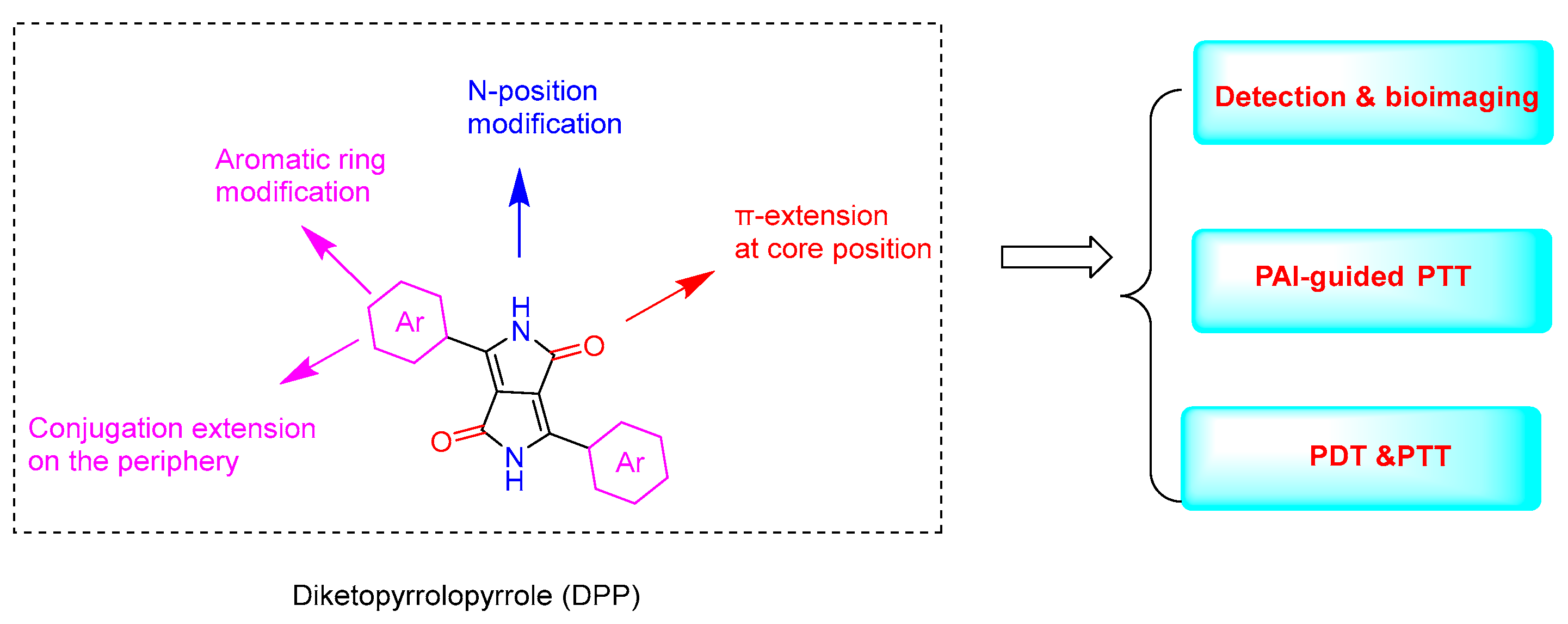
:1. Introduction
2. Application
2.1. Imaging and Detection

2.2. Detection and PDT
2.3. PTT and Photoacoustic Imaging (PAI)-Guided PTT
2.4. PDT/PTT Combination Therapy

3. Conclusions
- Both efficient ROS production efficiency and good photothermal conversion efficiency can be achieved via collaborative processing of PDT/PTT. Moreover, since immunotherapy is a promising cancer treatment approach, DPP-based dyes for combinational phototherapy and immunotherapy have been developed [60]. Moreover, multifunctional DPP materials created by combining optical/acoustic/magnetic imaging modes with other therapeutic modes (chemodynamic therapy or gene therapy, etc.) are promising in practical applications.
- NIR-II fluorescence imaging, as a non-invasive imaging technology that provides centimeter-level depth and micron-level resolution, has been investigated. In order to further broaden the application of DPP derivatives, it is necessary to further develop DPP derivatives that can be applied to NIR-II fluorescence bioimaging.
- The practical clinical application is limited by issues such as biocompatibility, cytotoxicity, targeting specificity, and biodegradability. For example, the mitochondria of normal cells and cancer cells are different, which has led to the development of DPP derivatives that can target mitochondria to improve the effectiveness of treatment. The targeting ability and response to external stimuli are also important during molecular design.
- Cancer cell membranes can be penetrated by nanomaterials, allowing accumulation in diseased areas and improvement of treatment effectiveness. Converting DPP derivatives into nanoreagents should be considered.
- A simpler synthesis route of DPP derivatives should be designed to achieve maximum effects at the lowest cost.
Author Contributions
Funding
Institutional Review Board Statement
Informed Consent Statement
Data Availability Statement
Conflicts of Interest
Abbreviations
References
- Patil, Y.; Misra, R. Rational Molecular Design towards NIR Absorption: Efficient Diketopyrrolopyrrole Derivatives for Organic Solar Cells and Photothermal Therapy. J. Mater. Chem. C 2019, 7, 13020–13031. [Google Scholar] [CrossRef]
- Li, W.; Wang, L.; Tang, H.; Cao, D. Diketopyrrolopyrrole-Based Fluorescent Probes for Detection and Bioimaging: Current Progresses and Perspectives. Dyes Pigments 2019, 162, 934–950. [Google Scholar] [CrossRef]
- Auwalu, M.A.; Cheng, S. Diketopyrrolopyrrole Fluorescent Probes, Photophysical and Biological Applications. Chemosensors 2021, 9, 44. [Google Scholar] [CrossRef]
- Yang, M.; Yang, T.; Mao, C. Enhancement of Photodynamic Cancer Therapy by Physical and Chemical Factors. Angew. Chem. Int. Ed. 2019, 58, 14066–14080. [Google Scholar] [CrossRef] [PubMed]
- Shen, Y.; Liang, L.; Zhang, S.; Huang, D.; Deng, R.; Zhang, J.; Qu, H.; Xu, S.; Liang, C.; Xu, W. Organelle-Targeting Gold Nanorods for Macromolecular Profiling of Subcellular Organelles and Enhanced Cancer Cell Killing. ACS Appl. Mater. Interfaces 2018, 10, 7910–7918. [Google Scholar] [CrossRef] [PubMed]
- Jung, H.S.; Verwilst, P.; Sharma, A.; Shin, J.; Sessler, J.L.; Kim, J.S. Organic Molecule-Based Photothermal Agents: An Expanding Photothermal Therapy Universe. Chem. Soc. Rev. 2018, 47, 2280–2297. [Google Scholar] [CrossRef]
- Cai, Y.; Liang, P.; Tang, Q.; Si, W.; Chen, P.; Zhang, Q.; Dong, X. Diketopyrrolopyrrole-Based Photosensitizers Conjugated with Chemotherapeutic Agents for Multimodal Tumor Therapy. ACS Appl. Mater. Interfaces 2017, 9, 30398–30405. [Google Scholar] [CrossRef]
- Ma, Q.; Sun, X.; Wang, W.; Yang, D.; Yang, C.; Shen, Q.; Shao, J. Diketopyrrolopyrrole-derived Organic Small Molecular Dyes for Tumor Phototheranostics. Chin. Chem. Lett. 2022, 33, 1681–1692. [Google Scholar] [CrossRef]
- Jiang, X.; Wang, L.; Tang, H.; Cao, D.; Chen, W. Diketopyrrolopyrrole: An Emerging Phototherapy Agent in Fighting Cancer. Dyes Pigments 2020, 181, 108599. [Google Scholar] [CrossRef]
- Wang, L.; Xin, S.; Tang, H.; Cao, D. Research Progress in Cancer Treatment by Diketopyrrolopyrrole-Based Photosensitizers and Photothermal Agents. Chin. J. Org. Chem. 2020, 40, 4155. [Google Scholar] [CrossRef]
- Zhang, Q.; Wang, Q.; Xu, X.; Liu, J.; Lu, X.; Huang, W.; Fan, Q. Diketopyrrolopyrrole Derivatives-Based NIR-II Fluorophores for Theranostics. Dyes Pigments 2021, 193, 109480. [Google Scholar] [CrossRef]
- Ding, A.X.; Tan, Z.L.; Shi, Y.D.; Song, L.; Gong, B.; Lu, Z.L. Tetraphenylethene End-Capped Diketopyrrolopyrrole Fluorogens with AIE and Large Two-Photon Absorption Cross-Sections Features and Application in Bioimaging. ACS Appl. Mater. Interfaces 2017, 9, 11546–11556. [Google Scholar] [CrossRef]
- Chiminazzo, A.; Borsato, G.; Favero, A.; Fabbro, C.; McKenna, C.E.; Dalle Carbonare, L.G.; Valenti, M.T.; Fabris, F.; Scarso, A. Diketopyrrolopyrrole Bis-Phosphonate Conjugate: A New Fluorescent Probe for In Vitro Bone Imaging. Chem. Eur. J. 2019, 25, 3617–3626. [Google Scholar] [CrossRef] [PubMed]
- Washington, K.E.; Du, J.; Kularatne, R.N.; Calubaquib, E.L.; Soltantabar, P.; Biewer, M.C.; Stefan, M.C. Diketopyrrolopyrrole and Benzodithiophene Based near Infrared-Emitting Small Molecule for Imaging Applications. Synth. Met. 2019, 256, 116123. [Google Scholar] [CrossRef]
- Du, C.; Fu, S.; Wang, X.; Sedgwick, A.C.; Zhen, W.; Li, M.; Li, X.; Zhou, J.; Wang, Z.; Wang, H.; et al. Diketopyrrolopyrrole-Based Fluorescence Probes for the Imaging of Lysosomal Zn 2+ and Identification of Prostate Cancer in Human Tissue. Chem. Sci. 2019, 10, 5699–5704. [Google Scholar] [CrossRef]
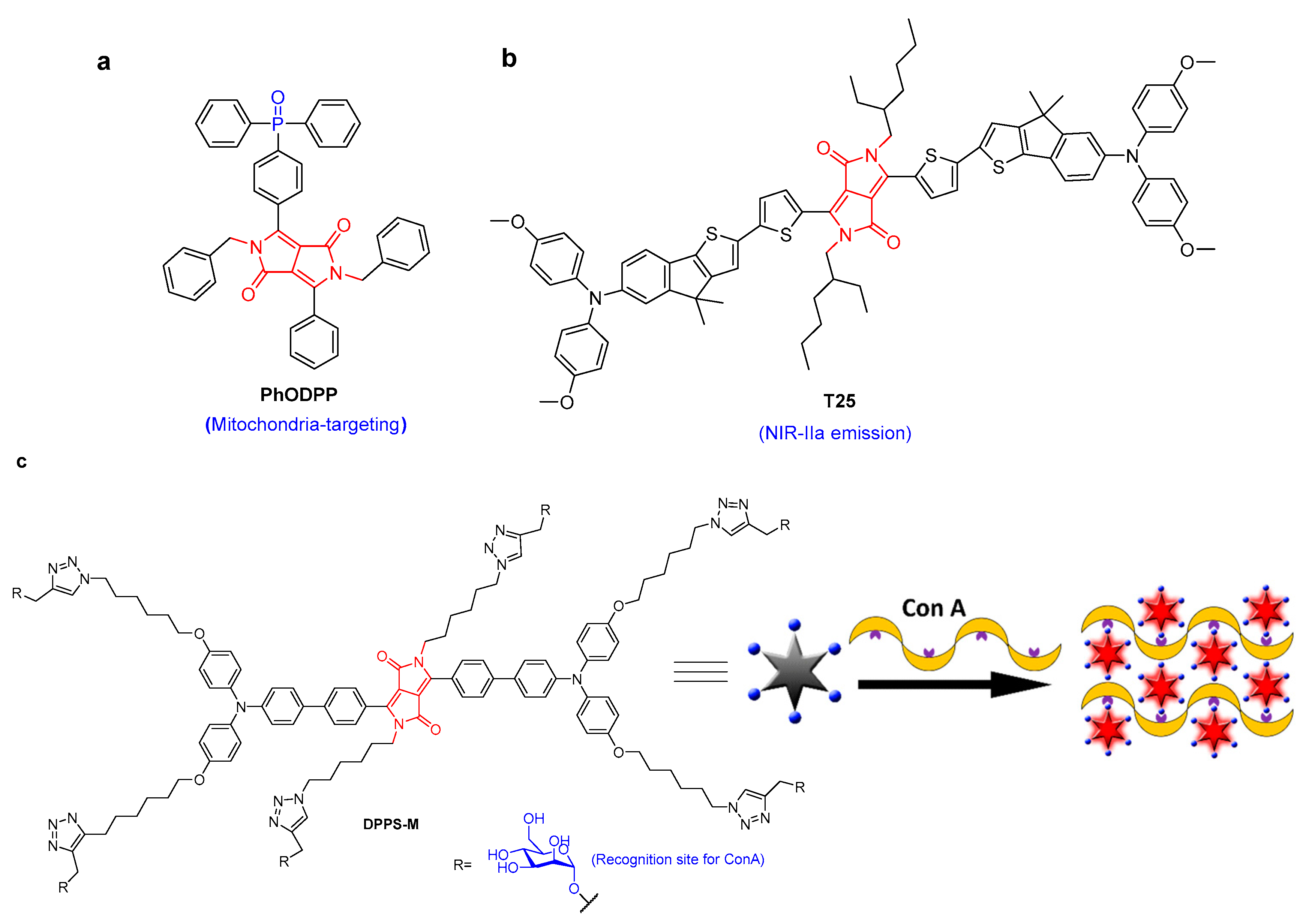
- Abelha, T.F.; Morris, G.; Lima, S.M.; Andrade, L.H.C.; McLean, A.J.; Alexander, C.; Calvo-Castro, J.; McHugh, C.J. Development of a Neutral Diketopyrrolopyrrole Phosphine Oxide for the Selective Bioimaging of Mitochondria at the Nanomolar Level. Chem. Eur. J. 2020, 26, 3173–3180. [Google Scholar] [CrossRef]
- Yang, Z.; Fan, X.; Li, H.; Li, X.; Li, S.; Zhang, Z.; Lin, H.; Qian, J.; Hua, J. A Small-Molecule Diketopyrrolopyrrole-Based Dye for in Vivo NIR-IIa Fluorescence Bioimaging. Chem. Eur. J. 2021, 27, 14240–14249. [Google Scholar] [CrossRef] [PubMed]
- Hang, Y.; He, X.-P.; Yang, L.; Hua, J. Probing Sugar–Lectin Recognitions in the near-Infrared Region Using Glyco-Diketopyrrolopyrrole with Aggregation-Induced-Emission. Biosens. Bioelectron. 2015, 65, 420–426. [Google Scholar] [CrossRef]
- Wang, J.; Hang, Y.; Hua, J. Mannose-Functionalized Diketopyrrolopyrrole as AIE-Active Probes for Lectin Detection and Cancer Cell Imaging. Sens. Actuators B Chem. 2019, 282, 232–242. [Google Scholar] [CrossRef]
- Chen, Y.; Li, K.; Zhang, S.; Xu, P.; Song, B. Turn-on Fluorescence Probe for BSA Detection and Selective Cell Imaging. Dyes Pigments 2022, 202, 110267. [Google Scholar] [CrossRef]
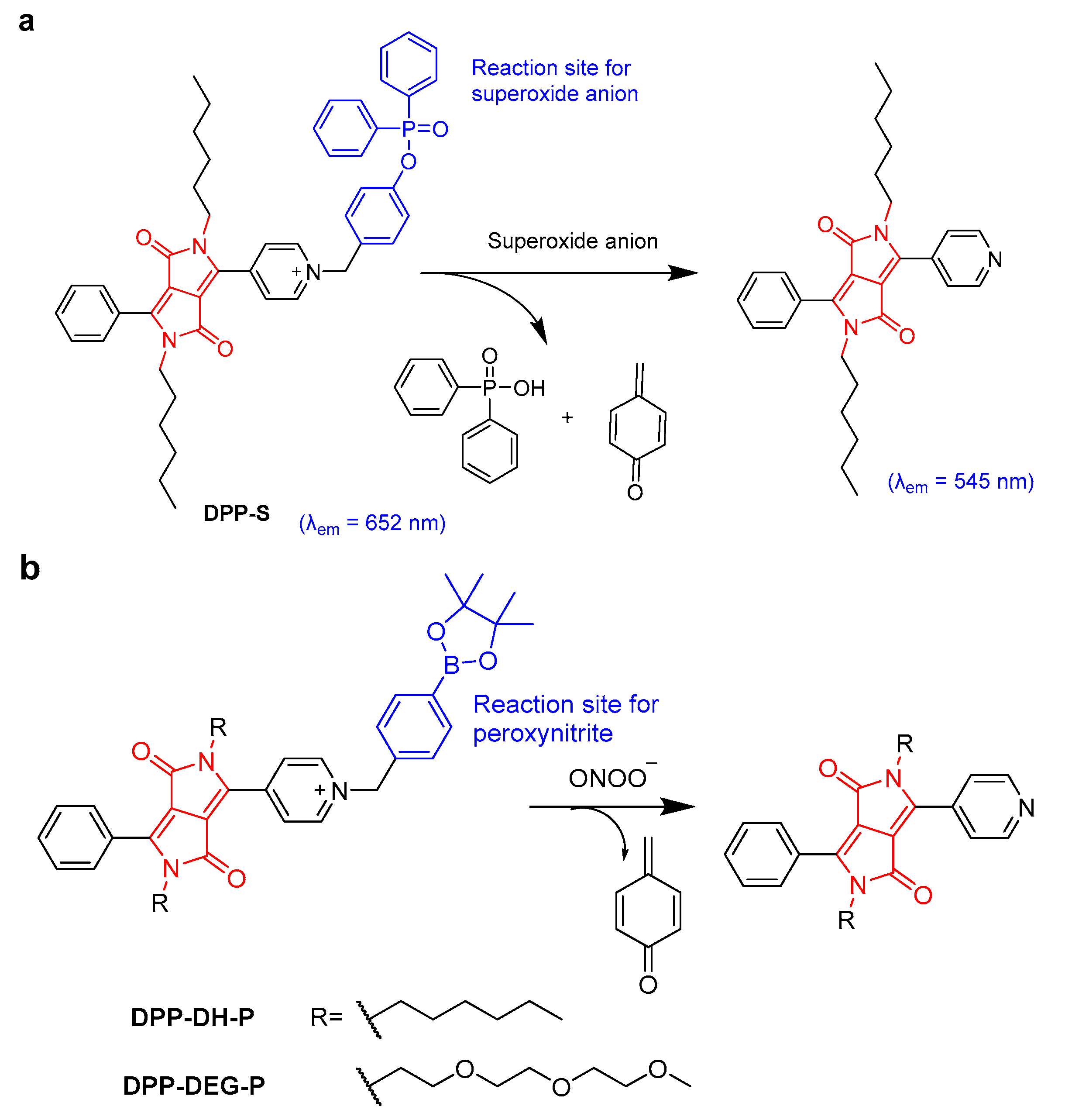
- Wang, J.; Liu, L.; Xu, W.; Yang, Z.; Yan, Y.; Xie, X.; Wang, Y.; Yi, T.; Wang, C.; Hua, J. Mitochondria-Targeted Ratiometric Fluorescent Probe Based on Diketopyrrolopyrrole for Detecting and Imaging of Endogenous Superoxide Anion in Vitro and in Vivo. Anal. Chem. 2019, 91, 5786–5793. [Google Scholar] [CrossRef]
- Wang, N.; Wang, H.; Zhang, J.; Ji, X.; Su, H.; Liu, J.; Wang, J.; Zhao, W. Diketopyrrolopyrrole-Based Sensor for over-Expressed Peroxynitrite in Drug-Induced Hepatotoxicity via Ratiometric Fluorescence Imaging. Sens. Actuators B Chem. 2022, 352, 13099292. [Google Scholar] [CrossRef]
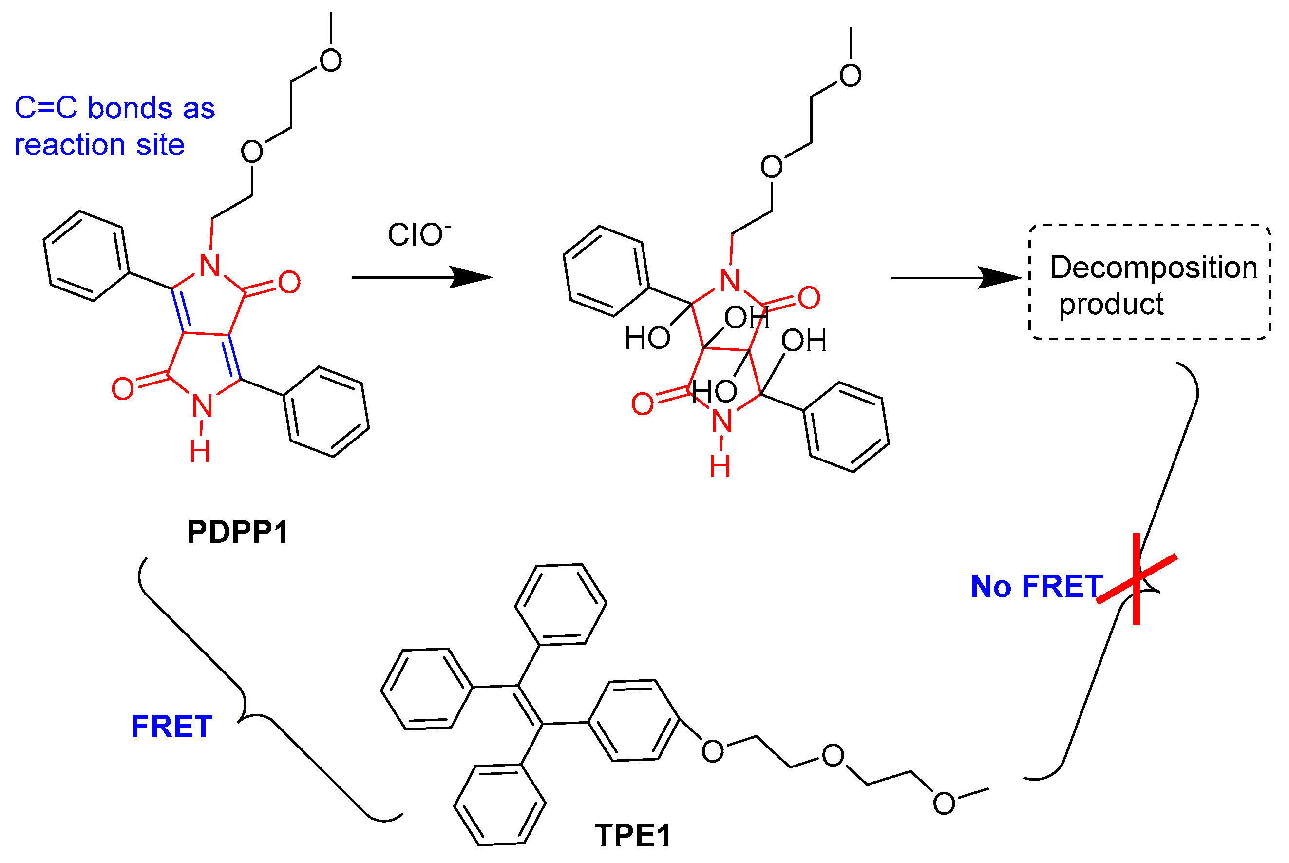
- Nie, K.; Yuan, Y.; Peng, X.; Song, J.; Qu, J. A Diketopyrrolopyrrole-Based Hybrid Organic Nanoprobe for Ratiometric Imaging of Endogenous Hypochlorite in Live Cells. Sens. Actuators B Chem. 2020, 307, 127632. [Google Scholar] [CrossRef]
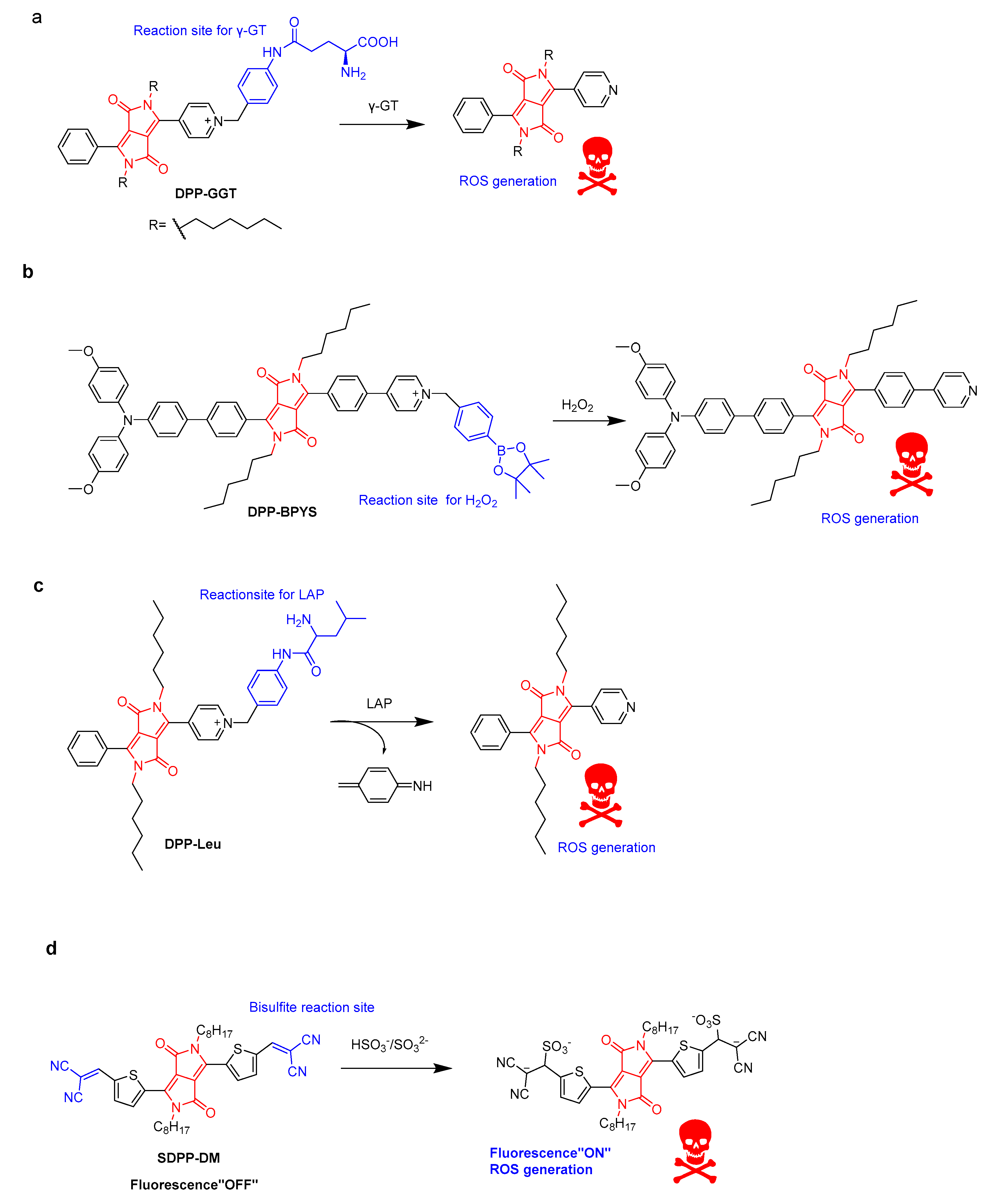
- Yang, Z.; Xu, W.; Wang, J.; Liu, L.; Chu, Y.; Wang, Y.; Hu, Y.; Yi, T.; Hua, J. Diketopyrrolopyrrole-Based Multifunctional Ratiometric Fluorescent Probe and γ-Glutamyltranspeptidase-Triggered Activatable Photosensitizer for Tumor Therapy. J. Mater. Chem. C 2020, 8, 8183–8190. [Google Scholar] [CrossRef]
- Li, X.; Xu, W.; Yang, Z.; Li, S.; Gu, X.; Yuan, T.; Li, C.; Wang, Y.; Hua, J. A Lipid Droplet-Targeted Multifunctional AIE-Active Fluorescent Probe for Hydrogen Peroxide Detection and Imaging-Guided Photodynamic Therapy. Sens. Actuators B Chem. 2023, 375, 132892. [Google Scholar] [CrossRef]
- Xu, W.; Wang, J.; Xu, C.; Hua, J.; Wang, Y. A Diketopyrrolopyrrole-Based Ratiometric Fluorescent Probe for Endogenous Leucine Aminopeptidase Detecting and Imaging with Specific Phototoxicity in Tumor Cells. J. Mater. Chem. B 2021, 9, 8842–8850. [Google Scholar] [CrossRef]
- Wang, J.; Xu, W.; Wang, Y.; Hua, J. Diketopyrrolopyrrole-Based Fluorescent Probe for Endogenous Bisulfite Detection and Bisulfite Triggered Phototoxicity Specific in Liver Cancer Cells. Spectrochim. Acta. A. Mol. Biomol. Spectrosc. 2021, 262, 120098. [Google Scholar] [CrossRef]
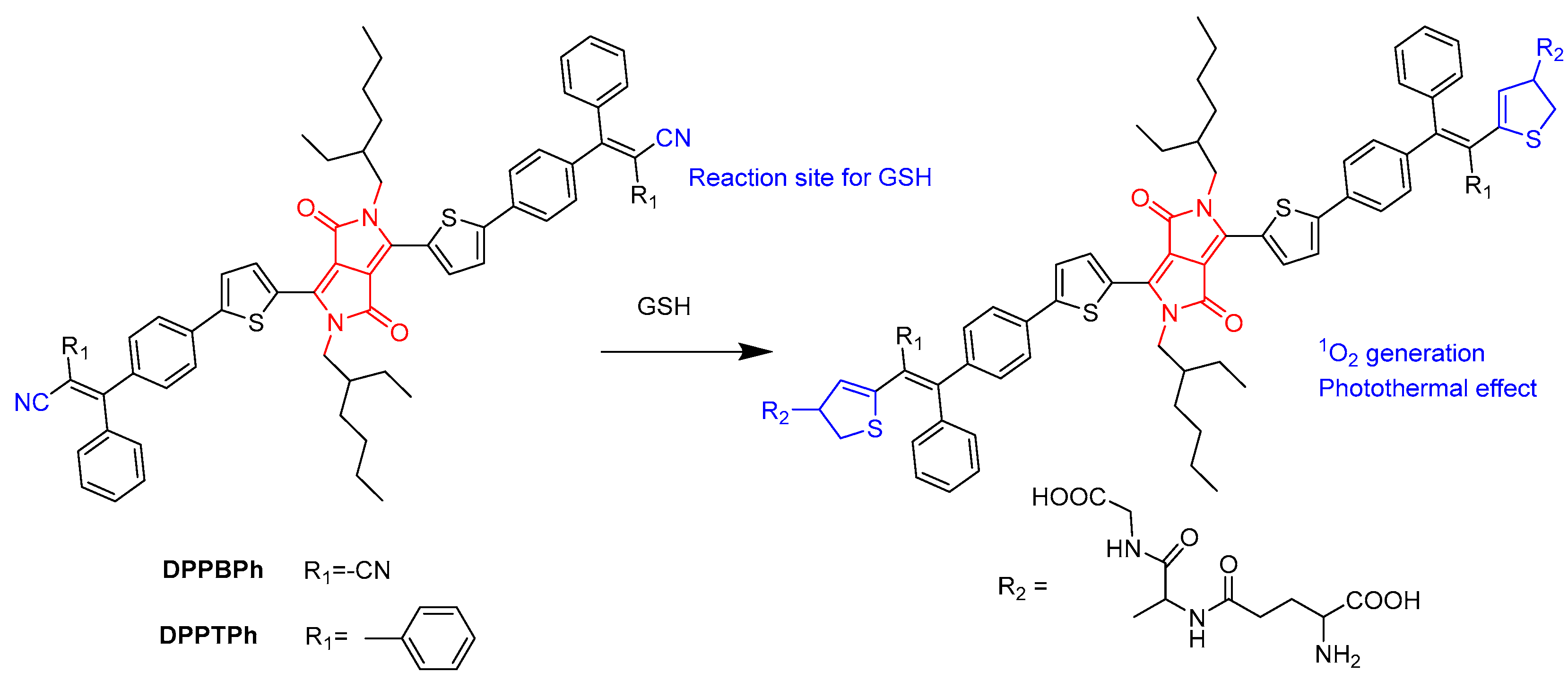
- Zou, J.; Xue, L.; Yang, N.; Ren, Y.; Fan, Z.; Wang, W.; Si, W.; Zhang, Y.; Huang, W.; Dong, X. A Glutathione Responsive Pyrrolopyrrolidone Nanotheranostic Agent for Turn-on Fluorescence Imaging Guided Photothermal/Photodynamic Cancer Therapy. Mater. Chem. Front. 2019, 3, 2143–2150. [Google Scholar] [CrossRef]
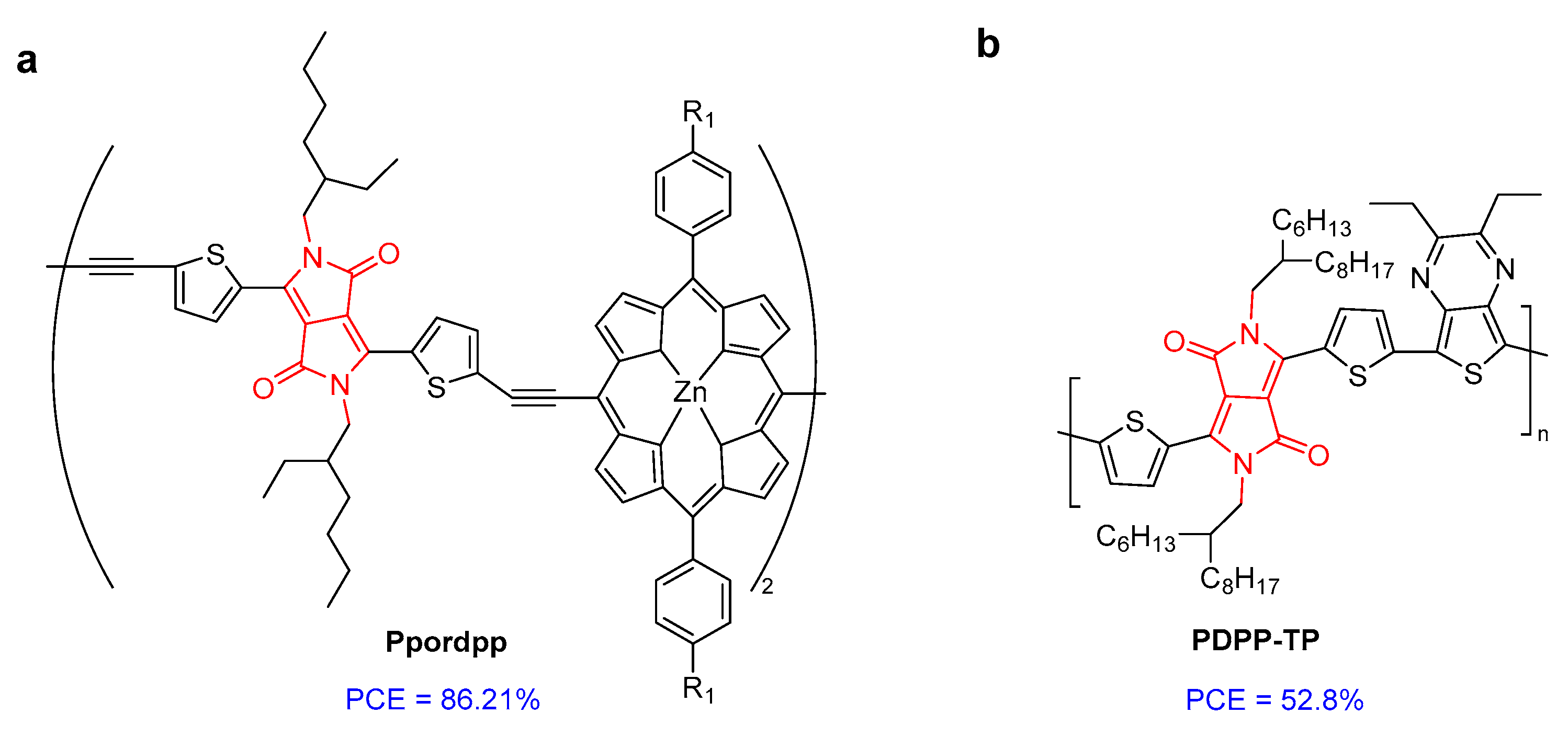
- Chen, L.; Li, X.; Xiong, M.; Zhao, Y.; Liu, S.; Li, C.; Wang, K. Development of Novel Nanoporphyrin Biomaterials for NIR-II Activated Photothermal Therapy against Tumor in Vivo. Mater. Des. 2023, 225, 111532. [Google Scholar] [CrossRef]
- Wu, Z.; Wang, J.; Zhao, L.; Li, C.; Lu, Y. A Novel Donor–Acceptor Structured Diketopyrrolopyrrole-Based Conjugated Polymer Synthesized by Direct Arylation Polycondensation (DArP) for Highly Efficient Antimicrobial Photothermal Therapy. Biomater. Sci. 2023, 11, 2151–2157. [Google Scholar] [CrossRef]
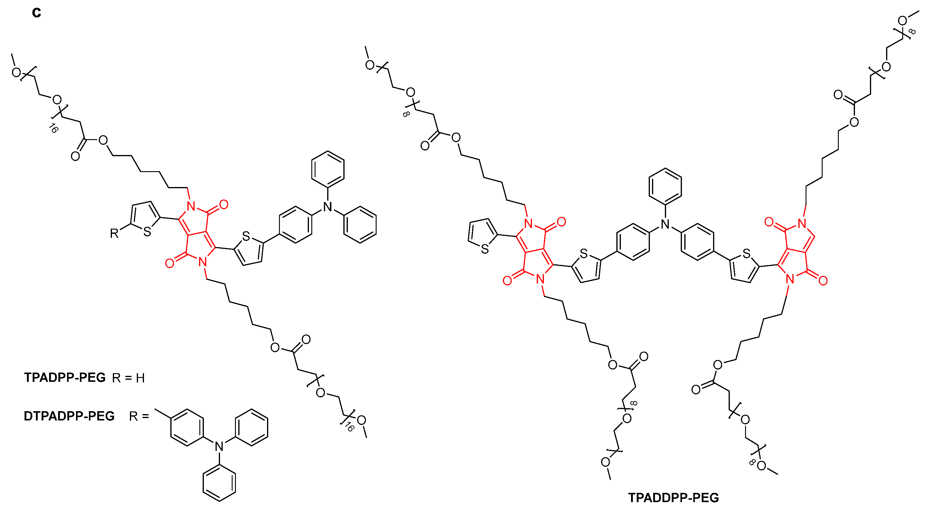
- Zheng, X.; Bian, S.; Liu, W.; Zhang, C.; Wu, J.; Ren, H.; Zhang, W.; Lee, C.-S.; Wang, P. Amphiphilic Diketopyrrolopyrrole Derivatives for Efficient Near-Infrared Fluorescence Imaging and Photothermal Therapy. ACS Omega 2021, 6, 26575–26582. [Google Scholar] [CrossRef] [PubMed]
- Jiang, Y.; Pu, K. Advanced Photoacoustic Imaging Applications of Near-Infrared Absorbing Organic Nanoparticles. Small 2017, 13, 1700710. [Google Scholar] [CrossRef] [PubMed]
- Chen, D.; Wang, C.; Nie, X.; Li, S.; Li, R.; Guan, M.; Liu, Z.; Chen, C.; Wang, C.; Shu, C.; et al. Photoacoustic Imaging Guided Near-Infrared Photothermal Therapy Using Highly Water-Dispersible Single-Walled Carbon Nanohorns as Theranostic Agents. Adv. Funct. Mater. 2014, 24, 6621–6628. [Google Scholar] [CrossRef]
- Wu, F.; Chen, L.; Yue, L.; Wang, K.; Cheng, K.; Chen, J.; Luo, X.; Zhang, T. Small-Molecule Porphyrin-Based Organic Nanoparticles with Remarkable Photothermal Conversion Efficiency for in Vivo Photoacoustic Imaging and Photothermal Therapy. ACS Appl. Mater. Interfaces 2019, 11, 21408–21416. [Google Scholar] [CrossRef] [PubMed]
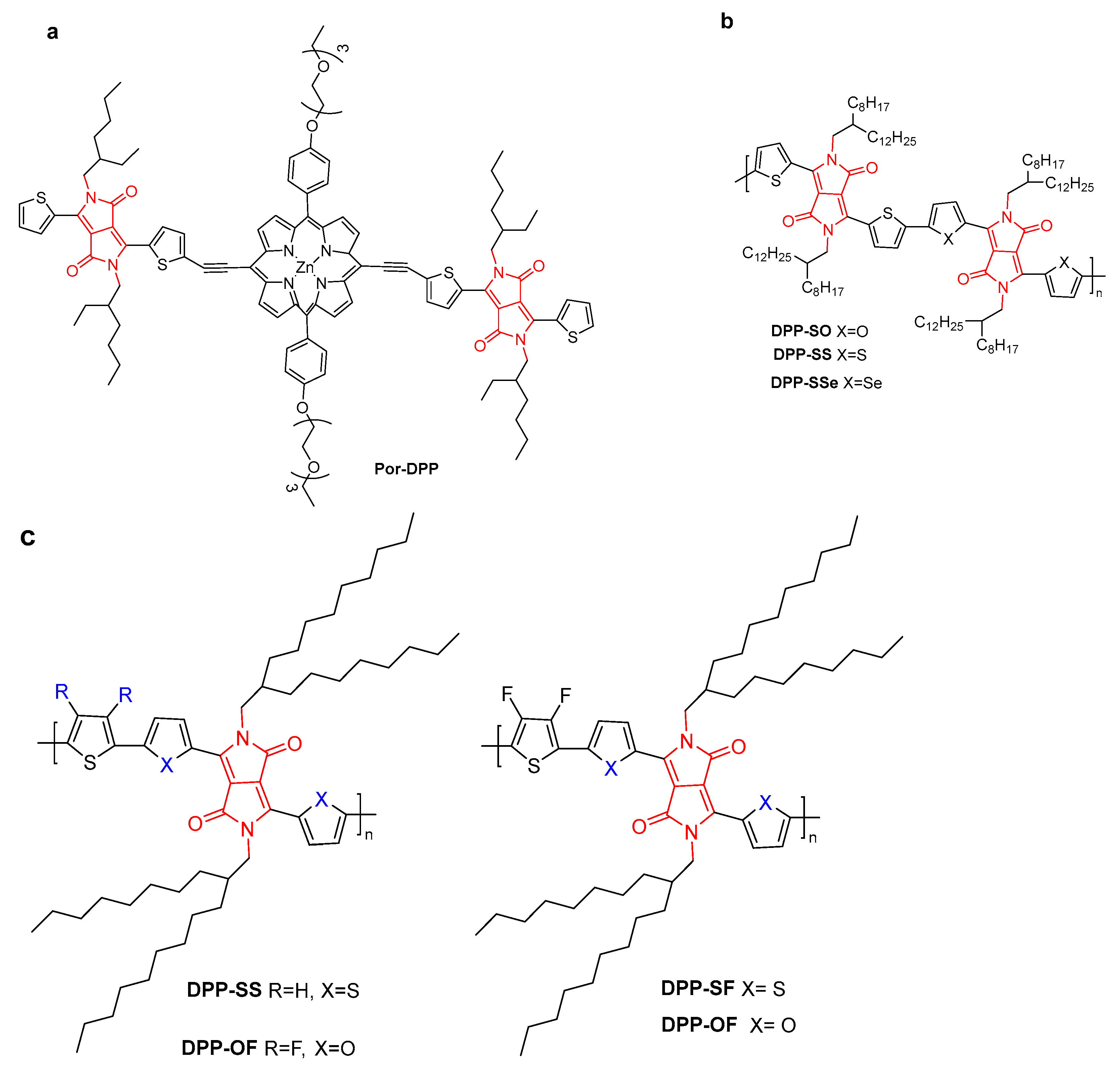
- Jin, X.; Xing, X.; Deng, Q.; Qing, W.; Liu, Z.; Huang, Y. Molecular Engineering of Diketopyrrolopyrrole-Conjugated Polymer Nanoparticles by Chalcogenide Variation for Photoacoustic Imaging Guided Photothermal Therapy. J. Mater. Chem. B 2021, 9, 3153–3160. [Google Scholar] [CrossRef]
- Liu, F.; Ma, F.; Chen, Q.; Zhou, E.; Zhang, P.; Cui, Z.; Liu, Z.; Huang, Y. Synergistic Non-Bonding Interactions Based on Diketopyrrolo-Pyrrole for Elevated Photoacoustic Imaging-Guided Photothermal Therapy. Biomater. Sci. 2021, 9, 908–916. [Google Scholar] [CrossRef]
- Pu, K.; Mei, J.; Jokerst, J.V.; Hong, G.; Antaris, A.L.; Chattopadhyay, N.; Shuhendler, A.J.; Kurosawa, T.; Zhou, Y.; Gambhir, S.S.; et al. Diketopyrrolopyrrole-Based Semiconducting Polymer Nanoparticles for In Vivo Photoacoustic Imaging. Adv. Mater. 2015, 27, 5184–5190. [Google Scholar] [CrossRef]
- Fu, X.; Huang, Y.; Zhao, H.; Zhang, E.; Shen, Q.; Di, Y.; Lv, F.; Liu, L.; Wang, S. Near-Infrared-Light Remote-Controlled Activation of Cancer Immunotherapy Using Photothermal Conjugated Polymer Nanoparticles. Adv. Mater. 2021, 33, 2102570. [Google Scholar] [CrossRef]
- Li, S.; Wang, X.; Hu, R.; Chen, H.; Li, M.; Wang, J.; Wang, Y.; Liu, L.; Lv, F.; Liang, X.-J.; et al. Near-infrared (NIR)-absorbing conjugated polymer dots as highly effective photothermal materials for in vivo cancer therapy. Chem. Mater. 2016, 28, 8669–8675. [Google Scholar] [CrossRef]
- Zhang, H.; Liang, Y.; Zhao, H.; Qi, R.; Chen, Z.; Yuan, H.; Liang, H.; Wang, L. Dual-Mode Antibacterial Conjugated Polymer Nanoparticles for Photothermal and Photodynamic Therapy. Macromol. Biosci. 2020, 20, 1900301. [Google Scholar] [CrossRef]
- Liu, J.; Xu, X.; Wang, J.; Sang, R.; Zhang, Z.; Chen, J.; Lu, X.; Wang, Q.; Fan, Q. A Diketopyrrolopyrrole-Based Conjugated Polymer for Efficient Photodynamic and Photothermal Combination Therapy under Single 808 Nm Laser Irradiation. Dyes Pigments 2021, 196, 109762. [Google Scholar] [CrossRef]
- Li, S.; Deng, Q.; Li, X.; Huang, Y.; Li, X.; Liu, F.; Wang, H.; Qing, W.; Liu, Z.; Lee, C.S. Bis-diketopyrrolopyrrole conjugated polymer nanoparticles as photothermic nanoagonist for specific and synergistic glioblastoma therapy. Biomaterials 2019, 216, 119252. [Google Scholar] [CrossRef] [PubMed]
- Huang, Y.-Q.; Liu, K.-L.; Ni, H.-L.; Zhang, R.; Liu, X.-F.; Fan, Q.-L.; Wang, L.-H.; Huang, W. Organic Theranostic Nanoplatform with Enhanced Fluorescence and Singlet Oxygen Quantum Yield for Tumor-Targeting Image-Guided Photodynamic/Photothermal Synergistic Therapy. ACS Appl. Polym. Mater. 2022, 4, 7739–7750. [Google Scholar] [CrossRef]
- Zhang, H.; Chen, X.; Li, S.; Shen, J.; Mao, Z.-W. An Enhanced Photothermal Therapeutic Iridium Hybrid Platform Reversing the Tumor Hypoxic Microenvironment. Molecules 2022, 27, 2629. [Google Scholar] [CrossRef] [PubMed]
- Li, Y.; Zhang, J.; Liu, S.; Zhang, C.; Chuah, C.; Tang, Y.; Kwok, R.T.K.; Lam, J.W.Y.; Ou, H.; Ding, D.; et al. Enlarging the Reservoir: High Absorption Coefficient Dyes Enable Synergetic Near Infrared-II Fluorescence Imaging and Near Infrared-I Photothermal Therapy. Adv. Funct. Mater. 2021, 31, 2102213. [Google Scholar] [CrossRef]
- Xiao, W.; Wang, P.; Ou, C.; Huang, X.; Tang, Y.; Wu, M.; Si, W.; Shao, J.; Huang, W.; Dong, X. 2-Pyridone-Functionalized Aza-BODIPY Photosensitizer for Imaging-Guided Sustainable Phototherapy. Biomaterials 2018, 183, 1–9. [Google Scholar] [CrossRef] [PubMed]
- Turan, I.S.; Yildiz, D.; Turksoy, A.; Gunaydin, G.; Akkaya, E.U. A Bifunctional Photosensitizer for Enhanced Fractional Photodynamic Therapy: Singlet Oxygen Generation in the Presence and Absence of Light. Angew. Chem. Int. Ed. 2016, 55, 2875–2878. [Google Scholar] [CrossRef]
- Zou, J.; Zhu, J.; Yang, Z.; Li, L.; Fan, W.; He, L.; Tang, W.; Deng, L.; Mu, J.; Ma, Y.; et al. A Phototheranostic Strategy to Continuously Deliver Singlet Oxygen in the Dark and Hypoxic Tumor Microenvironment. Angew. Chem. Int. Ed. 2020, 59, 8833–8838. [Google Scholar] [CrossRef]
- Li, C.; Zhang, W.; Liu, S.; Hu, X.; Xie, Z. Mitochondria-Targeting Organic Nanoparticles for Enhanced Photodynamic/Photothermal Therapy. ACS Appl. Mater. Interfaces 2020, 12, 30077–30084. [Google Scholar] [CrossRef]
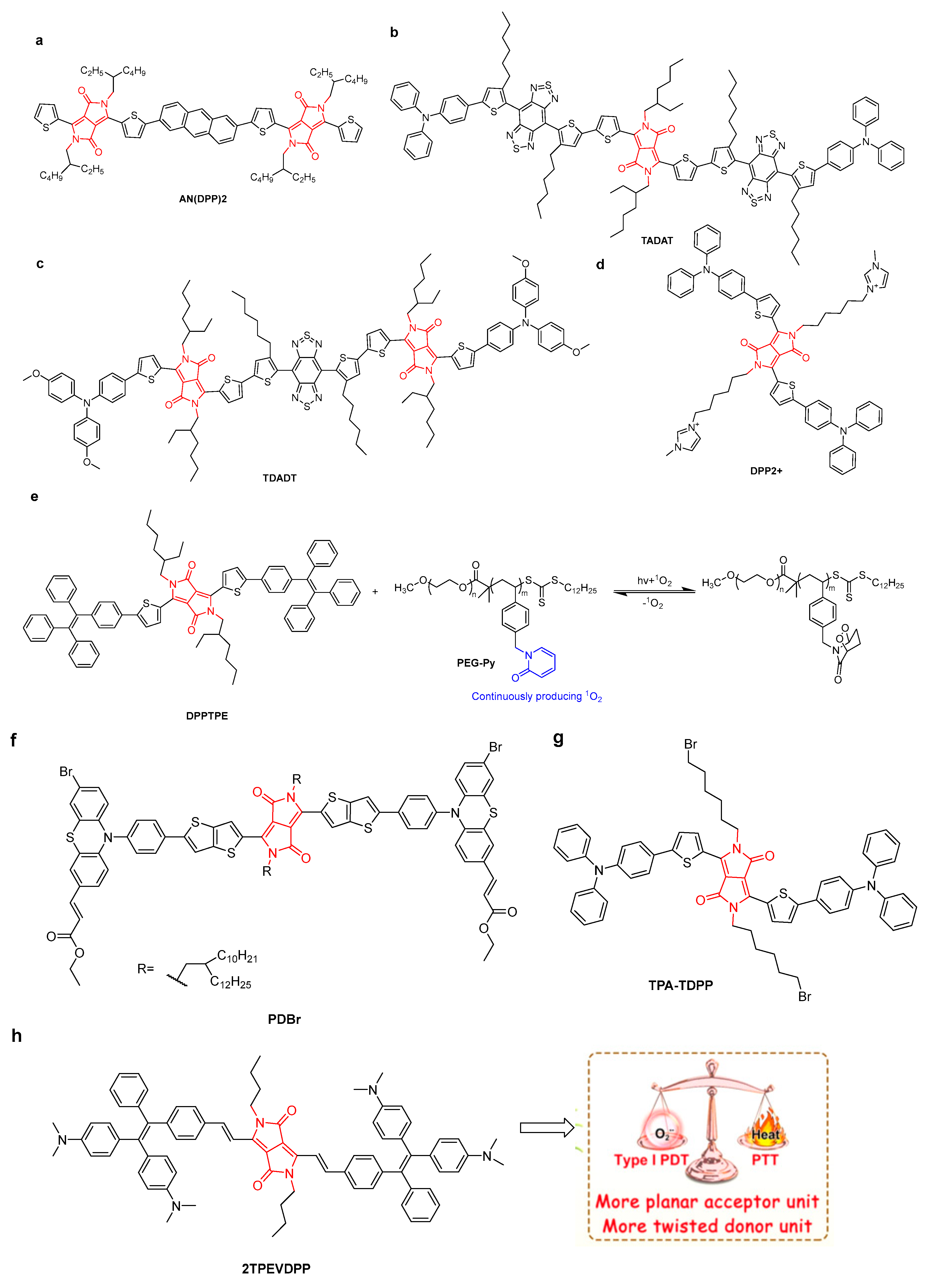
- Feng, L.; Li, C.; Liu, L.; Wang, Z.; Chen, Z.; Yu, J.; Ji, W.; Jiang, G.; Zhang, P.; Wang, J.; et al. Acceptor Planarization and Donor Rotation: A Facile Strategy for Realizing Synergistic Cancer Phototherapy via Type I PDT and PTT. ACS Nano 2022, 16, 4162–4174. [Google Scholar] [CrossRef] [PubMed]
- Yang, X.; Yu, Q.; Yang, N.; Xue, L.; Shao, J.; Li, B.; Shao, J.; Dong, X. Thieno[3,2-b]Thiophene-DPP Based near-Infrared Nanotheranostic Agent for Dual Imaging-Guided Photothermal/Photodynamic Synergistic Therapy. J. Mater. Chem. B 2019, 7, 2454–2462. [Google Scholar] [CrossRef]
- Sun, W.; Wang, X.; Cheng, Z.; Wang, X.; Fan, N.; Dong, P.; Tong, M.Q.; Liu, Y.; Sun, W. Phototheranostics for NIR Fluorescence Image Guided PDT/PTT with Extended Conjugation and Enhanced TICT. Biomed. Pharmacother. 2023, 158, 114071. [Google Scholar] [CrossRef] [PubMed]
- Li, Y.; Li, X.; Yu, J.; Wan, Z.; Hu, Y.; Huang, R.; Tao, Y.; Miao, W. A Versatile Diketopyrrolopyrrole-Based Photosensitizing Nanoplatform with High Photostability and Photoactivity for Metastatic Breast Cancer Treatment. Dyes Pigments 2022, 207, 110748. [Google Scholar] [CrossRef]
- Cai, Y.; Liang, P.; Tang, Q.; Yang, X.; Si, W.; Huang, W.; Zhang, Q.; Dong, X. Diketopyrrolopyrrole-Triphenylamine Organic Nanoparticles as Multifunctional Reagents for Photoacoustic ImagingGuided Photodynamic/Photothermal Synergistic Tumor Therapy. ACS Nano 2017, 11, 1054–1063. [Google Scholar] [CrossRef] [PubMed]
- Wang, Q.; Xia, B.; Xu, J.; Niu, X.; Cai, J.; Shen, Q.; Wang, W.; Huang, W.; Fan, Q. Biocompatible small organic molecule phototheranostics for NIR-II fluorescence/photoacoustic imaging and simultaneous photodynamic/photothermal combination therapy. Mater. Chem. Front. 2019, 3, 650–655. [Google Scholar] [CrossRef]
- Wang, P.; Wu, W.; Gao, R.; Zhu, H.; Wang, J.; Du, R.; Li, X.; Zhang, C.; Cao, S.; Xiang, R. Engineered Cell-Assisted Photoactive Nanoparticle Delivery for Image-Guided Synergistic Photodynamic/Photothermal Therapy of Cancer. ACS Appl. Mater. Interfaces 2019, 11, 13935–13944. [Google Scholar] [CrossRef] [PubMed]
- Liang, P.; Tang, Q.; Cai, Y.; Liu, G.; Si, W.; Shao, J.; Huang, W.; Zhang, Q.; Dong, X. Self-quenched ferrocenyl diketopyrrolopyrrole organic nanoparticles with amplifying photothermal effect for cancer therapy. Chem. Sci. 2017, 8, 7457–7463. [Google Scholar] [CrossRef]
- Lu, X.; Yuan, P.; Zhang, W.; Wu, Q.; Wang, X.; Zhao, M.; Sun, P.; Huang, W.; Fan, Q. A highly water-soluble triblock conjugated polymer for in vivo NIR-II imaging and photothermal therapy of cancer. Polym. Chem. 2018, 9, 3118–3126. [Google Scholar] [CrossRef]
- Jiang, Y.; Li, J.; Zhen, X.; Xie, C.; Pu, K. Dual-Peak Absorbing Semiconducting Copolymer Nanoparticles for First and Second NearInfrared Window Photothermal Therapy: A Comparative Study. Adv. Mater. 2018, 30, 1705980. [Google Scholar] [CrossRef]
- Cheng, X.; Zhang, C.; Shen, K.; Liu, H.; Bai, C.; Ding, Q.; Guan, M.; Wu, J.; Tian, Z.; Chen, D.; et al. Novel Diketopyrrolopyrrole NIR-II Fluorophores and DDR Inhibitors for in Vivo Chemo-Photodynamic Therapy of Osteosarcoma. Chem. Eng. J. 2022, 446, 136929. [Google Scholar] [CrossRef]
- Sun, W.; Liu, X.-Y.; Ma, L.-L.; Lu, Z.-L. Tumor Targeting Gene Vector for Visual Tracking of Bcl-2 SiRNA Transfection and Anti-Tumor Therapy. ACS Appl. Mater. Interfaces 2020, 12, 10193–10201. [Google Scholar] [CrossRef] [PubMed]
- Shen, L.; Zhou, T.; Fan, Y.; Chang, X.; Wang, Y.; Sun, J.; Xing, L.; Jiang, H. Recent Progress in Tumor Photodynamic Immunotherapy. Chin. Chem. Lett. 2020, 31, 1709–1716. [Google Scholar] [CrossRef]


Disclaimer/Publisher’s Note: The statements, opinions and data contained in all publications are solely those of the individual author(s) and contributor(s) and not of MDPI and/or the editor(s). MDPI and/or the editor(s) disclaim responsibility for any injury to people or property resulting from any ideas, methods, instructions or products referred to in the content. |
© 2023 by the authors. Licensee MDPI, Basel, Switzerland. This article is an open access article distributed under the terms and conditions of the Creative Commons Attribution (CC BY) license (https://creativecommons.org/licenses/by/4.0/).
Share and Cite
Wang, L.; Lai, B.; Ran, X.; Tang, H.; Cao, D. Recent Advances of Diketopyrrolopyrrole Derivatives in Cancer Therapy and Imaging Applications. Molecules 2023, 28, 4097. https://doi.org/10.3390/molecules28104097
Wang L, Lai B, Ran X, Tang H, Cao D. Recent Advances of Diketopyrrolopyrrole Derivatives in Cancer Therapy and Imaging Applications. Molecules. 2023; 28(10):4097. https://doi.org/10.3390/molecules28104097
Chicago/Turabian StyleWang, Lingyun, Bihong Lai, Xueguang Ran, Hao Tang, and Derong Cao. 2023. "Recent Advances of Diketopyrrolopyrrole Derivatives in Cancer Therapy and Imaging Applications" Molecules 28, no. 10: 4097. https://doi.org/10.3390/molecules28104097