
Application of Nanomaterials to Enhance Polymerase Chain Reaction

Abstract
:1. Introduction
2. Utilizing Different Nanomaterials to Enhance PCR Effects
2.1. Metal Nanomaterials
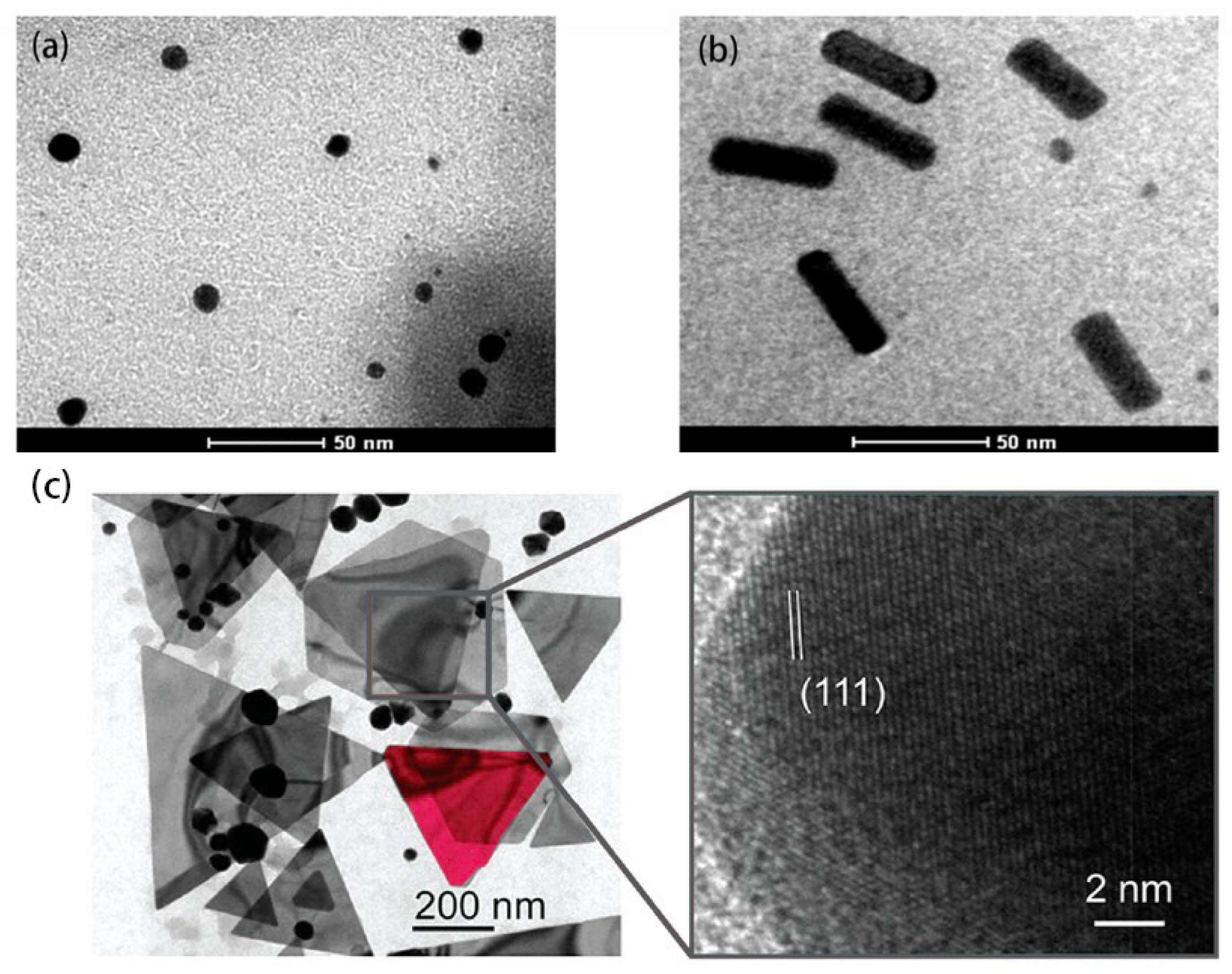
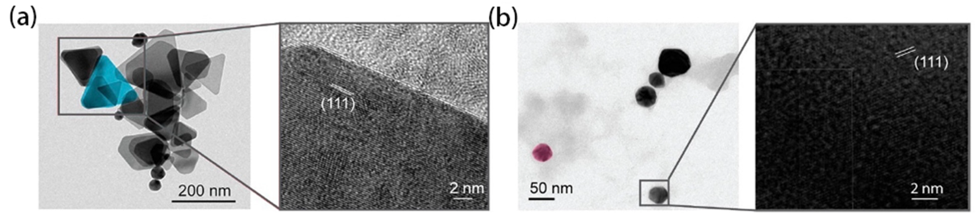
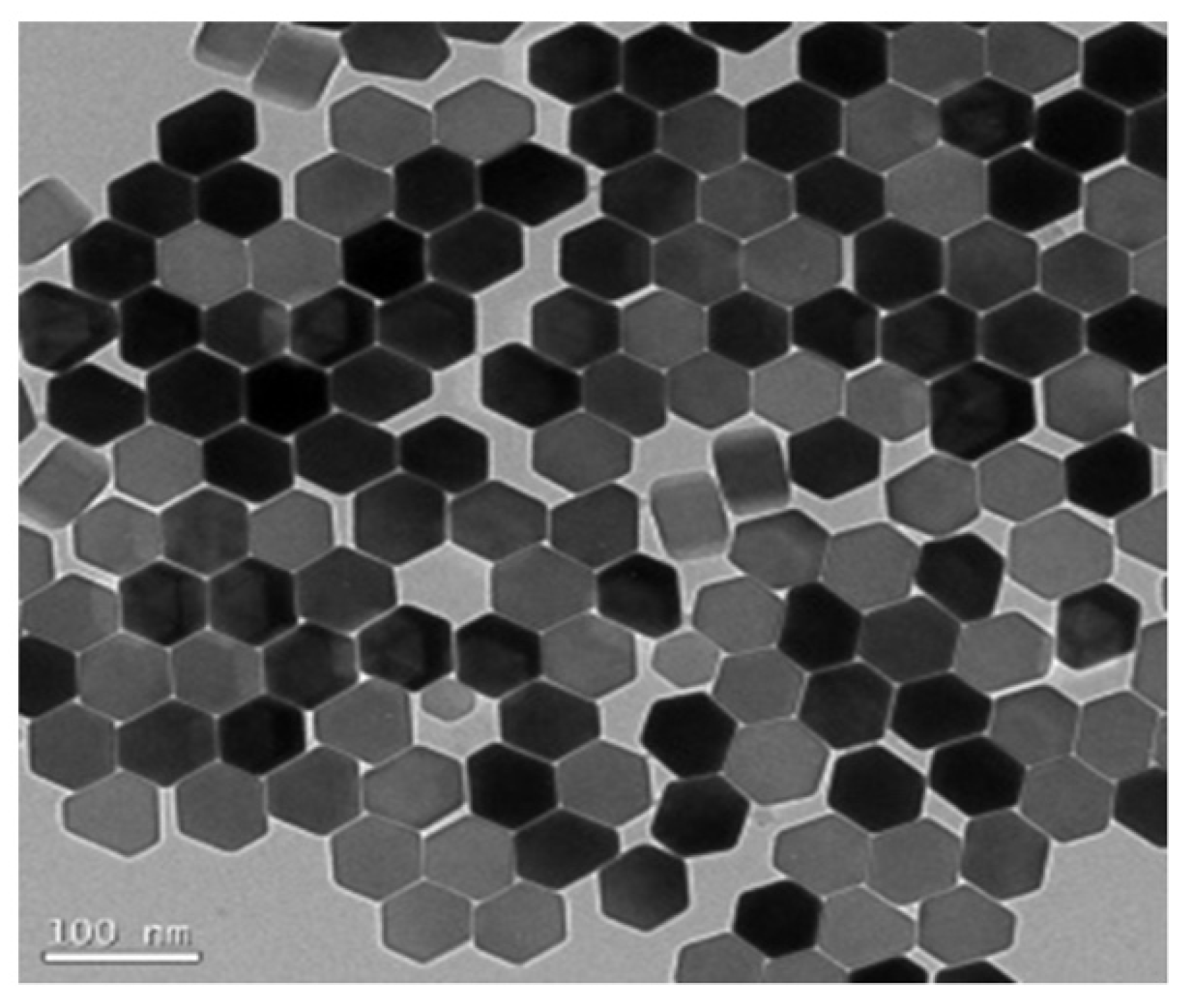
2.1.1. Au NPs
2.1.2. Ag NPs
2.2. Carbon-Based Nanomaterials
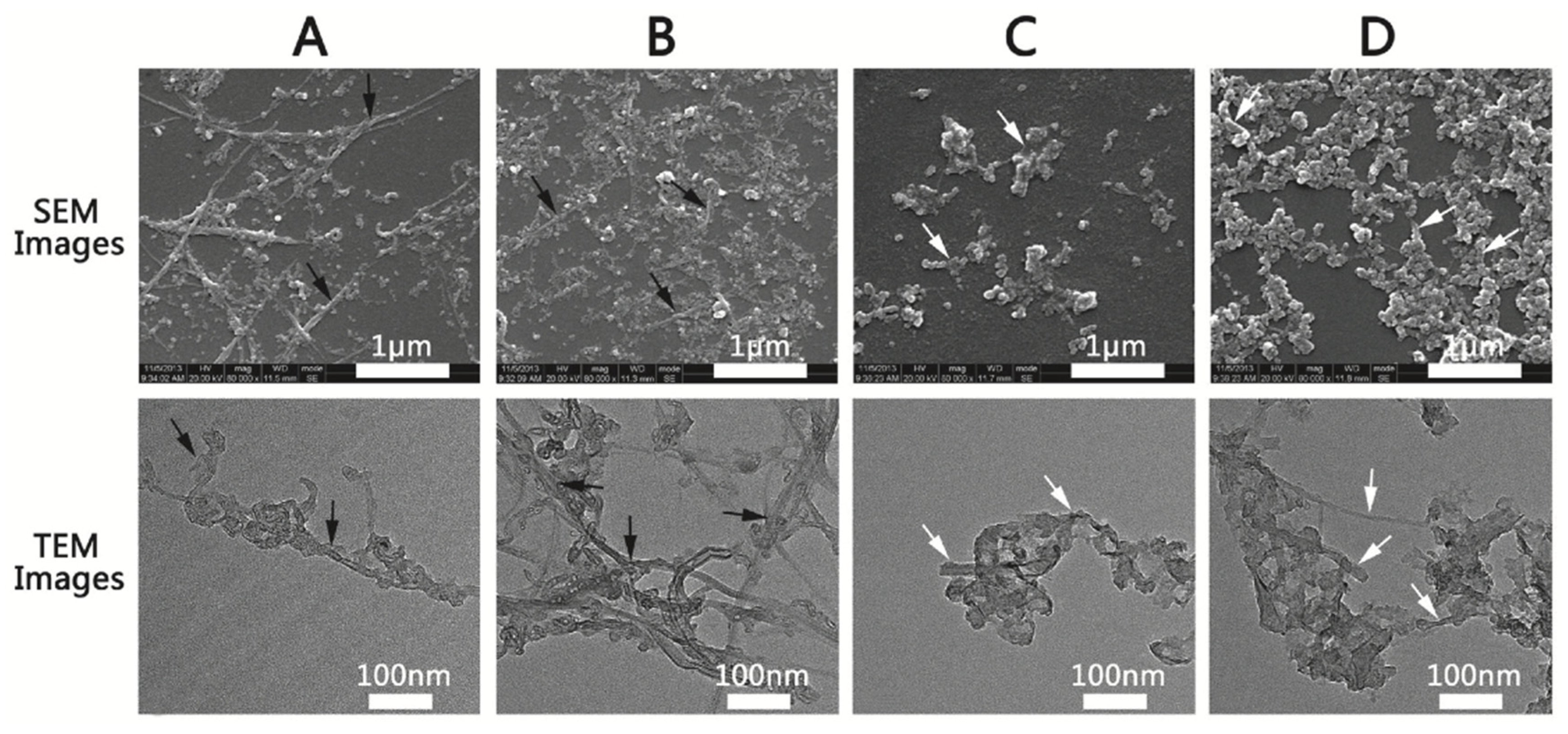
2.2.1. CNTs
2.2.2. CNP
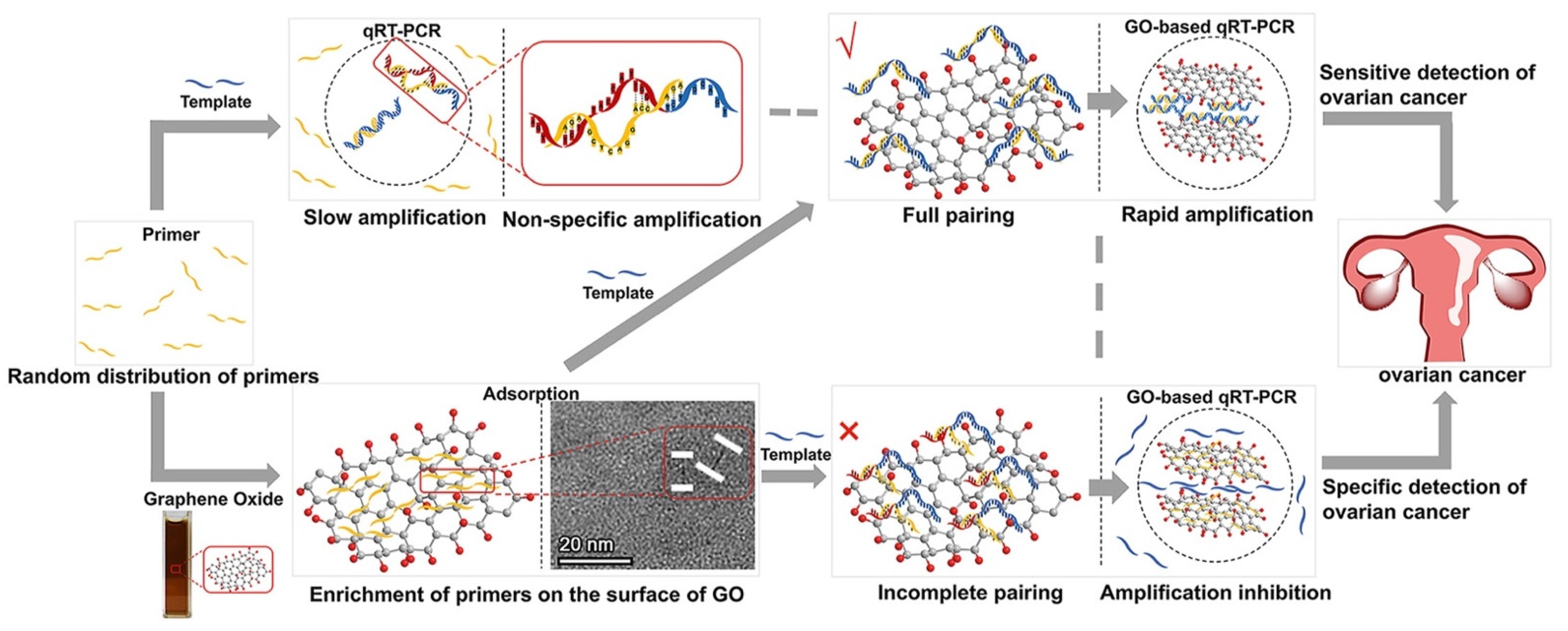
2.2.3. Graphene
2.3. Oxide Nanomaterials
2.3.1. TiO2
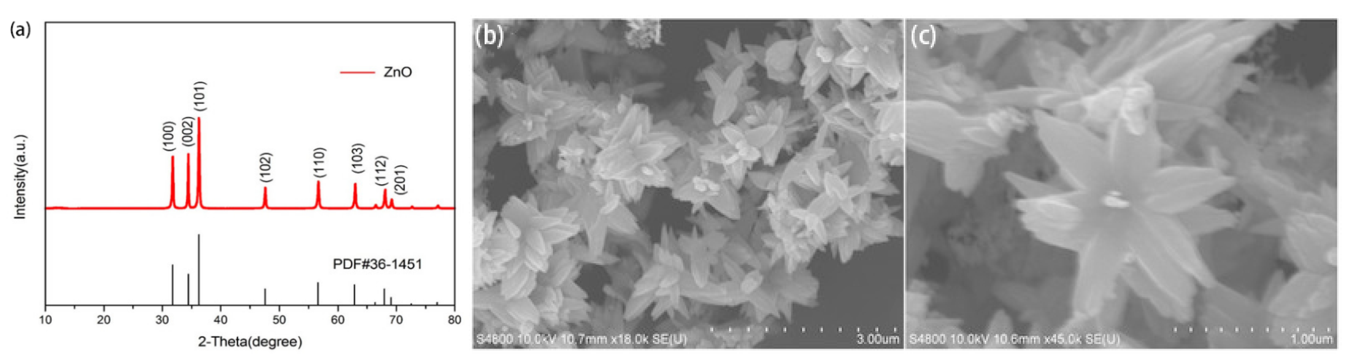
2.3.2. ZnO
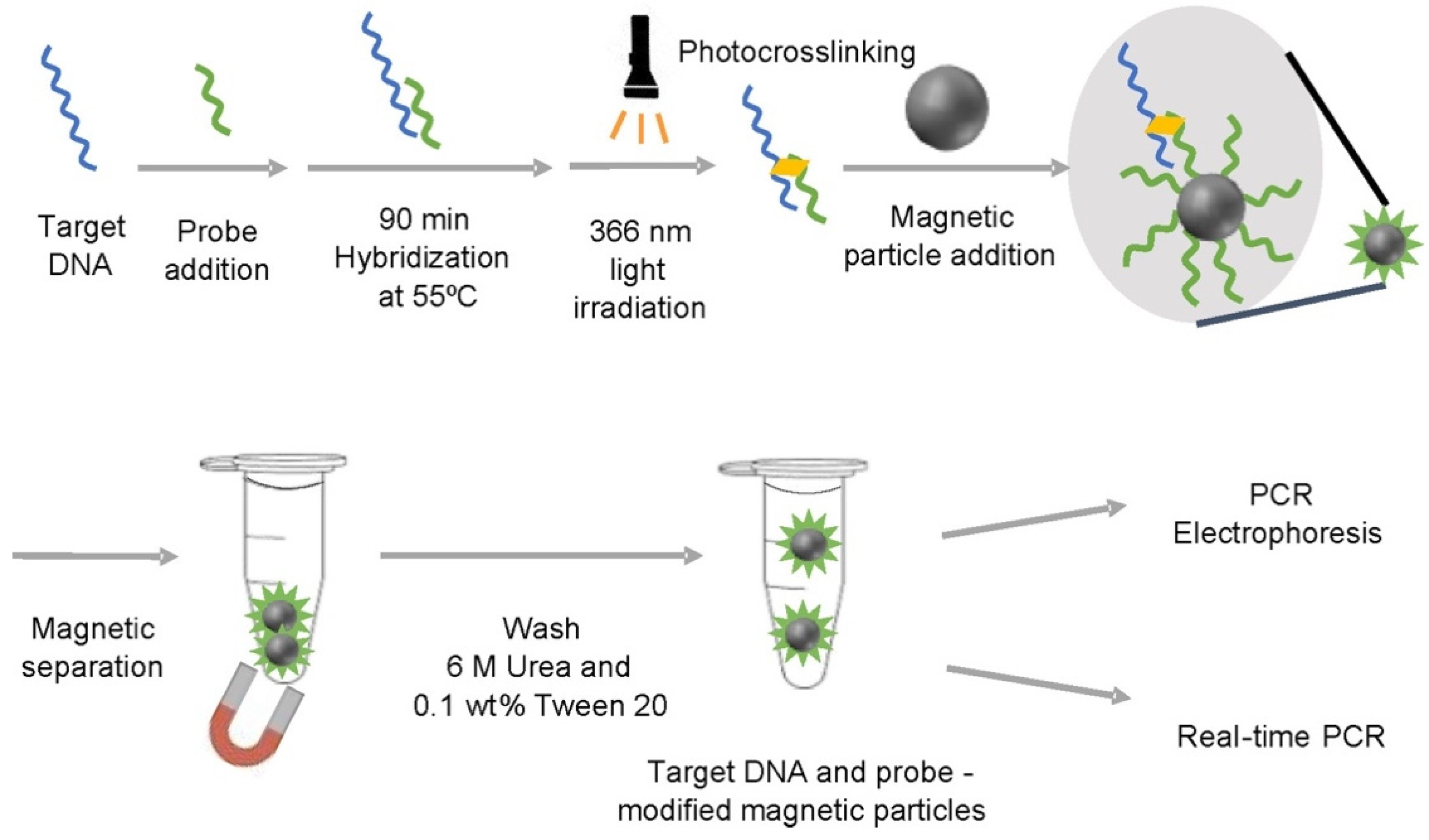
2.3.3. Fe3O4
2.3.4. MgO
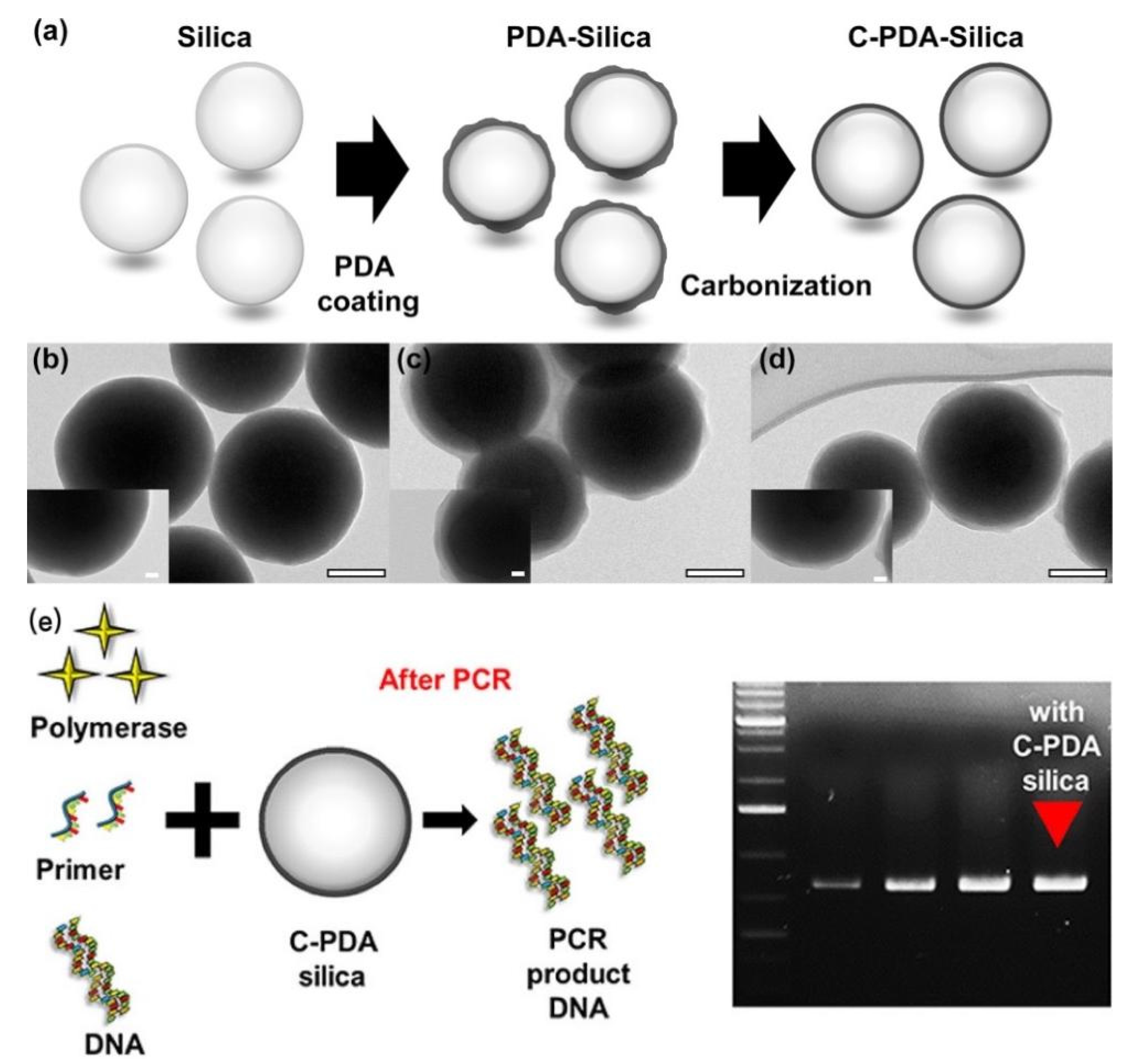
2.3.5. SiO2
2.4. Fluorescent Nanomaterials
2.4.1. QDs
2.4.2. Up-Conversion Nanomaterials
2.5. Others
2.5.1. Hybrid Nanocomposites
2.5.2. Other NPs
3. The Effects of NPs in Real-Time PCR
4. Mechanisms of Nanomaterials in PCR
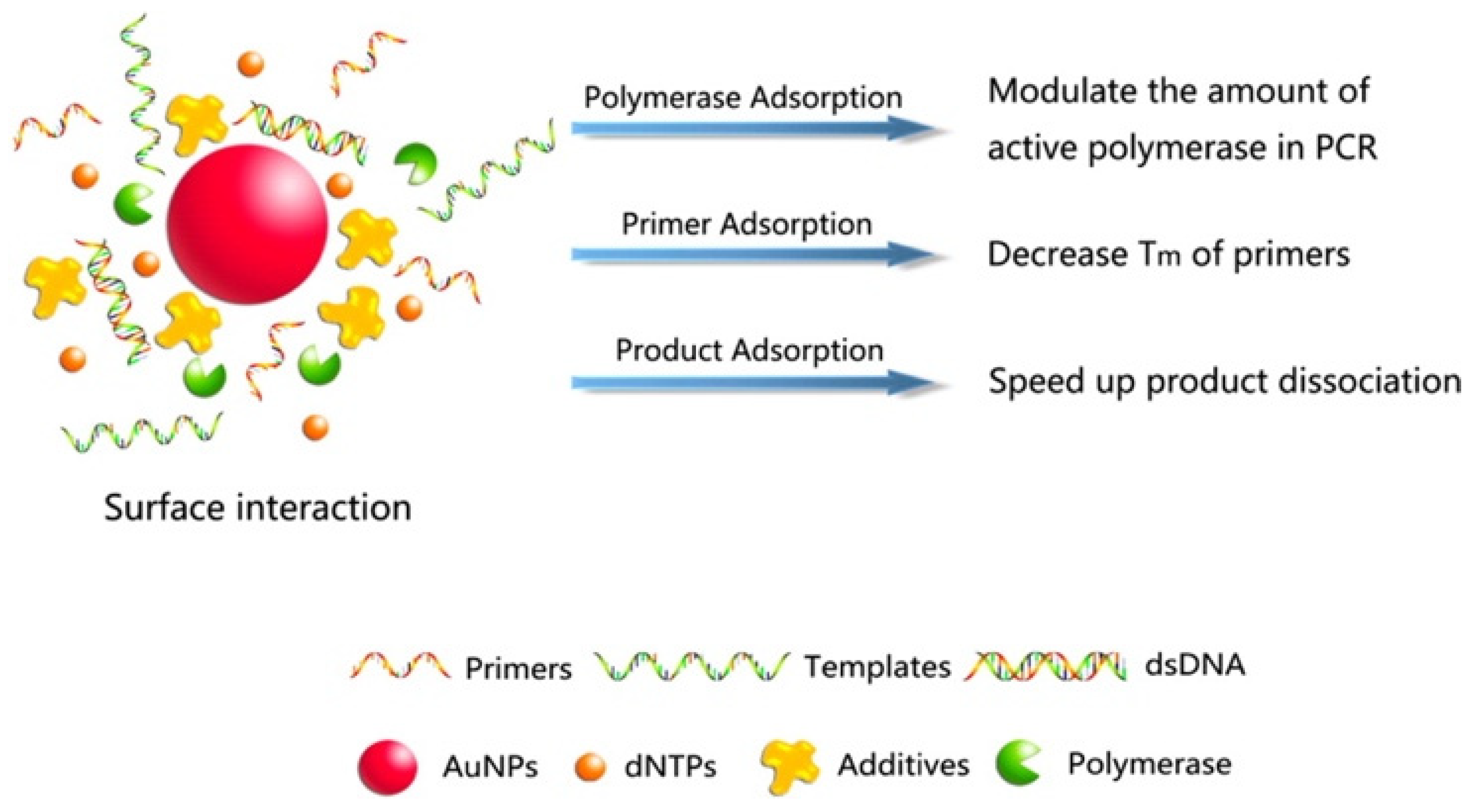
4.1. Surface Interactions
4.2. Thermal Conductivity
4.3. Electrostatic Interactions
4.4. Analogs to ssDNA Binding Protein (SSB)
4.5. Catalytic Activity
5. Application and Prospect of NanoPCR
6. Conclusions
Author Contributions
Funding
Institutional Review Board Statement
Informed Consent Statement
Data Availability Statement
Acknowledgments
Conflicts of Interest
References
- Kuypers, J.; Jerome, K.R. Applications of Digital PCR for Clinical Microbiology. J. Clin. Microbiol. 2017, 55, 1621–1628. [Google Scholar] [CrossRef] [Green Version]
- Elizaquivel, P.; Aznar, R.; Sanchez, G. Recent developments in the use of viability dyes and quantitative PCR in the food microbiology field. J. Appl. Microbiol. 2014, 116, 1–13. [Google Scholar] [CrossRef] [PubMed]
- Matheson, C.D.; Marion, T.E.; Hayter, S.; Esau, N.; Fratpietro, R.; Vernon, K.K. Technical note: Removal of metal ion inhibition encountered during DNA extraction and amplification of copper-preserved archaeological bone using size exclusion chromatography. Am. J. Phys. Anthropol. 2009, 140, 384–391. [Google Scholar] [CrossRef] [PubMed]
- Huang, Y.-H.; Hu, X.-X.; Xu, W.-Z.; Gao, Y.; Feng, J.-D.; Sun, H.; Li, N. The factors affecting the efficiency of mutiplex PCR. Yi Chuan = Hered. 2003, 25, 65–68. [Google Scholar]
- Yang, W.; Li, X.; Sun, J.; Shao, Z. Enhanced PCR amplification of GC-rich DNA templates by Au NPs. ACS Appl. Mater. Interfaces 2013, 5, 11520–11524. [Google Scholar] [CrossRef]
- Wang, L.; Zhu, Y.; Jiang, Y.; Qiao, R.; Zhu, S.; Chen, W.; Xu, C. Effects of QDs in Polymerase Chain Reaction. J. Phys. Chem. B 2009, 113, 7637–7641. [Google Scholar] [CrossRef]
- Cui, D.; Tian, F.; Kong, Y.; Titushikin, I.; Gao, H. Effects of SWCNTs on the polymerase chain reaction. Nanotechnology 2004, 15, 154–157. [Google Scholar] [CrossRef]
- Wang, Y.; Wang, F.; Wang, H.; Song, M. GO enhances the specificity of the polymerase chain reaction by modifying primer-template matching. Sci. Rep. 2017, 7, 16510. [Google Scholar] [CrossRef] [Green Version]
- Li, S.; Zhu, H.; Zhu, R.; Sun, X.; Yao, S.; Wang, S. Impact and mechanism of TiO2 NPs on DNA synthesis in vitro. Sci. China Ser. B Chem. 2008, 51, 367–372. [Google Scholar] [CrossRef]
- Nie, L.; Gao, L.; Yan, X.; Wang, T. Functionalized tetrapod-like ZnO nanostructures for plasmid DNA purification, polymerase chain reaction and delivery. Nanotechnology 2007, 18, 015101. [Google Scholar] [CrossRef]
- Yuan, L.; He, Y. Effect of surface charge of PDDA-protected Au NPs on the specificity and efficiency of DNA polymerase chain reaction. Analyst 2013, 138, 539–545. [Google Scholar] [CrossRef] [PubMed]
- Bai, Y.; Cui, Y.; Paoli, G.C.; Shi, C.; Wang, D.; Shi, X. Nanoparticles Affect PCR Primarily via Surface Interactions with PCR Components: Using Amino-Modified Silica-Coated Magnetic Nanoparticles as a Main Model. ACS Appl. Mater. Interfaces 2015, 7, 13142–13153. [Google Scholar] [CrossRef] [PubMed]
- Wang, Z.D.; Zhang, L.Y.; Wang, X. Molecular toxicity and defense mechanisms induced by silver nanoparticles in Drosophila melanogaster. J. Environ. Sci. 2023, 125, 616–629. [Google Scholar] [CrossRef]
- Wang, W.; Wang, X.; Liu, J.; Lin, C.; Liu, J.; Wang, J. The Integration of Gold Nanoparticles with Polymerase Chain Reaction for Constructing Colorimetric Sensing Platforms for Detection of Health-Related DNA and Proteins. Biosensors 2022, 12, 421. [Google Scholar] [CrossRef] [PubMed]
- Nunes, A.M.; da Silva Filho, R.C.; da Silva, K.R.M.; Bezerra, S.M.; de Figueiredo, R.C.B.Q.; Saraiva, K.L.A.; Leite, A.C.R.; Meneghetti, M.R. Gold nanoparticles with different shapes can cause distinct effect on mitochondria bioenergetics. J. Nanoparticle Res. 2022, 24, 31. [Google Scholar] [CrossRef]
- Kadu, P.; Pandey, S.; Neekhra, S.; Kumar, R.; Gadhe, L.; Srivastava, R.; Sastry, M.; Maji, S.K. Machine-Free Polymerase Chain Reaction with Triangular Au and Ag NPs. J. Phys. Chem. Lett. 2020, 11, 10489–10496. [Google Scholar] [CrossRef] [PubMed]
- Li, M.; Lin, Y.C.; Wu, C.C.; Liu, H.S. Enhancing the efficiency of a PCR using Au NPs. Nucleic Acids Res. 2005, 33, e184. [Google Scholar] [CrossRef] [Green Version]
- Pan, J.; Li, H.; Cao, X.; Huang, J.; Zhang, X.; Fan, C.; Hu, J. Nanogold-assisted multi-round polymerase chain reaction (PCR). J. Nanosci. Nanotechnol. 2007, 7, 4428–4433. [Google Scholar] [CrossRef]
- Binh, V. Vu, D.L., Richard C. Willson. Gold Nanoparticle Effects in Polymerase Chain Reaction: Favoring of Smaller Products by Polymerase Adsorption. Anal. Chem. 2008, 80, 5462–5467. [Google Scholar]
- Mi, L.; Zhu, H.; Zhang, X.; Hu, J.; Fan, C. Mechanism of the interaction between Au nanoparticles and polymerase in nanoparticle PCR. Chin. Sci. Bull. 2007, 52, 2345–2349. [Google Scholar] [CrossRef]
- Lin, Y.; Li, J.; Yao, J.; Liang, Y.; Zhang, J.; Zhou, Q.; Jiang, G. Mechanism of gold nanoparticle induced simultaneously increased PCR efficiency and specificity. Chin. Sci. Bull. 2013, 58, 4593–4601. [Google Scholar] [CrossRef]
- Lou, X.; Zhang, Y. Mechanism studies on nanoPCR and applications of Au NPs in genetic analysis. ACS Appl. Mater. Interfaces 2013, 5, 6276–6284. [Google Scholar] [CrossRef]
- Mandal, S.; Hossain, M.; Muruganandan, T.; Kumar, G.S.; Chaudhuri, K. Au NPs alter Taq DNA polymerase activity during polymerase chain reaction. RSC Adv. 2013, 3, 20793–20799. [Google Scholar] [CrossRef]
- Wang, Q.; Li, J.; Cao, X.; Zhang, C. Ag NPs Enhance the Specificity of Repeated Long PCR Amplification. J. Tianjin Univ. Sci. Technol. 2007, 22, 1–5. [Google Scholar] [CrossRef]
- Liu, P.; Guan, R.; Liu, M.; Huang, G.; Dai, X. Effect of PCR Amplification with Nano-silver on DNA Synthesis and Its Mechanism. J. Agric. Biotechnol. 2010, 18, 876–881. [Google Scholar] [CrossRef]
- Lee, K.-Y.; Pham, X.-H.; Rho, W.-Y.; Chang, H.; Lee, S.H.; Kim, J.; Hahm, E.; Lee, J.H.; Lee, Y.-S.; Jun, B.-H. Nanotechnology for Bioapplications. Adv. Exp. Med. Biol. 2021, 1309, 235–255. [Google Scholar]
- Suo, L.; Li, Z.; Luo, F.; Chen, J.; Jia, L.; Wang, T.; Pei, X.; Wan, Q. Effect of dentin surface modification using carbon nanotubes on dental bonding and antibacterial ability. Dent. Mater. J. 2018, 37, 229–236. [Google Scholar] [CrossRef] [Green Version]
- Zhang, Z.; Shen, C.; Wang, M.; Han, H.; Cao, X. Aqueous suspension of CNTs enhances the specificity of long PCR. Biotechniques 2008, 44, 537–538, 540, 542. [Google Scholar] [CrossRef] [Green Version]
- Cao, X.; Chen, J.; Wen, S.; Peng, C.; Shen, M.; Shi, X. Effect of surface charge of polyethyleneimine-modified MWCNTs on the improvement of polymerase chain reaction. Nanoscale 2011, 3, 1741–1747. [Google Scholar] [CrossRef]
- Yuce, M.; Budak, H. Dispersion quality of amine functionalized MWCNTs plays critical roles in polymerase chain reaction enhancement. J. Nanoparticle Res. 2014, 16, 2768. [Google Scholar] [CrossRef]
- Yüce, M.; Uysal, E.; Budak, H. Amplification yield enhancement of short DNA templates using bulk and surface-attached amine-functionalized SWCNTs. Appl. Surf. Sci. 2015, 349, 147–155. [Google Scholar] [CrossRef]
- Thong Le, B.; Bohus, M.; Lukacs, I.E.; Wongwises, S.; Grof, G.; Hernadi, K.; Szilagyi, I.M. Comparative Study of Carbon Nanosphere and Carbon Nanopowder on Viscosity and Thermal Conductivity of Nanofluids. Nanomaterials 2021, 11, 608. [Google Scholar] [CrossRef]
- Zhang, Z.; Wang, M.; An, H. An aqueous suspension of CNP enhances the efficiency of a polymerase chain reaction. Nanotechnology 2007, 18, 355706. [Google Scholar] [CrossRef]
- Wei, W.; Qu, X. Extraordinary physical properties of functionalized graphene. Small 2012, 8, 2138–2151. [Google Scholar] [CrossRef]
- Li, Y.; Li, J.-l.; Zhu, Q.-s.; Liang, J.-f.; Guo, J.-q.; Wang, X.-d. Research progress in graphene based thermal conductivity materials. J. Mater. Eng. 2021, 49, 1–13. [Google Scholar] [CrossRef]
- Jia, J.; Sun, L.; Hu, N.; Huang, G.; Weng, J. Graphene enhances the specificity of the polymerase chain reaction. Small 2012, 8, 2011–2015. [Google Scholar] [CrossRef] [Green Version]
- Abdul Khaliq, R.; Kafafy, R.; Salleh, H.M.; Faris, W.F. Enhancing the efficiency of polymerase chain reaction using GNFs. Nanotechnology 2012, 23, 455106. [Google Scholar] [CrossRef]
- Zhong, Y.; Huang, L.; Zhang, Z.; Xiong, Y.; Sun, L.; Weng, J. Enhancing the specificity of polymerase chain reaction by graphene oxide through surface modification: Zwitterionic polymer is superior to other polymers with different charges. Int. J. Nanomed. 2016, 11, 5989–6002. [Google Scholar] [CrossRef] [Green Version]
- Amadeh, A.; Ghazimirsaeed, E.; Shamloo, A.; Dizani, M. Improving the performance of a photonic PCR system using TiO2 nanoparticles. J. Ind. Eng. Chem. 2021, 94, 195–204. [Google Scholar] [CrossRef]
- Murshed, S.M.S.; Leong, K.C.; Yang, C. Enhanced thermal conductivity of TiO2-water based nanofluids. Int. J. Therm. Sci. 2005, 44, 367–373. [Google Scholar] [CrossRef]
- Abdul Khaliq, R.; Sonawane, P.J.; Sasi, B.K.; Sahu, B.S.; Pradeep, T.; Das, S.K.; Mahapatra, N.R. Enhancement in the efficiency of polymerase chain reaction by TiO2 NPs: Crucial role of enhanced thermal conductivity. Nanotechnology 2010, 21, 255704. [Google Scholar] [CrossRef] [PubMed] [Green Version]
- Lenka, G.; Weng, W.-H. Nanosized Paticles of TIitanium Dioxide Specifically Increase the Efficiency of Conventional Polymerase Chain Reaction. Dig. J. Nanomater. Biostructures 2013, 8, 1435–1445. [Google Scholar]
- Zhu, Y.F.; Yan, J.Y.; Zhou, L.; Feng, L.D. ZnO Nanorods Grown on Rhombic ZnO Microrods for Enhanced Photocatalytic Activity. Nanomaterials 2022, 12, 3085. [Google Scholar] [CrossRef]
- Upadhyay, A.; Yang, H.; Zaman, B.; Zhang, L.; Wu, Y.; Wang, J.; Zhao, J.; Liao, C.; Han, Q. ZnO Nanoflower-Based NanoPCR as an Efficient Diagnostic Tool for Quick Diagnosis of Canine Vector-Borne Pathogens. Pathogens 2020, 9, 122. [Google Scholar] [CrossRef]
- Kambli, P.; Kelkar-Mane, V. Nanosized Fe3O4 an efficient PCR yield enhancer Comparative study with Au, Ag nanoparticles. Colloids Surf. B-Biointerfaces 2016, 141, 546–552. [Google Scholar] [CrossRef]
- Ozalp, V.C.; Bayramoglu, G.; Arica, M.Y. Magnetic silica nanoparticle-Taq polymerase hybrids for multiple uses in polymerase chain reaction. Rsc Adv. 2015, 5, 87672–87678. [Google Scholar] [CrossRef]
- Yajima, S.; Koto, A.; Koda, M.; Sakamoto, H.; Takamura, E.; Suye, S.-i. Photo-Cross-Linked Probe-Modified Magnetic Particles for the Selective and Reliable Recovery of Nucleic Acids. Acs Omega 2022, 7, 12701–12706. [Google Scholar] [CrossRef]
- Narang, J.; Malhotra, N.; Narang, S.; Singhal, C.; Kansal, R.; Chandel, V.; Vastan, A.V.; Pundir, C.S. Replacement of magnesium chloride with magnesium NPs in polymerase chain reaction. Protoc. Exch. 2016. [Google Scholar] [CrossRef]
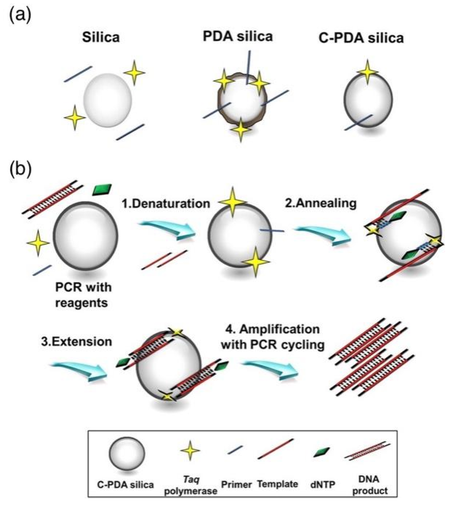
- Park, J.Y.; Back, S.H.; Chang, S.J.; Lee, S.J.; Lee, K.G.; Park, T.J. Dopamine-assisted synthesis of carbon-coated silica for PCR enhancement. ACS Appl. Mater. Interfaces 2015, 7, 15633–15640. [Google Scholar] [CrossRef]
- Fuming, S.; Yang, Y.; Hexiang, Z.; Meirong, M.; Zhizhou, Z. CdTe QDs accelerate the speed of Pfu-based polymerase chain reaction. J. Exp. Nanosci. 2013, 10, 476–482. [Google Scholar] [CrossRef] [Green Version]
- Sang, F.; Zhang, Z.; Yuan, L.; Liu, D. QDs for a high-throughput Pfu polymerase based multi-round polymerase chain reaction (PCR). Analyst 2018, 143, 1259–1267. [Google Scholar] [CrossRef]
- Zhu, M.; Luo, C.; Zhang, F.; Liu, F.; Zhang, J.; Guo, S. Interactions of the Primers and Mg2+ with GQDs Enhance PCR Performance. RSC Adv. 2015, 5, 74515–74522. [Google Scholar] [CrossRef]
- Hwang, S.H.; Im, S.G.; Hah, S.S.; Cong, V.T.; Lee, E.J.; Lee, Y.S.; Lee, G.K.; Lee, D.H.; Son, S.J. Effects of upconversion NPs on polymerase chain reaction. PLoS ONE 2013, 8, e73408. [Google Scholar] [CrossRef] [Green Version]
- Chen, J.; Cao, X.; Guo, R.; Shen, M.; Peng, C.; Xiao, T.; Shi, X. A highly effective polymerase chain reaction enhancer based on Au DENPs. Analyst 2012, 137, 223–228. [Google Scholar] [CrossRef]
- Li, A.; Zhou, B.; Alves, C.S.; Xu, B.; Guo, R.; Shi, X.; Cao, X. Mechanistic Studies of Enhanced PCR Using PEG-Au PENPs. ACS Appl. Mater. Interfaces 2016, 8, 25808–25817. [Google Scholar] [CrossRef]
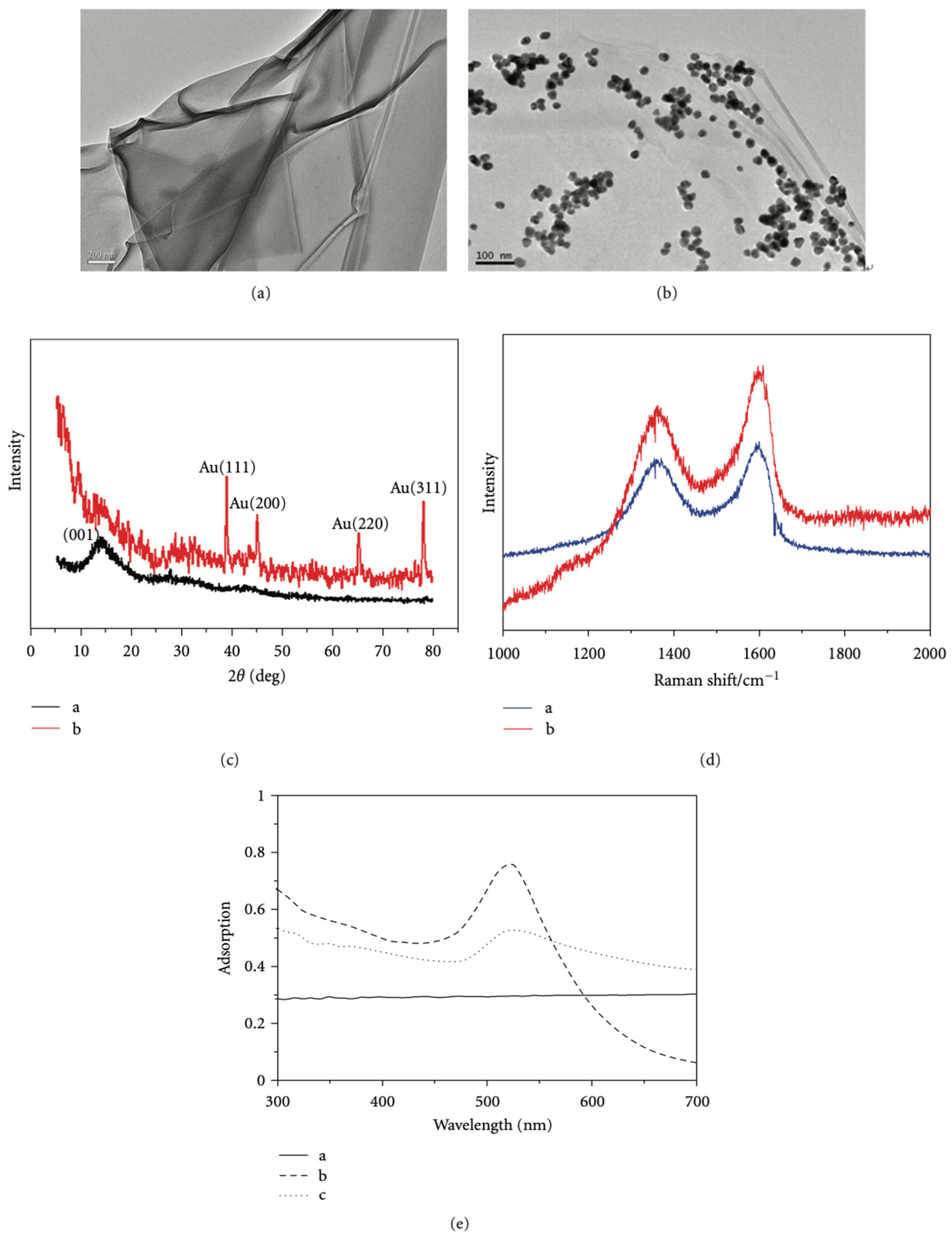
- Jeong, H.Y.; Baek, S.H.; Chang, S.-J.; Yang, M.; Lee, S.J.; Lee, K.G.; Park, T.J. A hybrid composite of gold and GO as a PCR enhancer. RSC Adv. 2015, 5, 93117–93121. [Google Scholar] [CrossRef]
- Song, M.; Yu, L.; Wu, Y. Simple Synthesis and Enhanced Performance of Graphene Oxide-Gold Composites. J. Nanomater. 2012, 2012, 135138. [Google Scholar] [CrossRef] [Green Version]
- Dao Van, Q.; Nguyen Minh, H.; Pham Thi, T.; Nguyen Hoang, N.; Nguyen Hoang, H.; Nguyen Thai, S.; Phan Tuan, N.; Nguyen Thi Van, A.; Tran Thi, H.; Nguyen Hoang, L. Synthesis of Silica-Coated Magnetic Nanoparticles and Application in the Detection of Pathogenic Viruses. J. Nanomater. 2013, 2013, 603940. [Google Scholar] [CrossRef] [Green Version]
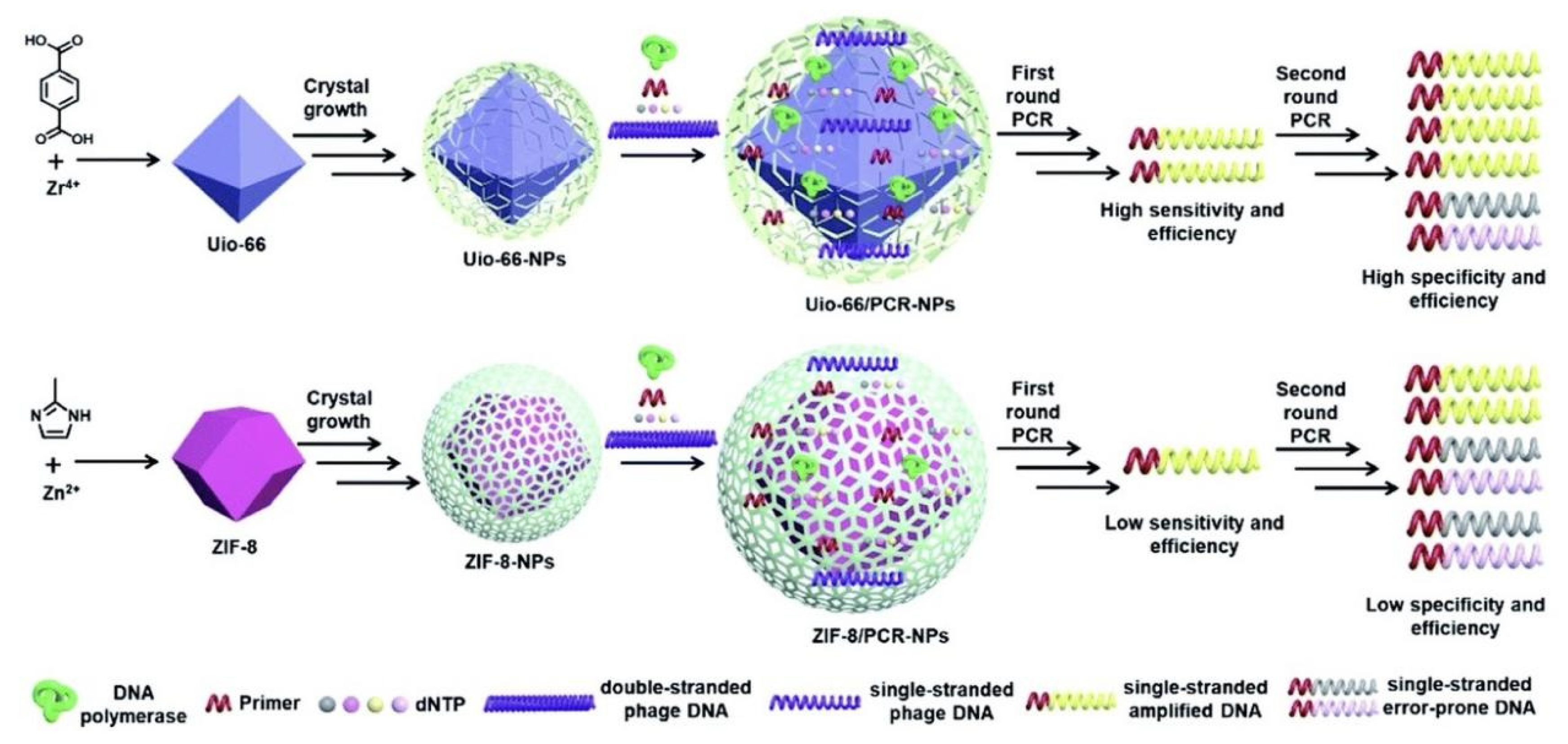
- Sun, C.; Cheng, Y.; Pan, Y.; Yang, J.; Wang, X.; Xia, F. Efficient polymerase chain reaction assisted by MOFs. Chem. Sci. 2019, 11, 797–802. [Google Scholar] [CrossRef] [Green Version]
- Rasheed, A.K.; Siddiqui, R.; Ahmed, S.M.K.; Gabriel, S.; Jalal, M.Z.; John, A.; Khan, N.A. hBN Nanoparticle-Assisted Rapid Thermal Cycling for the Detection of Acanthamoeba. Pathogens 2020, 9, 824. [Google Scholar] [CrossRef]
- Adams, G. A beginner’s guide to RT-PCR, qPCR and RT-qPCR. Biochemist 2020, 42, 48–53. [Google Scholar] [CrossRef]
- Namdari, M.; Negahdari, B.; Cheraghi, M.; Aiyelabegan, H.T.; Eatmadi, A. Cardiac failure detection in 30 minutes: New approach based on gold nanoparticles. J. Microencapsul. 2017, 34, 132–139. [Google Scholar] [CrossRef]
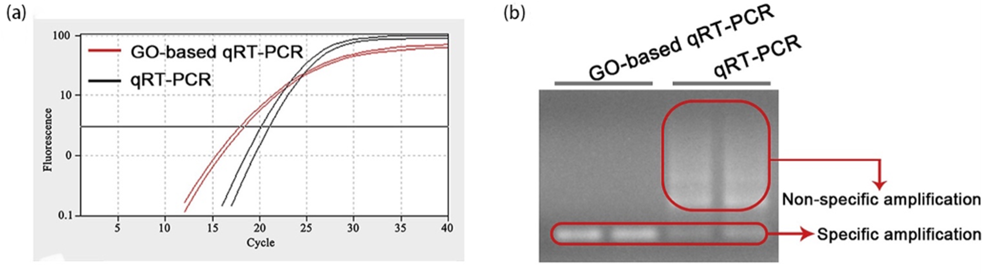
- Hu, C.; Zhang, L.; Yang, Z.; Song, Z.; Zhang, Q.; He, Y. Graphene oxide-based qRT-PCR assay enables the sensitive and specific detection of miRNAs for the screening of ovarian cancer. Anal. Chim. Acta 2021, 1174, 338715. [Google Scholar] [CrossRef]
- Chen, H.; Wu, Y.; Chen, Z.; Hu, Z.; Fang, Y.; Liao, P.; Deng, Y.; He, N. Performance Evaluation of a Novel Sample In–Answer Out (SIAO) System Based on Magnetic Nanoparticles. J. Biomed. Nanotechnol. 2017, 13, 1619–1630. [Google Scholar] [CrossRef]
- Li, B.; Ma, J.; Wang, L.; Xu, M. Novel Method for Rapid Detection of Staphylococcus Aureus and Its Enterotoxins in Patients with Diarrhea. Nanosci. Nanotechnol. Lett. 2019, 11, 593–599. [Google Scholar] [CrossRef]
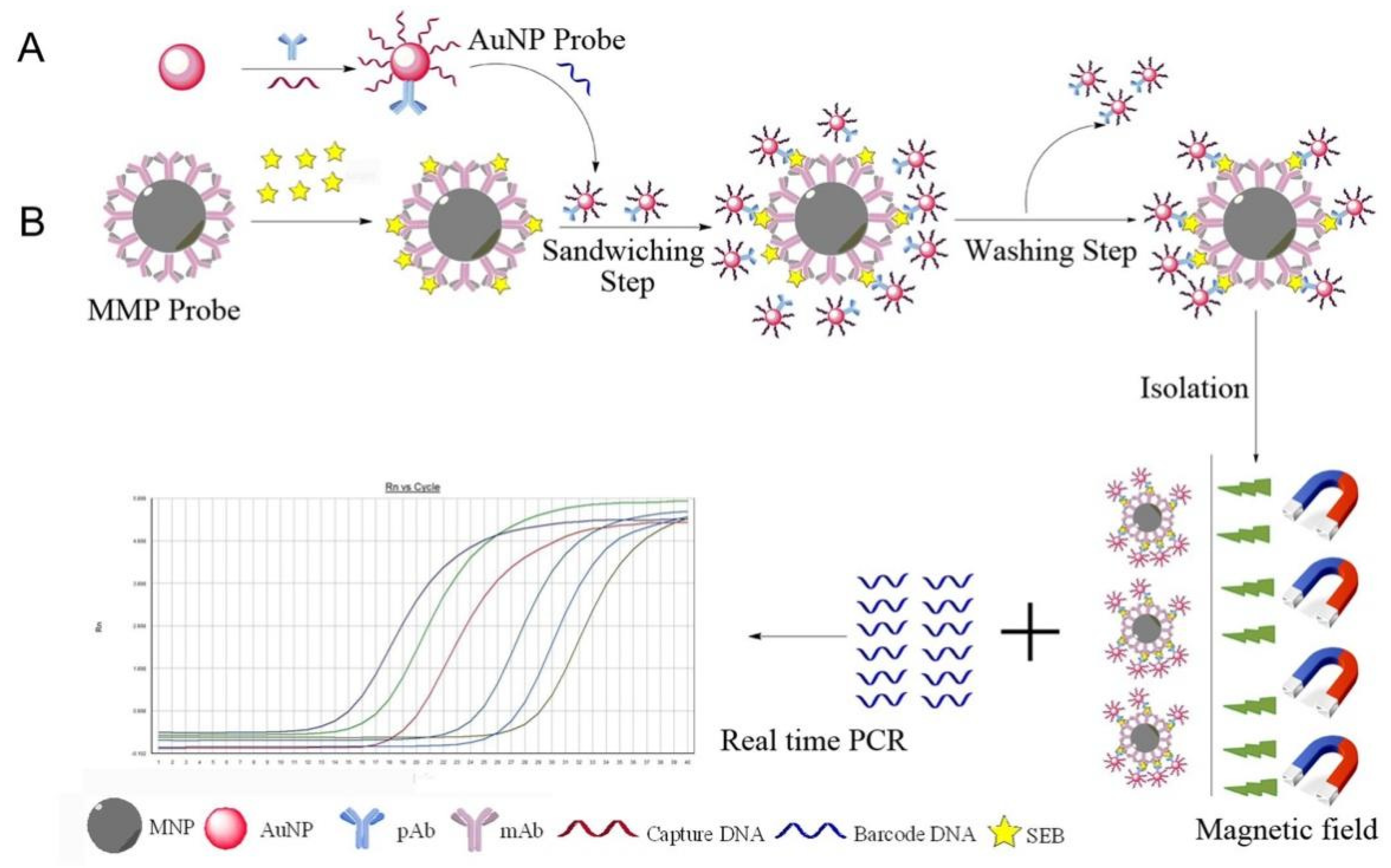
- Yang, H.; Qu, L.; Wimbrow, A.N.; Jiang, X.; Sun, Y. Rapid detection of Listeria monocytogenes by nanoparticle-based immunomagnetic separation and real-time PCR. Int. J. Food Microbiol. 2007, 118, 132–138. [Google Scholar] [CrossRef]
- Bakthavathsalam, P.; Rajendran, V.K.; Saran, U.; Chatterjee, S.; Jaffar Ali, B.M. Immunomagnetic nanoparticle based quantitative PCR for rapid detection of Salmonella. Microchim. Acta 2013, 180, 1241–1248. [Google Scholar] [CrossRef]
- Zhong, D.; He, W. Detection of Pseudomonas aeruginosa in the Skin by Immunomagnetic Isolation and Real-Time Quantitative PCR. J. Nanosci. Nanotechnol. 2019, 19, 5517–5521. [Google Scholar] [CrossRef]
- Yuan, J.; Chen, Q.Y.; Xu, X.J. Rapid Method for the Detection of Porphyromonas gingivalis in Chronic Periodontitis. Nanosci. Nanotechnol. Lett. 2019, 11, 689–695. [Google Scholar] [CrossRef]
- Ernst, C.; Bartel, A.; Elferink, J.W.; Huhn, J.; Eschbach, E.; Schönfeld, K.; Feßler, A.T.; Oberheitmann, B.; Schwarz, S. Improved DNA extraction and purification with magnetic nanoparticles for the detection of methicillin-resistant Staphylococcus aureus. Vet. Microbiol. 2019, 230, 45–48. [Google Scholar] [CrossRef]
- Wu, R.; Meng, B.; Corredig, M.; Griffiths, M.W. Efficient capturing and sensitive detection of hepatitis A virus from solid foods (green onion, strawberry, and mussel) using protamine-coated iron oxide (Fe3O4) magnetic nanoparticles and real-time RT-PCR. Food Microbiol. 2021, 102, 103921. [Google Scholar] [CrossRef] [PubMed]
- Xu, Y.; Huo, B.; Li, C.; Peng, Y.; Tian, S.; Fan, L.; Bai, J.; Ning, B.; Gao, Z. Ultrasensitive detection of staphylococcal enterotoxin B in foodstuff through dual signal amplification by bio-barcode and real-time PCR. Food Chem. 2019, 283, 338–344. [Google Scholar] [CrossRef] [PubMed]
- Rehman, A.; Sarwar, Y.; Raza, Z.A.; Hussain, S.Z.; Mustafa, T.; Khan, W.S.; Ghauri, M.A.; Haque, A.; Hussain, I. Metal nanoparticle assisted polymerase chain reaction for strain typing of Salmonella Typhi. Analyst 2015, 140, 7366–7372. [Google Scholar] [CrossRef] [PubMed]
- Xu, S.; Yao, M. NanoPCR detection of bacterial aerosols. J. Aerosol. Sci. 2013, 65, 1–9. [Google Scholar] [CrossRef]
- Gabriel, S.; Rasheed, A.K.; Siddiqui, R.; Appaturi, J.N.; Fen, L.B.; Khan, N.A. Development of nanoparticle-assisted PCR assay in the rapid detection of brain-eating amoebae. Parasitol. Res. 2018, 117, 1801–1811. [Google Scholar] [CrossRef]
- Cui, Y.; Wang, Z.; Ma, X.; Liu, J.; Cui, S. A sensitive and specific nanoparticle-assisted PCR assay for rapid detection of porcine parvovirus. Lett. Appl. Microbiol. 2014, 58, 163–167. [Google Scholar] [CrossRef]
- Ma, X.; Cui, Y.; Qiu, Z.; Zhang, B.; Cui, S. A nanoparticle-assisted PCR assay to improve the sensitivity for rapid detection and differentiation of wild-type pseudorabies virus and gene-deleted vaccine strains. J. Virol. Methods 2013, 193, 374–378. [Google Scholar] [CrossRef]
- Wang, X.; Bai, A.; Zhang, J.; Kong, M.; Cui, Y.; Ma, X.; Ai, X.; Tang, Q.; Cui, S. A new nanoPCR molecular assay for detection of porcine bocavirus. J. Virol. Methods 2014, 202, 106–111. [Google Scholar] [CrossRef]
- Yuan, W.; Li, Y.; Li, P.; Song, Q.; Li, L.; Sun, J. Development of a nanoparticle-assisted PCR assay for detection of porcine epidemic diarrhea virus. J. Virol. Methods 2015, 220, 18–20. [Google Scholar] [CrossRef]
- Wang, J.; Cheng, Y.; Zhang, M.; Zhao, H.; Lin, P.; Yi, L.; Tong, M.; Cheng, S. Development of a nanoparticle-assisted PCR (nanoPCR) assay for detection of mink enteritis virus (MEV) and genetic characterization of the NS1 gene in four Chinese MEV strains. Bmc Vet. Res. 2015, 11, 1. [Google Scholar] [CrossRef] [Green Version]
- Luo, Y.; Liang, L.; Zhou, L.; Zhao, K.; Cui, S. Concurrent infections of pseudorabies virus and porcine bocavirus in China detected by duplex nanoPCR. J. Virol. Methods 2015, 219, 46–50. [Google Scholar] [CrossRef] [PubMed]
- El-Husseini, D.M.; Helmy, N.M.; Tammam, R.H. The effect of gold nanoparticles on the diagnostic polymerase chain reaction technique for equine herpes virus 1 (EHV-1). Rsc Adv. 2016, 6, 54898–54903. [Google Scholar] [CrossRef]
- Yuan, W.; Li, Y.; Wang, J.; Wang, J.; Sun, J. A nanoparticle-assisted PCR assay for the detection of encephalomyocarditis virus. Vet. Arh. 2016, 86, 1–8. [Google Scholar]
- Zhu, Y.; Liang, L.; Luo, Y.; Wang, G.; Wang, C.; Cui, Y.; Ai, X.; Cui, S. A sensitive duplex nanoparticle-assisted PCR assay for identifying porcine epidemic diarrhea virus and porcine transmissible gastroenteritis virus from clinical specimens. Virus Genes 2017, 53, 71–76. [Google Scholar] [CrossRef] [PubMed]
- Liu, Z.; Li, J.; Liu, Z.; Li, J.; Li, Z.; Wang, C.; Wang, J.; Guo, L. Development of a nanoparticle-assisted PCR assay for detection of bovine respiratory syncytial virus. BMC Vet. Res. 2019, 15, 110. [Google Scholar] [CrossRef] [PubMed]
- Wang, M.; Yan, Y.; Wang, R.; Wang, L.; Zhou, H.; Li, Y.; Tang, L.; Xu, Y.; Jiang, Y.; Cui, W.; et al. Simultaneous Detection of Bovine Rotavirus, Bovine Parvovirus, and Bovine Viral Diarrhea Virus Using a Gold Nanoparticle-Assisted PCR Assay With a Dual-Priming Oligonucleotide System. Front. Microbiol. 2019, 10, 2884. [Google Scholar] [CrossRef] [Green Version]
- Ma, X.; Li, Y.; Liu, R.; Wei, W.; Ding, C. Development of a sensitive and specific nanoparticle-assisted PCR assay for detecting HPV-16 and HPV-18 DNA. J. Med. Virol. 2020, 92, 3793–3798. [Google Scholar] [CrossRef]
- Qin, T.; Wang, J.; Cui, S.-J. Development of a nanoparticle-assisted PCR assay to distinguish canine coronaviruses I and II. J. Vet. Diagn. Investig. 2021, 33, 104–107. [Google Scholar] [CrossRef]
- Wang, Y.; Wang, Y.; Chen, Z.; Liu, G.; Jiang, S.; Li, C. A multiplex nanoparticle-assisted polymerase chain reaction assay for detecting three canine epidemic viruses using a dual priming oligonucleotide system. J. Virol. Methods 2021, 298. [Google Scholar] [CrossRef]
- Ma, H.; Gao, X.; Fu, J.; Xue, H.; Song, Y.; Zhu, K. Development and Evaluation of NanoPCR for the Detection of Goose Parvovirus. Vet. Sci. 2022, 9, 460. [Google Scholar] [CrossRef]
- Ye, J.; Li, Z.; Sun, F.Y.; Guo, L.; Feng, E.; Bai, X.; Cheng, Y. Development of a triple NanoPCR method for feline calicivirus, feline panleukopenia syndrome virus, and feline herpesvirus type I virus. Bmc Vet. Res. 2022, 18, 379. [Google Scholar] [CrossRef]
- Xue, Z.; You, M.; Peng, P.; Tong, H.; He, W.; Li, A.; Mao, P.; Xu, T.; Xu, F.; Yao, C. Taqman-MGB nanoPCR for Highly Specific Detection of Single-Base Mutations. Int. J. Nanomed. 2021, 16, 3695–3705. [Google Scholar] [CrossRef] [PubMed]
- Lee, J.-H.; Cheglakov, Z.; Yi, J.; Cronin, T.M.; Gibson, K.J.; Tian, B.; Weizmann, Y. Plasmonic Photothermal Gold Bipyramid Nanoreactors for Ultrafast Real-Time Bioassays. J. Am. Chem. Soc. 2017, 139, 8054–8057. [Google Scholar] [CrossRef] [PubMed]
- Wang, L.; Huang, Z.; Wang, R.; Liu, Y.; Qian, C.; Wu, J.; Liu, J. Transition Metal Dichalcogenide Nanosheets for Visual Monitoring PCR Rivaling a Real-Time PCR Instrument. Acs Appl. Mater. Interfaces 2018, 10, 4409–4418. [Google Scholar] [CrossRef] [PubMed]
- Lee, C.Y.; Degani, I.; Cheong, J.; Weissleder, R.; Lee, J.-H.; Cheon, J.; Lee, H. Development of Integrated Systems for On-Site Infection Detection. Acc. Chem. Res. 2021, 54, 3991–4000. [Google Scholar] [CrossRef]
- Uchehara, G.; Kirk, A.G.; Trifiro, M.; Paliouras, M.; Mohammadyousef, P. Real time label-free monitoring of plasmonic polymerase chain reaction products. In Proceedings of the Conference on Nano-, Bio-, Info-Tech Sensors and 3D Systems III, Denver, CO, USA, 4–6 March 2019. [Google Scholar]
- Kim, S.-K.; Oh, Y.-H.; Ko, D.-H.; Sung, H.; Oh, H.-B.; Hwang, S.-H. Nanoparticle-Based Visual Detection of Amplified DNA for Diagnosis of Hepatitis C Virus. Biosensors 2022, 12, 744. [Google Scholar] [CrossRef]


| Category | Material | Type of DNA Molecule | Mechanism | Effect | References |
|---|---|---|---|---|---|
| Metal nanomaterials | Au NPs | cDNA from bladder cancer cell line and lung cancer tissue, BNIP3 cDNA from bladder cancer cells and colorectal tissue | Thermal conductivity | Increase the yield by 104–106 times; Shorten the reaction time | [17] |
| 309 bp fragment from pBR322 DNA | Surface interactions | Increase the yield of PCR product; Specificity; Efficiency | [18] | ||
| 283-bp λ-DNA | / | Enhance sensitivity and specificity in multi-round PCR | [19] | ||
| 309 bp λ-DNA and genomic cDNA | Surface interactions | Specificity; Efficiency (favor smaller products) | [20] | ||
| pBR322 DNA template | Thermal conductivity | Specificity; Efficiency | [21] | ||
| Human male genomic DNA | Surface interactions; Catalytic activity | Specificity; Efficiency | [22] | ||
| Housekeeping gene GAPDH from the human DNA template | / | Increase the yield of PCR product | [23] | ||
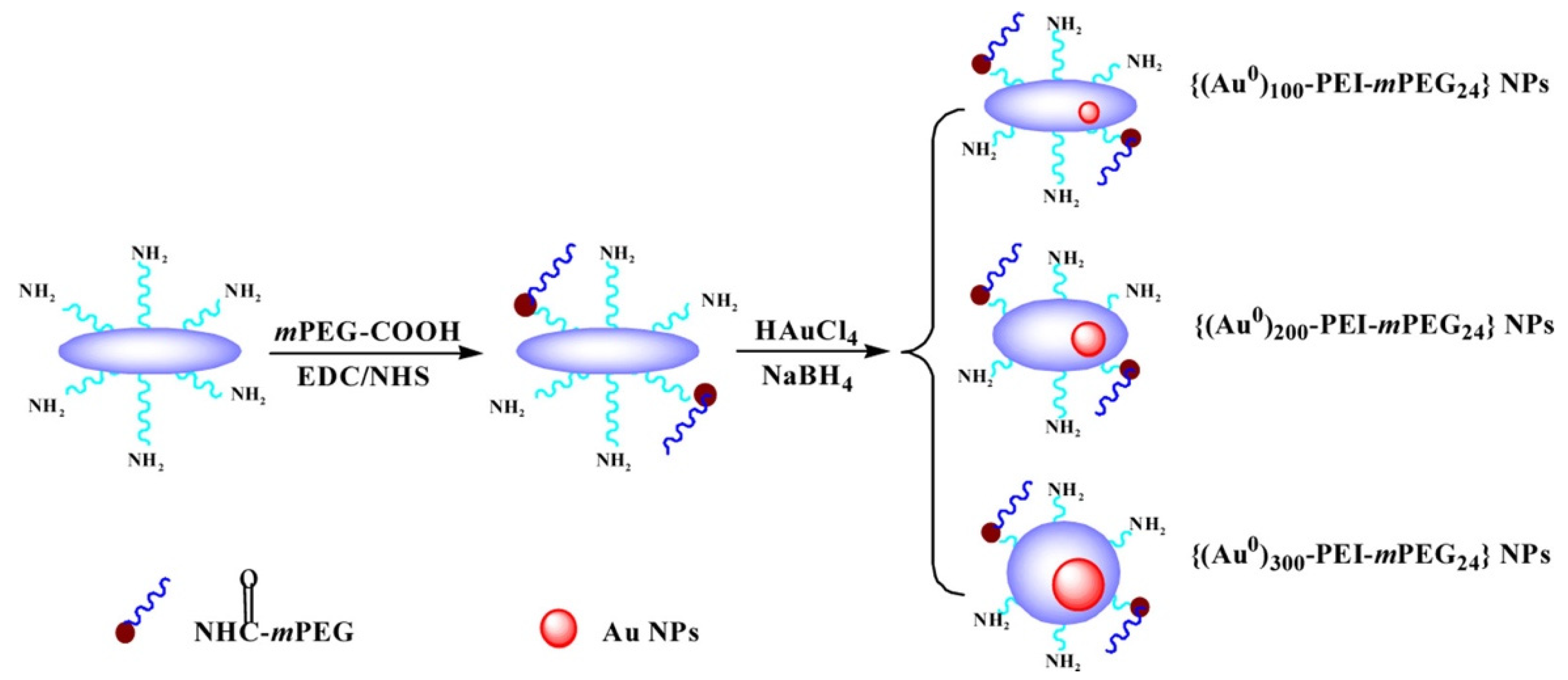
| Au DENPs | 283-bp λ-DNA | Electrostatic interactions | Specificity; Efficiency | [54] | |
| PDDA-Au NPs | 283-bp λ-DNA | Electrostatic interactions | Specificity; Efficiency | [11] | |
| PEG−Au PENPs | 283-bp λ-DNA | Electrostatic interactions; Thermal conductivity | Specificity; Efficiency | [55] | |
| Ag NPs | g-DNA, λ-DNA (kb) | Thermal conductivity | Increased PCR efficiency with long DNA and repeated amplification | [24] | |
| 714 bp GFP gene | Surface interactions | Inhibition | [25] | ||
| 750 bp mCherry containing plasmid | / | Efficiency | [16] | ||
| Carbon-based nanomaterials | CNTs | 410 bp DNA | Surface interactions; Catalytic activity | Increase the yield of PCR product | [7] |
| 14.3 kb λ-DNA | / | Specificity; Efficiency | [28] | ||
| CNT/PEI | 283 bp λ-DNA | Electrostatic interactions; Thermal conductivity | Specificity; Efficiency | [29] | |
| NH2-MWCNTs | 94 mer random DNA oligonucleotide library | Surface interactions | Specificity; Efficiency (filtered NH2-MWCNT) | [30] | |
| SWCNTs, NH2-SWCNTs | 283 bp λ-DNA | Electrostatic interactions | Increase the yield of PCR product | [31] | |
| CNP | 540 bp g-DNA | Surface interactions | Increased PCR specificity and efficiency with long DNA and repeated amplification | [33] | |
| Graphene | 300 bp fragment from pET-32a plasmid DNA | Surface interactions; Electrostatic interactions; Thermal conductivity | Specificity | [36] | |
| 283 bp λ-DNA | Surface interactions | Specificity | [8] | ||
| GNFs | 1248 bp g-DNA | Thermal conductivity | Reduce cycles, Efficiency | [37] | |
| GO | pET-32a plasmid | Electrostatic interactions | Specificity | [73] | |
| Oxide nanomaterials | TiO2 | 650 bp DNA | Surface interactions | Inhibition | [9] |
| Mouse and human genomic DNA, plasmid DNA, and mouse complementary DNA [cDNA] | Thermal conductivity | Efficiency | [41] | ||
| cDNA or gDNA | / | Increase the yield of PCR product | [42] | ||
| Silica-coated and amino-modified ZnO | Plasmid DNA | Electrostatic interactions | Increase the yield of PCR product | [10] | |
| ZnO | 619 bp and 666 bp DNA | / | Specificity; Efficiency; Reduce reaction time | [74] | |
| Fe3O4 | 800 bp prokaryotic DNA | Surface interactions; Thermal conductivity | Efficiency | [75] | |
| MgO | / | / | Efficiency | [48] | |
| SiO2 | Genomic DNA of E. coli (eae1, 248 bp) and pEGFP-C1 plasmid (egfp, 800 bp) | Surface interactions | Increase the final quantity of PCR product | [49] | |
| Fluorescent nanomaterials | CdTe QDs | λ-DNA | Analogous to ssDNA binding protein (SSB); Surface interactions | Specificity | [6] |
| 1000 bp human genomic DNA | Surface interactions | Reduce reaction time | [50] | ||
| Human DNA, plasmid DNA or marine fouling organism DNA | Surface interactions | Retained specificity in the ninth-round amplification | [51] | ||
| GQDs | 80 bp fragment from a GC-rich DNA | Surface interactions | Specificity; Efficiency; Increase the yield of PCR product | [52] | |
| UCNPs | 120 bp 5S rRNA | / | Specificity | [53] | |
| Others | GO-Au composites | Genomic DNA of Listeria monocyte (200 bp) and Scomber japonicas (800 bp) | Surface interactions | Specificity; Efficiency; Broad annealing temperatures | [56] |
| MOFs | λ-DNA | Surface interactions | Specificity; Efficiency; Wide annealing temperatures | [59] |
| Additives | ζ-potential (mV) | Optinimum Concentration (mg/L) | Maxima Efficiency a | Maximal Specificity a |
|---|---|---|---|---|
| PEI | 24.07 ± 1.45 | 0.47 | 1.5 | 1 |
| {(Au0)100-PEI-mPEG24} NPs | 28.93 ± 0.85 | 0.38 | 2.2 | 1 |
| {(Au0)200-PEI-mPEG24} NPs | 33.46 ± 1.28 | 0.34 | 3.6 | 1 |
| {(Au0)300-PEI-mPEG24} NPs | 34.23 ± 1.09 | 0.38 | 1.9 | 1 |
| {(Au0)200-PEI·NHAc-mPEG24} NPs | 6.34 ± 1.13 | 60 | 1.4 | 1 |
| Category | Type or Purpose of Detection | NPs | Effect | References |
|---|---|---|---|---|
| Bacteria detection | Strain Typing of Salmonella typhi | Citrate stabilized Au NPs, rhamnolipid stabilized Au and Ag NPs, and magnetic iron oxide NPs | Reduce non-specific amplification (Au and Ag NPs); Increase PCR yield (Au NPs, Au and Ag NPs); Inhibition (magnetic iron oxide NPs) | [73] |
| Bacterial aerosols | Ag NPs, TiO2 NPs and their combination | The detection limit down to 40 pg/μL | [74] | |
| Brain-eating amoebae | GO, CuO and Al2O3 NPs | Enhanced PCR efficiency | [75] | |
| Virus detection | Porcine parvovirus | Solid NPs (1–100 nm diameter) | Enhanced PCR sensitivity (100-fold more sensitive) | [76] |
| Detection and differentiation of wild-type pseudorabies virus and gene-deleted vaccine strains | Solid Au NPs (1–100 nm) | Enhanced PCR sensitivity (100–1000-fold more sensitive) | [77] | |
| Porcine bocavirus | Solid Au NPs (1–100 nm) form colloidal nanofluids | Enhanced PCR sensitivity (100-fold more sensitive); The detection limit down to 6.70 × 101 copies | [78] | |
| Porcine epidemic diarrhea virus | Solid Au NPs(1–100 nm) form colloidal nanofluids | Enhanced PCR sensitivity (100-fold more sensitive); The detection limit down to 2.7 × 10⁻⁶ ng/μL | [79] | |
| Mink enteritis virus (MEV) | No instructions | The detection limit down to 8.75 × 101 copies recombinant plasmids per reaction | [80] | |
| Concurrent infections of pseudorabies virus and porcine bocavirus | Solid Au NPs (1–100 nm) form colloidal nanofluids | Enhanced PCR efficiency; The detection limit of 6 copies for PRV and 95 copies for PBoV | [81] | |
| A diagnostic technique for equine herpes virus-1 (EHV-1) | Au NPs | Increase PCR yield; The detection limit down to 102 DNA copies | [82] | |
| Encephalomyocarditis virus | Solid Au NPs(1–100 nm) form colloidal nanofluids | Enhanced PCR sensitivity and specificity;Detection limit down to 1.2 × 10² copies/μL | [83] | |
| Porcine epidemic diarrhea virus and porcine transmissible gastroenteritis virus | Solid NPs (1–100 nm diameter) | Enhanced PCR sensitivity (10-fold more sensitive) | [84] | |
| Bovine respiratory syncytial virus | Au NPs | Enhanced PCR sensitivity; Detection limit down to 1.43 × 10² copies recombinant plasmids per reaction | [85] | |
| Bovine Rotavirus, Bovine Parvovirus, and Bovine Viral Diarrhea Virus | Au NPs | Enhanced PCR sensitivity and specificity | [86] | |
| Quick Diagnosis of Canine Vector-Borne Pathogens | ZnO Nanoflower | Reduce the reaction time; Enhanced PCR sensitivity and specificity | [44] | |
| HPV-16 and HPV-18 DNA | Solid Au NPs(1–100 nm) | Enhanced PCR sensitivity (10-fold more sensitive) and specificity | [87] | |
| Distinguishing canine coronaviruses I and II | Solid Au NPs (1–100 nm) form colloidal nanofluids | Enhanced PCR sensitivity (100-fold more sensitive) and specificity | [88] | |
| Canine distemper virus (CDV), canine parvovirus (CPV) and canine coronavirus (CCV) | Solid Au NPs(1–100 nm) | Enhanced PCR sensitivity and specificity | [89] | |
| Goose Parvovirus | Au NPs | Enhanced PCR sensitivity (100-fold more sensitive) | [90] | |
| Feline calicivirus, feline panleukopenia syndrome virus, and feline herpesvirus type I virus | Au NPs | Enhanced PCR sensitivity (10–100-fold more sensitive) and specificity | [91] | |
| Tumor monitoring | Single-base mutations to monitor tumor | Au NPs | Enhanced PCR sensitivity and specificity | [92] |
| Detection of miRNAs to screen ovarian cancer | GO | Enhanced PCR sensitivity and specificity | [63] | |
| No machine PCR | Plasmonic photothermal gold bipyramid banoreactors | Gold bipyramid nanoparticles (Au BPs) | Achieved ultrafast thermocycling | [93] |
| To realize on-site and instant analysis | GO, rGO, molybdenum disulfide (MoS2), and tungsten disulfide (WS2) | Achieved visual detection (MoS2 and WS2) | [94] | |
| point of care (POC) settings | Core−shell magnetoplasmonic nanoparticles (MPNs) | Detected SARS-CoV-2 RNA down to 3.2 copy/μL within 17 min | [95] | |
| Detection of health-related DNA and proteins | Au NPs | High sensitivity, visual detection, capability for on-site detection | [14] | |
| Real time label-free monitoring of plasmonic | Au NPs | The detection limit down to 10,000 genome copies/μL | [96] | |
| Diagnosis of Hepatitis C Virus | Streptavidin-coated magnetic particles (1μm) and anti-digoxigenin antibody-coated polystyrene particles (250–350 nm) | Visual detection; High sensitivity and specificity | [97] |
Publisher’s Note: MDPI stays neutral with regard to jurisdictional claims in published maps and institutional affiliations. |
© 2022 by the authors. Licensee MDPI, Basel, Switzerland. This article is an open access article distributed under the terms and conditions of the Creative Commons Attribution (CC BY) license (https://creativecommons.org/licenses/by/4.0/).
Share and Cite
Yang, Z.; Shen, B.; Yue, L.; Miao, Y.; Hu, Y.; Ouyang, R. Application of Nanomaterials to Enhance Polymerase Chain Reaction. Molecules 2022, 27, 8854. https://doi.org/10.3390/molecules27248854
Yang Z, Shen B, Yue L, Miao Y, Hu Y, Ouyang R. Application of Nanomaterials to Enhance Polymerase Chain Reaction. Molecules. 2022; 27(24):8854. https://doi.org/10.3390/molecules27248854
Chicago/Turabian StyleYang, Zhu, Bei Shen, Lihuan Yue, Yuqing Miao, Yihong Hu, and Ruizhuo Ouyang. 2022. "Application of Nanomaterials to Enhance Polymerase Chain Reaction" Molecules 27, no. 24: 8854. https://doi.org/10.3390/molecules27248854