1. Introduction
Healthy coral reefs are one of the most valuable ecosystems on earth as they provide food and coastal protection; however, some chemicals or active ingredients within sunscreens can cause permanent damage to coral reefs [
1]. The most known harmful chemical compounds are oxybenzone and octinoxate; nonetheless, other chemicals included in commercial sunscreens are benzophenone-1, benzophenone-8, OD-PABA, 4-methylbenzylidene camphor, 3-benzylidene camphor, nano-titanium dioxide, and nano-zinc oxide [
2,
3,
4]. Cosmetic ingredients are highly regulated to assure safety and efficiency standards, besides the fact that not allowed compounds may be present in a cosmetic at trace levels due to processing conditions, the prevalence of synthetic ingredients over naturals is related to the fact that the final composition of a commercial cosmetic must satisfy the corresponding legislation [
5]. It is common practice that a synthetic compound may be chosen over a natural source due to the stability of the molecule of interest. However, a natural form of astaxanthin (a microalgal pigment of current interest) may compete against the synthetic molecule, not only due to its safety and reduced environmental stress but also due to the higher antioxidant activity related to the esterified form in comparison to synthetic astaxanthin. Related to this condition high-value bioactive molecules from microalgae such as astaxanthin are being explored for pharmaceutical and cosmetic products [
6,
7,
8]. The formulation of sunscreens based on microalgae may have the necessary protection but need proper carriers due to the solubility of MAAs; they are not suitable for being on the skin for a long period. It has been reported that the use of microalgae extracts is beneficial for the ecosystem and human beings. Such extracts are useful in the formulation of skin care products offering daily sun protection that oils in other formulations do not have. However, there is a need to improve the formulation to carry, deliver, and extend the retention period on the skin [
9,
10]. This work looks to report bioactive compounds from algae with UV-protection activity and new methods to formulate longer-lasting topic formulation.
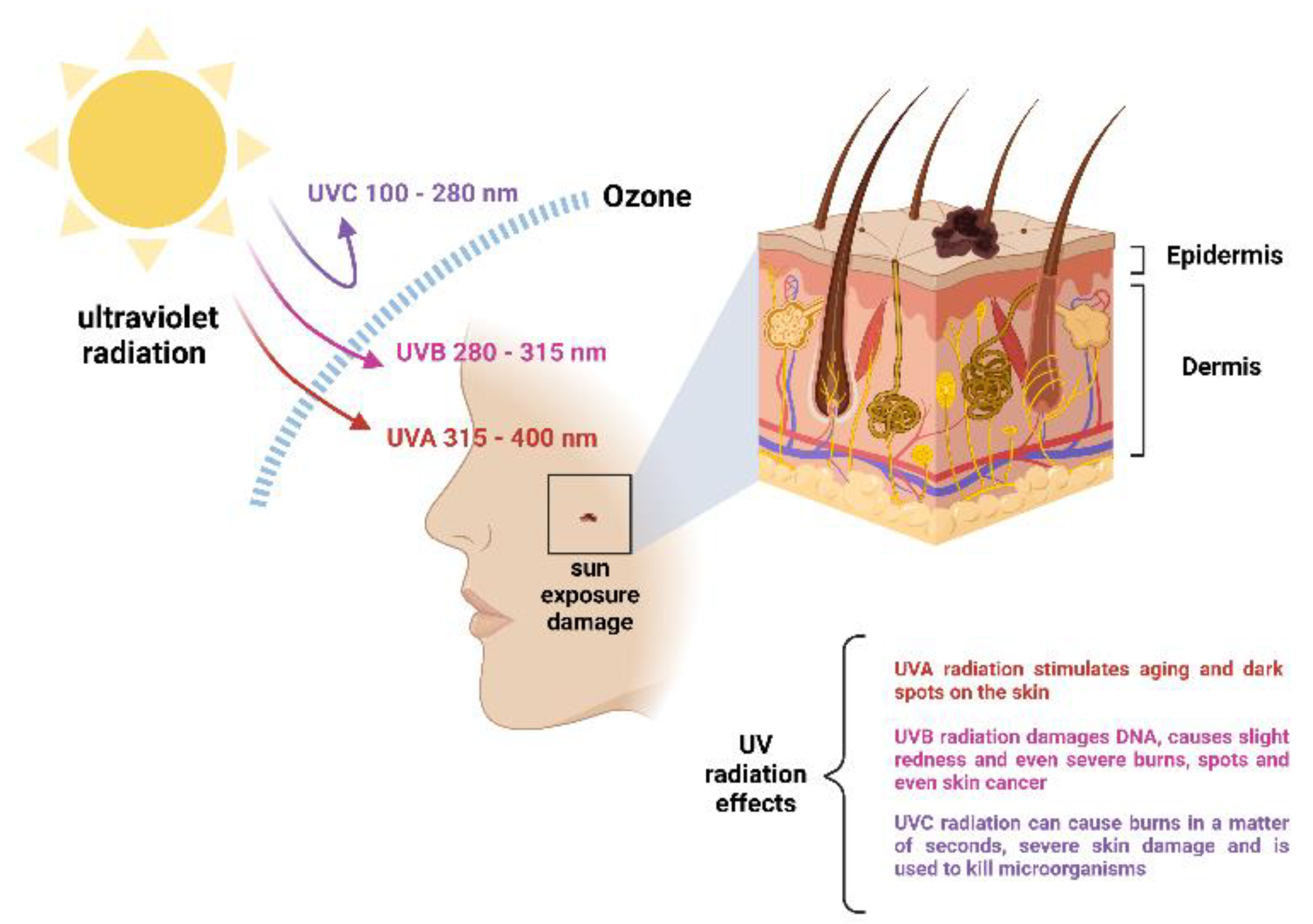
2. Solar Radiation
The level of solar radiation that reaches the earth’s surface has increased dramatically in recent years due to the decrease and changes in the permeability of the ozone layer. The UV index published by the Environmental Protection Agency of the United States (EPA), presents projections of the risk of overexposure to UV radiation; just as an example of the severity of the change in radiation levels, in 1994 in New York City, the forecast for the number of days per year with high UV exposure risk was 29, with no expected days with a UV index of very high risk, but in 2017, the number of days with high UV exposure prediction was 66, and even 58 days were predicted to feature very high levels of UV radiation [
11]. With this data in mind, the current importance of photoprotection becomes obvious. Ultraviolet radiation is defined as the electromagnetic energy emitted at wavelengths shorter than those corresponding to that visible to the human eye, but greater than what characterizes X-rays, between 100 and 400 nm. UV radiation is biologically harmful, damaging the DNA of cells, and it can cause genetic defects on external surfaces if received in high doses. UV radiation can damage human skin, causing slight redness to burns over time. It can cause serious discomfort, moles, blemishes, and even skin cancer; when the body perceives damage from sunlight, it sends it to the affected cells to avoid further damage and darkens the skin. It has harmful effects in the short and medium term. The reddening of the skin (solar erythema), from mild to severe burns, is the main immediate harmful effect. The medium-term effects include the most frequent skin cancers and premature aging of the skin and changes in the DNA of living beings [
12,
13]. Depending on the wavelength, UV radiation can be classified into three categories, UVA, UVB, and UVC.
Occupational exposure to solar radiation is a current problem, as well as the geographic variation of UV exposure; these environmental characteristics determine risk levels that concern occupational safety and health departments [
14]. There are six phototypes according to their UVR skin response; melano-compromised (1), melano- compromised (2), melano-competent (3), melano competent (4), melano-protected (5), and melano protected (6). In this case, the skin responses to UVR radiation, correspondingly, are: always sunburns with no tan, often sunburns with light tan, sometimes sunburns with medium tan, rarely sunburns with dark tan, rarely sunburns with naturally dark skin, and no sunburns with naturally dark skin. These phototypes are according to a Standard Erythemal Dose (SED) of 100 J/m
2 [
14].
UVA radiation is responsible for tanning, and it is continuously visible radiation, varying between 400 and 320 nm. UVB radiation is very dangerous for life when exposed for long periods, being the cause of many skin and eye diseases (skin, cancer, carats, etc.), it varies between 320 and 280 nm. UVC radiation is the most dangerous for human life. It is absorbed by the ozone layer and is below 280 nm, causing irritation, high-grade burns, and continuous damage to the skin.
Figure 1 shows the harmful effects caused by solar radiation on human skin [
15].
3. Algae and UVA Stimulation
UVA or long wavelength radiation (315–400 nm) is not absorbed by the ozone layer and constitutes 90% of the total radiation that reaches the earth’s surface. UVA rays are also known as “aging rays”, capable of passing through the dermis and are not only inducing the appearance of wrinkles but are also associated with the development of skin pathologies and the formation of reactive oxygen species (ROS) [
16]. Studies by Huang et al. in 2018 about the UVA radiation induction of microalgae metabolites reported the increase in total xanthophylls and total mycosporine-like amino acids through a 365 nm UVA light with a culture of
Nitzschia closterium and
Isochrysis zhangjiangensis [
17]. Additionally, Huang et al. in 2018 investigated the effects of microalgae against UV radiation and explored fecosterol mechanisms in microalgae such as
Nitzschia closterium by using classical methods including a semi-quantitative reverse transcription polymerase chain reaction, HPLC (high-performance liquid chromatography, reducing the concentration of UV-induced matrix metalloproteinases (MMPs), and inflammation caused by cytokine expression. Fucoxanthin is a carotenoid that is a potent antioxidant because it reduces (ROS) and MMP-13 expression in addition to inhibiting UV-induced VEGF expression in the skin. Astaxanthin is a carotenoid derived from algae; it is widely used as an ingredient in skin care products for its immune system-stimulating properties. Álvarez-Gómez et al. in 2016 found that fucoidan inhibits melanin synthesis by down-regulating melanogenesis-associated transcription factor (MITF) and protein tyrosinase. This result suggests that fucoidan can be used as an anti-pigmentation ingredient in medical and cosmetic fields. It has been described with antioxidant and anti-angiogenic properties. Below,
Table 1 shows different compounds that are induced by exposure to UVA irradiation of macro- and microalgae understanding macroalgae as a multicellular form of an organism and microalgae as a unicellular form [
18,
19].
4. Algae and UVB Stimulation
Surface UVB irradiation (280–315 nm) is related not only to the stratospheric ozone layer but also to other tropospheric factors (aerosols, clouds, etc.), surface albedo, sea ice, and snow [
21]. As shortwave solar radiation, UVB irradiation can penetrate 20 to 30 m into the ocean water and cause serious deleterious effects on marine organisms. Therefore, there has been great concern about the increase in UVB irradiation and its consequences in the biosphere throughout the 21st century, although the ozone layer has improved in recent years due to the implementation of the Montreal protocol [
22]. Phytoplankton, similarly, to chlorophyll, is subjected to stress induced by UVB irradiation since they live in a habitat where UV radiation has been changing. In general, high doses of UVB irradiation have been widely reported to cause stress to microalgae, resulting in DNA damage, photosynthesis impairment, lipid peroxidation, cyclobutene pyrimidine dimer formation, and growth inhibition [
23,
24]. Although several physiological and biochemical studies have investigated the effects of UVB irradiation on phytoplankton, few studies have explored the impacts of UVB irradiation on algae community composition. Substantially reduce the rate of CO
2 assimilation and the amount of rubisco and decrease the absorption of nutrients such as phosphorus and nitrate [
25]. Below, in
Table 2, the effects of UVB irradiation on microalgae are listed, showing the different types of microalgae and the compounds they produce.
5. Algae and UVC Stimulation
The UVC, or short wavelength, radiation (100–280 nm) is of higher energy but is completely absorbed by oxygen and ozone in the atmosphere [
26,
27]. Continuous exposure to UV irradiation contributes to aging and aesthetic problems, with consequences such as sunburn, hyperpigmentation, or skin cancer. Several studies have shown that UVC irradiation causes a negative response in cellular processes, metabolism, and growth [
28,
29]. Despite the above, based on the knowledge that it is possible to obtain a beneficial effect with the sublethal application of an agent capable of inducing physical stress or chemical, it has been verified that controlled radiation with UVC in some organisms, using doses between 2 and 14 Kj/m
2 can produce positive responses like in seedlings, fruits, and other plant structures in the postharvest [
30,
31]. Previous studies have found that UVC irradiation could inhibit the growth as well as the release of microcystins from
Microcystis aeruginosa in artificial culture. In addition, UVC irradiation could cause marked damage to various targets of algae cells including the gene, photosynthesis system, cell membrane integrity, the inhibition of toxin production, and release.
M. aeruginosa and C. vulgaris were chosen as cyanobacteria and microalgae to investigate the suppression effects of microalgae against UVC irradiation, looking for its photoprotective effect against solar radiation [
32]. It has been reported that the ultrafiltration method is effective at a dose of 37–150 Kj/m
2 and 0.180 Kj/m
2 in the development of photoprotective metabolites ROM (reactive oxygen metabolites), resulting in antioxidant and protective effects. In
Table 3 are gathered known compounds produced from microalgae as defense mechanisms against UVC irradiation.
Table 2.
Algae compounds obtained by UVB irradiation.
Table 2.
Algae compounds obtained by UVB irradiation.
| Algae | Type of Algae | Compounds | Extraction Method | Yield
(%) | Dose (Kj/m2∗d) | Cell Protection Effect | Reference |
|---|
| Alexandrium catenella | Microalgae | Diatoxanthin | HPLC | NR | NR | Change in concentration of photosynthetic pigment. | [33] |
Characium terrestre
Coelastrum microporum
Enallax coelastroides
Enallax sp.
Scenedesmus sp.
Scotiella chlorelloide | Microalgae | Porphyra-334 | HPLC | NR | NR | Non-lethal UVB dose increase MAAs synthesis. | [34,35] |
Nannochlorop-sis oceanica
Dunaliella salina
Nannochloropsis limnetica | Microalgae | Carotenoids and vitamin D3 | CG | 24 ± 0.1 | 3–6 | Accumulation of α-tocopherol and β-carotene | [36] |
| Rhodomonas salina | Microalgae | Carotenoids | HPLC | 24.1 ± 0.1 | 16–22 | Photoreactivation mechanism in cells to survive UVB damage. | [37] |
6. Influence of Light on Microalgae Growth and Bioproducts
Visible-ultraviolet radiation (UV-vis) is a photon emission of visible spectrum regions, between ultraviolet and infrared (400–780 nm).
UV-vis radiation uses electromagnetic radiation in the visible regions. It is used to identify some functional groups, in this case seaweed, such as temperature, salinity, or CO2 concentration. Microalgae can survive different doses of UV-vis radiation in extreme conditions since their physiology is capable of changing, promoting their adaptation through the production of secondary metabolites that allow them to conquer different environments. The intensity of UV-vis radiation can exert a harmful effect on the photosynthetic process and on the cellular components of algae, but it also promotes protection and repair mechanisms against UV radiation. One of the main protection mechanisms is the biosynthesis and accumulation of molecules such as carotenoids and phycobiliproteins.
Microalgae and macroalgae are phototrophic organisms, where their primary and secondary metabolism strongly depends on lighting for their growth and the production of biomass and secondary metabolites, which has generated significant efforts to achieve adequate irradiation (lighting time, intensity, and wavelength) to achieve more efficient production of the metabolite of interest [
40]. The term chromatic acclimation proposed demonstrated all the mechanisms of algae to optimize light capture in response to variations in light irradiation variations [
41]. The effect of light on the growth and production of microalgal metabolites has been shown to depend on the species and genetics of each microorganism, with no possibility of transfer from one strain to another [
42].
Arthrospira maxima and echinenone were examined under UV-vis radiation at a dose of 400–490 nm to look for the different metabolites and effects caused by this process. Previous studies found that
A. maxima develops secondary metabolites capable of generating photoprotective agents that are useful for the protection of microalgae from UV-vis radiation and block photochemical reactions in the epidermis. Spirulina is the commercial name for the edible cyanobacteria of the genus
Arthrospira that contains high protein content, fatty acids, vitamins, and minerals [
43,
44]. It was examined by the high-power elevation system at a dose of 300–500 nm to identify the production of photoprotective agents, and it was concluded that improvement in the interaction of compounds with the polymer due to the presence of hydroxyl in its structure provided layers of protection against light [
45].
Different studies have shown that the joint supply of monochromatic lights provides more energy for the photosynthesis of microalgae cultures. The combination of blue and red light increases the speed with which microalgae reach the stationary phase, through lower doubling rates and generation times. The simultaneous application of red and blue light in
Dunaliella salina showed biomass growth and an increase of up to 35.33% in lipid production, and blue light also showed significant increases in protein, carbohydrate, and lipid production [
46].
Similarly, Severes et al. (2017), demonstrated in cultures of
Chlorella sp. that the mixture of red 660 nm and blue 465 nm wavelengths in a 1:1 ratio induces an increase in chlorophyll content up to 3.64 μg/mL, compared to 2.62 μg/mL obtained with red light. The combination of red:blue light in proportions of 5:5, 7:3, and 3:7 induced an increase in biomass production up to 494, 456, and 452 mg/L, respectively, against 300–330 mg/L obtained with monochrome light [
47].
The absence of blue light and the presence of green light significantly downregulated genes involved in carbon fixation and decreased photosynthetic efficiency in
Nannochloropsis gaditana, converting β-carotene as the main derivative pigment to low nutrient levels, mimicking the conditions of the absence of nutrients [
37,
40].
The maximum productivity of lipids in different cultures of microalgae strains has been achieved with light intensities ranging from 60 to 700 µmol photons m
−2 s
−1, the use of red and blue light in different proportions, as well as the photoperiod or pulsed light, achieving a regulatory effect on the synthesis of fatty acids and carotenoids such as lutein, β-carotene, and astaxanthin [
48].
The strategic management of different monochromatic wavelengths allows to achieve more sustainable cultures of photoautotrophic strains, through the first stage of growth and the accumulation of biomass at favorable wavelengths, followed by a stage of an abrupt change to an unfavorable wavelength, to induce the production of secondary metabolites such as lipids and bioactive compounds.
Table 4 list some compounds produced from microalgae as defense mechanisms against UV-vis irradiation.
7. UV Protective Metabolites
7.1. Mycosporine-like Amino Acids
MAA are found naturally in seaweed, and a high MAA content has been shown in red algae and brown algae. The structure of MAAs includes cyclohexanediones or cyclohexene aldehydes in the active site that are linked to another active group through the phenol hydroxyl. MAAs are hypothesized to absorb light at a wavelength of 320 to 360 nm; therefore, MAAs exhibit a strong ultraviolet absorption capacity. In this research, MAA from
Porphyra haitanensis was extracted, separated, and purified and the mechanisms responsible for its cutaneous anti-photoaging effect and as explored by using a mouse cutaneous photoaging model. These experiments, involving local treatments and washes administered with various doses of MAA, were effective against skin photoaging, and metalloproteinases (MMPs) content and expression were detected in skin tissue homogenates, combined with the pathological analysis of the anti-photoaging activity and underlying mechanisms of MAAs. The anti-photoaging activity and underlying mechanisms of MAAs were analyzed. Scytonemin is another UVA-, UVB-, and UVC-filtering compound; this is a highly stable, yellowish-brown, lipid-soluble, inducible pigment. Scytonemin is found exclusively in the polysaccharide sheath of some cyanobacteria as a protective mechanism. During periods of desiccation, scytonemin becomes more important as a UV protector due to the inactivation of other detection mechanisms. Its UV protection properties mean scytonemin is a powerful sunscreen material [
48,
54]. Exposure of
Chlamydomonas nivalis to ultraviolet light induced the production of bioactive compounds with antioxidant properties. The tolerance to UV rays of the snow algae
C. nivalis and the ability to produce under this radiation phenolic compounds, free proline, and antioxidant protection factors in response to UV-A and UV-C light generates a great potential for biotechnological and pharmaceutical application [
55,
56].
7.2. Carotenoids
Carotenoids are known as fat-soluble plant pigments widely distributed in nature that provide various colors such as yellow, red, and orange to fruits and vegetables. They are synthesized mainly by plants and algae, as well as by fungi and bacteria. Carotenoids can be found throughout the animal kingdom and in humans due to selective absorption throughout the food chain [
57]. These lipophilic molecules are based on the chemical structure classified as carotenes and xanthophylls, and both classes have a common C40 polyisoprenoid structure containing a series of centrally located conjugated double bonds that act as a light-absorbing chromophore. Carotenoids that exist as pure nonpolar hydrocarbons are called carotenoids (α-carotene, β-carotene, and lycopene); on the contrary, xanthophylls (β-cryptoxanthin, lutein, zeaxanthin, and astaxanthin) are more polar carotenoids that contain oxygen as a functional group in their structure, either as a hydroxyl group or a keto group as a terminal group [
58]. So far, more than 800 carotenoids have been identified, but only several are found in the human body, including α-carotene, β-carotene, lutein, and lycopene, as well as zeaxanthin and α- and β-cryptoxanthin. People are constantly exposed to ultraviolet (UV) light, some less, some more, depending on where they live, their activities, what they do for a living, hobbies, culture, but also their understanding of the importance of sun protection and its implementation. Exposure to solar ultraviolet light has been estimated to be 10% of the total available annual UVR for outdoor workers and 3% for adults working indoors [
58,
59]. It is crucial to keep in mind that sunlight stimulates blood circulation and bone health, since the exposure to some sunlight induces vitamin D production in the human body. According to the World Health Organization (WHO), 5 to 15 minutes of sun exposure per week is enough to maintain healthy vitamin D levels in the body [
60]. In fact, as the UV Index tends to be higher with closeness to the equator, sunlight exposure must be preferably less for countries nearby this area [
61].
8. Biomedical Applications
Human skin is the largest organ in the integumentary system that covers the entire body surface. The skin is a complex organ consisting of three primary layers: the epidermis, the dermis, and the hypodermis. The epidermis is the outermost layer of the skin, which plays a protective role against environmental damage and is resistant to water. The epidermis has no blood vessels, and the main cells contain keratinocytes (content around 95%), melanocytes, Merkel cells, and Langerhans cells. The epidermis could be subdivided into three cell layers, the upper one being a superficial corneal layer, which is composed of flattened cells that contain the proteinaceous and resistant factors keratin [
62]. Since the skin interacts directly with the environment, it is sensitive to stimuli and can even receive damage from chemical and physical substances, especially ultraviolet (UV) radiation [
63]. Continuous radiation exposure often has many complications, including sunburn, hypopigmentation, and even skin cancer. However adequate levels of sunlight are necessary for correct human body function, besides the production of D vitamin and their repercussion on rickets and osteoporosis prevention. Diseases such as lupus vulgaris (skin tuberculosis) were successfully cured with UVB stimuli. Psoriasis, an autoimmune disease, can be treated with UVA radiation, and the same with vitiligo [
60].
8.1. Algae against Acne
Bioactive compounds extracted from seaweed could be a natural and safe alternative. Macroalgae extracts have been reported to possess antibacterial properties and antifungal activities. Ruxton and Jenkins (2015) report a new algal zinc-oligosaccharide complex (SOZC) from the polysaccharide membrane of
Laminaria digitata through a series of double-blind, placebo-clinical trials. The findings suggest that SOZC may relieve acne symptoms [
64].
8.2. Algae Protect the Skin from Damage by UV Radiation
Photoaging caused by excessive exposure to sunlight has become a huge problem in recent years. Marine organisms, especially. The bioactive compounds of macroalgae can absorb UVA and UVB; some of them eliminate ROS and inhibit the formation of MMP. Various extracts from different algae exhibit photoprotective functions [
65,
66]. Compounds in those extracts that have confirmed photoprotective activity include shinorine, Porphyra-334, palythene, eckstolonol, eckol, Mycosporine-glycine, Mycosporine methylamine-serine, sargacromenol, fucoxanthin, tetraprenyltoluquinol chromane meroterpenoid, tetraprenyltoluquinol chromane meroterpenoid, and sargaquinoic acids [
67]. The most efficient UV absorber compounds are MAA, which are water-soluble substances found in many organisms, such as cyanobacteria and algae. Porphyra-334 can downregulate intracellular UV-activated ROS and controls MMP expression by eliminating damaged HDF overdoses [
68]. Therefore, the search for safe and effective skin whitening agents from seaweed can be beneficial for the cosmetic industry. To find new anti-browning and bleaching agents, scientists screened various seaweeds for tyrosinase inhibitors and found some potential algae. Several species of microalgae are exploited in the cosmetic industry, especially in the skincare market, the main ones being
Arthrospira and
Chlorella [
69]. The phlorotannins, active ingredients of nutraceuticals, are the most abundant polyphenols found in brown marine algae; their health applications are due to antioxidant activity [
70,
71]. Polysaccharides have been shown to have several important properties. However, attempts to establish a relationship between polysaccharides’ structures and their bioactivities have been challenging due to the complexity of this type of polymer. The polysaccharides produced by algae are presented according to their group of macroalgae, phaeophytes, rhodophytes, and chlorophytes, which are relative to microalgae. However, there are always some similarities between the polysaccharides of each group of algae: fucoidans are often extracted from brown algal species, agaroides come from red macroalgae, and ulvans are obtained from green algae [
72].
9. Discussion
The problem of the entire cosmetology industry is the development of sunscreens without full knowledge about their use, compatibility, performance, and photostability. It has been detected that different chemical substances such as octyl methoxycinnamate and benzophenone-3 are used solely to increase the sun protection factor (SPF) levels in sunscreens. This is not possible, and they want to do it only by using these two sunscreens but using a higher concentration in their formula. This is serious, since in the case of octyl methoxycinnamate, the maximum concentration of use allowed worldwide is 10%, and with this concentration, only an SPF of 8 of protection can be achieved. In the spectrum of UVB radiation and with benzophenone-3, the maximum concentration allowed worldwide is 10%, and more than 90% of customers who are currently developing sunscreens still have the belief that this agent protects from UVA radiation. Additionally, their harmful effects in marine environments have been reported. Those effects emerge by accumulation in swimming pools and reactions with chlorine-producing hazardous by-products along with municipal, residential, and boat/ship wastewater discharge producing coral bleaching due to the endocrinal disruptor direct effects of oxybenzone [
73,
74,
75]. Since the absorption spectrum of benzophenone-3 barely touches the spectrum of UVA radiation (320 to 400 nm), this harms the consumer.
On the other hand, the use of marine algae as sunscreens is more efficient, durable, and biodegradable than other chemical components traditionally used, which contribute less contamination of the water and also contribute to greater efficiency in the absorption of ultraviolet rays, which provides better protection against different ailments. The possibility of including MAAs molecules that naturally contain algae in sunscreen creams has been shown to allow them to be more effective and last. It has been confirmed that the stability of MAAs lasts up to 270 °C, which represents the stability of 100% after 6 h under the sun and can increase the degree of photoprotection. Otherwise, legislation is needed to include algae-derived photoprotectans metabolites as part of the GRAS list. This includes several assays to guarantee their safety as an active ingredient in sunscreen formulation.
The information available on the protective effects of microalgae on human skin suggests that they can be implemented in the dermatological field as cosmetics and sunscreens since some of the main advantages are that they stimulate and improve blood circulation, revitalize and firm the skin, they are toning, they are rebalancing, detoxifying, and naturally moisturizing. Red algae are used for acne treatments, meanwhile, brown algae for masks, creams, shampoos, or lotions. Greenish-brown algae are good for obesity treatments and green algae relieve stress and can be used for toners, creams, and exfoliants. Green algae are enriched with calcium, iron, potassium, magnesium, phosphorus, and vitamins A, B, C, E, F, and K. Microalgae are useful to protect the population from the damage caused by UVA, UVB, UVC, and UV-vis radiation with high risks such as outdoor works, high-grade burns, skin cancer, etc. The Department of Medicine and Dermatology at the University of Malaga and the Department of Photochemistry at the University of La Roja have developed compounds that provide greater stability and duration to sunscreens. They have been inspired by substances produced by certain fungi and algae naturally. It offers a new formulation with greater guarantees against the sun. Researchers have confirmed the absence of allergies and a longer duration of its effect to avoid application several times a day during sun exposure.
Processes for purification and extraction became a challenge due to the nature of the different bioactive metabolites and the strain characteristics. New, sustainable protocols are needed to improve raw biomass processing [
76]. As a result of microalgae bioremediation processes such as CO
2 phycocapture, water treatment, and phycopigments production, microalgae biomass will be generated, and its content properties can be used to develop application products that allowed the microalgae production and processing to be profitable to implement [
77,
78,
79,
80].
10. Conclusions
This work explores the protective mechanisms of microalgae against different types of radiation (UVA, UVB, UVC, and UV-vis). Radiation is a growing problem for human beings; its effects are of a greater degree which causes damage to the skin and health of living beings. The effects on marine algae and their characteristics, including their secondary metabolites, can be used as photoprotective agents against UV radiation. Microalgae have been shown to inhibit photoaging properties, and they can potentially work to prevent skin damage. Developing compounds based on microalgae is a good option for its multiple benefits on the skin (tonifying, detoxifying, etc.), helping to prevent diseases caused by long periods of light exposition such as skin cancer, and burns. The importance of this type of compound based on micro and macroalgae is key for medical use and the production of bio-based materials in life sciences such as pharmaceuticals, cosmetics, and medical devices. This research looks to resume the effects of UV and UV-vis radiation on the production of different compounds at different doses in algae species. Light is an essential requirement for photoautotrophic growth and establishing the spectral composition, light intensity, time, and frequency of illumination. It is a powerful tool to regulate the growth of microalgae biomass and induce the production of value-added metabolites for the development of photobioprotectors and biomedical products.
Author Contributions
Conceptualization, R.G.A., B.A.-R. and M.M.-R.; methodology E.R.M.-S.; validation, R.G.A.; formal analysis, R.G.A.; investigation, R.G.A., M.M.-R. and E.R.M.-S.; resources, R.P.-S.; data curation, E.R.M.-S. and J.E.S.-H.; writing—original draft preparation, R.G.A., B.A.-R. and M.M.-R.; writing—review and editing, R.G.A., M.M.-R. and M.A.M.-P.; visualization, E.R.M.-S.; supervision, M.M.-R., J.E.S.-H. and M.A.M.-P.; project administration, M.M.-R., M.A.M.-P., R.P.-S. and H.M.N.I.; funding acquisition, M.M.-R., R.P.-S. and H.M.N.I. All authors have read and agreed to the published version of the manuscript.
Funding
This research was supported by NET ZERO RESEARCH FUND (2021 call) from The Bank of Nova Scotia. The funding number is not applicable.
Institutional Review Board Statement
Not applicable.
Informed Consent Statement
Not applicable.
Data Availability Statement
Not applicable.
Acknowledgments
This work was also partially supported by Consejo Nacional de Ciencia y Tecnología (CONACYT) under the Sistema Nacional de Investigadores (SNI) program awarded to Manuel Martínez Ruiz (CVU: 418151), Rafael G. Araújo (CVU: 714118), Juan Eduardo Sosa Hernández (CVU: 375202), Roberto Parra Saldívar (CVU: 35753), María Adriana Martínez Prado (CVU: 85841), and Hafiz M.N. Iqbal (CVU: 735340). The figures were created with BioRender.com.
Conflicts of Interest
The authors declare no conflict of interest.
References
- National Oceanic and Atmospheric Administration Coral Reefs: Essential and Threatened. Available online: https://www.noaa.gov/explainers/coral-reefs-essential-and-threatened (accessed on 19 July 2022).
- Chang, N.I.; Yoo, M.; Lee, S. Determination of fourteen sunscreen agents in cosmetics using high-performance liquid chromatography. Int. J. Cosmet. Sci. 2014, 37, 175–180. [Google Scholar] [CrossRef] [PubMed]
- Subramaniam, V.D.; Prasad, S.V.; Banerjee, A.; Gopinath, M.; Murugesan, R.; Marotta, F.; Sun, X.-F.; Pathak, S. Health hazards of nanoparticles: Understanding the toxicity mechanism of nanosized ZnO in cosmetic products. Drug Chem. Toxicol. 2018, 42, 84–93. [Google Scholar] [CrossRef] [PubMed]
- Smijs, T.; Pavel, S. Chapter 2.8—A Case Study: Nano-sized Titanium Dioxide in Sunscreens; Dolez, P.I.B.T.-N., Ed.; Elsevier: Amsterdam, The Netherlands, 2015; pp. 375–423. ISBN 978-0-444-62747-6. [Google Scholar]
- Chisvert, A.; Salvador, A. Cosmetic ingredients: From the cosmetic to the human body and the environment. Anal. Methods 2013, 5, 309–310. [Google Scholar] [CrossRef]
- Natural Algae Astaxanthin Association Natural, vs. Synthetic. Available online: https://www.astaxanthin.org/about/natural-vs-synthetic/ (accessed on 20 July 2022).
- Villaró, S.; Ciardi, M.; Morillas-España, A.; Sánchez-Zurano, A.; Acién-Fernández, G.; Lafarga, T. Microalgae Derived Astaxanthin: Research and Consumer Trends and Industrial Use as Food. Foods 2021, 10, 2303. [Google Scholar] [CrossRef] [PubMed]
- Aoi, W.; Maoka, T.; Abe, R.; Fujishita, M.; Tominaga, K. Comparison of the effect of non-esterified and esterified astaxanthins on endurance performance in mice. J. Clin. Biochem. Nutr. 2018, 62, 161–166. [Google Scholar] [CrossRef] [Green Version]
- Farahin, A.W.; Yusoff, F.M.; Basri, M.; Nagao, N.; Shariff, M. Use of microalgae: Tetraselmis tetrathele extract in formulation of nanoemulsions for cosmeceutical application. J. Appl. Phycol. 2019, 31, 1743–1752. [Google Scholar] [CrossRef]
- Dianursanti; Prakasa, M.B.; Nugroho, P. The effect of adding microalgae extract Spirulina platensis containing flavonoid in the formation of Sunscreen towards cream stability and SPF values. AIP Conf. Proc. 2020, 2255, 040022. [Google Scholar] [CrossRef]
- Climate Prediction Center Internet Team Climate Prediction Center—Stratosphere: UV Index: Annual Time Series. Available online: https://www.cpc.ncep.noaa.gov/products/stratosphere/uv_index/uv_annual.shtml (accessed on 20 July 2022).
- Stamnes, K. Ultraviolet Radiation. Holton, J.R.B.T.-E.A.S., Ed.; Academic Press: Oxford, UK, 2003; pp. 2467–2473. ISBN 978-0-12-227090-1. [Google Scholar]
- Gilbertz, K.-P.; Meineke, V.; Placzek, M. Ultraviolet Exposure: Health Effects; Nriagu, J.O.B.T.-E.E.H., Ed.; Elsevier: Burlington, Germany, 2011; pp. 452–460. ISBN 978-0-444-52272-6. [Google Scholar]
- Grandi, C.; Borra, M.; Militello, A.; Polichetti, A. Impact of climate change on occupational exposure to solar radiation. Ann. Ist. Super. Sanita 2016, 52, 343–356. [Google Scholar] [CrossRef]
- Hart, P.H.; Norval, M. More Than Effects in Skin: Ultraviolet Radiation-Induced Changes in Immune Cells in Human Blood. Front. Immunol. 2021, 12, 694086. [Google Scholar] [CrossRef]
- Bajgar, R.; Moukova, A.; Chalupnikova, N.; Kolarova, H. Differences in the Effects of Broad-Band UVA and Narrow-Band UVB on Epidermal Keratinocytes. Int. J. Environ. Res. Public Health 2021, 18, 12480. [Google Scholar] [CrossRef]
- Huang, J.J.; Lin, S.; Xu, W.; Cheung, P.C.K. Enhancement of the Production of Bioactive Microalgal Metabolites by Ultraviolet Radiation (UVA 365 nm). J. Agric. Food Chem. 2018, 66, 10215–10224. [Google Scholar] [CrossRef] [PubMed]
- Álvarez-Gomez, F.; Korbee, N.; Figueroa, F.L. Analysis of antioxidant capacity and bioactive compounds in marine macroalgal and lichenic extracts using different solvents and evaluation methods. Mar. Sci. 2016, 42, 271–288. [Google Scholar] [CrossRef] [Green Version]
- Ryu, J.; Park, S.-J.; Kim, I.-H.; Choi, Y.H.; Nam, T.-J. Protective effect of porphyra-334 on UVA-induced photoaging in human skin fibroblasts. Int. J. Mol. Med. 2014, 34, 796–803. [Google Scholar] [CrossRef] [PubMed] [Green Version]
- Saewan, N.; Jimtaisong, A. Natural products as photoprotection. J. Cosmet. Dermatol. 2015, 14, 47–63. [Google Scholar] [CrossRef]
- Madronich, S.; Tilmes, S.; Kravitz, B.; MacMartin, D.G.; Richter, J.H. Response of Surface Ultraviolet and Visible Radiation to Stratospheric SO2 Injections. Atmosphere 2018, 9, 432. [Google Scholar] [CrossRef] [Green Version]
- Young, P.J.; Harper, A.B.; Huntingford, C.; Paul, N.D.; Morgenstern, O.; Newman, P.A.; Oman, L.D.; Madronich, S.; Garcia, R.R. The Montreal Protocol protects the terrestrial carbon sink. Nature 2021, 596, 384–388. [Google Scholar] [CrossRef]
- Wang, B.; Ye, T.; Li, C.; Li, X.; Chen, L.; Wang, G. Cell damage repair mechanism in a desert green algae Chlorella sp. against UV-B radiation. Ecotoxicol. Environ. Saf. 2022, 242, 113916. [Google Scholar] [CrossRef]
- Otogo, R.A.; Chia, M.A.; Uyovbisere, E.E.; Iortsuun, D.N.; Bittencourt-Oliveira, M.D.C. Effect of ultraviolet radiation (type B) and titanium dioxide nanoparticles on the interspecific interaction between Microcystis flos-aquae and Pseudokirchneriella subcapitata. Sci. Total Environ. 2021, 779, 146561. [Google Scholar] [CrossRef]
- Gao, K.; Zhang, Y.; Häder, D.-P. Individual and interactive effects of ocean acidification, global warming, and UV radiation on phytoplankton. J. Appl. Phycol. 2017, 30, 743–759. [Google Scholar] [CrossRef]
- Herndon, J.M.; Hoisington, R.D.; Whiteside, M. Deadly Ultraviolet UV-C and UV-B Penetration to Earth’s Surface: Human and Environmental Health Implications. J. Geogr. Environ. Earth Sci. Int. 2018, 14, 1–11. [Google Scholar] [CrossRef]
- Claus, H. Ozone Generation by Ultraviolet Lamps †. Photochem. Photobiol. 2021, 97, 471–476. [Google Scholar] [CrossRef] [PubMed]
- McDaniel, D.; Farris, P.; Valacchi, G. Atmospheric skin aging-Contributors and inhibitors. J. Cosmet. Dermatol. 2018, 17, 124–137. [Google Scholar] [CrossRef]
- Suman, G.; Suman, S. Ultraviolet Radiation-Induced Immunomodulation: Skin Ageing and Cancer. In Skin Aging & Cancer: Ambient UV-R Exposure; Springer: Singapore, 2020; pp. 47–58. ISBN 9789811325410. [Google Scholar]
- Rai, A.; Kumari, K.; Vashistha, P. Umbrella review on chilling injuries: Post-harvest issue, cause, and treatment in tomato. Sci. Hortic. 2022, 293, 110710. [Google Scholar] [CrossRef]
- Kan, J.; Hui, Y.; Lin, X.; Liu, Y.; Jin, C. Postharvest ultraviolet-C treatment of peach fruit: Changes in transcriptome profile focusing on genes involved in softening and senescence. J. Food Process. Preserv. 2021, 45, e15813. [Google Scholar] [CrossRef]
- Li, S.; Dao, G.-H.; Tao, Y.; Zhou, J.; Jiang, H.-S.; Xue, Y.-M.; Yu, W.-W.; Yong, X.-L.; Hu, H.-Y. The growth suppression effects of UV-C irradiation on Microcystis aeruginosa and Chlorella vulgaris under solo-culture and co-culture conditions in reclaimed water. Sci. Total Environ. 2020, 713, 136374. [Google Scholar] [CrossRef] [PubMed]
- Carreto, J.I.; Carignan, M.O.; Montoya, N.G. Short-term effects of ultraviolet radiation on the dinoflagellate Alexandrium catenella. Pigment bleaching and inhibition of MAA synthesis. In Aquaculture, Environment and Marine Phytoplankton: Proceedings of a Symposium Held in Brest, 21–23 May 2001, 1st ed.; Ifremer: Plouzané, France, 2001; p. 173. [Google Scholar]
- Xiong, F.; Komenda, J.; Kopecký, J.; Nedbal, L. Strategies of ultraviolet-B protection in microscopic algae. Physiol. Plant. 1997, 100, 378–388. [Google Scholar] [CrossRef]
- Klisch, M.; Häder, D.P. Mycosporine-like amino acids and marine toxins-The common and the different. Mar. Drugs 2008, 6, 147–163. [Google Scholar] [CrossRef] [PubMed]
- Ljubic, A.; Thulesen, E.T.; Jacobsen, C.; Jakobsen, J. UVB exposure stimulates production of vitamin D3 in selected microalgae. Algal Res. 2021, 59, 102472. [Google Scholar] [CrossRef]
- Ljubic, A.; Holdt, S.L.; Jakobsen, J.; Bysted, A.; Jacobsen, C. Fatty acids, carotenoids, and tocopherols from microalgae: Targeting the accumulation by manipulating the light during growth. J. Appl. Phycol. 2021, 33, 2783–2793. [Google Scholar] [CrossRef]
- Colina, F.; Carbó, M.; Meijón, M.; Cañal, M.J.; Valledor, L. Low UV-C stress modulates Chlamydomonas reinhardtii biomass composition and oxidative stress response through proteomic and metabolomic changes involving novel signalers and effectors. Biotechnol. Biofuels 2020, 13, 110. [Google Scholar] [CrossRef]
- Sharma, K.; Schenk, P.M. Rapid induction of omega-3 fatty acids (EPA) in Nannochloropsis sp. by UV-C radiation. Biotechnol. Bioeng. 2015, 112, 1243–1249. [Google Scholar] [CrossRef] [PubMed]
- Leonardi, R.J.; Niizawa, I.; Irazoqui, H.A.; Heinrich, J.M. Modeling and simulation of the influence of fractions of blue and red light on the growth of the microalga Scenedesmus quadricauda. Biochem. Eng. J. 2018, 129, 16–25. [Google Scholar] [CrossRef]
- Dubinsky, Z.; Stambler, N. Photoacclimation Processes in Phytoplankton: Mechanisms, Consequences, and Applications. Aquat. Microb. Ecol. 2009, 56, 163–176. [Google Scholar] [CrossRef]
- Furmaniak, M.A.; Misztak, A.E.; Franczuk, M.D.; Wilmotte, A.; Waleron, M.; Waleron, K.F. Edible Cyanobacterial Genus Arthrospira: Actual State of the Art in Cultivation Methods, Genetics, and Application in Medicine. Front. Microbiol. 2017, 8, 2541. [Google Scholar] [CrossRef]
- Baer, S.; Heining, M.; Schwerna, P.; Buchholz, R.; Hübner, H. Optimization of spectral light quality for growth and product formation in different microalgae using a continuous photobioreactor. Algal Res. 2016, 14, 109–115. [Google Scholar] [CrossRef]
- Ghosh, T.; Paliwal, C.; Maurya, R.; Mishra, S. Microalgal Rainbow Colours for Nutraceutical and Pharmaceutical Applications; Springer: Berlin/Heidelberg, Germany, 2015; Volume 1, pp. 777–791. [Google Scholar] [CrossRef]
- Jin, C.; Yu, B.; Qian, S.; Liu, Q.; Zhou, X. Impact of combined monochromatic light on the biocomponent productivity of Dunaliella salina. J. Renew. Sustain. Energy 2021, 13, 023101. [Google Scholar] [CrossRef]
- Severes, A.; Hegde, S.; D’Souza, L.; Hegde, S. Use of light emitting diodes (LEDs) for enhanced lipid production in micro-algae based biofuels. J. Photochem. Photobiol. B Biol. 2017, 170, 235–240. [Google Scholar] [CrossRef]
- Maltsev, Y.; Maltseva, K.; Kulikovskiy, M.; Maltseva, S. Influence of Light Conditions on Microalgae Growth and Content of Lipids, Carotenoids, and Fatty Acid Composition. Biology 2021, 10, 1060. [Google Scholar] [CrossRef]
- Fuentes-Tristan, S.; Parra-Saldivar, R.; Iqbal, H.M.N.; Carrillo-Nieves, D. Bioinspired Biomolecules: Mycosporine-like Amino Acids and Scytonemin from Lyngbya Sp with UV-Protection Potentialities. J. Photochem. Photobiol. B Biol. 2019, 201, 111684. [Google Scholar] [CrossRef]
- Ghaeni, M.; Roomiani, L.; Moradi, Y. Evaluation of Carotenoids and Chlorophyll as Natural Resources for Food in Spirulina Microalgae. Appl. Food Biotechnol. 2014, 2, 39–44. [Google Scholar] [CrossRef]
- Ma, H.; Cao, L.; Wei, Z.; Zheng, J.; Zou, S.; Zhao, L.; Li, Y.; Hu, Q.; Han, D. Type I diacylglycerol acyltransferase (DGAT1) from the unicellular green alga Haematococcus pluvialis is a xanthophyll acyltransferase. Algal Res. 2022, 64, 102720. [Google Scholar] [CrossRef]
- Paliwal, C.; Ghosh, T.; George, B.; Pancha, I.; Maurya, R.; Chokshi, K.; Ghosh, A.; Mishra, S. Microalgal carotenoids: Potential nutraceutical compounds with chemotaxonomic importance. Algal Res. 2016, 15, 24–31. [Google Scholar] [CrossRef]
- Rodrigues, D.B.; Flores, M.; Barin, J.S.; Mercadante, A.Z.; Jacob-Lopes, E.; Zepka, L.Q. Production of carotenoids from microalgae cultivated using agroindustrial wastes. Food Res. Int. 2014, 65, 144–148. [Google Scholar] [CrossRef]
- Careri, M.; Furlattini, L.; Mangia, A.; Musci, M.; Anklam, E.; Theobald, A.; von Holst, C. Supercritical fluid extraction for liquid chromatographic determination of carotenoids in Spirulina Pacifica algae: A chemometric approach. J. Chromatogr. A 2001, 912, 61–71. [Google Scholar] [CrossRef]
- Pathak, J.; Pandey, A.; Maurya, P.K.; Rajneesh, R.; Sinha, R.P.; Singh, S.P. Cyanobacterial Secondary Metabolite Scytonemin: A Potential Photoprotective and Pharmaceutical Compound. Proc. Natl. Acad. Sci. India Sect. B Biol. Sci. 2020, 90, 467–481. [Google Scholar] [CrossRef]
- Gorton, H.L.; Vogelmann, T.C. Ultraviolet Radiation and the Snow Alga Chlamydomonas nivalis (Bauer) Willey. Photochem. Photobiol. 2003, 77, 608–615. [Google Scholar] [CrossRef]
- Duval, B.; Shetty, K.; Thomas, W.H. Phenolic compounds and antioxidant properties in the snow alga Chlamydomonas nivalis after exposure to UV light. J. Appl. Phycol. 1999, 11, 559. [Google Scholar] [CrossRef]
- Maoka, T. Carotenoids as natural functional pigments. J. Nat. Med. 2020, 74, 1–16. [Google Scholar] [CrossRef] [PubMed] [Green Version]
- Barreiro, C.; Barredo, J.-L. Carotenoids Production: A Healthy and Profitable Industry. In Methods in Molecular Biology; Humana Press Inc.: New York, NY, USA, 2018; Volume 1852, pp. 45–55. [Google Scholar] [CrossRef]
- Khoo, H.-E.; Prasad, N.; Kong, K.-W.; Jiang, Y.; Ismail, A. Carotenoids and Their Isomers: Color Pigments in Fruits and Vegetables. Molecules 2011, 16, 1710–1738. [Google Scholar] [CrossRef]
- Radiation: The Known Health Effects of Ultraviolet Radiation. Available online: https://www.who.int/news-room/questions-and-answers/item/radiation-the-known-health-effects-of-ultraviolet-radiation (accessed on 19 July 2022).
- UV INDEX. Available online: https://neo.gsfc.nasa.gov/view.php?datasetId=AURA_UVI_CLIM_M (accessed on 17 May 2022).
- Juturu, V.; Bowman, J.P.; Deshpande, J. Overall skin tone and skin-lightening-improving effects with oral supplementation of lutein and zeaxanthin isomers: A double-blind, placebo-controlled clinical trial. Clin. Cosmet. Investig. Dermatol. 2016, 9, 325–332. [Google Scholar] [CrossRef] [Green Version]
- Davinelli, S.; Nielsen, M.E.; Scapagnini, G. Astaxanthin in Skin Health, Repair, and Disease: A Comprehensive Review. Nutrients 2018, 10, 522. [Google Scholar] [CrossRef] [PubMed] [Green Version]
- Ruxton, C.H.S.; Jenkins, G. A novel topical ingredient derived from seaweed significantly reduces symptoms of acne vulgaris: A general literature review. J. Cosmet. Sci. 2013, 64, 219. [Google Scholar] [PubMed]
- Vega, J.; Schneider, G.; Moreira, B.R.; Herrera, C.; Bonomi-Barufi, J.; Figueroa, F.L. Mycosporine-like Amino Acids from Red Macroalgae: Uv-Photoprotectors with Potential Cosmeceutical Applications. Appl. Sci. 2021, 11, 5112. [Google Scholar] [CrossRef]
- Pangestuti, R.; Shin, K.H.; Kim, S.K. Anti-Photoaging and Potential Skin Health Benefits of Seaweeds. Marine Drugs 2021, 19, 172. [Google Scholar] [CrossRef] [PubMed]
- Balboa, E.; Li, Y.-X.; Ahn, B.-N.; Eom, S.-H.; Domínguez, H.; Jiménez, C.; Rodríguez, J. Photodamage attenuation effect by a tetraprenyltoluquinol chromane meroterpenoid isolated from Sargassum muticum. J. Photochem. Photobiol. B Biol. 2015, 148, 51–58. [Google Scholar] [CrossRef]
- Velatooru, L.R.; Baggu, C.B.; Janapala, V.R. Spatane Diterpinoid from the Brown Algae, Stoechospermum Marginatum Induces Apoptosis via ROS Induced Mitochondrial Mediated Caspase Dependent Pathway in Murine B16F10 Melanoma Cells. Mol. Carcinog. 2016, 55, 2222–2235. [Google Scholar] [CrossRef]
- Daniel, S.; Cornelia, S.; Fred, Z. UV-A sunscreen from red algae for protection against 21 premature skin aging. Cosmet Toilet. Manuf. Worldw. 2004, 2004, 139–143. [Google Scholar]
- Venkatesan, J.; Keekan, K.K.; Anil, S.; Bhatnagar, I.; Kim, S.K. Phlorotannins. Encycl. Food Chem. 2019, 2019, 515–527. [Google Scholar] [CrossRef]
- Leyton, A.; Pezoa-Conte, R.; Barriga, A.; Buschmann, A.; Mäki-Arvela, P.; Mikkola, J.-P.; Lienqueo, M. Identification and efficient extraction method of phlorotannins from the brown seaweed Macrocystis pyrifera using an orthogonal experimental design. Algal Res. 2016, 16, 201–208. [Google Scholar] [CrossRef]
- Wang, S.-H.; Huang, C.-Y.; Chen, C.-Y.; Chang, C.-C.; Huang, C.-Y.; Dong, C.-D.; Chang, J.-S. Structure and Biological Activity Analysis of Fucoidan Isolated from Sargassum siliquosum. ACS Omega 2020, 5, 32447–32455. [Google Scholar] [CrossRef]
- Dinardo, J.C.; Downs, C.A. Dermatological and environmental toxicological impact of the sunscreen ingredient oxybenzone/benzophenone-3. J. Cosmet. Dermatol. 2018, 17, 15–19. [Google Scholar] [CrossRef] [PubMed]
- Caloni, S.; Durazzano, T.; Franci, G.; Marsili, L. Sunscreens’ UV Filters Risk for Coastal Marine Environment Biodiversity: A Review. Diversity 2021, 13, 374. [Google Scholar] [CrossRef]
- Downs, C.A.; Kramarsky-Winter, E.; Segal, R.; Fauth, J.; Knutson, S.; Bronstein, O.; Ciner, F.R.; Jeger, R.; Lichtenfeld, Y.; Woodley, C.M.; et al. Toxicopathological effects of the sunscreen UV filter, oxybenzone (benzophenone-3), on coral planulae and cultured primary cells and its environmental contamination in Hawaii and the US Virgin Islands. Arch. Environ. Contam. Toxicol. 2016, 70, 265–288. [Google Scholar] [CrossRef]
- Martínez-Ruiz, M.; Martínez-González, C.A.; Kim, D.H.; Santiesteban-Romero, B.; Reyes-Pardo, H.; Villaseñor-Zepeda, K.R.; Parra-Saldivar, R. Microalgae Bioactive Compounds to Topical Applications Products—A Review. Molecules 2022, 27, 3512. [Google Scholar] [CrossRef]
- Seth, J.R.; Wangikar, P.P. Challenges and opportunities for microalgae-mediated CO2capture and biorefinery. Biotechnol. Bioeng. 2015, 112, 1281–1296. [Google Scholar] [CrossRef] [PubMed]
- Martínez-Ruiz, M.; Molina-Vázquez, A.; Santiesteban-Romero, B.; Reyes-Pardo, H.; Villaseñor-Zepeda, K.R.; Meléndez-Sánchez, E.R.; Araújo, R.G.; Sosa-Hernández, J.E.; Bilal, M.; Iqbal, H.M.N.; et al. Micro-algae assisted green bioremediation of water pollutants rich leachate and source products recovery. Environ. Pollut. 2022, 306, 119422. [Google Scholar] [CrossRef] [PubMed]
- López-Pacheco, I.Y.; Rodas-Zuluaga, L.I.; Fuentes-Tristan, S.; Castillo-Zacarías, C.; Sosa-Hernández, J.E.; Barceló, D.; Iqbal, H.M.; Parra-Saldívar, R. Phycocapture of CO2 as an option to reduce greenhouse gases in cities: Carbon sinks in urban spaces. J. CO2 Util. 2021, 53, 101704. [Google Scholar] [CrossRef]
- López-Pacheco, I.Y.; Castillo-Vacas, E.I.; Castañeda-Hernández, L.; Gradiz-Menjivar, A.; Rodas-Zuluaga, L.I.; Castillo-Zacarías, C.; Sosa-Hernández, J.E.; Barceló, D.; Iqbal, H.M.; Parra-Saldívar, R. CO2 biocapture by Scenedesmus sp. grown in industrial wastewater. Sci. Total Environ. 2021, 790, 148222. [Google Scholar] [CrossRef]
| Publisher’s Note: MDPI stays neutral with regard to jurisdictional claims in published maps and institutional affiliations. |
© 2022 by the authors. Licensee MDPI, Basel, Switzerland. This article is an open access article distributed under the terms and conditions of the Creative Commons Attribution (CC BY) license (https://creativecommons.org/licenses/by/4.0/).


 ,
, 


