Article

Correlation between Irradiation Treatment and Metabolite Changes in Bactrocera dorsalis (Diptera: Tephritidae) Larvae Using Solid-Phase Microextraction (SPME) Coupled with Gas Chromatography-Mass Spectrometry (GC-MS)

1
Institute of Equipment Technology, Chinese Academy of Inspection and Quarantine, No. A3, Gaobeidianbeilu, Chaoyang District, Beijing 100123, China
2
College of Science, Health, Engineering and Education, Murdoch University, Perth 6150, Australia
*
Authors to whom correspondence should be addressed.
Molecules 2022, 27(14), 4641; https://doi.org/10.3390/molecules27144641
Submission received: 4 July 2022 / Revised: 12 July 2022 / Accepted: 17 July 2022 / Published: 20 July 2022

Abstract

:
The metabolites produced by the larvae of Bactrocera dorsalis (Diptera: Tephritidae) exposed to different doses of irradiation were analyzed using solid phase microextraction (SPME) and gas chromatography-mass spectrometry (GC-MS), and a metabonomic analysis method of irradiated insects based on GC-MS was established. The analysis revealed 67 peaks, of which 23 peaks were identified. The metabolites produced by larvae treated with different irradiation doses were compared by multivariate statistical analysis, and eight differential metabolites were selected. Irradiation seriously influenced the fatty acid metabolic pathway in larvae. Using the R platform combined with the method of multivariate statistical analysis, changes to metabolite production under four irradiation doses given to B. dorsalis larvae were described. Differential metabolites of B. dorsalis larvae carried chemical signatures that indicated irradiation dose, and this method is expected to provide a reference for the detection of irradiated insects.

1. Introduction

Bactrocera dorsalis (Hendel), called oriental fruit fly, is one of the world’s most damaging agricultural pests. Because they have a wide range of hosts, primarily fruits, and are highly invasive. The fruit fly was initially recorded in Taiwan in 1912, and it has since widely distributed throughout the Asia-Pacific region and much of sub-Saharan Africa [1,2]. Through its larvae, which feed on fruit, the species can cause massive direct crop losses to agricultural production. Simultaneously, significant indirect loss is caused by quarantine restrictions on potentially infested fruits [3]. Therefore, B. dorsalis is known as a quarantine pest, and it is critical to be aware of quarantine requirements throughout the world.
Irradiation using high-energy rays causes sterility in insects. Irradiation can be generated by gamma rays, high-energy X-rays or high-energy electron beams released by cobalt as the energy source [1,4]. The dose range is generally 100–300 Gy, which is clean, fast and suitable for refrigerated fruits. Irradiation is a proven phytosanitary treatment method. Irradiation of fruit flies in the quarantine environment has been successfully used for many years [5,6,7]. The International Plant Protection Convention Organization (IPPC) has issued a variety of international standards for irradiation treatment doses of fruit flies and is considering 150 Gy as a general dose for fruit flies [8].
Irradiation treatment of insects can prevent their successful reproduction. A difference from other methods of controlling insects is that irradiated insects remain alive, though sterile. Live insects discovered in quarantine situations are assumed to be fertile, and large costs may be required to contain them [9,10,11]. Sterile insects are not a threat to biosecurity, whereas fertile insects are. Thus, it is important to know the radiation status of potentially invasive insects found in biosecurity checks [11]. The development of a protocol to quantify radiation dose was the aim of the research described in this paper.
Various approaches were developed with the aim of determining radiation dosage in insects, but they largely remain at the experimental concept stage [12]. One approach is to observe the increase in abundance of insect gut bacteria, another assesses the damage to genes and chromosomes, and another records changes to the structure of sperm and mitochondria [13,14,15,16,17]. Problems with such approaches are large individual differences in histomorphology; therefore, the sensitivity and application of these approaches on a high-throughput level is problematic. There remains a need to identify a robust, sensitive and reproducible approach to measuring irradiation dose in insects.
In recent years, more attention has been paid to the study of volatile compounds using solid phase microextraction (SPME) which is a rapid sample treatment technology [18,19,20,21]. Compared with traditional detection methods, solid phase microextraction coupled with gas chromatography-mass spectrometry (GC-MS) technology provides increased extract purity, reproducibility and sensitivity [18]. By studying the composition of lipids in the stratum corneum and internal tissues of Tribolium castaneum (Herbst) and Rhyzopertha dominica (Fabricius), Alnajim et al. (2019) found that SPME-GC-MS is an efficient extraction and sensitive analytical method for the determination of non-derivative insect fats in stratum corneum and homogenate tissues [22]. Al-Khshemawee et al. (2018) used GC-MS to analyze the representative metabolites of insects captured using SPME in order to better understand the biological changes in Ceratitis capitata (Wiedemann) during mating [23]. Tanaka et al. (2021) used solid phase microextraction technology to show that volatile isopentenols and polysulfides were biomarkers for early the detection of brown rice diseases and insect pests [24]. Lijun Cai et al. (2022) used SPME-GC-MS to analyze and compare the composition of volatile compounds produced by T. castaneum, R. dominica and Sitophilus ranarius (L.) alone and together with wheat, and identified the biomarkers released by three kinds of wheat stored grain pests [13].
As barriers to international trade in primary produce (food, feed, fibers, timber, etc) fall around the world, prevention of the spread of exotic insect pests and their control has become a high priority [1,25,26]. Irradiation treatment technology has a broad application because of its cleanliness and high efficiency [27,28,29], but some insects survive this treatment. There is a need to rapidly determine the impact that irradiation has had on the reproductive fitness of those insects. Existing detection techniques are cumbersome, insensitive and with poor reproducibility, and so there is an urgent need to develop convenient and fast detection techniques [10]. The purpose of this study was to evaluate the irradiation dose to B. dorsalis larvae based on SPME-GC-MS as an alternative approach. We discuss its theoretical basis and its potential benefits over existing methods.

2. Results and Discussion

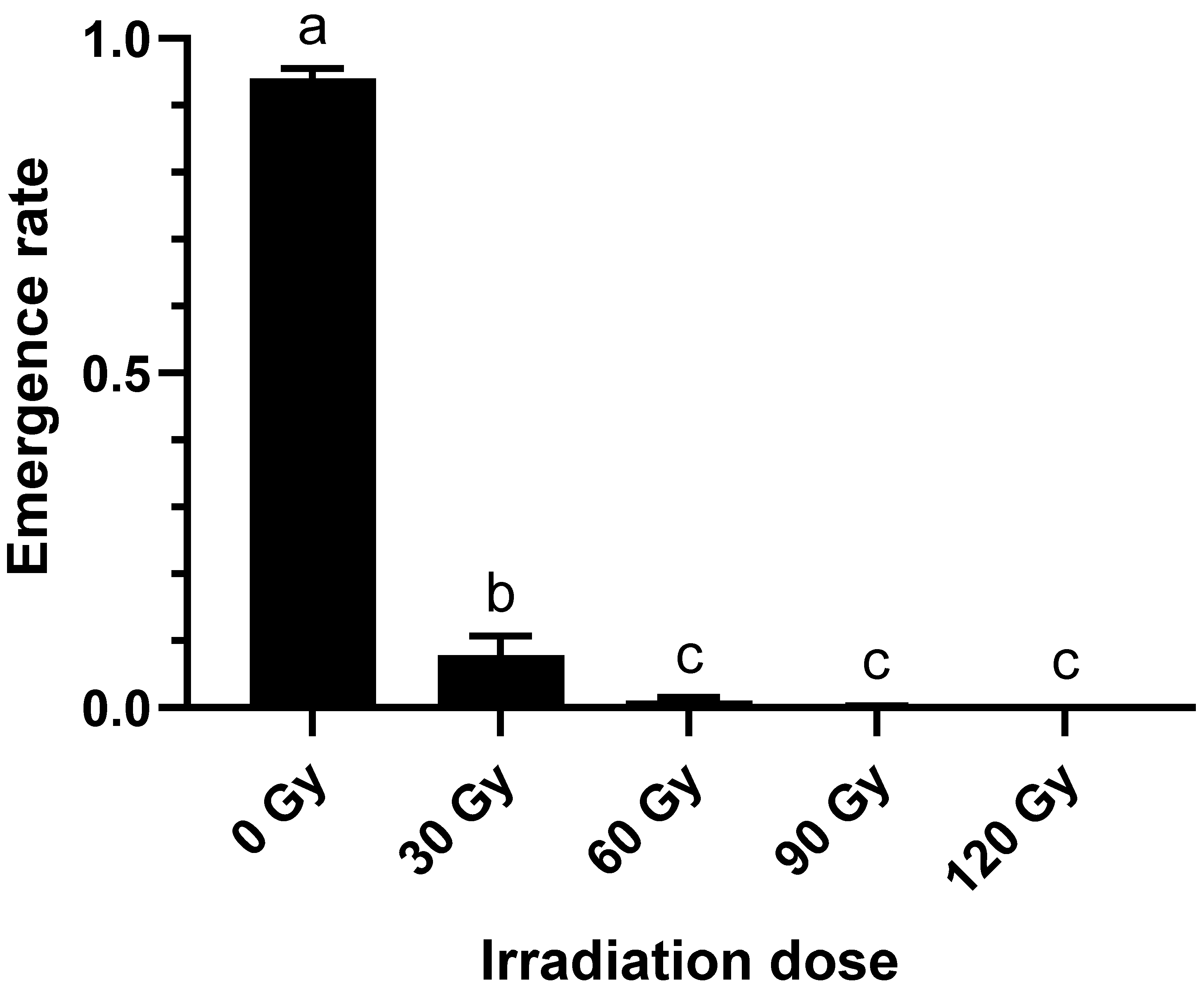
2.1. Effect of Irradiation Doses on Emergence Rate of B. dorsalis Larvae

As irradiation dose increases from 0 Gy to 60 Gy, the emergence rate of the 3rd instar of B. dorsalis larvae sharply declines (Figure 1), a function of disruption to metabolic pathways [30,31]. When the irradiation dose is 0 Gy, the emergence rate of the 3rd instar of B. dorsalis larvae is 94.01 ± 1.23%; when the irradiation dose is 30 Gy, the emergence rate of the 3rd instar of B. dorsalis larvae is only 7.84 ± 2.33%; when the irradiation dose reaches or exceeds 60 Gy, the 3rd instar of B. dorsalis larvae can hardly emerge normally.

2.2. Metabolite Expression in Response to Irradiation Dose

In all samples there were 67 peaks, and 23 metabolites were identified from these by GC-MS raw data (Table 1). For the B. dorsalis larvae treated with four levels of irradiation compared with untreated controls, the range of different metabolites produced decreased with the increase in irradiation dose. At the same time, the content of specific metabolites varied with the irradiation dose. Compared with the control group, when the irradiation dose reached or exceeded 30 Gy, the content of benzaldehyde, 3-ethyl-, hexanedioic acid, dioctyl ester, sulfurous acid, 2-ethylhexyl hexyl ester, 1,2,4-Benzenetricarboxylic acid and 1,2-dimethyl ester were significantly down-regulated, while the content of tetradecanoic acid increased. When the irradiation dose reached or exceeded 60 Gy, the content of octadecanoic acid suddenly increased, while the content of 2-Hydroxy-gamma-butyrolactone, Dodecanoic acid, Diethyl Phthalate, n-Hexadecanoic acid and Di-n-decylsulfone rapidly decreased. When the irradiation dose reached or exceeded 90 Gy, the amount of n-Decanoic acid, Phthalic acid and cyclobutyl isobutyl ester increased. When the irradiation dose reached 120 Gy, 2-Hexen-1-ol and (E)- increased. Pentadecanoic acid, Butyric acid, 2,2-dimethyl- and vinyl ester were detected only in unirradiated larvae, so this is a marker indicating no treatment. Other compounds could stably exist at the irradiation dose of 0 Gy to 120 Gy, and the content of 6-methylmethyl and Oleic acid slightly decreased with the increase in irradiation dose.

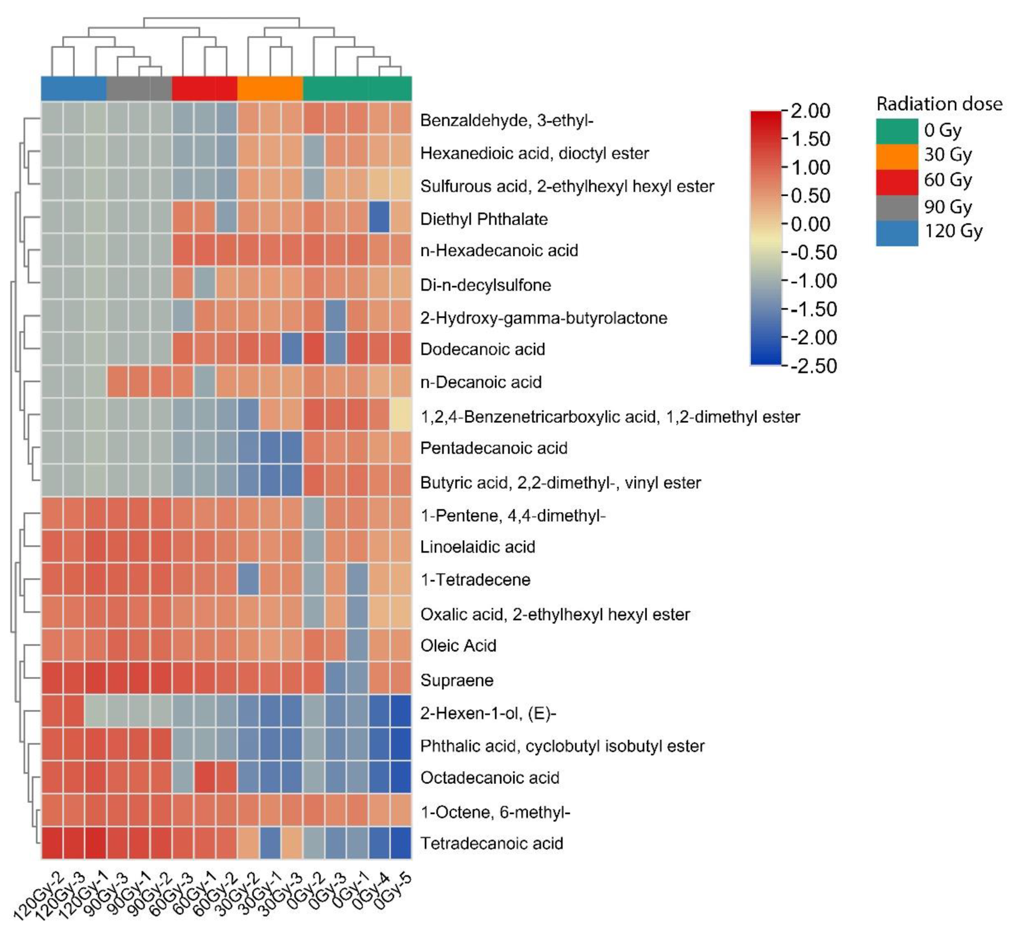
2.3. Hierarchical Cluster Analysis

The data set was scaled using the heatmap software package in the R software (Version: 4.1.3), and the samples and metabolites were analyzed using two-way cluster analysis [32]. Figure 2 is a hierarchical cluster diagram of relative quantification of metabolites of B. dorsalis larvae. The heatmap is divided into five areas: green, yellow, red, grey and blue, indicating that the content of metabolites greatly varies, and the difference between them is obvious. At the top of the graph, the samples of metabolites of B. dorsalis larvae treated with different irradiation doses are clustered. The clustering results clearly show two main irradiation dose clusters (cluster 1): 0 Gy and 30 Gy (cluster 1), and 60 Gy, 90 Gy and 120 Gy (cluster 2). For cluster 1, when the irradiation doses were 0 Gy and 30 Gy, the content of Benzaldehyde, 3-ethyl-, Hexanedioic acid, dioctyl ester, Sulfurous acid, 2-ethylhexyl hexyl ester, Diethyl Phthalate, n-Hexadecanoic acid, Di-n-decylsulfone, 2-Hydroxy-gamma-butyrolactone, Dodecanoic acid, n-Decanoic acid, 1,2,4-Benzenetricarboxylic acid, 1,2-dimethyl ester, Pentadecanoic acid, Butyric acid, 2,2-dimethyl- and vinyl ester metabolites produced significantly increased. For cluster 2, the content of metabolites included 1-Pentene, 4,4-dimethyl-, Linoelaidic acid, 1-Tetradecene, Oxalic acid, 2-ethylhexyl hexyl ester, Oleic acid, Supraene, 2-Hexen-1-ol, (E)-, Phthalic acid, cyclobutyl isobutyl ester, Octadecanoic acid, 1-Octene, 6-methyl- and Tetradecanoic acid, and these metabolites were significantly upregulated when the irradiation doses were 60 Gy, 90 Gy and 120 Gy. These results are consistent with the results of the GC-MS analysis in Table 1. The above analyses show that different irradiation doses received by the larvae can be inferred by the presence of different metabolites.

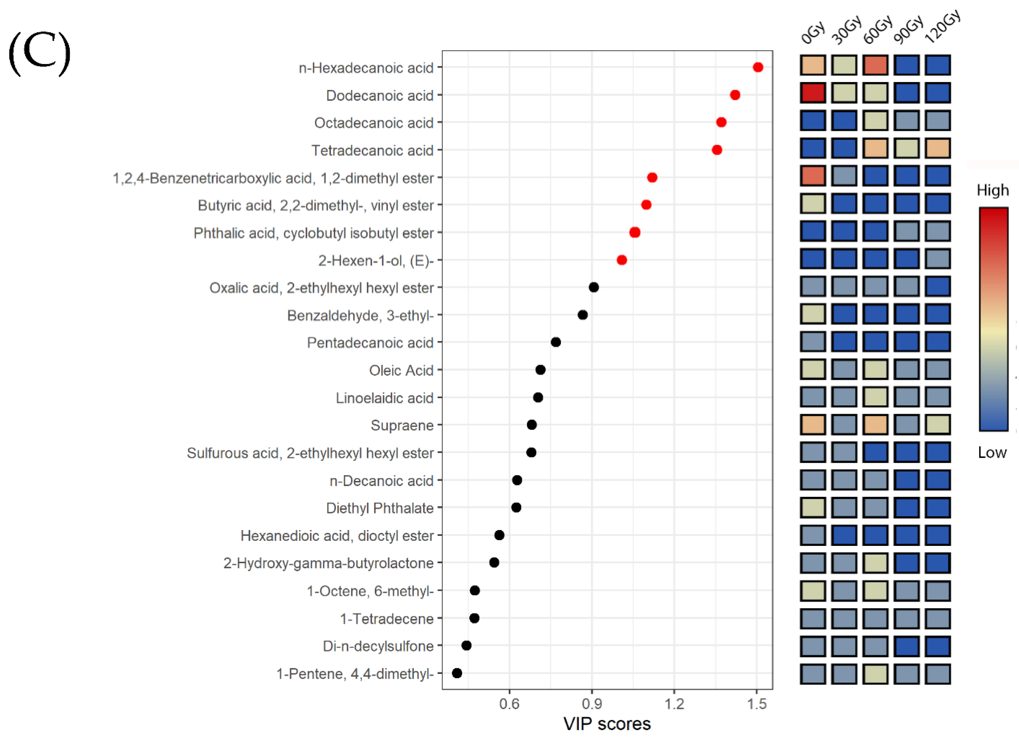
2.4. Multivariate Analysis of Metabolites in B. dorsalis Larvae Exposed to Different Irradiation Doses

Multivariate statistical analysis can simplify and reduce the dimensionality of high-dimensional and complex data while retaining a large amount of original information [33]. An unsupervised clustering method, PCA, is implemented on the screened GC-MS data. Six principal components are obtained with a cumulative contribution rate of 92.29%, indicating that the fitting degree of the PCA model is high, and the results of multidimensional statistical analysis are reliable. Therefore, the PCA model can be used to analyze the overall differences between treatment groups and the differences between samples within groups. The PCA scores chart (Figure 3A) showed that there were significant differences between larvae treated with different irradiation doses (30 Gy, 60 Gy, 90 Gy and 120 Gy) and the control group (0 Gy). There was significant separation between the treatment group and the control group on the first principal component (Component 1), which could explain 49.36% of the total variance. However, the separation effect between the treatment groups with irradiation dose of 30 Gy and 60 Gy was not significant.
Partial least-squares discriminant analysis (PLS-DA) is a multidimensional statistical analysis method for supervised pattern recognition [34,35]. Compared with PCA, PLS-DA not only reduces the dimension, but also combines the regression model to make a discriminant analysis of the regression results with a certain discriminant threshold, which is helpful to identify differences in compounds between groups. To maximize separation between groups, PLS-DA was carried out on the basis of the above GC-MS data, which better illustrated the differences in metabolites produced by the larvae upon exposure to different irradiation doses. Figure 3B shows the classification pattern using the partial least square model, and the variance explains 60.63% and 13.51% of Component 1 and Component 2, respectively. Using the different compounds produced by the larvae after different irradiation doses, all larvae can be effectively isolated. Larvae treated with 60 Gy, 90 Gy and 120 Gy gathered on the left side of the score map, while those treated with 0 Gy and 30 Gy gathered on the right side of the score map. Thus, the metabolite analysis using PCA and PLS-DA completely correspond to that using hierarchical cluster analysis (HCA).
A variable importance projection (VIP) score was constructed, in which VIP > 1 represents important distinguishing compounds to further identify the key compounds that are differentially present in larva treated with different irradiation doses [36,37]. The partial least square VIP scores of 23 metabolites are shown in Figure 3C. Table 2 shows irradiation doses and eight metabolites with significant changes determined using partial least square VIP score and t-test p value, which are n-Hexadecanoic acid, Dodecanoic acid, Octadecanoic acid, Tetradecanoic acid, 1,2,4-Benzenetricarboxylic acid, 1,2-dimethyl ester, Butyric acid, 2,2-dimethyl-, vinyl ester, Phthalic acid, cyclobutyl isobutyl ester, 2-Hexen-1-ol and (E)-. These compounds can be used as biomarkers to identify the irradiation dose of B. dorsalis larvae during quarantine treatment. When the irradiation dose reached 30 Gy and 120 Gy, the contents of Tetradecanoic acid, Octadecanoic acid, Phthalic acid, cyclobutyl isobutyl ester, 2-Hexen-1-ol, (E)- significantly increased, but the changes in Phthalic acid, cyclobutyl isobutyl ester, 2-Hexen-1-ol and (E)- contributed relatively little to the discrimination in irradiation dose. When irradiation dose exceeded 0 Gy, 30 Gy and 60 Gy, the contents of Butyric acid, 2,2-dimethyl-, vinyl ester, 1,2,4-Benzenetricarboxylic acid, 1,2-dimethyl ester, Dodecanoic acid and n-Hexadecanoic acid decreased, enabling the prediction of irradiation dose. These compounds significantly changed with the change in irradiation dose, indicating that irradiation treatment disturbed the normal metabolism of B. dorsalis larvae. Therefore, we can use the information contained in these differential metabolites to provide a reference for the detection of irradiation dose.

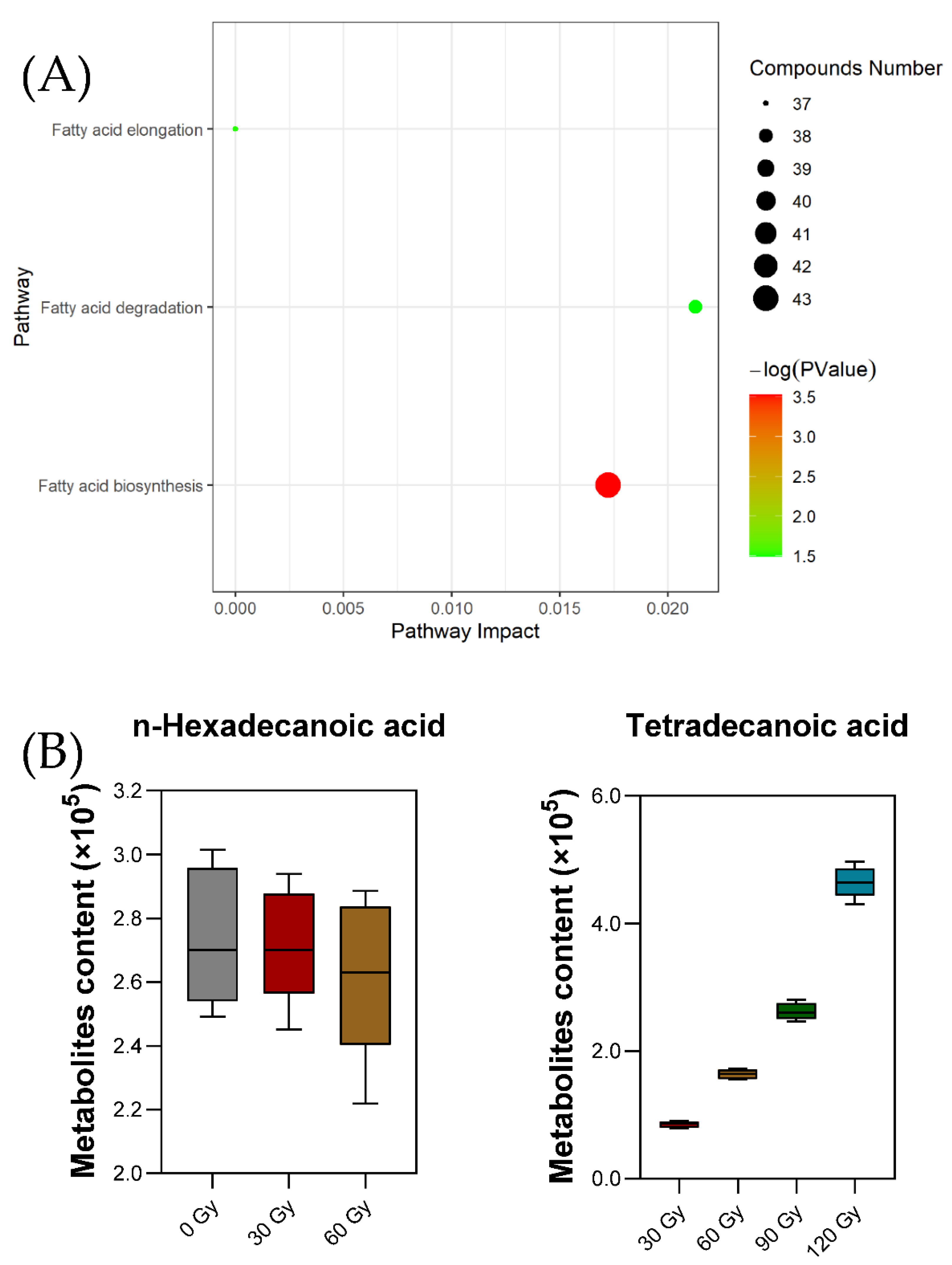
2.5. Preliminary Pathway Analysis of Differential Metabolites

The metabolic pathways in insects is regulated by a variety of compounds and reactions. Seven metabolic pathways were examined using the pathway database at the Kyoto Encyclopedia of Genes and Genomes (KEGG) [38,39]. The correlation between differential metabolites and three key metabolic pathways of fatty acid biosynthesis, fatty acid elongation and fatty acid degradation were screened, as shown in Table 3. The metabolic pathways of differential metabolites after treatment with different irradiation doses were analyzed using enrichment analysis and KEGG metabolic pathway retrieval. In Figure 4A, the number of bubbles represents the number of metabolic pathways, the color of the bubbles represents the degree of enrichment and the size of the bubbles represents the total number of metabolites contained in the metabolic pathway.
Metabolic pathway analysis revealed that irradiation affected fatty acid metabolism, indicating that there was a high correlation between the difference in fatty acid content and irradiation dose, but the mechanism for this was unclear [40]. According to previous research, fatty acids are basic substances present during insect embryonic development, metamorphosis and other life activities involved in growth, development and reproduction. [41,42]. Development of B. dorsalis larvae can be indirectly regulated by irradiation dose through the change in fatty acid content in the body, which is consistent with the results shown in Figure 1.
Two differential metabolites found in the target metabolic pathway were n-Hexadecanoic acid and Tetradecanoic acid (Figure 4B). The metabolites n-Hexadecanoic acid and Tetradecanoic acid are involved in the metabolism of fatty acid biosynthesis; n-Hexadecanoic acid is a common key metabolite in the two metabolic pathways of fatty acid elongation and fatty acid degradation. With the increase in irradiation dose, the content of n-Hexadecanoic acid released decreased, while the content of Tetradecanoic acid showed the opposite trend. This may be due to n-Hexadecanoic acid being partly converted to Tetradecanoic acid and ethyl easter at a high dose of irradiation treatment. Considering the pathways and mechanisms of fatty acid synthesis in insects, this result may be caused by the enhancement of the activity of key enzymes such as Acetyl CoA carboxylase or fatty acid synthase, leading to a certain degree of lipid accumulation [43,44,45,46]. Moreover, n-Hexadecanoic acid and Tetradecanoic acid were found to be responsible for the insect growth inhibitory and insecticide activity [47,48,49,50]. Therefore, irradiation treatment can lead to fatty profile changes and was found to be lethal to treated insects, but the mechanism was more complicated than we expected.

3. Materials and Methods

3.1. Insect Culture

Insects used in this study were collected from a mango orchard in Guangxi Zhuang Autonomous Region, China. They were reared in a laboratory at the Chinese Academy of Inspection and Quarantine. Late 3rd instar B. dorsalis larvae that emerged from mango fruit were transferred to moist sterile sand for pupariation. The pupae were placed in rearing cages (40 × 40 × 50 cm). Adults were fed with orange slices and a solid mixture of sucrose and hydrolyzed yeast (3:1) [51]. Eggs were collected from two weeks after adults had emerged from the puparium and mated. The adult females laid eggs through the sides of the cage cloths and the eggs fall into a distilled water (26 ± 1 °C) collector. Between 20–30 mL of egg suspension containing more than 7000 eggs were produced in 8 to 12 h. The larvae were reared on the artificial diet described by Vargas et al. (1984) [52]. At all stages, the larvae were reared at 25 ± 2 °C and relative humidity 70 ± 5% with a photoperiod of 12:12 (D: L) h.

3.2. Irradiation Treatment

All irradiation treatments were performed in an RS-2000 ProX irradiator (Rad SourceTechnologies, Inc., Coral Springs, FL, USA). The operation parameters were 220 KV and 17.6 mA [53]. A plastic box containing larvae was placed in the irradiation room with given irradiation doses of 0 Gy, 30 Gy, 60 Gy, 90 Gy and 120 Gy. The dose rate was 5.0 Gy/min for irradiation exposure time of 6 min, 12 min, 18 min and 24 min. Three or five plastic boxes (as replicates) were placed in the irradiation chamber and were irradiated at the same time. After irradiation, larvae were placed in a constant temperature incubator for 1 h before metabolites were extracted using SPME.

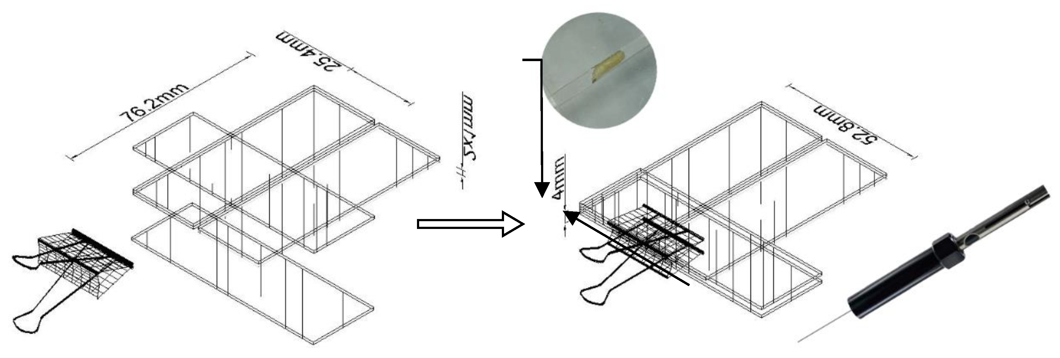
3.3. Solid Phase Microextraction (SPME) Procedure and Sampling Setup

The cuticular compounds profile of the late 3rd instar of B. dorsalis larvae were obtained by gently rubbing the body surface with a 50/30 um DVB/CAR/PDMS fiber solid phase microextraction sampler. For sampling the surface compounds, a new tool was designed which allows fixing larvae and adjusting the fiber position (Figure 5). Six slide glasses were placed as shown in Figure 5 using a hot-melt adhesive to hold one side of the slide glasses and then adjusting the slide glass on the other side to form an angle between these two slides [54]. With this structure, the larvae were fixed onto this tool and the SPME syringe was pushed into the slit. The fiber was slowly moved toward the larvae until the absorbent portion of the fibers was fully attached to the entire body segments of the larvae. Then, the slide was moved back and forth so that the body surface of the larvae constantly rubbed against the absorbent portion of fiber for 5 min.

3.4. Gas Chromatography-Mass Spectrometry (GC-MS) Conditions

Agilent 8890 gas chromatograph (GC) was used with an HP-5MS capillary column (30 m × 0.25 mm, 0.25 μm; Agilent J&W Scientific) and a 5977B mass selective detector (MSD). The carrier gas used was 99.999% purified helium with a constant flow rate of 1 mL/min. The GC conditions were: injection temperature of 270 °C and column temperature program of 60 °C for 5 min, which was then increased to 180 °C at the rate of 5 °C/min, and was finally increased to 280 °C at the rate of 10 °C/min, and maintained for 5 min. The MS parameters were: the transmission line temperature of the ion source was 280 °C and the quadrupole temperature was 150 °C. The information was collected using the full scanning mode of the mass spectrometry, the mass scanning range of the mass spectrometry was 30,500 atomic mass units (Amu) and the solvent delay time was 4.5 min. The total running time was 49 min.

3.5. Statistical Analysis

The GC-MS data were preliminarily identified using Aglient MasterHunter Qualitative Analysis 10.0 and recorded and sorted in Microsoft Excel. Most metabolites were further identified using the National Institute of Standards and Technology (NIST) and Wiley Registry of mass spectral data, as well as the retention index provided by the compound database of NIST Chemistry Web Book [55].
The XCMS package in RStudio (Version: 2021.9.0) was used to extract and analyze the feature data of GC-MS data [39]. The edited data matrix was imported into RStudio, and the unsupervised PCA and supervised PLS-DA were analyzed in R (Version: 4.1.3) prcomp package and mixOmics package. The differential metabolites were screened according to VIP-value and Student t-test, and the significant differences among the experimental groups were analyzed [35,56]. The selected differential metabolites were annotated in KEGG, and all the pathways of differential metabolites mapping were retrieved [57]. Then, the pathways of differential metabolites were further screened using enrichment analysis in order to find the key pathways with the highest correlation with differential metabolites.

4. Conclusions

In this experiment, a simple tool was used to fix B. dorsalis larvae, and SPME was used to extract metabolites from larvae exposed to different doses of irradiation treatment. Metabolites of different treatment groups were identified using GC-MS. By comparing the metabolites produced after five different irradiation doses, it was found that there were eight differential metabolites produced, of which four were fatty acids. The differential presence of metabolites occurred with different irradiation doses, and it was feasible to infer the irradiation dose of larvae using the difference in metabolites. This method is feasible to separate and identify the metabolites from B. dorsalis larvae in the process of quarantine treatment.
In summary, irradiation dose influences the spectrum of metabolites present in B. dorsalis larvae. Metabolic pathway analysis showed that different irradiation doses significantly changed fatty acid-related metabolic pathways in the species tested. Specifically, it was found for the first time that opposite trends occurred to n-Hexadecanoic acid and Tetradecanoic acid; that is, when n-Hexadecanoic acid decreased, Tetradecanoic acid increased. These findings provide a basis for understanding the molecular mechanism of irradiation damage in insects, and a basis for developing biomarkers corresponding to different irradiation doses.

Author Contributions

Conceptualization, C.S., Y.R. and T.L.; methodology, C.S., Y.R. and T.L.; software, C.S.; validation, C.S. and B.L. (Baishu Li); formal analysis, C.S. and B.L. (Beibei Li); investigation, C.S.; resources, C.S., B.L. (Baishu Li) and L.L.; data curation, C.S. and T.L.; writing—original draft preparation, C.S.; writing—review and editing, C.S., Y.R. and T.L.; visualization, C.S.; supervision, Y.R. and T.L.; project administration, C.S., Y.R. and T.L.; funding acquisition, T.L. All authors have read and agreed to the published version of the manuscript.

Funding

This research was funded by the Scientific Research Fund of the Chinese Academy of Inspection and Quarantine (2020JK045), and the technical support fund on postharvest control of biological contaminants of State Administration for Market Regulation (No. 2022).

Institutional Review Board Statement

Not applicable.

Informed Consent Statement

Not applicable.

Data Availability Statement

All data are contained within the article.

Conflicts of Interest

The authors declare no conflict of interest.

References

  1. Wan, X.; Liu, Y.; Zhang, B. Invasion history of the oriental fruit fly, Bactrocera dorsalis, in the Pacific-Asia region: Two main invasion routes. PLoS ONE 2012, 7, e36176. [Google Scholar] [CrossRef] [PubMed] [Green Version]
  2. Heve, W.K.; Adjadeh, T.A.; Billah, M.K. Overview and future research needs for development of effective biocontrol strategies for management of Bactrocera dorsalis Hendel (Diptera: Tephritidae) in sub-Saharan Africa. Pest Manag. Sci. 2021, 77, 4224–4237. [Google Scholar] [CrossRef] [PubMed]
  3. Dohino, T.; Hallman, G.J.; Grout, T.G.; Clarke, A.R.; Follett, P.A.; Cugala, D.R.; Minh Tu, D.; Murdita, W.; Hernandez, E.; Pereira, R.; et al. Phytosanitary Treatments Against Bactrocera dorsalis (Diptera: Tephritidae): Current Situation and Future Prospects. J. Econ. Entomol. 2017, 110, 67–79. [Google Scholar] [PubMed] [Green Version]
  4. Hallman, G.J. Phytosanitary Applications of Irradiation. Compr. Rev. Food Sci. Food Saf. 2011, 10, 143–151. [Google Scholar] [CrossRef]
  5. McDonald, H.; Arpaia, M.L.; Caporaso, F.; Obenland, D.; Were, L.; Rakovski, C.; Prakash, A. Effect of gamma irradiation treatment at phytosanitary dose levels on the quality of ‘Lane Late’ navel oranges. Postharvest Biol. Technol. 2013, 86, 91–99. [Google Scholar] [CrossRef]
  6. Drake, S.R.; Neven, L.G. Irradiation as an Alternative to Methyl Bromide for Quarantine Treatment of Stone Fruits. J. Food Qual. 1998, 21, 529–538. [Google Scholar] [CrossRef]
  7. Zahran, N.F.; Hamza, A.F.; Ramadan, M.H. Gamma Irradiation as an Alternative Treatment for Controlling of Lyctus africanus Lesne (Coleoptera: Bostrichidae) in Dry Wood. Egypt. J. Biol. Pest Control 2016, 26, 97–101. [Google Scholar]
  8. ISPM 28; International Plant Protection Convention (IPPC). Irradiation treatment for fruit flies of the family Tephritidae (generic). Food and Agricultural Organization: Rome, Italy, 2009. [Google Scholar]
  9. Hallman, G.J. Expanding radiation quarantine treatments beyond fruit flies. Agric. For. Entomol. 2000, 2, 85–95. [Google Scholar] [CrossRef]
  10. Hallman, G.J.; Levang-Brilz, N.M.; Zettler, J.L.; Winborne, I.C. Factors affecting ionizing radiation phytosanitary treatments, and implications for research and generic treatments. J. Econ. Entomol. 2010, 103, 1950–1963. [Google Scholar] [CrossRef]
  11. von Sonntag, C. The Chemical Basis of Radiation Biology; Taylor & Francis: London, UK, 1987. [Google Scholar]
  12. Hallman, G.J.; Loaharanu, P. Phytosanitary irradiation—Development and application. Radiat. Phys. Chem. 2016, 129, 39–45. [Google Scholar] [CrossRef]
  13. Woruba, D.N.; Morrow, J.L.; Reynolds, O.L.; Chapman, T.A.; Collins, D.P.; Riegler, M. Diet and irradiation effects on the bacterial community composition and structure in the gut of domesticated teneral and mature Queensland fruit fly, Bactrocera tryoni (Diptera: Tephritidae). BMC Microbiol. 2019, 19, 281. [Google Scholar] [CrossRef] [PubMed] [Green Version]
  14. Tanaka, Y.; Furuta, M. Biological effects of low-dose γ-ray irradiation on chromosomes and DNA of Drosophila melanogaster. J. Radiat. Res. 2021, 62, 1–11. [Google Scholar] [CrossRef] [PubMed]
  15. Oinata, K.; Chambers, D.L.; Fujimoto, M.; Kashiwai, S.; Miyabara, R. Sterilization of the Mediterranean fruit fly by irradiation: Comparative mating effectiveness of treated pupae and adults. J. Econ. Entomol. 1971, 64, 781–787. [Google Scholar] [CrossRef]
  16. Baye, Z.K. Determine the Gamma Radiation Technique to Sterilization of Insects from Risk Factor. World J. Appl. Phys. 2021, 6, 35–40. [Google Scholar]
  17. Siddiqui, M.S.; Filomeni, E.; François, M.; Collins, S.R.; Cooper, T.; Glatz, R.V.; Taylor, P.W.; Fenech, M.; Leifert, W.R. Exposure of insect cells to ionising radiation in vivo induces persistent phosphorylation of a H2AX homologue (H2AvB). Mutagenesis 2013, 28, 531–541. [Google Scholar] [CrossRef] [Green Version]
  18. Arthur, C.L.; Pawliszyn, J.B. Solid phase microextraction with thermal desorption using fused silica optical fibers. Anal. Chem. 1990, 62, 2145–2148. [Google Scholar] [CrossRef]
  19. Kataoka, H.; Heather, L.L.; Pawliszyn, J. Applications of solid-phase microextraction in food analysis. J. Chromatogr. A 2020, 880, 35–62. [Google Scholar] [CrossRef]
  20. Laopongsit, W.; Srzednicki, G.; Craske, J.D. Preliminary study of solid phase micro-extraction (SPME) as a method for detecting insect infestation in wheat grain. J. Stored Prod. Res. 2014, 59, 88–95. [Google Scholar] [CrossRef]
  21. Cai, L.; Macfadyen, S.; Hua, B.; Zhang, H.; Xu, W.; Ren, Y. Identification of Biomarker Volatile Organic Compounds Released by Three Stored-Grain Insect Pests in Wheat. Molecules 2022, 27, 1963. [Google Scholar] [CrossRef]
  22. Alnajim, I.; Du, X.; Lee, B.; Agarwal, M.; Liu, T.; Ren, Y. New Method of Analysis of Lipids in Tribolium castaneum (Herbst) and Rhyzopertha dominica (Fabricius) Insects by Direct Immersion Solid-Phase Microextraction (DI-SPME) Coupled with GC–MS. Insects 2019, 10, 363. [Google Scholar] [CrossRef] [Green Version]
  23. Al-Khshemawee, H.; Du, X.; Agarwal, M.; Yang, J.; Ren, Y. Application of Direct Immersion Solid-Phase Microextraction (DI-SPME) for Understanding Biological Changes of Mediterranean Fruit Fly (Ceratitis capitata) During Mating Procedures. Molecules 2018, 23, 2951. [Google Scholar] [CrossRef] [PubMed] [Green Version]
  24. Tanaka, F.; Magariyama, Y.; Miyanoshita, A. Volatile biomarkers for early-stage detection of insect-infested brown rice: Isopentenols and polysulfides. Food Chem. 2019, 303, 125381. [Google Scholar] [CrossRef] [PubMed]
  25. Fulano, A.M.; Lengai, G.M.; Muthomi, J.W. Phytosanitary and Technical Quality Challenges in Export Fresh Vegetables and Strategies to Compliance with Market Requirements: Case of Smallholder Snap Beans in Kenya. Sustainability 2021, 13, 1546. [Google Scholar] [CrossRef]
  26. Nawaz, A.; Sufyan, M.; Gogi, M.D.; Javed, M.W. Sustainable Management of Insect-Pests. Innov. Sustain. Agric. 2019, 10, 287–335. [Google Scholar]
  27. Heather, N.W.; Hallman, G.J. Pest Management and Phytosanitary Trade Barriers; CABI: Wallingford, UK, 2008. [Google Scholar]
  28. Hallman, G.J. Process control in phytosanitary irradiation of fresh fruits and vegetables as a model for other phytosanitary treatment processes. Food Control 2017, 72, 372–377. [Google Scholar] [CrossRef]
  29. Hallman, G.J.; Blackburn, C.M. Phytosanitary Irradiation. Foods 2016, 5, 8. [Google Scholar] [CrossRef] [Green Version]
  30. Barry, J.D.; McInnis, D.O.; Gates, D.; Morse, J.G. Effects of irradiation on Mediterranean fruit flies (Diptera: Tephritidae): Emergence, survivorship, lure attraction, and mating competition. J. Econ. Entomol. 2003, 96, 615–622. [Google Scholar] [CrossRef]
  31. Benelli, M.; Ponton, F.; Lallu, U.; Mitchell, K.A.; Taylor, P.W. Cool storage of Queensland fruit fly pupae for improved management of mass production schedules. Pest Manag. Sci. 2019, 75, 3184–3192. [Google Scholar] [CrossRef]
  32. Srivastava, S.; Mishra, G.; Mishra, H.N. Identification and differentiation of insect infested rice grains varieties with FTNIR spectroscopy and hierarchical cluster analysis. Food Chem. 2018, 268, 402–410. [Google Scholar] [CrossRef]
  33. Eriksson, L.; Johansson, E.; Antti, H.; Holmes, E. Multi- and Megavariate Data Analysis: Finding and Using Regularities in Metabonomics Data; CRC Press: Boca Raton, FL, USA, 2005. [Google Scholar]
  34. Sharin, S.N.; Sani, M.S.A.; Jaafar, M.A.; Yuswan, M.H.; Kassim, N.K.; Manaf, Y.N. Discrimination of Malaysian stingless bee honey from different entomological origins based on physicochemical properties and volatile compound profiles using chemometrics and machine learning. Food Chem. 2020, 346, 128654. [Google Scholar] [CrossRef]
  35. Feng, Y.; Fu, T.; Zhang, L.; Wang, C.; Zhang, D. Research on Differential Metabolites in Distinction of Rice (Oryza sativa L.) Origin Based on GC-MS. J. Chem. 2019, 2019, 1614504. [Google Scholar] [CrossRef] [Green Version]
  36. Sun, J.; Feng, X.; Lyu, C.; Zhou, S.; Liu, Z. Effects of different processing methods on the lipid composition of hazelnut oil: A lipidomics analysis. Food Sci. Hum. Wellness 2022, 11, 427–435. [Google Scholar] [CrossRef]
  37. Hu, Y.; Li, Y.; Li, X.; Zhang, H.; Chen, Q.; Kong, B. Application of lactic acid bacteria for improving the quality of reduced-salt dry fermented sausage: Texture, color, and flavor profiles. LWT 2021, 154, 112723. [Google Scholar] [CrossRef]
  38. Kanehisa, M.; Goto, S. KEGG: Kyoto Encyclopedia of Genes and Genomes. Nucleic Acids Res. 2000, 28, 27–30. [Google Scholar] [CrossRef]
  39. Sun, L.; Fan, K.; Wang, L.; Ma, D.; Wang, Y.; Kong, X.; Li, H.; Ren, Y.; Ding, Z. Correlation among Metabolic Changes in Tea Plant Camellia sinensis (L.) Shoots, Green Tea Quality and the Application of Cow Manure to Tea Plantation Soils. Molecules 2021, 26, 6180. [Google Scholar] [CrossRef]
  40. Zhang, L.; Zhang, X.; Ji, H.; Wang, W.; Liu, J.; Wang, F.; Xie, F.; Yu, Y.; Qin, Y.; Wang, X. Metabolic profiling of tobacco leaves at different growth stages or different stalk positions by gas chromatography–mass spectrometry. Ind. Crops Prod. 2018, 116, 46–55. [Google Scholar] [CrossRef]
  41. Stanley-Samuelson, D.W.; Jurenka, R.; Cripps, C.; Blomquist, G.J.; Renobales, M.D. Fatty acids in insects: Composition, metabolism, and biological significance. Arch. Insect Biochem. Physiol. 1988, 9, 1–33. [Google Scholar] [CrossRef]
  42. Beenakkers, A.M.; Van der Horst, D.J.; Van Marrewijk, W. Insect lipids and lipoproteins, and their role in physiological processes. Prog. Lipid Res. 1985, 24, 19–67. [Google Scholar] [CrossRef]
  43. Parvy, J.P.; Napal, L.; Rubin, T.; Poidevin, M.; Perrin, L.; Wicker, T.C.; Montagne, J. Drosophila melanogaster Acetyl-CoA-Carboxylase Sustains a Fatty Acid–Dependent Remote Signal to Waterproof the Respiratory System. PLoS Genet. 2012, 8, e1002925. [Google Scholar] [CrossRef] [Green Version]
  44. Urbanski, J.; Benoit, J.; Michaud, M.; Denlinger, D.; Armbruster, P. The molecular physiology of increased egg desiccation resistance during diapause in the invasive mosquito, Aedes albopictus. Proc. Biol. Sci. 2010, 277, 2683–2692. [Google Scholar] [CrossRef] [Green Version]
  45. Li, L.; Jiang, Y.; Liu, Z.; You, L.; Wu, Y.; Xu, B.; Ge, L.; Stanley, D.; Song, Q.; Wu, J. Jinggangmycin increases fecundity of the brown planthopper, Nilaparvata lugens (Stål) via fatty acid synthase gene expression. J. Proteom. 2016, 130, 140–149. [Google Scholar] [CrossRef] [PubMed]
  46. Sim, C.; Denlinger, D.L. Transcription profiling and regulation of fat metabolism genes in diapausing adults of the mosquito Culex pipiens. Physiol. Genom. 2009, 39, 202–209. [Google Scholar] [CrossRef] [PubMed] [Green Version]
  47. Thompson, S.N. A review and comparative characterization of the fatty acid compositions of seven insect orders. Comp. Biochem. Physiol. Part B Comp. Biochem. 1973, 45, 467–482. [Google Scholar] [CrossRef]
  48. Abdullah, R. Insecticidal Activity of Secondary Metabolites of Locally Isolated Fungal Strains against some Cotton Insect Pests. J. Plant Prot. Pathol. 2019, 10, 647–653. [Google Scholar] [CrossRef] [Green Version]
  49. Alagarmalai, J.; Chinnamani, T.C. Chemical composition and growth inhibitory activities of Solonum pseudocapsicum against Spodoptera litura and Helicoverpa armigera (Lepidoptera: Noctuidae). Int. J. Entomol. Res. 2017, 2, 2455–4758. [Google Scholar]
  50. Sivakumar, R.; Jebanesan, A.; Govindarajan, M.; Rajasekar, P. Larvicidal and repellent activity of tetradecanoic acid against Aedes aegypti (Linn.) and Culex quinquefasciatus (Say.) (Diptera:Culicidae). Asian Pac. J. Trop. Med. 2011, 4, 706–710. [Google Scholar] [CrossRef] [Green Version]
  51. Zhao, J.; Ma, J.; Mutao, W.; Jiao, X.; Wang, Z.; Liang, F.; Zhan, G. Gamma radiation as a phytosanitary treatment against larvae and pupae of Bactrocera dorsalis (Diptera: Tephritidae) in guava fruits. Food Control 2017, 72, 360–366. [Google Scholar] [CrossRef]
  52. Vargas, R.I.; Miyashita, D.H.; Nishida, T. Life History and Demographic Parameters of Three Laboratory-reared Tephritids (Diptera: Tephritidae). Ann. Entomol. Soc. Am. 1984, 77, 651–656. [Google Scholar] [CrossRef]
  53. Zhan, G.; Zhao, J.; Ma, F.; Liu, B.; Zhong, Y.; Song, Z.; Zhao, Q.; Chen, N.; Ma, C. Radioprotective Effects on Late Third-Instar Bactrocera dorsalis (Diptera: Tephritidae) Larvae in Low-Oxygen Atmospheres. Insects 2020, 11, 526. [Google Scholar] [CrossRef]
  54. Geiselhardt, S.F.; Geiselhardt, S.; Peschke, K. Comparison of tarsal and cuticular chemistry in the leaf beetle Gastrophysa viridula (Coleoptera: Chrysomelidae) and an evaluation of solid-phase microextraction and solvent extraction techniques. Chemoecology 2009, 19, 185–193. [Google Scholar] [CrossRef]
  55. Ismarti, I.; Handoko, D.D.; Triyana, K.; Salleh, H.M.; Fadzillah, N.A.; Nordin, N.F. Study on Volatile Compounds of Gelatine and The Maillard Reaction Products from Different Species Using SPME-GCMS. Sci. Technol. Indones. 2022, 7, 132–139. [Google Scholar] [CrossRef]
  56. Du, W.; Liu, X.; Zhao, L.; Xu, Y.; Yin, Y.; Wu, J.; Ji, R.; Sun, Y.; Guo, H. Response of cucumber (Cucumis sativus) to perfluorooctanoic acid in photosynthesis and metabolomics. Sci. Total Environ. 2020, 724, 138257. [Google Scholar] [CrossRef] [PubMed]
  57. Zhang, J.D.; Wiemann, S. KEGG graph: A graph approach to KEGG PATHWAY in R and bioconductor. Bioinformatics 2009, 25, 1470–1471. [Google Scholar] [CrossRef] [PubMed]
Figure 1. Emergence rates of the 3rd instar of B. dorsalis larvae in response to irradiation dosage (Different lower case letters indicate that emergence rates has significant differences between irradiation dosages at p < 0.05).
Molecules 27 04641 g001
Figure 2. Clustering heatmap of metabolites in B. dorsalis larvae exposed to different doses of irradiation treatment.
Molecules 27 04641 g002
Figure 3. Principal component analysis (PCA) score plot (A), partial least squares-discriminant analysis (PLS-DA) loading plot (B), and variable importance projection (VIP) scores plot (C) of all metabolites in B. dorsalis larvae exposed to different doses of irradiation treatment at 0 Gy, 30 Gy, 60 Gy, 90 Gy and 120 Gy.
Molecules 27 04641 g003aMolecules 27 04641 g003b
Figure 4. Effects of hit metabolites on metabolic pathways in B. dorsalis larvae (A). Content changes in metabolites in key metabolic pathways (B).
Molecules 27 04641 g004
Figure 5. Experimental setup for SPME sampling of B. dorsalis larvae specimens. The larvae are fixed in a slit assembled by slide glasses and contacted with an SPME fiber.
Molecules 27 04641 g005
Table 1. Metabolites produced by B. dorsalis larvae treated with different irradiation doses.
MetabolitesRetention TimeRetention IndexGC Response (105) ± SDCAS Number
0 Gy30 Gy60 Gy90 Gy120 Gy
2-Hydroxy-gamma-butyrolactone8.404997.261.2 ± 0.061.12 ± 0.131.06 ± 0.14N.D.N.D.19444-84-9
1-Pentene, 4,4-dimethyl-9.8251037.861.41 ± 0.121.25 ± 0.151.14 ± 0.131.26 ± 0.161.01 ± 0.10762-62-9
Benzaldehyde, 3-ethyl-14.3051061.021.36 ± 0.170.91 ± 0.11N.D.N.D.N.D.34246-54-3
2-Hexen-1-ol, (E)-15.581102.61N.D.N.D.N.D.N.D.2.11 ± 0.09928-95-0
n-Decanoic acid20.6861274.261.14 ± 0.161.09 ± 0.130.89 ± 0.100.67 ± 0.02N.D.334-48-5
1-Octene, 6-methyl-21.2881295.341.55 ± 0.161.54 ± 0.121.29 ± 0.261.16 ± 0.121.05 ± 0.1413151-10-5
1-Tetradecene23.281369.701.7 ± 0.101.62 ± 0.011.27 ± 0.121.23 ± 0.081.39 ± 0.161120-36-1
Dodecanoic acid24.9661435.123.72 ± 0.253.31 ± 0.171.13 ± 0.13N.D.N.D.143-07-7
Diethyl Phthalate25.7721467.361.32 ± 0.121.06 ± 0.081.04 ± 0.06N.D.N.D.84-66-2
Tetradecanoic acid29.3651739.12N.D.0.85 ± 0.101.64 ± 0.102.62 ± 0.234.6 ± 0.43544-63-8
Pentadecanoic acid31.941817.301.45 ± 0.13N.D.N.D.N.D.N.D.1002-84-2
Phthalic acid, cyclobutyl isobutyl ester35.3061947.82N.D.N.D.N.D.1.66 ± 0.171.61 ± 0.221000314-91-1
n-Hexadecanoic acid35.232058.072.73 ± 0.192.72 ± 0.122.63 ± 0.28N.D.N.D.57-10-3
Linoelaidic acid35.9692135.051.54 ± 0.211.53 ± 0.191.14 ± 0.091.28 ± 0.111.19 ± 0.03506-21-8
Oleic acid38.6642181.341.68 ± 0.231.35 ± 0.181.09 ± 0.10.98 ± 0.090.89 ± 0.03112-80-1
Octadecanoic acid39.0142187.36N.D.N.D.1.08 ± 0.041.23 ± 0.011.56 ± 0.2157-11-4
Oxalic acid, 2-ethylhexyl hexyl ester40.5852221.021.22 ± 0.161.08 ± 0.100.85 ± 0.210.83 ± 0.070.91 ± 0.081000309-38-9
Hexanedioic acid, dioctyl ester41.7592350.941.27 ± 0.130.79 ± 0.11N.D.N.D.N.D.123-79-5
Di-n-decylsulfone42.0462439.441.57 ± 0.171.16 ± 0.140.96 ± 0.07N.D.N.D.111530-37-1
Sulfurous acid, 2-ethylhexyl hexyl ester43.3092561.141.32 ± 0.170.9 ± 0.04N.D.N.D.N.D.1000309-20-2
1,2,4-Benzenetricarboxylic acid, 1,2-dimethyl ester44.342678.853.26 ± 0.211.04 ± 0.11N.D.N.D.N.D.54699-35-3
Butyric acid, 2,2-dimethyl-, vinyl ester45.2192793.952.13 ± 0.14N.D.N.D.N.D.N.D.13170-00-8
Supraene45.5682867.192.69 ± 0.292.82 ± 0.012.04 ± 0.252.65 ± 0.262.39 ± 0.317683-64-9
SD: standard deviation. N.D.: metabolite not detected.
Table 2. Significantly changed metabolites in B. dorsalis larvae exposed to different doses of irradiation treatment.
MetabolitesVIP Scoresp ValueFDRClass
n-Hexadecanoic acid1.5050.0490.069Acid
Dodecanoic acid1.4210.0030.008
Octadecanoic acid1.3710.0010.006
Tetradecanoic acid1.3550.0050.012
1,2,4-Benzenetricarboxylic acid, 1,2-dimethyl ester1.1200.0030.008Ester
Butyric acid, 2,2-dimethyl-, vinyl ester1.0980.0030.008
Phthalic acid, cyclobutyl isobutyl ester1.0560.0000.000
2-Hexen-1-ol, (E)-1.0090.0170.029Alcohol
Table 3. The key metabolic pathways of differential metabolites in B. dorsalis larvae.
Pathway NameTotalExpectedHitsRaw pImpactHit Metabolites
Fatty acid biosynthesis430.2828920.0294540.01724C00249: n-Hexadecanoic acidC06424: Tetradecanoic acid
Fatty acid elongation370.2434210.220010C00249: n-Hexadecanoic acid
Fatty acid degradation380.2510.225320.02128C00249: n-Hexadecanoic acid
Publisher’s Note: MDPI stays neutral with regard to jurisdictional claims in published maps and institutional affiliations.

