
Synthesis, Molecular Docking Study, and Cytotoxic Activity against MCF Cells of New Thiazole–Thiophene Scaffolds

 ,
,  ,
,
Abstract
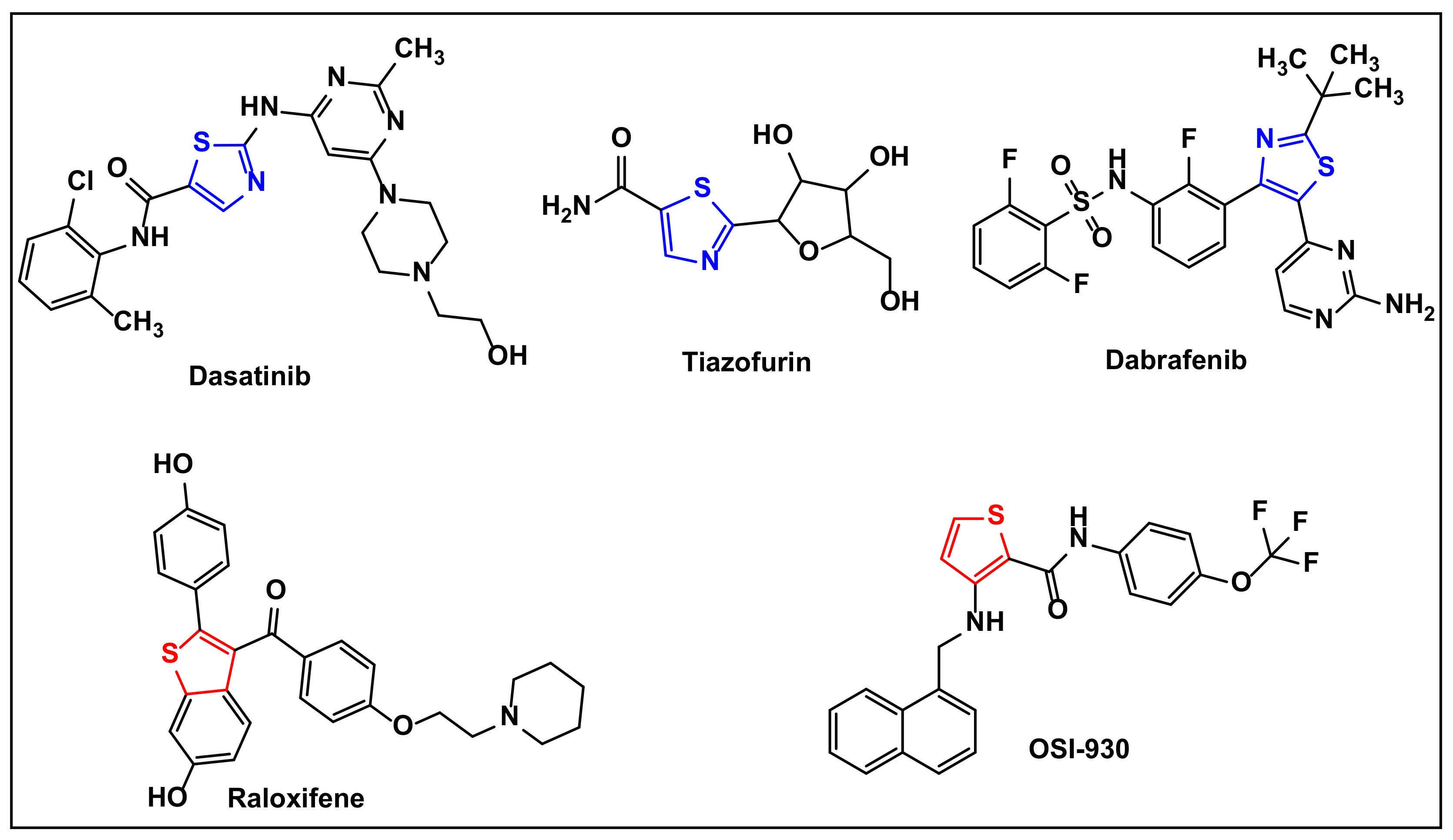
:1. Introduction
2. Results and Discussion
2.1. Chemistry
2.2. Cytotoxic Potential
- The 1,3-thiazole derivatives 4b and 13a (IC50 = 10.2 ± 0.7 and 11.5 ± 0.8 μM, respectively) have promising antitumor activity against the breast carcinoma cell line (MCF-7), and showed greater activities than the cisplatin reference drug (IC50 = 13.3 ± 0.61 μM);
- The 1,3-thiazole derivatives 11c and 11d have poor antitumor activity (IC50 > 38 μM), while the rest of the evaluated thiazoles have moderate activity (IC50 = 13.6–23.7 μM);
- For 1,3-thiazoles 4, 8, and 11: the introduction of an electron-donating group (eg. methyl group) into phenyl group at position 5 in the 1,3-thiazole ring enhances the antitumor activity, while the introduction of an electron-withdrawing group (chlorine) decreases the activity (4b > 4a > 4c > 4d; 8a > 8b; and 11b >11a > 11c >11d);
- For the substituent at position 5 of the 1,3-thiazoles: an acetyl group (Ac) gives higher activity than an ester group (CO2Et). 13a (IC50 = 11.5 ± 0.7 μM) > 13b (IC50 = 16.3 ± 1.4 μM).
2.3. Molecular Docking Studies
2.4. Toxicity Radar
2.5. SwissADME Studies
2.6. Pred-hERG
3. Experimental Section
3.1. Chemistry
3.1.1. Experimental Instrumentation
3.1.2. General Procedure for Synthesizing the Thiazole Derivatives 4a–d, 8a,b, 11a–d, and 13a,b
3.1.3. Alternate Synthesis of 4a and 8a
3.2. In Vitro Cytotoxic Activity
4. Conclusions
Supplementary Materials
Author Contributions
Funding
Institutional Review Board Statement
Informed Consent Statement
Data Availability Statement
Acknowledgments
Conflicts of Interest
References
- Balasubramanian, R.; Rolph, R.; Morgan, C.; Hamed, H. Genetics of breast cancer: Management strategies and risk-reducing surgery. Br. J. Hosp. Med. 2019, 80, 720–725. [Google Scholar] [CrossRef] [PubMed]
- Bregoli, L.; Movia, D.; Gavigan-Imedio, J.D.; Lysaght, J.; Reynolds, J.; Prina-Mello, A. Nanomedicine applied to translational oncology: A future perspective on cancer treatment. Nanomedicine 2016, 12, 81–103. [Google Scholar] [CrossRef] [PubMed]
- De Vasconcelos, A.; Campos, V.F.; Nedel, F.; Seixas, F.K.; Dellagostin, O.A.; Smith, K.R.; De Pereira, C.M.P.; Stefanello, F.M.; Collares, T.; Barschak, A.G. Cytotoxic and apoptotic effects of chalcone derivatives of 2-acetyl thiophene on human colon adenocarcinoma cells. Cell Biochem. Funct. 2013, 31, 289–297. [Google Scholar] [CrossRef] [PubMed]
- Schmitt, C.; Kail, D.; Mariano, M.; Empting, M.; Weber, N.; Paul, T.; Hartmann, R.W.; Engel, M. Design and Synthesis of a Library of Lead-Like 2,4-Bisheterocyclic Substituted Thiophenes as Selective Dyrk/Clk Inhibitors. PLoS ONE 2014, 9, e87851. [Google Scholar] [CrossRef]
- Al-Said, M.S.; Bashandy, M.S.; Al-Qasoumi, S.I.; Ghorab, M.M. Anti-breast cancer activity of some novel 1,2-dihydropyridine, thiophene and thiazole derivatives. Eur. J. Med. Chem. 2011, 46, 137–141. [Google Scholar] [CrossRef]
- Dallemagne, P.; Khanh, L.P.; Alsaidi, A.; Renault, O.; Varlet, I.; Collot, V.; Bureau, R.; Rault, S. Synthesis and biological evaluation of cyclopenta[c]thiophene related compounds as new antitumor agents. Bioorg. Med. Chem. 2002, 10, 2185–2191. [Google Scholar] [CrossRef]
- Venkataramireddy, V.; Shankaraiah, M.; Allaka, T.R.; Kalyani, C.; Narasu, M.L.; Varala, R.; Anireddy, A. Synthesis and anticancer Activity of novel 3-aryl thiophene-2-carbaldehydes and their aryl/heteroaryl chalcone derivatives. Rasayan J. Chem. 2016, 9, 31–39. [Google Scholar]
- Gulipalli, K.C.; Bodige, S.; Ravula, P.; Endoori, S.; Vanaja, G.R.; Babu, G.S.; Chandra, J.N.N.; Seelam, N. Design, synthesis, in silico and in vitro evaluation of thiophene derivatives: A potent tyrosine phosphatase 1B inhibitor and anticancer activity. Bioorg. Med. Chem. Lett. 2017, 27, 3558–3564. [Google Scholar] [CrossRef]
- AbdElhameid, M.K.; Labib, M.B.; Negmeldin, A.T.; Al-Shorbagy, M.; Mohammed, M.R. Design, synthesis, and screening of ortho-amino thiophene carboxamide derivatives on hepatocellular carcinomaas VEGFR-2Inhibitors. J. Enzym. Inhib. Med. Chem. 2018, 33, 1472–1493. [Google Scholar] [CrossRef]
- Sujatha, K.; Vedula, R.R. Novel one-pot expeditious synthesis of 2,4-disubstituted thiazoles through a three-component reaction under solvent free conditions. Synth. Commun. 2018, 48, 302–308. [Google Scholar] [CrossRef]
- Abu-Melha, S.; Edrees, M.M.; Salem, H.H.; Kheder, N.A.; Gomha, S.M.; Abdelaziz, M.R. Synthesis and Biological Evaluation of Some Novel Thiazole-Based Heterocycles as Potential Anticancer and Antimicrobial Agents. Molecules 2019, 24, 539. [Google Scholar] [CrossRef]
- Nayak, S.; Gaonkar, S.L. A Review on Recent Synthetic Strategies and Pharmacological Importance of 1,3-Thiazole Deriva-tives. Mini. Rev. Med. Chem. 2019, 19, 215–238. [Google Scholar] [CrossRef]
- Kumar, S.; Aggarwal, R. Thiazole: A Privileged Motif in Marine Natural Products. Mini Rev. Org. Chem. 2019, 16, 26–34. [Google Scholar] [CrossRef]
- Gomha, S.M.; Salah, T.A.; Abdelhamid, A.O. Synthesis, characterization, and pharmacological evaluation of some novel thiadiazoles and thiazoles incorporating pyrazole moiety as anticancer agents. Monatsh. Chem. 2014, 146, 149–158. [Google Scholar] [CrossRef]
- Dos Santos Silva, T.D.; Bomfim, L.M.; da Cruz Rodrigues, A.C.B.; Dias, R.B.; Sales, C.B.S.; Rocha, C.A.G.; Soares, M.B.P.; Bezerra, D.P.; de Oliveira Cardoso, M.V.; Leite, A.C.; et al. Anti-liver cancer activity in vitro and in vivo induced by 2-pyridyl 2, 3-thiazole derivatives. Toxicol. Appl. Pharmacol. 2017, 329, 212. [Google Scholar] [CrossRef]
- Morigi, R.; Locatelli, A.; Leoni, A.; Rambaldi, M. Recent Patents on Thiazole Derivatives Endowed with Antitumor Activity. Recent Pat. Anti-Cancer Drug Discov. 2015, 10, 280–297. [Google Scholar] [CrossRef]
- Gul, H.I.; Yamali, C.; Sakagami, H.; Angeli, A.; Leitans, J.; Kazaks, A.; Tars, K.; Ozgun, D.O.; Supuran, C.T. New anticancer drug candidates sulfonamides as selective hCA IX or hCA XII inhibitors. Bioorg. Chem. 2018, 77, 411–419. [Google Scholar] [CrossRef] [PubMed]
- Zhang, H.-Z.; Kasibhatla, S.; Kuemmerle, J.; Kemnitzer, W.; Ollis-Mason, K.; Qiu, L.; Crogan-Grundy, C.; Tseng, B.; Drewe, J.; Cai, S.X. Discovery and Structure−Activity Relationship of 3-Aryl-5-aryl-1,2,4-oxadiazoles as a New Series of Apoptosis Inducers and Potential Anticancer Agents. J. Med. Chem. 2005, 48, 5215–5223. [Google Scholar] [CrossRef]
- Ahsan, M.J.; Agarwal, M.; Singh, V.; Sharma, S.K.; Sharma, P.; Ansari, M.Y.; Jadav, S.S.; Yasmin, S.; Sreenivasulu, R.; Hassan, M.Z.; et al. Design and synthesis of new 2,5-disubstituted-1,3,4-oxadiazole analogues as anticancer agents. Med. Chem. Res. 2016, 25, 2289–2303. [Google Scholar] [CrossRef]
- Elmetwally, S.A.; Saied, K.F.; Eissa, I.H.; Elkaeed, E.B. Design, synthesis and anticancer evaluation of thieno[2,3-d]pyrimidine derivatives as dual EGFR/HER2 inhibitors and apoptosis inducers. Bioorg. Chem. 2019, 88, 102944. [Google Scholar] [CrossRef] [PubMed]
- Milik, S.N.; Abdel-Aziz, A.K.; Lasheen, D.S.; Serya, R.A.; Minucci, S.; Abouzid, K.A.M. Surmounting the resistance against EGFR inhibitors through the development of thieno[2,3-d]pyrimidine-based dual EGFR/HER2 inhibitors. Eur. J. Med. Chem. 2018, 155, 316–336. [Google Scholar] [CrossRef] [PubMed]
- Hirsch, F.R.; Witta, S. Biomarkers for prediction of sensitivity to EGFR inhibitors in non-small cell lung cancer. Curr. Opin. Oncol. 2005, 17, 118–122. [Google Scholar] [CrossRef] [PubMed]
- Giri, R.S.; Thaker, H.M.; Giordano, T.; Williams, J.; Rogers, D.; Vasu, K.K.; Sudarsanam, V. Design, synthesis and evaluation of novel 2-thiophen-5-yl-3H-quinazolin-4-one analogues as inhibitors of transcription factors NF-κB and AP-1 mediated transcriptional activation: Their possible utilization as anti-inflammatory and anti-cancer agents. Bioorg. Med. Chem. 2010, 18, 2796–2808. [Google Scholar] [CrossRef] [PubMed]
- Radwan, A.S.M.; Khalid, M. Synthesis, Docking, and Anticancer Activity of New Thiazole Clubbed Thiophene, Pyridine, or Chromene Scaffolds. J. Heterocycl. Chem. 2019, 56, 1063–1074. [Google Scholar] [CrossRef]
- Gomha, S.M.; Muhammad, Z.A.; Abdel-Aziz, M.R.; Abdel-Aziz, H.M.; Gaber, H.M.; Elaasser, M.M. One-Pot Synthesis of New Thiadiazolyl-Pyridines as Anticancer and Antioxidant Agents. J. Heterocycl. Chem. 2018, 55, 530–536. [Google Scholar] [CrossRef]
- Gomha, S.M.; Kheder, N.A.; Abdelaziz, M.R.; Mabkhot, Y.N.; Alhajoj, A.M. A facile synthesis and anticancer activity of some novel thiazoles carrying 1,3,4-thiadiazole moiety. Chem. Cent. J. 2017, 11, 25. [Google Scholar] [CrossRef]
- Gomha, S.M.; Edrees, M.M.; Muhammad, Z.A.; El-Reedy, A.A.M. 5-(Thiophen-2-yl)-1,3,4-thiadiazole derivatives: Synthesis, molecular docking and in vitro cytotoxicity evaluation as potential anticancer agents. Drug Des. Dev. Ther. 2018, 12, 1511–1523. [Google Scholar] [CrossRef]
- Edrees, M.M.; Abu-Melha, S.; Saad, A.M.; Kheder, N.A.; Gomha, S.M.; Muhammad, Z.A. Eco-Friendly Synthesis, Characterization and Biological Evaluation of Some Novel Pyrazolines Containing Thiazole Moiety as Potential Anticancer and Antimicrobial Agents. Molecules 2018, 23, 2970. [Google Scholar] [CrossRef]
- Sathishkumar, N.; Sathiyamoorthy, S.; Ramya, M.; Yang, D.; Lee, H.N.; Yang, D. Molecular docking studies of anti-apoptotic BCL-2, BCL-XL, and MCL-1 proteins with ginsenosides from Panax ginseng. J. Enz. Inh. Med. Chem. 2012, 27, 685–692. [Google Scholar] [CrossRef]
- Williams, M.M.; Cook, R.S. Bcl-2 family proteins in breast development and cancer: Could Mcl-1 targeting overcome therapeutic resistance? Oncotarget 2015, 6, 3519–3530. [Google Scholar] [CrossRef]
- Murad, H.A.S.; Alqurashi, M.M.; Hussien, M.A. Interactions of selected cardiovascular active natural compounds with CXCR4 and CXCR7 receptors: A molecular docking, molecular dynamics, and pharmacokinetic/toxicity prediction study. BMC Complement. Med. Ther. 2022, 22, 35. [Google Scholar] [CrossRef]
- Gomha, S.M.; Riyadh, S.M. Multicomponent synthesis of novel penta-heterocyclic ring systems incorporating benzopyranopyridines scaffold. Synthesis 2014, 46, 258–262. [Google Scholar] [CrossRef]
- Abbas, E.M.H.; Gomha, S.M.; Farghaly, T.A. Multicomponent reactions for synthesis of bioactive polyheterocyclic ring systems under controlled microwave irradiation. Arab. J. Chem. 2014, 7, 623–629. [Google Scholar] [CrossRef]
- Kaplancikli, Z.A.; Turan-Zitouni, G.; Ozdemir, A.D.; Altintop, M.D.; Tunali, Y. Synthesis of Some Thienyl-Triazine Derivatives and Antimicrobial Activity. Asian J. Chem. 2010, 22, 6701–6707. [Google Scholar]
- Farghaly, T.A.; Abdallah, M.A.; Masaret, G.S.; Muhammad, Z.A. New and efficient approach for synthesis of novel bioactive [1,3,4]thiadiazoles incorporated with 1,3-thiazole moiety. Eur. J. Med. Chem. 2015, 97, 320–333. [Google Scholar] [CrossRef]
- Al-Mutabagani, L.A.; Abdelrazek, F.M.; Gomha, S.M.; Hebishy, A.S.; Abdelfattah, M.S.; Hassan, S.M.; Sayed, A.R.; Elaasser, M.M. Synthesis and Biological Evaluation of Thiazolyl-ethylidene hydrazino-thiazole Derivatives: A Novel Heterocyclic System. Appl. Sci. 2021, 11, 8908. [Google Scholar] [CrossRef]
- Mashat, K.H.; Babgi, B.A.; Hussien, M.A.; Arshad, M.N.; Abdellattif, M.H. Synthesis, structures, DNA-binding and anticancer activities of some copper(I)-phosphine complexes. Polyhedron 2019, 158, 164–172. [Google Scholar] [CrossRef]
- Kumari, R.; Kumar, R.; Lynn, A. g_mmpbsa—A GROMACS tool for high-throughput MM-PBSA calculations. J. Chem. Inf. Model. 2014, 54, 1951–1962. [Google Scholar] [CrossRef]
- Skehan, P.; Storeng, R.; Scudiero, D.; Monks, A.; McMahon, J.; Vistica, D.; Warren, J.T.; Bokesch, H.; Kenney, S.; Boyd, M.R. New Colorimetric Cytotoxicity Assay for Anticancer-Drug Screening. J. Natl. Cancer Inst. 1990, 82, 1107–1112. [Google Scholar] [CrossRef]
- Da Costa, R.M.; Bastos, J.K.; Costa, M.C.A.; Ferreira, M.M.C.; Mizuno, C.S.; Caramori, G.F.; Nagurniak, G.R.; Simão, M.R.; Dos Santos, R.A.; Veneziani, R.C.S.; et al. In vitro cytotoxicity and structure-activity relationship approaches of ent-kaurenoic acid derivatives against human breast carcinoma cell line. Phytochemistry 2018, 15, 214–223. [Google Scholar] [CrossRef]


| Tested Compounds | IC50 (μM) MCF-7 | CC50 (μM) LLC-MK2 | SI Values (CC50/IC50) |
|---|---|---|---|
| 4a | 16.3 ± 1.0 | 183.05 ± 21.31 | 11.23 |
| 4b | 10.2 ± 0.8 | 175.92 ± 18.24 | 17.25 |
| 4c | 19.7 ± 1.3 | - | - |
| 4d | 19.8 ± 0.9 | - | - |
| 8a | 13.6 ± 0.9 | 149.46 ± 15.86 | 10.99 |
| 8b | 23.7 ± 1.6 | - | - |
| 11a | 21.0 ± 0.7 | - | - |
| 11b | 17.9 ± 0.8 | 231.45 ± 25.03 | 12.93 |
| 11c | 38.2 ± 1.4 | - | - |
| 11d | 54.8 ± 1.6 | - | - |
| 13a | 11.5 ± 0.7 | 162.65 ± 19.06 | 14.14 |
| 13b | 16.3 ± 1.4 | 135.22 ± 9.58 | 8.29 |
| Cisplatin | 13.3 ± 0.61 | 158.75 ± 4.67 | 11.93 |
| Compound | Ligand | Receptor | Interaction | Distance | E (kcal/mol) |
|---|---|---|---|---|---|
| 4a | No measurable interaction | ||||
| 4b | S (8) | O ARG 66 (B) | H-donor | 3.69 | −0.5 |
| 8a | No measurable interaction | ||||
| 11b | No measurable interaction | ||||
| 13a | No measurable interaction | ||||
| 13b | No measurable interaction | ||||
| Carbo-Pt | No measurable interaction | ||||
| 4a | 4b | 8a | 11b | 13a | 13b | |
|---|---|---|---|---|---|---|
| Predicted LD50 (mg/kg) | 525 | 3200 | 1000 | 1000 | 300 | 1000 |
| Predicted toxicity class | 4 | 5 | 4 | 4 | 3 | 4 |
| Average similarity (%) | 29.02 | 29.63 | 33.11 | 38.09 | 32.55 | 37.66 |
| Prediction accuracy (%) | 12 | 12 | 23 | 23 | 23 | 23 |
| Property | Pred-hERG | |
|---|---|---|
| 4b | 13a | |
| Prediction/Potency | Weak or Moderate | Weak or Moderate |
| Confidence (%) | 60 | 70 |
| Applicability domain (AD) | No (Value = 0.19 and limit = 0.26) | No (Value = 0.23 and limit = 0.26) |
Publisher’s Note: MDPI stays neutral with regard to jurisdictional claims in published maps and institutional affiliations. |
© 2022 by the authors. Licensee MDPI, Basel, Switzerland. This article is an open access article distributed under the terms and conditions of the Creative Commons Attribution (CC BY) license (https://creativecommons.org/licenses/by/4.0/).
Share and Cite
Gomha, S.M.; Riyadh, S.M.; Huwaimel, B.; Zayed, M.E.M.; Abdellattif, M.H. Synthesis, Molecular Docking Study, and Cytotoxic Activity against MCF Cells of New Thiazole–Thiophene Scaffolds. Molecules 2022, 27, 4639. https://doi.org/10.3390/molecules27144639
Gomha SM, Riyadh SM, Huwaimel B, Zayed MEM, Abdellattif MH. Synthesis, Molecular Docking Study, and Cytotoxic Activity against MCF Cells of New Thiazole–Thiophene Scaffolds. Molecules. 2022; 27(14):4639. https://doi.org/10.3390/molecules27144639
Chicago/Turabian StyleGomha, Sobhi M., Sayed M. Riyadh, Bader Huwaimel, Mohie E. M. Zayed, and Magda H. Abdellattif. 2022. "Synthesis, Molecular Docking Study, and Cytotoxic Activity against MCF Cells of New Thiazole–Thiophene Scaffolds" Molecules 27, no. 14: 4639. https://doi.org/10.3390/molecules27144639