
Insights into Terminal Sterilization Processes of Nanoparticles for Biomedical Applications

 , , , , and
Abstract
:
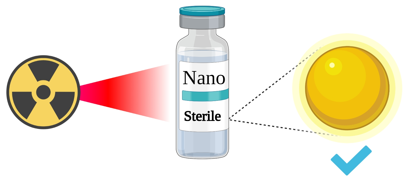
1. Introduction
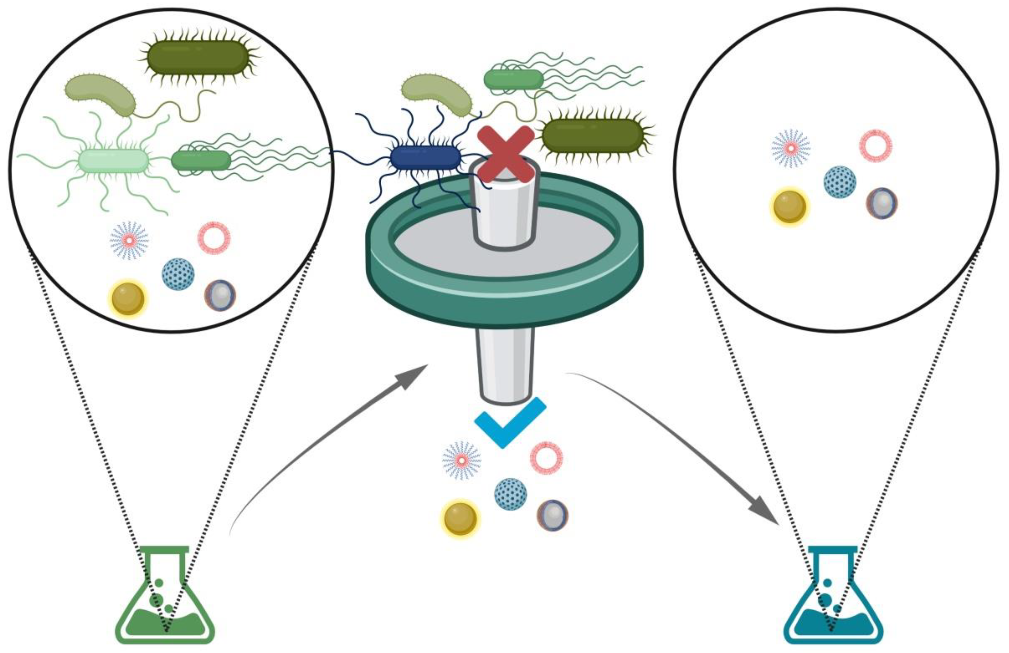
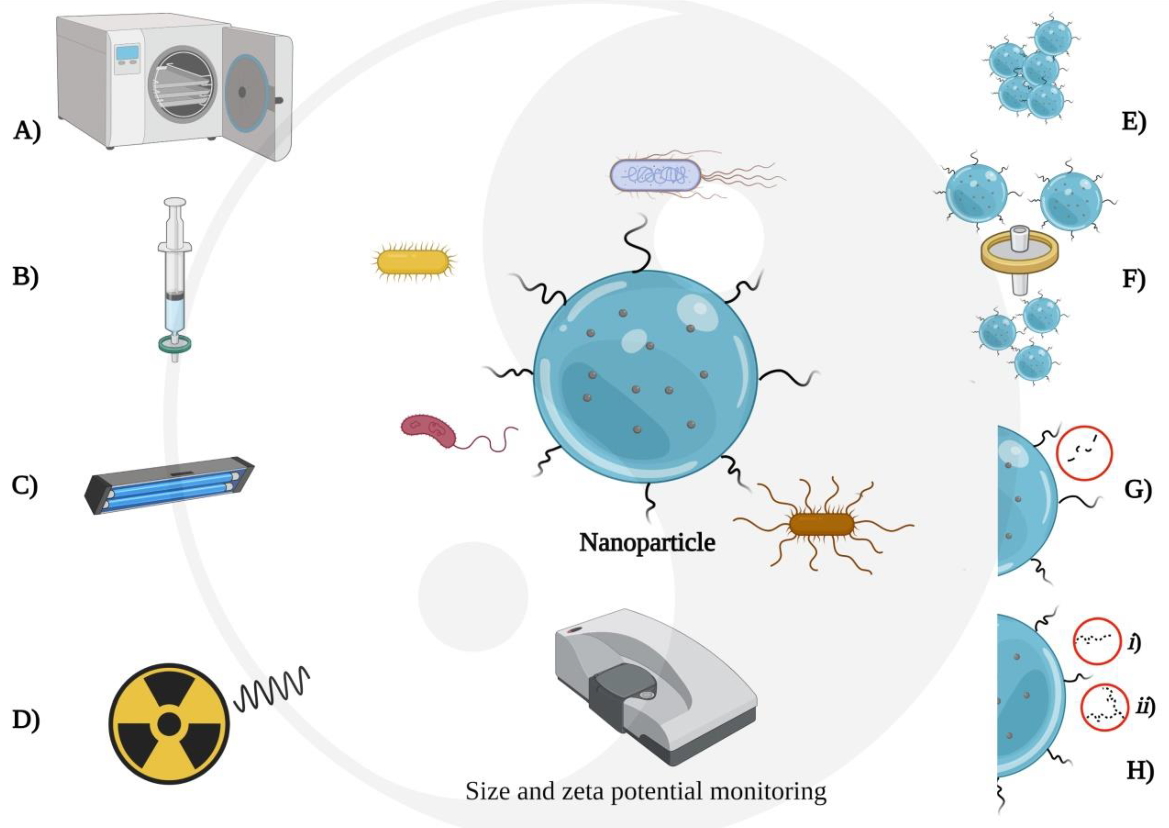
2. Sterile Filtration
2.1. Fundament
2.2. Applications
2.3. Advantages and Disadvantages
3. Autoclaving
3.1. Fundament
3.2. Applications
3.3. Advantages and Disadvantages

| Nanoparticle | Nanoparticle Size (nm) | Autoclaving Conditions | Effect of Sterilization on Nanoparticle | Ref. |
|---|---|---|---|---|
| Gold Nanoparticles capped with PEG or Tiopronin | 2–60 | 134 °C/40 min | The PEG shell had better chemical stability around metal cores after autoclaving than the tiopronin shell. | [8] |
| Citrate-stabilized Silver nanoparticles | 20–80 | 121 °C/30 min | No changes in particle integrity and hemocompatibility were found. Nanoparticles did not vary their sizes after autoclaving. | [30] |
| Dextran-coated magnetic iron oxide nanoparticles | 131.6 | 121 °C/20 min | The dextran shell did not undergo alteration or destruction. No significant difference in mean sizes was detected. No apparent influences from autoclaving on nanoparticles magnetic behavior were found. | [11] |
| ZnO and mesoporous silica-ZnO nanoparticles | 5–20 | --- | Autoclaving plus ultrasound stimulation decreased the bacterial concentration of the nanoparticles. | [32] |
| Silver nanoparticles | 40–80 | 121 °C/15 min | Nanoparticles presented a typical X-ray diffraction pattern for silver nanoparticles. | [42] |
| Trialurine and phospholipids | 200–300 | 121 °C/20 min | Particle size and Z potential were stable. A slight reduction of the incorporated drug was detected, probably due to drug hydrolysis and the formation of a drug’s hydrophilic form. | [46] |
| Trimyristin, tripalmitin or Tristearin, with soy lecithin, poloxamer 188, and stearylamine | 60–170 | 121 °C/20 min | Sizes presented increases, and the Z potential changed to positive. The EE did not significantly change. SLNs stabilized with polymer presented a partial collapse of surface adsorbed polymer and particle aggregation. | [13] |
| Compritol 888ATO, Poloxamer 188 | 200–250 | 121 °C/15 min Or 110 °C/30 min. | The particle size increased, whereas Z potential decreased from −16.9 ± 0.7 to −20.5 ± 0.5. The size increase was attributed to a distortion of the mechanical properties of the surfactant film. | [47] |
| Compritol 888ATO, Poloxamer 188, Tween 80, glycerin | 149 | 110 °C/30 min | The size, Z potential, pH, and EE did not significantly change. | [10] |
| Liposomes DPPC/DPPG EPC/EPG | 200 | 121 °C/15 min. N2 presence. | The particle size of the liposome did not change. Liposomes prepared at pH 7.4 presented a slight change in the gel-sol transition. | [48] |
| PCL with Cremophor RH40, Synperonics, Tonc P787, or MPS. | 130–230 | 121 °C/20 min | Nanoparticles stabilized with cremophor RH40 presented massive aggregation. A decrease of the pH was detected in all preparations, probably by the oxidation of the surfactants. | [9] |
| Polybutylcyanoacrylate. Dextran, Poloxamer or Polysorbate | 200–300 | 121 °C/20 min | Particle sizes did not show a significant difference. Dextran nanoparticles did not show an increased particle size, but the size increased without cooling. The polysorbate nanoparticles agglomerated in scarcely suspendable sediment. | [37] |
| Chitosan-carboximethyl dextran | 538 | 121 °C/30 min | Sizes presented a decrease. No apparent changes in the structure of the polymer. | [49] |
| PEGylated poly (y-benzyl-l-glutamate) | 120 | 121 °C/20 min | Increased nanoparticle size and polydispersity index accompanied with massive aggregation and precipitation. | [15] |
| PEG-b-polycaprolactone. | 45–105 | 121 °C/20 min | The presence of medium-chain triglycerides reduced drug leakage in the sterilization process. The drug loading content did not present a significant reduction | [36] |
| Hydroxyapatite nanoparticles | 100 | 120 °C/20 min | Nanoparticles did not present chemical structure alterations. Nanoparticles synthesized by the wet chemical method showed agglomeration. | [34] |
| Curcumin-Hydroxypropyl-β-cyclodextrin complex and curcumin-Sulfobutylether-β-cyclodextrin) complex. | 200–300 | 121 °C/30 min steaming phase followed by a 30 min drying phase. | The cyclodextrin complex could entrap the curcumin efficiently. 1H-NMR spectra indicated chemically stable curcumin. Possible isomerization of the curcumin was detected in the Raman spectra after the sterilization-synthesis process. | [35] |
| Amphiphilic β-cyclodextrin | 170 | 121 °C/20 min | Particle size and polydispersity showed increases. Nanoparticles exhibited aggregation at the autoclaving temperature. | [24] |
4. Nonionizing Radiation
4.1. Fundament
4.2. Applications
| Time of Exposition | Nanoparticle Type | Loaded Drug | Effect of Sterilization on Nanoparticle | Ref. |
|---|---|---|---|---|
| 30 min | PEG-PLGA | Curcumin | No effect reported | [67] |
| 30 min | PLGA | C-glycosylflavonoid enriched fraction of Cecropia glaziovii | No effect reported | [68] |
| 45 min | PEG-AuNRs | - | The absence of bacterial colonies was verified after the culture onto agar plates | [69] |
| 1 h | Chitosan coated magnetic SLN | Letrozole | No effect reported | [70] |
| 12 h | Au@tiopronin NPs and Au@PEG NPs | - | No detectable changes observed | [8] |
| 12 h | Dextran-coated iron oxide NPs | - | No detectable changes observed | [11] |
| 2 h | PCL/PVA and PLGA/PVA | - | No detectable changes observed | [64] |
| 15 min | PLGA-PEG NPs | - | No effect reported | [71] |
| 3 h | Ag NPs and Au NPs | - | No effect reported | [72] |
4.3. Advantages and Disadvantages
5. Ionizing Radiation
5.1. Fundament
5.2. Applications
| Type of Nanoparticle | Nanoparticle Size (nm) before Irradiation | Radiation Conditions | Effect of Sterilization on Nanoparticle | Ref. |
|---|---|---|---|---|
| Chitosan hydrogel nanoparticles | 288 ± 15 | Gamma irradiation (cobalt-60 at doses of 8, 13, and 25 kGy) |
| [14] |
| Doxorubicin-loaded poly(butyl cyanoacrylate) nanoparticles | 245 ± 83 | Gamma irradiation (cobalt-60 with a dose rate of 0.9–1.0 kGy/s) and electron beams irradiation (linear electron accelerator with doses of 10, 15, 25, and 35 kGy) |
| [85] |
| Diclofenac sodium loaded- N-trimethyl chitosan nanoparticles | 129.3 ± 3.8 | Gamma irradiation (cobalt-60 at doses of 5, 10, 20, and 25 kGy) |
| [89] |
| Silver nanoparticles | 20–80 | Gamma irradiation (cobalt-60 at doses of 15, 25, and 50 kGy) |
| [30] |
| Lyophilised oligodeoxynucleotide-loaded gelatin nanoparticles | 200–280 | Gamma irradiation (cobalt-60 with a dose of 25 kGy) |
| [12,90] |
| Papain nanoparticles | 7.7 ± 0.9 | Gamma irradiation (cobalt-60 with a dose of 10 kGy) |
| [91] |
| Poly-ε-caprolactone and poly(d,l-lactide-co-glycolide) nanoparticles | 228.8 ± 11.60 and 243.1 ± 3.06 respectively. | Gamma irradiation (cobalt-60 at doses of 5 and 10 kGy) |
| [64] |
| Ciprofloxacin HCl-loaded poly(d,l-lactide-glycolide) nanoparticles | 226.1 ± 1.30 | Gamma irradiation (cobalt-60 with a dose of 25 kGy) |
| [92] |
5.3. Advantages and Disadvantages
6. Challenges in Choosing the Sterilization Method
6.1. Factors Related to the Formulation
6.2. Issues Related to Batch Volume
6.3. Aspects Related to Available Methods
6.4. The Limitation of Terminal Sterilization
7. Conclusions
Author Contributions
Funding
Institutional Review Board Statement
Informed Consent Statement
Conflicts of Interest
References
- Lee, B.K.; Yun, Y.H.; Park, K. Smart Nanoparticles for Drug Delivery: Boundaries and Opportunities. Chem. Eng. Sci. 2015, 125, 158–164. [Google Scholar] [CrossRef] [PubMed] [Green Version]
- Zahin, N.; Anwar, R.; Tewari, D.; Kabir, M.T.; Sajid, A.; Mathew, B.; Uddin, M.S.; Aleya, L.; Abdel-Daim, M.M. Nanoparticles and its biomedical applications in health and diseases: Special focus on drug delivery. Environ. Sci. Pollut. Res. 2020, 27, 19151–19168. [Google Scholar] [CrossRef] [PubMed]
- Konan, Y.N.; Cerny, R.; Favet, J.; Berton, M.; Gurny, R.; Allémann, E. Preparation and characterization of sterile sub-200 nm meso-tetra(4-hydroxylphenyl)porphyrin-loaded nanoparticles for photodynamic therapy. Eur. J. Pharm. Biopharm. 2003, 55, 115–124. [Google Scholar] [CrossRef]
- Leyva-Gómez, G.; Cortés, H.; Magaña, J.J.; Leyva-García, N.; Quintanar-Guerrero, D.; Florán, B. Nanoparticle technology for treatment of Parkinson’s disease: The role of surface phenomena in reaching the brain. Drug Discov. Today 2015, 20, 824–837. [Google Scholar] [CrossRef]
- Cortes, H.; Alcalá-Alcalá, S.; Ávalos-Fuentes, A.; Mendoza-Muñoz, N.; Quintanar-Guerrero, D.; Leyva-Gomez, G.; Florán, B. Nanotechnology as Potential Tool for siRNA Delivery in Parkinson’s Disease. Curr. Drug Targets 2017, 18, 1866–1879. [Google Scholar] [CrossRef] [PubMed]
- Cortés, H.; Alcalá-Alcalá, S.; Caballero-Florán, I.H.; Bernal-Chávez, S.A.; Ávalos-Fuentes, A.; González-Torres, M.; Carmen, M.G.D.; Figueroa-González, G.; Reyes-Hernández, O.D.; Floran, B.; et al. A Reevaluation of Chitosan-Decorated Nanoparticles to Cross the Blood-Brain Barrier. Membranes 2020, 10, 212. [Google Scholar] [CrossRef]
- Vetten, M.A.; Yah, C.S.; Singh, T.; Gulumian, M. Challenges facing sterilization and depyrogenation of nanoparticles: Effects on structural stability and biomedical applications. Nanomed. Nanotechnol. Biol. Med. 2014, 10, 1391–1399. [Google Scholar] [CrossRef]
- França, Á.; Pelaz, B.; Moros, M.; Sánchez-Espinel, C.; Hernández, A.; Fernández-López, C.; Grazú, V.; De La Fuente, J.M.; Pastoriza-Santos, I.; Liz-Marzán, L.M.; et al. Sterilization Matters: Consequences of Different Sterilization Techniques on Gold Nanoparticles. Small 2010, 6, 89–95. [Google Scholar] [CrossRef]
- Masson, V.; Maurin, F.; Fessi, H.; Devissaguet, J.P. Influence of sterilization processes on poly(ε-caprolactone) nanospheres. Biomaterials 1997, 18, 327–335. [Google Scholar] [CrossRef]
- Hippalgaonkar, K.; Adelli, G.R.; Hippalgaonkar, K.; Repka, M.A.; Majumdar, S. Indomethacin-Loaded Solid Lipid Nanoparticles for Ocular Delivery: Development, Characterization, and In Vitro Evaluation. J. Ocul. Pharmacol. Ther. 2013, 29, 216–228. [Google Scholar] [CrossRef] [Green Version]
- Li, L.; Mak, K.Y.; Shi, J.; Leung, C.H.; Wong, C.M.; Leung, C.W.; Mak, C.S.K.; Chan, K.Y.; Chan, N.M.M.; Wu, E.X.; et al. Sterilization on dextran-coated iron oxide nanoparticles: Effects of autoclaving, filtration, UV irradiation, and ethanol treatment. Microelectron. Eng. 2013, 111, 310–313. [Google Scholar] [CrossRef]
- Geh, K.J.; Hubert, M.; Winter, G. Progress in formulation development and sterilisation of freeze-dried oligodeoxynucleotide-loaded gelatine nanoparticles. Eur. J. Pharm. Biopharm. 2018, 129, 10–20. [Google Scholar] [CrossRef] [PubMed]
- Venkateswarlu, V.; Manjunath, K. Preparation, characterization and in vitro release kinetics of clozapine solid lipid nanoparticles. J. Control. Release 2004, 95, 627–638. [Google Scholar] [CrossRef] [PubMed]
- Galante, R.; Rediguieri, C.F.; Kikuchi, I.S.; Vasquez, P.A.S.; Colaço, R.; Serro, A.P.; Pinto, T.J.A. About the Sterilization of Chitosan Hydrogel Nanoparticles. PLoS ONE 2016, 11, e0168862. [Google Scholar] [CrossRef]
- Özcan, I.; Bouchemal, K.; Sánchez, F.S.; Abaci, Ö.; Özer, Ö.; Güneri, T.; Ponchel, G. Effects of sterilization techniques on the PEGylated poly (gamma-benzyl-L-glutamate) (PBLG) nanoparticles. Acta Pharm. Sci. 2009, 51, 211–218. [Google Scholar]
- Tsukada, Y.; Hara, K.; Bando, Y.; Huang, C.C.; Kousaka, Y.; Kawashima, Y.; Morishita, R.; Tsujimoto, H. Particle size control of poly(dl-lactide-co-glycolide) nanospheres for sterile applications. Int. J. Pharm. 2009, 370, 196–201. [Google Scholar] [CrossRef]
- Darole, P.S.; Hegde, D.D.; Nair, H.A. Formulation and Evaluation of Microemulsion Based Delivery System for Amphotericin B. AAPS PharmSciTech 2008, 9, 122–128. [Google Scholar] [CrossRef]
- Li, C.; Deng, Y. A novel method for the preparation of liposomes: Freeze drying of monophase solutions. J. Pharm. Sci. 2004, 93, 1403–1414. [Google Scholar] [CrossRef] [PubMed]
- Bos, G.W.; Trullas-Jimeno, A.; Jiskoot, W.; Crommelin, D.J.A.; Hennink, W.E. Sterilization of poly(dimethylamino) ethyl methacrylate-based gene transfer complexes. Int. J. Pharm. 2000, 211, 79–88. [Google Scholar] [CrossRef]
- Cheng, J.; Davis, M.; Khin, T. Cyclodextrin-Based Polymers for Therapeutics Delivery. U.S. Patent 8,475,781B2, 2 July 2013. [Google Scholar]
- Zhang, Z.; Almarsson, U.; Chen, H. Aqueous 2,6-Diisopropylphenol Pharmaceutical Compositions. U.S. Patent 7,915,317B2, 29 March 2011. [Google Scholar]
- Mori, Y. Size-Selective Separation Techniques for Nanoparticles in Liquid. KONA Powder Part. J. 2015, 32, 102–144. [Google Scholar] [CrossRef] [Green Version]
- Dutz, S.; Wojahn, S.; Gräfe, C.; Weidner, A.; Clement, J.H. Influence of Sterilization and Preservation Procedures on the Integrity of Serum Protein-Coated Magnetic Nanoparticles. Nanomaterials 2017, 7, 453. [Google Scholar] [CrossRef] [Green Version]
- Memisoglu-Bilensoy, E.; Hincal, A.A. Sterile, injectable cyclodextrin nanoparticles: Effects of gamma irradiation and autoclaving. Int. J. Pharm. 2006, 311, 203–208. [Google Scholar] [CrossRef]
- Bai, Y.; Moeinzadeh, S.; Kim, S.; Park, Y.; Lui, E.; Tan, H.; Zhao, W.; Zhou, X.; Yang, Y.P. Development of PLGA-PEG-COOH and Gelatin-Based Microparticles Dual Delivery System and E-Beam Sterilization Effects for Controlled Release of BMP-2 and IGF-1. Part. Part. Syst. Charact. 2020, 37, 1–11. [Google Scholar] [CrossRef] [PubMed]
- Rawal, M.; Singh, A.; Amiji, M.M. Quality-by-Design Concepts to Improve Nanotechnology-Based Drug Development. Pharm. Res. 2019, 36, 1–20. [Google Scholar] [CrossRef] [PubMed]
- Rutala, W.A.; Weber, D.J. Infection control: The role of disinfection and sterilization. J. Hosp. Infect. 1999, 43, S43–S55. [Google Scholar] [CrossRef]
- Pišlová, M.; Kolářová, K.; Vokatá, B.; Brož, A.; Ulbrich, P.; Bačáková, L.; Kolská, Z.; Švorčík, V. A new way to prepare gold nanoparticles by sputtering—Sterilization, stability and other properties. Mater. Sci. Eng. C 2020, 115, 111087. [Google Scholar] [CrossRef] [PubMed]
- Pourali, P.; Badiee, S.H.; Manafi, S.; Noorani, T.; Rezaei, A.; Yahyaei, B. Biosynthesis of gold nanoparticles by two bacterial and fungal strains, Bacillus cereus and Fusarium oxysporum, and assessment and comparison of their nanotoxicity in vitro by direct and indirect assays. Electron. J. Biotechnol. 2017, 29, 86–93. [Google Scholar] [CrossRef]
- Zheng, J. Sterilization of Silver Nanoparticles Using Standard Gamma Irradiation Procedure Affects Particle Integrity and Biocompatibility. J. Nanomed. Nanotechnol. 2011, S5. [Google Scholar] [CrossRef] [Green Version]
- Kummer, K.M.; Taylor, E.N.; Durmas, N.G.; Tarquinio, K.M.; Ercan, B.; Webster, T.J. Effects of different sterilization techniques and varying anodized TiO 2 nanotube dimensions on bacteria growth. J. Biomed. Mater. Res. Part B Appl. Biomater. 2013, 101, 677–688. [Google Scholar] [CrossRef] [PubMed]
- Rokbani, H.; Daigle, F.; Ajji, A. Combined Effect of Ultrasound Stimulations and Autoclaving on the Enhancement of Antibacterial Activity of ZnO and SiO2/ZnO Nanoparticles. Nanomaterials 2018, 8, 129. [Google Scholar] [CrossRef] [PubMed] [Green Version]
- Mancini, G.; Lopes, R.M.; Clemente, P.; Raposo, S.; Gonçalves, L.M.D.; Bica, A.; Ribeiro, H.M.; Almeida, A.J. Lecithin and parabens play a crucial role in tripalmitin-based lipid nanoparticle stabilization throughout moist heat sterilization and freeze-drying. Eur. J. Lipid Sci. Technol. 2015, 117, 1947–1959. [Google Scholar] [CrossRef]
- Santos, C.; Gomes, P.S.; Duarte, J.A.; Franke, R.P.; Almeida, M.M.; Costa, M.E.V.; Fernandes, M.H. Relevance of the sterilization-induced effects on the properties of different hydroxyapatite nanoparticles and assessment of the osteoblastic cell response. J. R. Soc. Interface 2012, 9, 3397–3410. [Google Scholar] [CrossRef] [Green Version]
- Hagbani, T.; Nazzal, S. Curcumin complexation with cyclodextrins by the autoclave process: Method development and characterization of complex formation. Int. J. Pharm. 2017, 520, 173–180. [Google Scholar] [CrossRef] [PubMed]
- Gou, J.; Chao, Y.; Liang, Y.; Zhang, N.; He, H.; Yin, T.; Zhang, Y.; Xu, H.; Tang, X. Humid Heat Autoclaving of Hybrid Nanoparticles Achieved by Decreased Nanoparticle Concentration and Improved Nanoparticle Stability Using Medium Chain Triglycerides as a Modifier. Pharm. Res. 2016, 33, 2140–2151. [Google Scholar] [CrossRef]
- Sommerfeld, P.; Schroeder, U.; Sabel, B.A. Sterilization of unloaded polybutylcyanoacrylate nanoparticles. Int. J. Pharm. 1998, 164, 113–118. [Google Scholar] [CrossRef]
- Lichther, J.; Trammel, A.; Piu, U.; Ye, Q.; Scaife, M.C.; Vollrath, B.; Duron, G.; Dellamary, L.; Lebel, C.; Harris, J. Controlled Release Delivery Devices for the Treatment of Otic Disorders. U.S. Patent 2019/0192425A1, 27 September 2019. [Google Scholar]
- Groman, E.; Paul, K.; Frigo, T.; Bengele, H.; Lewis, J. Heat Stable Colloidal Iron Oxides Coated with Reduced Carbohydrates and Carbohydrate Derivatives. U.S. Patent 6,599,498B1, 29 July 2003. [Google Scholar]
- Mirkin, C.A.; Giljohann, D.A.; Seferos, D.S.; Prigodich, A.E.; Patel, P. Polyvalent RNA-Nanoparticle Compositions. U.S. Patent 10,391,116B2, 29 March 2019. [Google Scholar]
- Yonfeng Zhang, J.; Ziping Luo, M. Sterile Pharmaceutical Composition and Process for a Solution of Propofol Emulsion Having Microbial Growth Retardation. U.S. Patent 7,468,394B1, 23 December 2008. [Google Scholar]
- Selvi, N.T.; Navamathavan, R.; Kim, H.Y.; Nirmala, R. Autoclave Mediated Synthesis of Silver Nanoparticles Using Aqueous Extract of Canna indica L. Rhizome and Evaluation of Its Antimicrobial Activity. Macromol. Res. 2019, 27, 1155–1160. [Google Scholar] [CrossRef]
- Zielińska, A.; Soles, B.B.; Lopes, A.R.; Vaz, B.F.; Rodrigues, C.M.; Alves, T.F.R.; Klensporf-Pawlik, D.; Durazzo, A.; Lucarini, M.; Severino, P.; et al. Nanopharmaceuticals for Eye Administration: Sterilization, Depyrogenation and Clinical Applications. Biology 2020, 9, 336. [Google Scholar] [CrossRef] [PubMed]
- Subbarao, N. Nanoparticle Sterility and Sterilization of Nanomaterials. Handb. Immunol. Prop. Eng. Nanomater. 2016, 1, 53–75. [Google Scholar]
- Vauthier, C.; Bouchemal, K. Methods for the Preparation and Manufacture of Polymeric Nanoparticles. Pharm. Res. 2009, 26, 1025–1058. [Google Scholar] [CrossRef]
- Heiati, H.; Tawashi, R.; Phillips, N.C. Drug retention and stability of solid lipid nanoparticles containing azidothymidine palmitate after autoclaving, storage and lyophilization. J. Microencapsul. 1998, 15, 173–184. [Google Scholar] [CrossRef]
- Gokce, E.H.; Sandri, G.; Bonferoni, M.C.; Rossi, S.; Ferrari, F.; Güneri, T.; Caramella, C. Cyclosporine A loaded SLNs: Evaluation of cellular uptake and corneal cytotoxicity. Int. J. Pharm. 2008, 364, 76–86. [Google Scholar] [CrossRef] [PubMed]
- Zuidam, N.J.; Lee, S.S.L.; Crommelin, D.J.A. Sterilization of Liposomes by Heat Treatment. Pharm. Res. 1993, 10, 1591–1596. [Google Scholar] [CrossRef]
- Lin, Y.S.; Renbutsu, E.; Morimoto, M.; Okamura, Y.; Tsuka, T.; Saimoto, H.; Okamoto, Y.; Minami, S. Preparation of stable chitosan-carboxymethyl dextran nanoparticles. J. Nanosci. Nanotechnol. 2009, 9, 2558–2565. [Google Scholar] [CrossRef] [PubMed]
- Mu, W.; Akrofi, R.; Chen, Q. Near-infrared-driven Au-decorated polymer-metal protein microfibers with bacterial filtration ability for use in photothermal sterilization. Chem. Eng. J. 2020, 388, 124236. [Google Scholar] [CrossRef]
- Cai, S.; Qian, J.; Yang, S.; Kuang, L.; Hua, D. Acetylcysteine-decorated Prussian blue nanoparticles for strong photothermal sterilization and focal infection treatment. Colloids Surf. B Biointerfaces 2019, 181, 31–38. [Google Scholar] [CrossRef] [PubMed]
- Benson, R.S. Use of radiation in biomaterials science. Nucl. Instrum. Methods Phys. Res. Sect. B Beam Interact. Mater. At. 2002, 191, 752–757. [Google Scholar] [CrossRef]
- Diffey, B.L. Sources and measurement of ultraviolet radiation. Methods 2002, 28, 4–13. [Google Scholar] [CrossRef] [Green Version]
- Griffin, M.; Naderi, N.; Kalaskar, D.M.; Malins, E.; Becer, R.; Thornton, C.A.; Whitaker, I.S.; Mosahebi, A.; Butler, P.E.M.; Seifalian, A.M. Evaluation of Sterilisation Techniques for Regenerative Medicine Scaffolds Fabricated with Polyurethane Nonbiodegradable and Bioabsorbable Nanocomposite Materials. Int. J. Biomater. 2018, 2018. [Google Scholar] [CrossRef]
- Divya, K.; Smitha, V.; Jisha, M.S. Antifungal, antioxidant and cytotoxic activities of chitosan nanoparticles and its use as an edible coating on vegetables. Int. J. Biol. Macromol. 2018, 114, 572–577. [Google Scholar] [CrossRef]
- Kim, H.Y.; Park, S.S.; Lim, S.T. Preparation, characterization and utilization of starch nanoparticles. Colloids Surf. B Biointerfaces 2015, 126, 607–620. [Google Scholar] [CrossRef]
- Kwon, S.S.; Nam, Y.S.; Lee, J.S.; Ku, B.S.; Han, S.H.; Lee, J.Y.; Chang, I.S. Preparation and characterization of coenzyme Q10-loaded PMMA nanoparticles by a new emulsification process based on microfluidization. Colloids Surf. A Physicochem. Eng. Asp. 2002, 210, 95–104. [Google Scholar] [CrossRef]
- Borcia, C.; Borcia, G.; Dumitrascu, N. Surface treatment of polymers by plasma and UV radiation. Rom. Rep. Phys. 2011, 56, 224–232. [Google Scholar]
- Eve, S.; Mohr, J. Study of the surface modification of the PMMA by UV-radiation. Procedia Eng. 2009, 1, 237–240. [Google Scholar] [CrossRef] [Green Version]
- Kowalonek, J. Studies of chitosan/pectin complexes exposed to UV radiation. Int. J. Biol. Macromol. 2017, 103, 515–524. [Google Scholar] [CrossRef]
- Liu, Q.; Jing, Y.; Han, C.; Zhang, H.; Tian, Y. Encapsulation of curcumin in zein/caseinate/sodium alginate nanoparticles with improved physicochemical and controlled release properties. Food Hydrocoll. 2019, 93, 432–442. [Google Scholar] [CrossRef]
- Lian, Y.; Wang, X.; Guo, P.; Li, Y.; Raza, F.; Su, J.; Qiu, M. Erythrocyte Membrane-Coated Arsenic Trioxide-Loaded Sodium Alginate Nanoparticles for Tumor Therapy. Pharmaceutics 2020, 12, 21. [Google Scholar] [CrossRef] [PubMed] [Green Version]
- Chansoria, P.; Narayanan, L.K.; Wood, M.; Alvarado, C.; Lin, A.; Shirwaiker, R.A. Effects of Autoclaving, EtOH, and UV Sterilization on the Chemical, Mechanical, Printability, and Biocompatibility Characteristics of Alginate. ACS Biomater. Sci. Eng. 2020, 6, 5191–5201. [Google Scholar] [CrossRef]
- Tapia-Guerrero, Y.S.; Del Prado-Audelo, M.L.; Borbolla-Jiménez, F.V.; Giraldo Gomez, D.M.; García-Aguirre, I.; Colín-Castro, C.A.; Morales-González, J.A.; Leyva-Gómez, G.; Magaña, J.J. Effect of UV and Gamma Irradiation Sterilization Processes in the Properties of Different Polymeric Nanoparticles for Biomedical Applications. Materials 2020, 13, 1090. [Google Scholar] [CrossRef] [Green Version]
- Ripolles-Avila, C.; Martinez-Garcia, M.; Hascoët, A.S.; Rodríguez-Jerez, J.J. Bactericidal efficacy of UV activated TiO2 nanoparticles against Gram-positive and Gram-negative bacteria on suspension. CYTA J. Food 2019, 17, 408–418. [Google Scholar] [CrossRef] [Green Version]
- Yang, Y.; Guan, C.; Chen, S. Structural characterization and catalytic sterilization performance of a TiO2 nano-photocatalyst. Food Sci. Nutr. 2020, 8, 3638–3646. [Google Scholar] [CrossRef]
- Orunoğlu, M.; Kaffashi, A.; Pehlivan, S.B.; Şahin, S.; Söylemezoğlu, F.; Karlı-Oğuz, K.; Mut, M. Effects of curcumin-loaded PLGA nanoparticles on the RG2 rat glioma model. Mater. Sci. Eng. C 2017, 78, 32–38. [Google Scholar] [CrossRef] [PubMed]
- Caldas dos Santos, T.; Rescignano, N.; Boff, L.; Reginatto, F.H.; Simões, C.M.O.; de Campos, A.M.; Mijangos, C. In vitro antiherpes effect of C-glycosyl flavonoid enriched fraction of Cecropia glaziovii encapsulated in PLGA nanoparticles. Mater. Sci. Eng. C 2017, 75, 1214–1220. [Google Scholar] [CrossRef]
- Mahmoud, N.N.; Hikmat, S.; Abu Ghith, D.; Hajeer, M.; Hamadneh, L.; Qattan, D.; Khalil, E.A. Gold nanoparticles loaded into polymeric hydrogel for wound healing in rats: Effect of nanoparticles’ shape and surface modification. Int. J. Pharm. 2019, 565, 174–186. [Google Scholar] [CrossRef] [PubMed]
- Ahmadifard, Z.; Ahmeda, A.; Rasekhian, M.; Moradi, S.; Arkan, E. Chitosan-coated magnetic solid lipid nanoparticles for controlled release of letrozole. J. Drug Deliv. Sci. Technol. 2020, 57, 101621. [Google Scholar] [CrossRef]
- Zhang, B.; Sai Lung, P.; Zhao, S.; Chu, Z.; Chrzanowski, W.; Li, Q. Shape dependent cytotoxicity of PLGA-PEG nanoparticles on human cells. Sci. Rep. 2017, 7, 1–8. [Google Scholar] [CrossRef] [PubMed]
- Loutfy, S.A.; Al-Ansary, N.A.; Abdel-Ghani, N.T.; Hamed, A.R.; Mohamed, M.B.; Craik, J.D.; Salah Eldin, T.A.; Abdellah, A.M.; Hussein, Y.; Hasanin, M.T.M.; et al. Anti-proliferative Activities of Metallic Nanoparticles in an In Vitro Breast Cancer Model. Asian Pac. J. Cancer Prev. 2015, 16, 6039–6046. [Google Scholar] [CrossRef] [Green Version]
- Silva, C.A.; De Andrade, N.J.; Soares, N.D.F.F.; Ferreira, S.O. Evaluation of ultraviolet radiation to control microorganisms adhering to low-density polyethylene films. Braz. J. Microbiol. 2003, 34, 175–178. [Google Scholar] [CrossRef]
- Lambert, P.A. Radiation processes. In Principles and Practice of Disinfection, Preservation and Sterilization; Fraise, A.P., Lambert, P.A., Maillard, J.-Y., Eds.; Wiley: Hoboken, NJ, USA, 2012; pp. 294–305. [Google Scholar]
- De Freitas, L.F.; Varca, G.; Batista, J.D.S.; Lugão, A.B. An Overview of the Synthesis of Gold Nanoparticles Using Radiation Technologies. Nanomaterials 2018, 8, 939. [Google Scholar] [CrossRef] [PubMed] [Green Version]
- Conti, B.; Dorati, R.; Colonna, C.; Genta, I. Effects of ionizing radiation sterilization on microparticulate drug delivery systems based on poly-α-hydroxyacids: An overview. J. Drug Deliv. Sci. Technol. 2009, 19, 99–112. [Google Scholar] [CrossRef]
- Pirker, L.; Krajnc, A.P.; Malec, J.; Radulović, V.; Gradišek, A.; Jelen, A.; Remškar, M.; Mekjavić, I.B.; Kovač, J.; Mozetič, M.; et al. Sterilization of polypropylene membranes of facepiece respirators by ionizing radiation. J. Memb. Sci. 2021, 619, 118756. [Google Scholar] [CrossRef]
- Toh, M.-R.; Chiu, G.N.C. Liposomes as sterile preparations and limitations of sterilisation techniques in liposomal manufacturing. Asian J. Pharm. Sci. 2013, 8, 88–95. [Google Scholar] [CrossRef] [Green Version]
- Kalia, V.; Joseph, D. Medicine: Sterilization. In Reference Module in Earth Systems and Environmental Sciences; Elsevier: Amsterdam, The Netherlands, 2020. [Google Scholar]
- Domańska, I.M.; Oledzka, E.; Sobczak, M. Sterilization process of polyester based anticancer-drug delivery systems. Int. J. Pharm. 2020, 587, 119663. [Google Scholar] [CrossRef] [PubMed]
- International Atomic Energy Agency. Trends in Radiation Sterilization of Health Care Products; IAEA Library Cataloguing in Publication Data: Vienna, Austria, 2008. [Google Scholar]
- Sedlacek, O.; Kucka, J.; Monnery, B.D.; Slouf, M.; Vetrik, M.; Hoogenboom, R.; Hruby, M. The effect of ionizing radiation on biocompatible polymers: From sterilization to radiolysis and hydrogel formation. Polym. Degrad. Stab. 2017, 137, 1–10. [Google Scholar] [CrossRef]
- Buchalla, R.; Schüttler, C.; Bögl, K.W. Radiation sterilization of medical devices. Effects of ionizing radiation on ultra-high molecular-weight polyethylene. Radiat. Phys. Chem. 1995, 46, 579–585. [Google Scholar] [CrossRef]
- Yasunaga, M.; Kobayashi, F.; Hara, Y.; Yamazaki, M.; Ohno, T.; Ito, A. Biological activity of terminally gamma-ray-sterilized titanium and hydroxyapatite coated with a growth factor−apatite composite layer. Mater. Today Commun. 2020, 24, 101098. [Google Scholar] [CrossRef]
- Maksimenko, O.; Pavlov, E.; Toushov, E.; Molin, A.; Stukalov, Y.; Prudskova, T.; Feldman, V.; Kreuter, J.; Gelperina, S. Radiation sterilisation of doxorubicin bound to poly(butyl cyanoacrylate) nanoparticles. Int. J. Pharm. 2008, 356, 325–332. [Google Scholar] [CrossRef] [PubMed]
- Hasanain, F.; Guenther, K.; Mullett, W.M.; Craven, E. Gamma Sterilization of Pharmaceuticals—A Review of the Irradiation of Excipients, Active Pharmaceutical Ingredients, and Final Drug Product Formulations. PDA J. Pharm. Sci. Technol. 2014, 68, 113–137. [Google Scholar] [CrossRef]
- Nanobiotix Home Page. Available online: https://www.nanobiotix.com/ (accessed on 20 March 2021).
- QuikClot Home Page. Available online: https://quikclot.com/ (accessed on 21 March 2021).
- Asasutjarit, R.; Theerachayanan, T.; Kewsuwan, P.; Veeranondha, S.; Fuongfuchat, A.; Ritthidej, G.C. Gamma sterilization of diclofenac sodium loaded- N-trimethyl chitosan nanoparticles for ophthalmic use. Carbohydr. Polym. 2017, 157, 603–612. [Google Scholar] [CrossRef]
- Timofejeva, A.; D’Este, M.; Loca, D. Calcium phosphate/polyvinyl alcohol composite hydrogels: A review on the freeze-thawing synthesis approach and applications in regenerative medicine. Eur. Polym. J. 2017, 95, 547–565. [Google Scholar] [CrossRef]
- Fazolin, G.N.; Varca, G.H.C.; de Freitas, L.F.; Rokita, B.; Kadlubowski, S.; Lugão, A.B. Simultaneous intramolecular crosslinking and sterilization of papain nanoparticles by gamma radiation. Radiat. Phys. Chem. 2020, 171, 108697. [Google Scholar] [CrossRef]
- Bozdag, S.; Dillen, K.; Vandervoort, J.; Ludwig, A. The effect of freeze-drying with different cryoprotectants and gamma-irradiation sterilization on the characteristics of ciprofloxacin HCl-loaded poly(D,L-lactide-glycolide) nanoparticles. J. Pharm. Pharmacol. 2005, 57, 699–707. [Google Scholar] [CrossRef] [PubMed]
- Postek, M.T.; Poster, D.L.; Vládar, A.E.; Driscoll, M.S.; LaVerne, J.A.; Tsinas, Z.; Al-Sheikhly, M.I. Ionizing radiation processing and its potential in advancing biorefining and nanocellulose composite materials manufacturing. Radiat. Phys. Chem. 2018, 143, 47–52. [Google Scholar] [CrossRef] [PubMed]
- Gu, L.; Zablocki, K.; Lavelle, L.; Bodnar, S.; Halperin, F.; Harper, I.; Moghe, P.V.; Uhrich, K.E. Impact of ionizing radiation on physicochemical and biological properties of an amphiphilic macromolecule. Polym. Degrad. Stab. 2012, 97, 1686–1689. [Google Scholar] [CrossRef] [PubMed] [Green Version]
- Leyva-Gómez, G.; Santillan-Reyes, E.; Lima, E.; Madrid-Martínez, A.; Krötzsch, E.; Quintanar-Guerrero, D.; Garciadiego-Cázares, D.; Martínez-Jiménez, A.; Hernández Morales, M.; Ortega-Peña, S.; et al. A novel hydrogel of poloxamer 407 and chitosan obtained by gamma irradiation exhibits physicochemical properties for wound management. Mater. Sci. Eng. C 2017, 74, 36–46. [Google Scholar] [CrossRef] [PubMed]
- González-Torres, M.; Leyva-Gómez, G.; Rivera, M.; Krötzsch, E.; Rodríguez-Talavera, R.; Rivera, A.L.; Cabrera-Wrooman, A. Biological activity of radiation-induced collagen-polyvinylpyrrolidone-PEG hydrogels. Mater. Lett. 2017, 214, 224–227. [Google Scholar] [CrossRef]
- Le, V.H.; Weiss, S.; Lundahl, B.; Lam, S. Terminal sterilization. Assur. Steril. Sensitive Comb. Prod. Mater. 2019, 23–38. [Google Scholar]


Publisher’s Note: MDPI stays neutral with regard to jurisdictional claims in published maps and institutional affiliations. |
© 2021 by the authors. Licensee MDPI, Basel, Switzerland. This article is an open access article distributed under the terms and conditions of the Creative Commons Attribution (CC BY) license (https://creativecommons.org/licenses/by/4.0/).
Share and Cite
Bernal-Chávez, S.A.; Del Prado-Audelo, M.L.; Caballero-Florán, I.H.; Giraldo-Gomez, D.M.; Figueroa-Gonzalez, G.; Reyes-Hernandez, O.D.; González-Del Carmen, M.; González-Torres, M.; Cortés, H.; Leyva-Gómez, G. Insights into Terminal Sterilization Processes of Nanoparticles for Biomedical Applications. Molecules 2021, 26, 2068. https://doi.org/10.3390/molecules26072068
Bernal-Chávez SA, Del Prado-Audelo ML, Caballero-Florán IH, Giraldo-Gomez DM, Figueroa-Gonzalez G, Reyes-Hernandez OD, González-Del Carmen M, González-Torres M, Cortés H, Leyva-Gómez G. Insights into Terminal Sterilization Processes of Nanoparticles for Biomedical Applications. Molecules. 2021; 26(7):2068. https://doi.org/10.3390/molecules26072068
Chicago/Turabian StyleBernal-Chávez, Sergio A., María Luisa Del Prado-Audelo, Isaac H. Caballero-Florán, David M. Giraldo-Gomez, Gabriela Figueroa-Gonzalez, Octavio D. Reyes-Hernandez, Manuel González-Del Carmen, Maykel González-Torres, Hernán Cortés, and Gerardo Leyva-Gómez. 2021. "Insights into Terminal Sterilization Processes of Nanoparticles for Biomedical Applications" Molecules 26, no. 7: 2068. https://doi.org/10.3390/molecules26072068