
Ethiopian Medicinal Plants Traditionally Used for the Treatment of Cancer; Part 3: Selective Cytotoxic Activity of 22 Plants against Human Cancer Cell Lines

 , ,
, , 
Abstract
:
1. Introduction
2. Results and Discussion
3. Materials and Methods
3.1. Plant Material
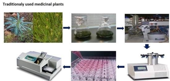
3.2. Preparation of Crude Extract
3.3. Cell Culture
3.4. Peripheral Blood Mononuclear Cell (PBMC) Isolation
3.5. Crystal Violet Cell Proliferation Assay
3.6. MTT Cell Viability Assay
3.7. Statistical Analysis
4. Conclusions
Author Contributions
Funding
Institutional Review Board Statement
Informed Consent Statement
Data Availability Statement
Acknowledgments
Conflicts of Interest
Sample Availability
References
- Pilleron, S.; Sarfati, D.; Janssen-Heijnen, M.; Vignat, J.; Ferlay, J.; Bray, F.; Soerjomataram, I. Global cancer incidence in older adults, 2012 and 2035: A population-based study. Int. J. Cancer 2019, 144, 49–58. [Google Scholar] [CrossRef] [PubMed]
- Bray, F.; Ferlay, J.; Soerjomataram, I.; Siegel, R.L.; Torre, L.A.; Jemal, A. Global cancer statistics 2018: GLOBOCAN estimates of incidence and mortality worldwide for 36 cancers in 185 countries. CA Cancer J. Clin. 2018, 68, 394–424. [Google Scholar] [CrossRef] [PubMed] [Green Version]
- Tesfaye, S.; Belete, A.; Engidawork, E.; Gedif, T.; Asres, K. Ethnobotanical Study of Medicinal Plants Used by Traditional Healers to Treat Cancer-Like Symptoms in Eleven Districts, Ethiopia. Evid.-Based Complement. Altern. Med. 2020, 2020, 1–23. [Google Scholar] [CrossRef] [PubMed] [Green Version]
- Yohannis, S.W.; Asfaw, Z.; Kelbessa, E. Ethnobotanical Study of Medicinal Plants Used by Local People in Menz Gera Midir District, North Shewa Zone, Amhara Regional State, Ethiopia. J. Med. Plant. Res. 2018, 12, 296–314. [Google Scholar]
- Tuasha, N.; Petros, B.; Asfaw, Z. Medicinal plants used by traditional healers to treat malignancies and other human ailments in Dalle District, Sidama Zone, Ethiopia. J. Ethnobiol. Ethnomed. 2018, 14, 1–21. [Google Scholar] [CrossRef] [PubMed] [Green Version]
- Yong, Y.; Tesso, H.; Terfa, A.; Dekebo, A.; Dinku, W.; Lee, Y.H.; Shin, S.Y.; Lim, Y. Biological evaluation of the diterpenes from Croton macrostachyus. Appl. Biol. Chem. 2017, 60, 615–621. [Google Scholar] [CrossRef]
- Liu, J.-Q.; Yang, Y.-F.; Li, X.-Y.; Liu, E.-Q.; Li, Z.-R.; Zhou, L.; Li, Y.; Qiu, M.-H. Cytotoxicity of Naturally Occurring Rham-nofolane Diterpenes from Jatropha curcas. Phytochemistry 2013, 96, 265–272. [Google Scholar] [CrossRef]
- Okoye, F.B.C.; Debbab, A.; Wray, V.; Esimone, C.O.; Osadebe, P.O.; Proksch, P. A phenyldilactone, bisnorsesquiterpene, and cytotoxic phenolics from Maytenus senegalensis leaves. Tetrahedron Lett. 2014, 55, 3756–3760. [Google Scholar] [CrossRef]
- Balkrishna, A.; Das, S.K.; Pokhrel, S.; Joshi, A.; Laxmi Verma, S.; Sharma, V.K.; Sharma, V.; Sharma, N.; Joshi, C.S. Colchicine: Isolation, LC–MS QTof Screening, and Anticancer Activity Study of Gloriosa superba Seeds. Molecules 2019, 24, 2772. [Google Scholar] [CrossRef] [Green Version]
- Tesfaye, S.; Tadesse, S.; Engidawork, E.; Belete, A.; Schaufler, K.; Guenther, S.; Asres, K. Screening of Twenty-one Ethiopian Medicinal Plants for their Antiproliferative Activity against Human Acute Myeloid Leukemia (MV4-11) Cell Line. Ethiop. Pharm. J. 2019, 35, 143–148. [Google Scholar] [CrossRef]
- Addo, E.M.; Chai, H.-B.; Hymete, A.; Yeshak, M.Y.; Slebodnick, C.; Kingston, D.G.I.; Rakotondraibe, L.H. Antiproliferative Constituents of the Roots of Ethiopian Podocarpus falcatus and Structure Revision of 2α-Hydroxynagilactone F and Nagilactone, I. J. Nat. Prod. 2015, 78, 827–835. [Google Scholar] [CrossRef]
- Habtemariam, S. Knipholone anthrone from Kniphofia foliosa induces a rapid onset of necrotic cell death in cancer cells. Fitoterapia 2010, 81, 1013–1019. [Google Scholar] [CrossRef]
- Habtemariam, S. Cytotoxicity of Diterpenes from Premna schimperi and Premna oligotricha. Planta Med. 1995, 61, 368–369. [Google Scholar] [CrossRef]
- Galati, G.; O’brien, P. Potential Toxicity of Flavonoids and Other Dietary Phenolics: Significance for Their Chemopreventive and Anticancer Properties. Free Radic. Biol. Med. 2004, 37, 287–303. [Google Scholar] [CrossRef]
- Tadic Jeremic, I.; Dobric, S.; Isakovic, A.; Markovic, I.; Trajkovic, V.; Bojovic, D.; Arsic, I. Anti-Inflammatory, Gastroprotective, and Cytotoxic Effects of Sideritis scardica Extracts. Planta Med. 2012, 78, 415–427. [Google Scholar] [CrossRef]
- Sudeep, N.; Nithya, M.N.; Kiranmayee, P. Evaluation of in vitro Cytotoxic Effects of Three Medicinal Plants on Peripheral Blood Mononuclear Cells (PBMC). J. Chem. Pharm. Res. 2017, 9, 18–26. [Google Scholar]
- Cassels, B.K. Analysis of a Maasai arrow poison. J. Ethnopharmacol. 1985, 14, 273–281. [Google Scholar] [CrossRef]
- Giday, M.; Teklehaymanot, T.; Animut, A.; Mekonnen, Y. Medicinal plants of the Shinasha, Agew-awi and Amhara peoples in northwest Ethiopia. J. Ethnopharmacol. 2007, 110, 516–525. [Google Scholar] [CrossRef] [PubMed]
- Chekole, G.; Asfaw, Z.; Kelbessa, E. Ethnobotanical study of medicinal plants in the environs of Tara-gedam and Amba remnant forests of Libo Kemkem District, northwest Ethiopia. J. Ethnobiol. Ethnomed. 2015, 11, 1–38. [Google Scholar] [CrossRef] [Green Version]
- Belayneh, A.; Asfaw, Z.; Demissew, S.; Bussa, N.F. Medicinal Plants Potential and Use by Pastoral and Agro-Pastoral Com-munities in Erer Valley of Babile Wereda, Eastern Ethiopia. J. Ethnobiol. Ethnomed. 2012, 8, 42. [Google Scholar] [CrossRef] [PubMed] [Green Version]
- Gebre-Mariam, T.; Neubert, R.; Schmidt, P.C.; Wutzler, P.; Schmidtke, M. Antiviral activities of some Ethiopian medicinal plants used for the treatment of dermatological disorders. J. Ethnopharmacol. 2006, 104, 182–187. [Google Scholar] [CrossRef] [PubMed]
- Tadeg, H.; Mohammed, E.; Asres, K.; Gebre-Mariam, T. Antimicrobial Activities of Some Selected Traditional Ethiopian Me-dicinal Plants Used in the Treatment of Skin Disorders. J. Ethnopharmacol. 2005, 100, 168–175. [Google Scholar] [CrossRef] [PubMed]
- Mohammed, T.; Erko, B.; Giday, M. Evaluation of antimalarial activity of leaves of Acokanthera schimperi and Croton macrostachyus against Plasmodium berghei in Swiss albino mice. BMC Complem. Altern. Med. 2014, 14, 1–10. [Google Scholar] [CrossRef] [PubMed] [Green Version]
- Nibret, E.; Wink, M. Trypanocidal and Cytotoxic Effects of 30 Ethiopian Medicinal Plants. Z. Naturforsch. C 2011, 66, 541–546. [Google Scholar] [CrossRef]
- Abrha, B.; Krishna Chaithanya, K.; Gopalakrishnan, V.K.; Hagos, Z.; Hiruy, M.; Devaki, K. Phytochemical Screening and in vitro Antioxidants Activities of Ethanolic Extract of Acokanthera schimperi Leaves. J. Pharm. Res. 2018, 12, 660. [Google Scholar]
- Almehdar, H.; Abdallah, H.M.; Osman, A.-M.M.; Abdelsattar, E.A. In vitro cytotoxic screening of selected Saudi medicinal plants. J. Nat. Med. 2011, 66, 406–412. [Google Scholar] [CrossRef]
- Jafarian, A.; Aliomrani, M.; Zolfaghari, B. Phytochemical Screening and Cytotoxic Evaluation of Euphorbia turcomanica on Hela and HT-29 Tumor Cell Lines. Adv. Biomed. Res. 2017, 6, 68. [Google Scholar] [CrossRef]
- Alves, M.G.; de Sousa Cabral, L.G.; Stolz, L.; Cruz, F.L.B.; de Lima Luna, A.C.; Maria, D.A. Induced Antiproliferative Re-sponsive by Fraction of Euphorbia umbellata Latex Inhibits Melanoma Tumor Cells. Int. J. Herb. Med. 2020, 8, 125–133. [Google Scholar]
- Silva, V.A.O.; Rosa, M.N.; Tansini, A.; Oliveira, R.J.; Martinho, O.; Lima, J.P.; Pianowski, L.F.; Reis, R.M. In vitro screening of cytotoxic activity of euphol from Euphorbia tirucalli on a large panel of human cancer-derived cell lines. Exp. Ther. Med. 2018, 16, 557–566. [Google Scholar] [CrossRef] [Green Version]
- Shaker, K.; Al-Shehri, B.M.; Oteef, M.; Moustafa, M. Antioxidant Compounds from Euphorbia schimperiana Scheele in Aseer Region, Saudi Arabia. Int. J. Pharm. Sci. Rev. Res. 2015, 32, 117–122. [Google Scholar]
- Abebe, W. An Overview of Ethiopian Traditional Medicinal Plants Used for Cancer Treatment. Eur. J. Med. Plants 2016, 1, 16. [Google Scholar] [CrossRef]
- Kuete, V.; Fokou, F.W.; Karaosmanoğlu, O.; Beng, V.P.; Sivas, H. Cytotoxicity of the methanol extracts of Elephantopus mollis, Kalanchoe crenata and 4 other Cameroonian medicinal plants towards human carcinoma cells. BMC Complement. Altern. Med. 2017, 17, 280. [Google Scholar] [CrossRef] [Green Version]
- Lai, Z.-R.; Ho, Y.-L.; Huang, S.-C.; Huang, T.-H.; Lai, S.-C.; Tsai, J.-C.; Wang, C.-Y.; Huang, G.-J.; Chang, Y.-S. Antioxidant, Anti-inflammatory and Antiproliferative Activities of Kalanchoe gracilis (L.) DC Stem. Am. J. Chin. Med. 2011, 39, 1275–1290. [Google Scholar] [CrossRef] [PubMed]
- Yamagishi, T.; Haruna, M.; Yan, X.-Z.; Chang, J.-J.; Lee, K.-H. Antitumor Agents, 110, Bryophyllin B, a Novel Potent Cytotoxic Bufadienolide from Bryophyllum pinnatum. J. Nat. Prod. 1989, 52, 1071–1079. [Google Scholar] [CrossRef]
- Huang, H.-C.; Lin, M.-K.; Yang, H.-L.; Hseu, Y.-C.; Liaw, C.-C.; Tseng, Y.-H.; Tsuzuki, M.; Kuo, Y.-H. Cardenolides and Bufadienolide Glycosides from Kalanchoe tubiflora and Evaluation of Cytotoxicity. Planta Medica 2013, 79, 1362–1369. [Google Scholar] [CrossRef] [Green Version]
- Haute, G.V.; Caberlon, E.; Squizani, E.; De Mesquita, F.C.; Pedrazza, L.; Martha, B.A.; da Silva Melo, D.A.D.S.; Cassel, E.; Czepielewski, R.S.; Bitencourt, S.; et al. Gallic acid reduces the effect of LPS on apoptosis and inhibits the formation of neutrophil extracellular traps. Toxicol. In Vitro 2015, 30, 309–317. [Google Scholar] [CrossRef] [PubMed] [Green Version]
- Mothana, R.A.A.; Grünert, R.; Lindequist, U.; Bednarski, P.J. Study of the anticancer potential of Yemeni plants used in folk medicine. Die Pharmazie-Int. J. Pharm. Sci. 2007, 62, 305–307. [Google Scholar]
- Sa̧czewski, F.; Reszka, P.; Gdaniec, M.; Grünert, R.; Bednarski, P.J. Synthesis, X-ray Crystal Structures, Stabilities, and in vitro Cytotoxic Activities of New Heteroarylacrylonitriles. J. Med. Chem. 2004, 47, 3438–3449. [Google Scholar] [CrossRef] [PubMed]
- Yao, H.; Chen, B.; Zhang, Y.; Ou, H.; Li, Y.; Li, S.; Shi, P.; Lin, X. Analysis of the total biflavonoids extract from Selaginella doederleinii by HPLC-QTOF-MS and its in vitro and in vivo anticancer effects. Molecules 2017, 22, 325. [Google Scholar] [CrossRef] [Green Version]


| Extract | T/Ccorr. (%) | |||
|---|---|---|---|---|
| Cell Lines | ||||
| A427 | MCF-7 | RT-4 | SiSo | |
| A. schimperi | −5.15 | −14.96 | −10.08 | −1.37 |
| K. foliosa | −5.76 | −11.42 | −7.19 | −1.41 |
| K. petitiana | −6.27 | −7.64 | −7.58 | −2.13 |
| E. schimperiana | −0.95 | ˃50 | −10.41 | 30.77 |
| C. abyssinica | 29.29 | ˃50 | −8.69 | 22.69 |
| G. involucrata | 49.65 | ˃50 | 29.17 | ˃50 |
| A. debrana | 26.76 | ˃50 | ˃50 | ˃50 |
| S. nilotica | 26.41 | ˃50 | ˃50 | ˃50 |
| C. simensis | ˃50 | ˃50 | ˃50 | 27.37 |
| T. schimperi | ˃50 | ˃50 | ˃50 | 32.79 |
| S. schimperiana | 46.62 | ˃50 | ˃50 | ˃50 |
| P. insipidum | ˃50 | ˃50 | ˃50 | ˃50 |
| A. caulirhiza | ˃50 | ˃50 | ˃50 | ˃50 |
| L. ocymifolia | ˃50 | ˃50 | ˃50 | ˃50 |
| D. barnimiana | ˃50 | ˃50 | ˃50 | ˃50 |
| R. nervosus | ˃50 | ˃50 | ˃50 | ˃50 |
| C. anisata | ˃50 | ˃50 | ˃50 | ˃50 |
| H. mannii | ˃50 | ˃50 | ˃50 | ˃50 |
| A. leucantha | ˃50 | ˃50 | ˃50 | ˃50 |
| V. auriculifera | ˃50 | ˃50 | ˃50 | ˃50 |
| C. brachycarpa | ˃50 | ˃50 | ˃50 | ˃50 |
| C. macrostachyus | ˃50 | ˃50 | ˃50 | ˃50 |
| Cell Lines | Mean ± Standard Error of Mean (µg/mL) | |||
|---|---|---|---|---|
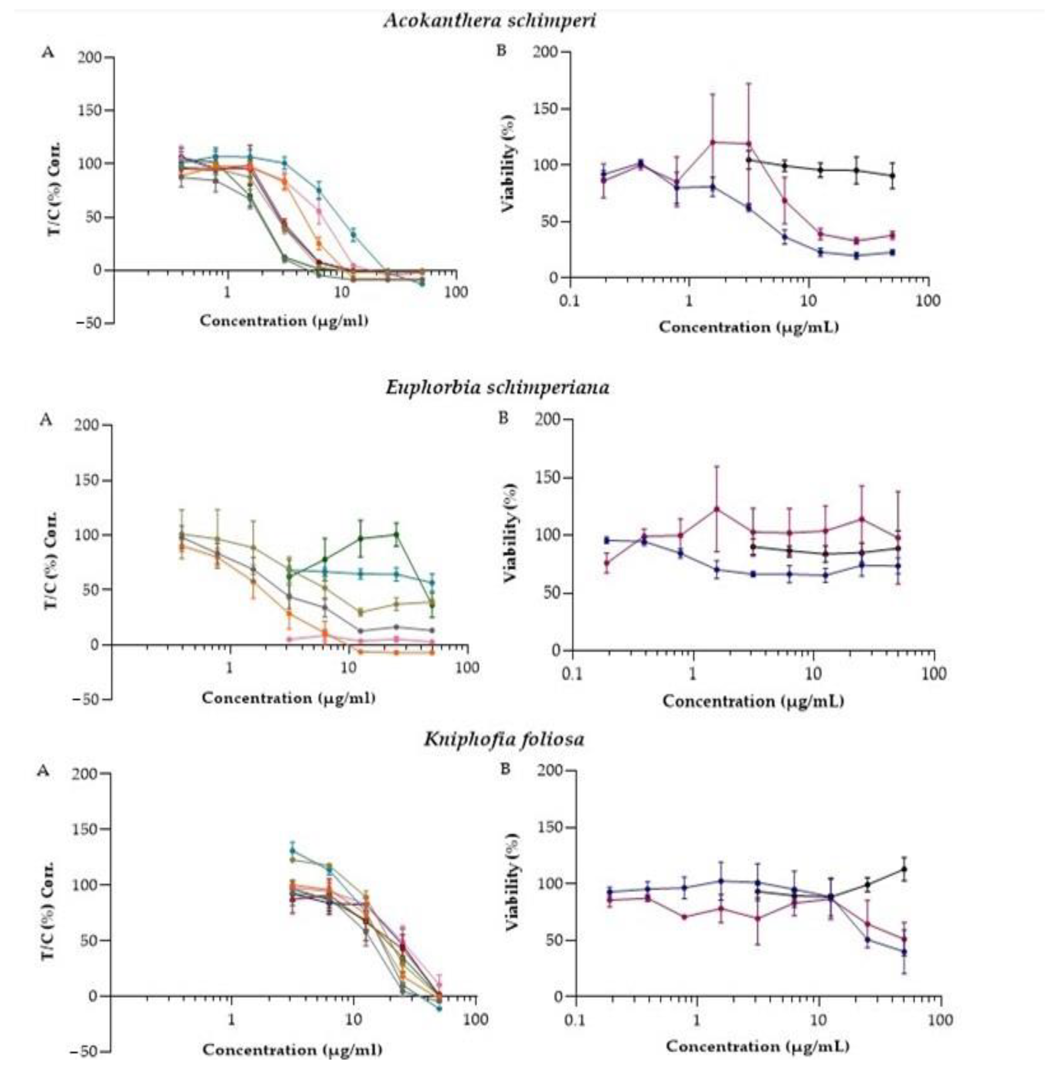
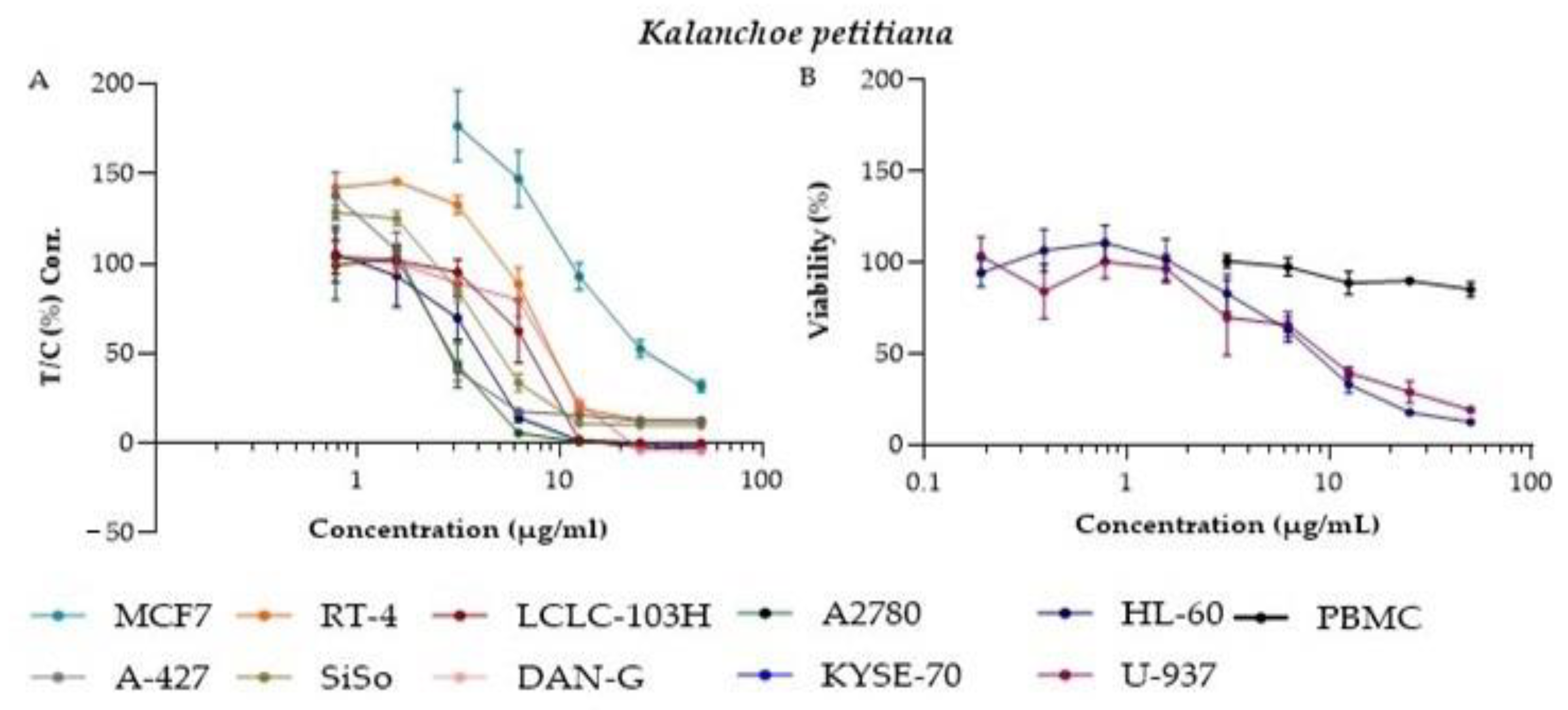
| A. schimperi | E. schimperiana | K. petitiana | K. foliosa | |
| A427 | 2.17 ± 0.41 | 1.85 ± 0.44 | 2.09 ± 0.43 | 14.54 ± 4.14 |
| MCF-7 | 10.31 ± 3.45 | Nd | 10.41 ± 5.59 | 14.89 ± 2.38 |
| RT-4 | 5.18 ± 0.69 | 2.13 ± 3.78 | 6.83 ± 0.79 | 17.3 ± 5.44 |
| SiSo | 2.86 ± 0.29 | 3.28 ± 1.2 | 3.79 ± 0.49 | 17.8 ± 2.31 |
| LCLC-103H | 3.06 ± 0.3 | 0.086 | 7.33 ± 2.7 | 24.16 ± 0.4 |
| DAN-G | 5.23 ± 1.7 | Nd | 9.6 ± 1.6 | 27.06 ± 10.8 |
| KYSE-70 | 2.87 ± 0.3 | 30.37 | 3.45 ± 1.6 | 22.03 ± 3.4 |
| A2780 | 1.87 ± 0.4 | 26.54 ± 18.5 | 2.35 ± 0.9 | 16.77 ± 4.6 |
| HL-60 | 4.08 ± 1.4 | Nd | 8.0 ± 1.7 | 24.2 ± 0.3 |
| U-937 | 9.76 ± 6.8 | 47.68 | 8.58 ± 3.5 | 16.9 |
| PBMC | ˃50 | ˃50 | ˃50 | ˃50 |
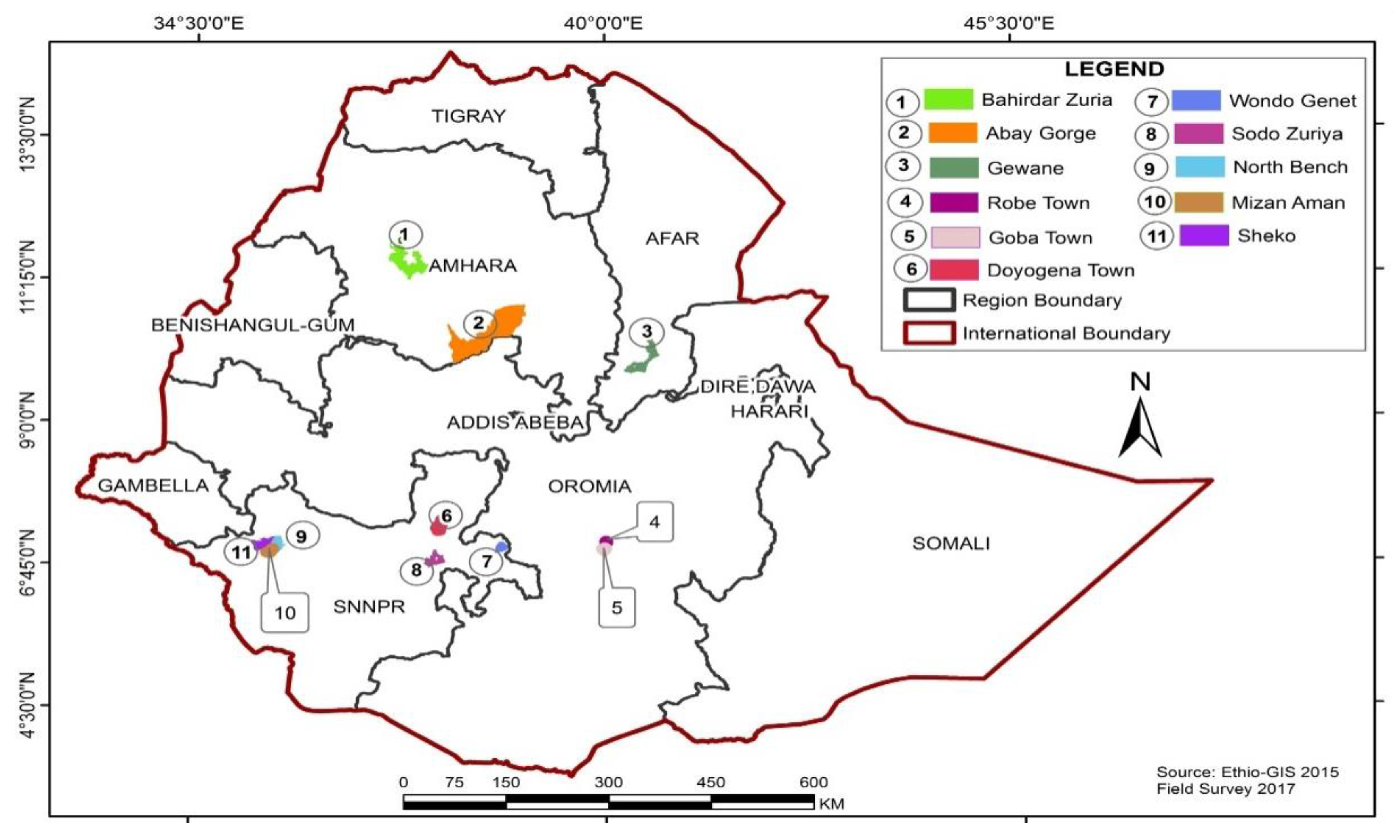
| Voucher Number | Botanical Name (Family) | Vernacular Name | Districts | Growth Form | Parts Used |
|---|---|---|---|---|---|
| Bele-060 | Aloe debrana (Xanthorrhoeaceae) | Gurtawaqota | DoyoGena | Shrub | Roots |
| Bel-002 | Hydrocotyle mannii Hook.f (Apiaceae) | Ye’timedhanit | North Bench | Herb | Leaves |
| Bel-003 | Acokanthera schimperi (A.DC.) Schweinf. (Apocynaceae) | Merenz | Bahir Dar Zuria | Shrub | Leaves |
| Bel-036 | Pentarrhinum insipidum E.Mey. (Asclepiadaceae) | Barohula | Gewane | Shrub | Roots |
| Bel-020 | Kniphofia foliosa Hochst. (Asphodelaceae) | Shushube | Bale Goba | Shrub | Roots |
| Bel-045 | Acmella caulirhiza Delile (Asteraceae) | Kustasht | MizanAman | Shrub | Leaves |
| Bel-025 | Vernonia auriculifera Hiern (Asteraceae) | Barawa | DoyoGena | Shrub | Leaves |
| Bel-021 | Cineraria abyssinica Sch.Bip. ex A.Rich. (Asteraceae) | Esemefirh | Bale Robe | Herb | Leaves |
| Bel-039 | Cleome brachycarpa (Forssk.) Vahl ex DC. (Capparidaceae) | Berbere | Gewane | Herb | Leaves |
| Bel-019 | Kalanchoe petitiana A. Rich. (Crassulaceae) | Anchura | Bale Goba | Shrub | Leaves |
| Bel-032 | Euphorbia schimperiana Scheele (Euphorbiaceae) | Gendalelata | DoyoGena | Shrub | Roots |
| Bel-035 | Croton macrostachyus Hochst. ex Delile (Euphorbiaceae) | Besana | DoyoGena | Tree | Bark |
| Bel-043 | Ajuga leucantha Lukhoba (Lamiaceae) | Tiksasht | North Bench | Herb | Leaves |
| Bel-024 | Leonotis ocymifolia (Burm.f.) Iwarsson (Lamiaceae) | Armagusa | Bale Goba | Herb | Leaves |
| Bel-042 | Salvia nilotica Juss. ex Jacq. (Lamiaceae) | Barnbanch | North Bench | Shrub | Whole plant |
| Bel-022 | Thymus schimperi Ronniger (Lamiaceae) | Tosigne | Bale Goba | Herb | Leaves |
| Bel-051 | Sida schimperiana Hochst. ex A. Rich. (Malvaceae) | Kotijebessa | Wondo Genet | Shrub | Roots and leaves |
| Bel-008 | Dorstenia barnimiana Schweinf. (Moraceae) | Work Bemeda | Bahir Dar Zuria | Herb | Roots |
| Bel-018 | Rumex nervosus Vahl (Polygonaceae) | Emboacho | Abay Gorge | Shrub | Roots |
| Bel-010 | Clematis simensis Fresen. (Ranunculaceae) | YeazoHareg | Bahir Dar Zuria | Climber | Leaves |
| Bel-016 | Clausena anisata (Willd.) Hook. f. ex Benth. (Rutaceae) | Limich | Abay Gorge | Shrub | Leaves |
| Bel-055 | Gnidia involucrata Steud. ex A.Rich. (Thymelaeaceae) | Bito | Bahir Dar Zuria | Herb | Roots |
| Adherent Cell Lines | Corresponding Organ/Tissue |
|---|---|
| MCF-7 | Breast Adenocarcinoma |
| A427 | Lung Cancer |
| RT-4 | Urinary bladder cancer |
| SiSo | Cervical Cancer |
| LCLC-103H | Large cell lung carcinoma |
| DAN-G | Pancreatic Cancer |
| A2780 | Ovarian Cancer |
| KYSE-70 | Squamous cell carcinoma of the esophagus |
| Suspension Cell lines | |
| HL-60 | Acute myeloid leukemia |
| U-937 | Histiocytic lymphoma |
Publisher’s Note: MDPI stays neutral with regard to jurisdictional claims in published maps and institutional affiliations. |
© 2021 by the authors. Licensee MDPI, Basel, Switzerland. This article is an open access article distributed under the terms and conditions of the Creative Commons Attribution (CC BY) license (https://creativecommons.org/licenses/by/4.0/).
Share and Cite
Tesfaye, S.; Braun, H.; Asres, K.; Engidawork, E.; Belete, A.; Muhammad, I.; Schulze, C.; Schultze, N.; Guenther, S.; Bednarski, P.J. Ethiopian Medicinal Plants Traditionally Used for the Treatment of Cancer; Part 3: Selective Cytotoxic Activity of 22 Plants against Human Cancer Cell Lines. Molecules 2021, 26, 3658. https://doi.org/10.3390/molecules26123658
Tesfaye S, Braun H, Asres K, Engidawork E, Belete A, Muhammad I, Schulze C, Schultze N, Guenther S, Bednarski PJ. Ethiopian Medicinal Plants Traditionally Used for the Treatment of Cancer; Part 3: Selective Cytotoxic Activity of 22 Plants against Human Cancer Cell Lines. Molecules. 2021; 26(12):3658. https://doi.org/10.3390/molecules26123658
Chicago/Turabian StyleTesfaye, Solomon, Hannah Braun, Kaleab Asres, Ephrem Engidawork, Anteneh Belete, Ilias Muhammad, Christian Schulze, Nadin Schultze, Sebastian Guenther, and Patrick J. Bednarski. 2021. "Ethiopian Medicinal Plants Traditionally Used for the Treatment of Cancer; Part 3: Selective Cytotoxic Activity of 22 Plants against Human Cancer Cell Lines" Molecules 26, no. 12: 3658. https://doi.org/10.3390/molecules26123658