
Bioactive Components of Salvia and Their Potential Antidiabetic Properties: A Review


Abstract
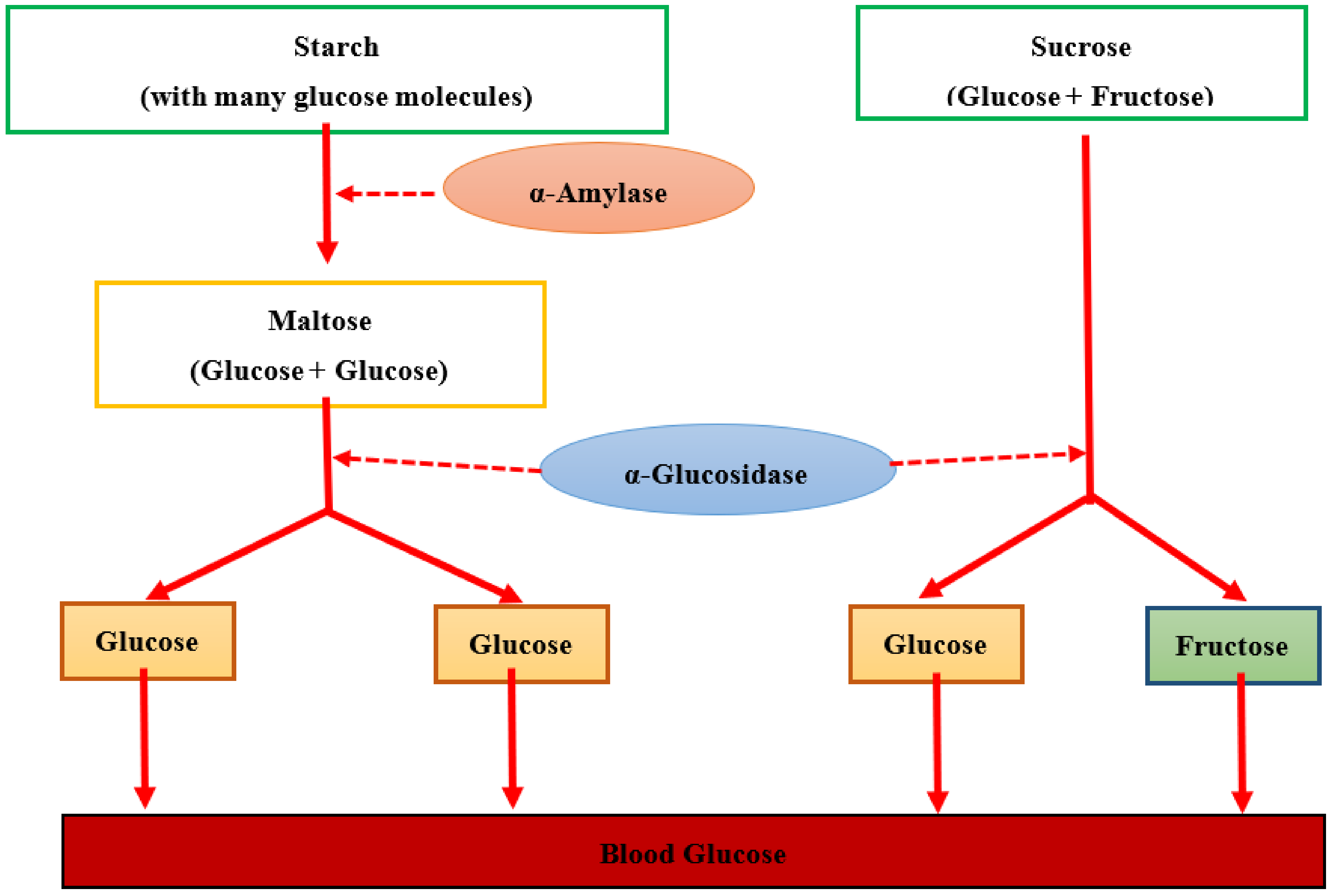
:1. Introduction
2. Materials and Methods
Search Strategy
3. Results
4. Discussion
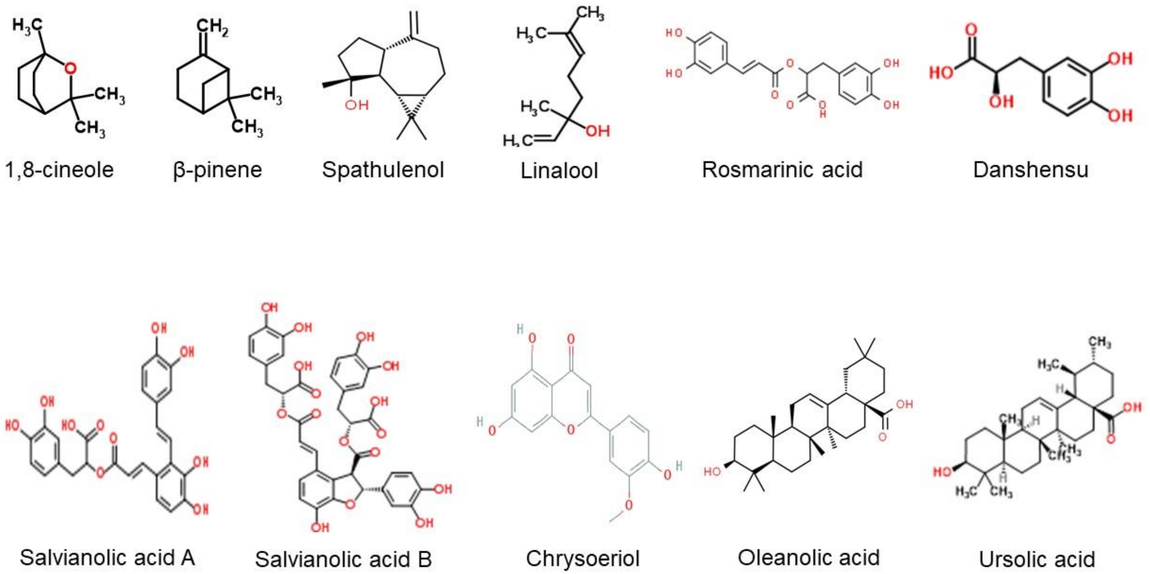
4.1. Chemical Composition of Salvia spp. and Their Antidiabetic Effect via In Vitro and In Vivo Studies
4.2. Salvia spp. in Intervention Studies
5. Conclusions
Author Contributions
Funding
Acknowledgments
Conflicts of Interest
Abbreviation
| ACAE | Acarbose equivalent |
| ACh | Acetylcholine |
| AGE | Advanced glycation end |
| Akt | Protein kinase B |
| ALT | Alanine aminotransferase |
| AMPK | AMP-activated protein kinase |
| ANG | Angiotensin |
| AST | Aspartate aminotransferase |
| ATP | Adenosine triphosphate |
| BBM | Brush-border membrane |
| BUN | Blood urea nitrogen |
| CaMKKβ | Calmodulin-dependent protein kinase kinase β |
| CDDP | Compound danshen dripping pill |
| ColIV | Collagen IV |
| COX-2 | Cyclooxygenase 2 |
| DAD | Diode array detector |
| DM | Diabetes mellitus |
| db/db | Type 2 diabetes mice |
| DN | Diabetic nephropathy |
| DR | Diabetic retinopathy |
| DSS | Danshensu |
| EO | Essential oil |
| ER | Endoplasmic reticulum |
| FBG | Fasting blood glucose |
| FBS | Fasting blood sugar |
| FFA | Fluorescence fundus angiography |
| FID | Flame ionization detector |
| FINS | Fasting blood insulin |
| FN | Fibronectin |
| GC | Gas chromatography |
| GLP-1 | Glucagon-like peptide-1 |
| GLUT | Glucose transporter |
| GSH | Glutathione |
| GSK | Glycogen synthase kinase |
| HbA1c | Glycosylated hemoglobin |
| HDD | Huangqi-danshen decoction |
| HDL | High-density lipoprotein |
| HEK | Human embryonic kidney |
| HFD | High-fat diet |
| HPLC | High-performance liquid chromatography |
| hs-CRP | High-sensitivity C-reactive protein |
| ILK1 | Integrin-linked kinase 1 |
| IL-6 | Interleukin 6 |
| IP | Intraperitoneal |
| IRS-1 | Insulin receptor substrate-1 |
| ISI | Insulin sensitivity index |
| JTD | Jiangtang decoction |
| LAB | Lithospermic acid B |
| LDL | Low-density lipoprotein |
| LKB1 | Liver kinase 1 |
| MCP1 | Monocyte chemotactic protein-1 |
| MDA | Malondialdehyde |
| MNCV | Motor nerve conduction velocity |
| MnSOD | Manganese superoxide dismutase |
| mRNA | Messenger ribonucleic acid |
| MS | Mass spectrometry |
| NMR | Nuclear magnetic resonance |
| NOS | Nitric oxide synthase |
| NPDR | Nonproliferative diabetic retinopathy |
| NF-κB | Nuclear factor kappa-light-chain-enhancer of B cells |
| OGTT | Oral glucose tolerance test |
| OLETF | Otsuka Long–Evans Tokushima Fatty |
| p-ACC | Phosphorylated acetyl CoA carboxylase |
| Pae | Paeonol |
| PAF | Polyphenolic acids fraction |
| PGC1α | Proliferator-activated receptor-gamma coactivator-1α |
| PI3K | phosphoinositide 3-kinas |
| PINK1 | PTEN-induced putative kinase 1 |
| PKG-1 | Protein kinase G |
| PPARα | Proliferator-activated receptor alpha |
| PPHG | Postprandial hyperglycemia |
| PGE₂ | Prostaglandin E2 |
| PWMT | Paw withdrawal mechanical threshold |
| RA | Rosmarinic acid |
| RAGE | Receptor of glycation end |
| RPTEC | Renal proximal tubular epithelial cells |
| Sal | Salvianolic acid |
| SFT | S. fruticosa tea |
| SGLT1 | Intestinal Na+/glucose cotransporter-1 |
| Sirt3/Sirtuin | Silent information regulator protein3 |
| SS-Bt | S. sclarea Beirut |
| SS-Tl | S. sclarea Taanayel |
| STAT3 | Signal transducer and activator of transcription 3 |
| STZ | Streptozotocin |
| TSIIA | Tanshinone IIA |
| TC | Total cholesterol |
| TENS | Transcutaneous electrical nerve stimulation |
| TG | Triglyceride |
| TGF- β1 | Transforming growth factor-β1 |
| TNFα | Tumor necrosis factor-alpha |
| TSP1 | Thrombospondin 1 |
| T2DM | Type 2 diabetes mellitus |
| UAE | Ultrasound-assisted extraction |
| VED | Vascular endothelial dysfunction |
| VEGF | Vascular endothelial growth factor |
| 2hPPG | 2 h postprandial glucose |
| 8-OHdG | 8-Hydroxy-2′-deoxyguanosine |
References
- American Diabetes Association. Diagnosis and Classification of Diabetes Mellitus. Diabetes Care 2009, 32, S62–S67. [Google Scholar] [CrossRef] [Green Version]
- Afrisham, R.; Aberomand, M.; Ghaffari, M.A.; Siahpoosh, A.; Jamalan, M. Inhibitory Effect of Heracleum persicum and Ziziphus jujuba on Activity of Alpha-Amylase. J. Bot. 2015, 2015, 824683. [Google Scholar] [CrossRef] [Green Version]
- Kooti, W.; Moradi, M.; Aliakbari, S.; Sharafi-Ahvazi, N.; Asadi-Samani, M.; Ashtary-Larky, D. Therapeutic and pharmacological potential of Foeniculum vulgare Mill: A review. J. Herb. Med. Pharmacol. 2015, 4, 1–9. [Google Scholar]
- Chen, S.T.; Dou, J.; Temple, R.; Agarwal, R.; Wu, K.M.; Walker, S. New therapies from old medicines. Nat. Biotechnol. 2008, 26, 1077–1083. [Google Scholar] [CrossRef]
- Pelkonen, O.; Xu, Q.; Fan, T.-P. Why is Research on Herbal Medicinal Products Important and How Can We Improve Its Quality? J. Tradit. Complement. Med. 2014, 4, 1–7. [Google Scholar] [CrossRef] [Green Version]
- Longaray Delamare, A.P.; Moschen-Pistorello, I.T.; Artico, L.; Atti-Serafini, L.; Echeverrigaray, S. Antibacterial activity of the essential oils of Salvia officinalis L. and Salvia triloba L. cultivated in South Brazil. Food Chem. 2007, 100, 603–608. [Google Scholar] [CrossRef]
- Lu, Y.; Foo, L.Y. Polyphenolics of Salvia-a review. Phytochemistry 2002, 59, 117–140. [Google Scholar] [CrossRef]
- Zengin, G.; Llorent-Martínez, E.; Fernández de Cordova, M.L.; Bahadori, M.B.; Mocan, A.; Locatelli, M.; Aktumsek, A. Chemical composition and biological activities of extracts from three Salvia species: S. blepharochlaena, S. euphratica var. leiocalycina, and S. verticillata subsp. amasiaca. Ind. Crops Prod. 2018, 111, 11–21. [Google Scholar] [CrossRef]
- Asghari, B.; Salehi, P.; Sonboli, A.; Nejad Ebrahimi, S. Flavonoids from Salvia chloroleuca with α-Amylsae and α-Glucosidase Inhibitory Effect. Iran. J. Pharm. Res. 2015, 14, 609–615. [Google Scholar]
- Moradi-Afrapoli, F.; Asghari, B.; Saeidnia, S.; Ajani, Y.; Mirjani, M.; Malmir, M.; Dolatabadi Bazaz, R.; Hadjiakhoondi, A.; Salehi, P.; Hamburger, M.; et al. In vitro α-glucosidase inhibitory activity of phenolic constituents from aerial parts of Polygonum hyrcanicum. DARU J. Pharm. Sci. 2012, 20, 37. [Google Scholar] [CrossRef] [Green Version]
- Bahadori, M.B.; Salehi, P.; Sonboli, A. Comparative study of the essential oil composition of Salvia urmiensis and its enzyme inhibitory activities linked to diabetes mellitus and Alzheimer’s disease. Int. J. Food 2017, 20, 2974–2981. [Google Scholar] [CrossRef] [Green Version]
- Li, M.; Li, Q.; Zhang, C.; Zhang, N.; Cui, Z.; Huang, L.; Xiao, P. An ethnopharmacological investigation of medicinal Salvia plants (Lamiaceae) in China. Acta Pharm. Sin. B 2013, 3, 273–280. [Google Scholar] [CrossRef] [Green Version]
- Christensen, K.B.; Jørgensen, M.; Kotowska, D.; Petersen, R.K.; Kristiansen, K.; Christensen, L.P. Activation of the nuclear receptor PPARγ by metabolites isolated from sage (Salvia officinalis L.). J. Ethnopharmacol. 2010, 132, 127–133. [Google Scholar] [CrossRef]
- Chaudhury, A.; Duvoor, C.; Reddy Dendi, V.S.; Kraleti, S.; Chada, A.; Ravilla, R.; Marco, A.; Shekhawat, N.S.; Montales, M.T.; Kuriakose, K.; et al. Clinical Review of Antidiabetic Drugs: Implications for Type 2 Diabetes Mellitus Management. Front. Endocrinol. 2017, 8, 6. [Google Scholar] [CrossRef] [Green Version]
- United Kingdom Prospective Diabetes Study Group. United Kingdom Prospective Diabetes Study 24: A 6-year, randomized, controlled trial comparing sulfonylurea, insulin, and metformin therapy in patients with newly diagnosed type 2 diabetes that could not be controlled with diet therapy. United Kingdom Prospective Diabetes Study Group. Ann. Intern. Med. 1998, 128, 165–175. [Google Scholar] [CrossRef]
- Tundis, R.; Loizzo, M.R.; Menichini, F. Natural products as alpha-amylase and alpha-glucosidase inhibitors and their hypoglycaemic potential in the treatment of diabetes: An update. Mini Rev. Med. Chem. 2010, 10, 315–331. [Google Scholar] [CrossRef]
- Dineshkumar, B.; Mitra, A.; Manjunatha, M. A comparative study of alpha amylase inhibitory activities of common anti-diabetic plants at Kharagpur 1 block. Int. J. Green Pharm. 2010, 4, 115–121. [Google Scholar]
- Aung-Htut, M.T.; Ham, K.A.; Tchan, M.C.; Fletcher, S.; Wilton, S.D. Novel Mutations Found in Individuals with Adult-Onset Pompe Disease. Genes 2020, 11, 135. [Google Scholar] [CrossRef] [Green Version]
- Lebovitz, H.E. ALPHA-GLUCOSIDASE INHIBITORS. Endocrinol. Metab. Clin. N. Am. 1997, 26, 539–551. [Google Scholar] [CrossRef]
- Notkins, A.L. Immunologic and genetic factors in type 1 diabetes. J. Biol. Chem. 2002, 277, 43545–43548. [Google Scholar] [CrossRef] [Green Version]
- Liu, L.; Deseo, M.A.; Morris, C.; Winter, K.M.; Leach, D.N. Investigation of α-glucosidase inhibitory activity of wheat bran and germ. Food Chem. 2011, 126, 553–561. [Google Scholar] [CrossRef]
- Rahimzadeh, M.; Jahanshahi, S.; Moein, S.; Moein, M.R. Evaluation of alpha- amylase inhibition by Urtica dioica and Juglans regia extracts. Iran. J. Basic Med. Sci. 2014, 17, 465–469. [Google Scholar] [PubMed]
- Yang, C.Y.; Yen, Y.Y.; Hung, K.C.; Hsu, S.W.; Lan, S.J.; Lin, H.C. Inhibitory effects of pu-erh tea on alpha glucosidase and alpha amylase: A systemic review. Nutr. Diabetes 2019, 9, 23. [Google Scholar] [CrossRef] [PubMed] [Green Version]
- Ikebukuro, K.; Adachi, Y.; Yamada, Y.; Fujimoto, S.; Seino, Y.; Oyaizu, H.; Hioki, K.; Ikehara, S. Treatment of streptozotocin-induced diabetes mellitus by transplantation of islet cells plus bone marrow cells via portal vein in rats. Transplantation 2002, 73, 512–518. [Google Scholar] [CrossRef] [Green Version]
- Rastellini, C.; Shapiro, R.; Corry, R.; Fung, J.J.; Starzl, T.E.; Rao, A.S. An attempt to reverse diabetes by delayed islet cell transplantation in humans. Transpl. Proc. 1997, 29, 2238–2239. [Google Scholar] [CrossRef] [Green Version]
- Elias, D.; Prigozin, H.; Polak, N.; Rapoport, M.; Lohse, A.W.; Cohen, I.R. Autoimmune diabetes induced by the beta-cell toxin STZ. Immunity to the 60-kDa heat shock protein and to insulin. Diabetes 1994, 43, 992–998. [Google Scholar] [CrossRef]
- Weiss, R.B. Streptozocin: A review of its pharmacology, efficacy, and toxicity. Cancer Treat. Rep. 1982, 66, 427–438. [Google Scholar]
- Lenzen, S. The mechanisms of alloxan- and streptozotocin-induced diabetes. Diabetologia 2008, 51, 216–226. [Google Scholar] [CrossRef] [Green Version]
- Katsumata, K.; Katsumata, Y.; Ozawa, T.; Katsumata, K., Jr. Potentiating effects of combined usage of three sulfonylurea drugs on the occurrence of alloxan diabetes in rats. Horm. Metab. Res. 1993, 25, 125–126. [Google Scholar] [CrossRef]
- Szkudelski, T. The mechanism of alloxan and streptozotocin action in B cells of the rat pancreas. Physiol. Res. 2001, 50, 537–546. [Google Scholar]
- Nickavar, B.; Abolhasani, L. Bioactivity-Guided Separation of an α-Amylase Inhibitor Flavonoid from Salvia virgata. Iran. J. Pharm. Res. 2013, 12, 57–61. [Google Scholar]
- Zengin, G.; Mahomoodally, F.; Picot-Allain, C.; Diuzheva, A.; Jekő, J.; Cziáky, Z.; Cvetanović, A.; Aktumsek, A.; Zeković, Z.; Rengasamy, K.R.R. Metabolomic profile of Salvia viridis L. root extracts using HPLC–MS/MS technique and their pharmacological properties: A comparative study. Ind. Crops Prod. 2019, 131, 266–280. [Google Scholar] [CrossRef]
- Bahadori, M.B.; Dinparast, L.; Zengin, G.; Sarikurkcu, C.; Bahadori, S.; Asghari, B.; Movahhedin, N. Functional components, antidiabetic, anti-Alzheimer’s disease, and antioxidant activities of Salvia syriaca L. Int. J. Food Prop. 2017, 20, 1761–1772. [Google Scholar] [CrossRef] [Green Version]
- Bahadori, M.B.; Asghari, B.; Dinparast, L.; Zengin, G.; Sarikurkcu, C.; Abbas-Mohammadi, M.; Bahadori, S. Salvia nemorosa L.: A novel source of bioactive agents with functional connections. LWT Food Sci. Technol. 2017, 75, 42–50. [Google Scholar] [CrossRef]
- Kalaycıoğlu, Z.; Uzaşçı, S.; Dirmenci, T.; Erim, F.B. α-Glucosidase enzyme inhibitory effects and ursolic and oleanolic acid contents of fourteen Anatolian Salvia species. J. Pharm. Biomed. Anal. 2018, 155, 284–287. [Google Scholar] [CrossRef]
- Yang, X.-Y.; Qiang, G.-F.; Zhang, L.; Zhu, X.-M.; Wang, S.-B.; Sun, L.; Yang, H.-G.; Du, G.-H. Salvianolic acid A protects against vascular endothelial dysfunction in high-fat diet fed and streptozotocin-induced diabetic rats. J. Asian Nat. Prod. Res. 2011, 13, 884–894. [Google Scholar] [CrossRef]
- Yu, X.; Zhang, L.; Yang, X.; Huang, H.; Huang, Z.; Shi, L.; Zhang, H.; Du, G. Salvianolic Acid A Protects the Peripheral Nerve Function in Diabetic Rats through Regulation of the AMPK-PGC1α-Sirt3 Axis. Molecules 2012, 17, 11216–11228. [Google Scholar] [CrossRef] [Green Version]
- Qiang, G.; Yang, X.; Shi, L.; Zhang, H.; Chen, B.; Zhao, Y.; Zu, M.; Zhou, D.; Guo, J.; Yang, H.; et al. Antidiabetic Effect of Salvianolic Acid A on Diabetic Animal Models via AMPK Activation and Mitochondrial Regulation. Cell. Physiol. Biochem. 2015, 36, 395–408. [Google Scholar] [CrossRef]
- Raoufi, S.; Baluchnejadmojarad, T.; Roghani, M.; Ghazanfari, T.; Khojasteh, F.; Mansouri, M. Antidiabetic potential of salvianolic acid B in multiple low-dose streptozotocin-induced diabetes. Pharm. Biol. 2015, 53, 1803–1809. [Google Scholar] [CrossRef]
- Huang, M.Q.; Zhou, C.J.; Zhang, Y.P.; Zhang, X.Q.; Xu, W.; Lin, J.; Wang, P.J. Salvianolic Acid B Ameliorates Hyperglycemia and Dyslipidemia in db/db Mice through the AMPK Pathway. Cell. Physiol. Biochem. 2016, 40, 933–943. [Google Scholar] [CrossRef]
- Huang, M.; Wang, P.; Xu, S.; Xu, W.; Xu, W.; Chu, K.; Lu, J. Biological activities of salvianolic acid B from Salvia miltiorrhiza on type 2 diabetes induced by high-fat diet and streptozotocin. Pharm. Biol. 2015, 53, 1058–1065. [Google Scholar] [CrossRef] [PubMed] [Green Version]
- Azevedo, M.F.; Lima, C.F.; Fernandes-Ferreira, M.; Almeida, M.J.; Wilson, J.M.; Pereira-Wilson, C. Rosmarinic acid, major phenolic constituent of Greek sage herbal tea, modulates rat intestinal SGLT1 levels with effects on blood glucose. Mol. Nutr. Food Res. 2011, 55 (Suppl. 1), S15–S25. [Google Scholar] [CrossRef] [Green Version]
- Kim, D.Y.; Kang, M.K.; Park, S.H.; Lee, E.J.; Kim, Y.H.; Oh, H.; Choi, Y.J.; Kang, Y.H. Eucalyptol ameliorates Snail1/β-catenin-dependent diabetic disjunction of renal tubular epithelial cells and tubulointerstitial fibrosis. Oncotarget 2017, 8, 106190–106205. [Google Scholar] [CrossRef] [PubMed] [Green Version]
- Raafat, K.; Habib, J. Phytochemical Compositions and Antidiabetic Potentials of Salvia sclarea L. Essential Oils. J. Oleo Sci. 2018, 67, 1015–1025. [Google Scholar] [CrossRef] [PubMed]
- Jin, C.J.; Yu, S.H.; Wang, X.M.; Woo, S.J.; Park, H.J.; Lee, H.C.; Choi, S.H.; Kim, K.M.; Kim, J.H.; Park, K.S.; et al. The effect of lithospermic acid, an antioxidant, on development of diabetic retinopathy in spontaneously obese diabetic rats. PLoS ONE 2014, 9, e98232. [Google Scholar] [CrossRef] [PubMed]
- Lo, S.H.; Hsu, C.T.; Niu, H.S.; Niu, C.S.; Cheng, J.T.; Chen, Z.C. Cryptotanshinone Inhibits STAT3 Signaling to Alleviate Cardiac Fibrosis in Type 1-like Diabetic Rats. Phytother. Res. 2017, 31, 638–646. [Google Scholar] [CrossRef] [PubMed]
- Zhang, J.; Cai, Z.; Yang, M.; Tong, L.; Zhang, Y. Inhibition of tanshinone IIA on renin activity protected against osteoporosis in diabetic mice. Pharm. Biol. 2020, 58, 219–224. [Google Scholar] [CrossRef] [PubMed]
- Tao, S.; Chen, L.; Song, J.; Zhu, N.; Song, X.; Shi, R.; Ge, G.; Zhang, Y. Tanshinone IIA ameliorates diabetic cardiomyopathy by inhibiting Grp78 and CHOP expression in STZ-induced diabetes rats. Exp. Ther. Med. 2019, 18, 729–734. [Google Scholar] [CrossRef]
- Huang, M.; Xie, Y.; Chen, L.; Chu, K.; Wu, S.; Lu, J.; Chen, X.; Wang, Y.; Lai, X. Antidiabetic effect of the total polyphenolic acids fraction from Salvia miltiorrhiza Bunge in diabetic rats. Phytother. Res. 2012, 26, 944–948. [Google Scholar] [CrossRef]
- Hu, J.; Li, Y.L.; Li, Z.L.; Li, H.; Zhou, X.X.; Qiu, P.C.; Yang, Q.; Wang, S.W. Chronic supplementation of paeonol combined with danshensu for the improvement of vascular reactivity in the cerebral basilar artery of diabetic rats. Int. J. Mol. Sci. 2012, 13, 14565–14578. [Google Scholar] [CrossRef]
- Wang, T.; Fu, F.; Han, B.; Zhang, L.; Zhang, X. Danshensu ameliorates the cognitive decline in streptozotocin-induced diabetic mice by attenuating advanced glycation end product-mediated neuroinflammation. J. Neuroimmunol. 2012, 245, 79–86. [Google Scholar] [CrossRef]
- Hong, J.N.; Li, W.W.; Wang, L.L.; Guo, H.; Jiang, Y.; Gao, Y.J.; Tu, P.F.; Wang, X.M. Jiangtang decoction ameliorate diabetic nephropathy through the regulation of PI3K/Akt-mediated NF-κB pathways in KK-Ay mice. Chin. Med. 2017, 12, 13. [Google Scholar] [CrossRef] [Green Version]
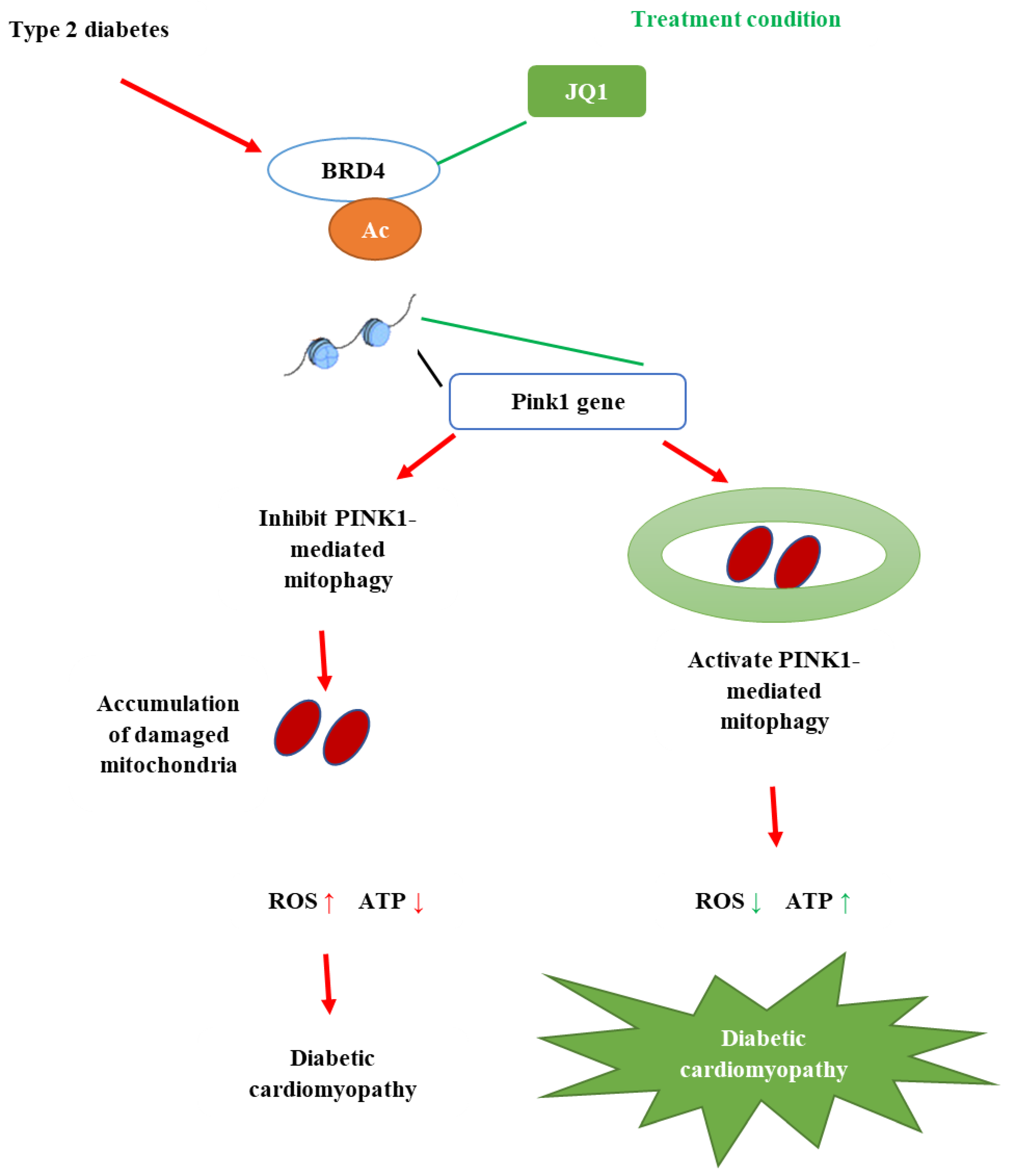
- Liu, X.; Lu, J.; Liu, S.; Huang, D.; Chen, M.; Xiong, G.; Li, S. Huangqi-Danshen decoction alleviates diabetic nephropathy in db/db mice by inhibiting PINK1/Parkin-mediated mitophagy. Am. J. Transl. Res. 2020, 12, 989–998. [Google Scholar]
- Lian, F.; Wu, L.; Tian, J.; Jin, M.; Zhou, S.; Zhao, M.; Wei, L.; Zheng, Y.; Wang, Y.; Zhang, M.; et al. The effectiveness and safety of a danshen-containing Chinese herbal medicine for diabetic retinopathy: A randomized, double-blind, placebo-controlled multicenter clinical trial. J. Ethnopharmacol. 2015, 164, 71–77. [Google Scholar] [CrossRef]
- Tassadaq, N.; Wahid, Y.S. Effectiveness of Tricardin (Danshenform 250 mg dripping pills capsules) along with physical rehabilitation in diabetic polyneuropathies. Rawal Med. J. 2019, 44, 49–52. [Google Scholar]
- Nie, J.M.; Li, H.F. Therapeutic effects of Salvia miltiorrhiza injection combined with telmisartan in patients with diabetic nephropathy by influencing collagen IV and fibronectin: A case-control study. Exp. Ther. Med. 2018, 16, 3405–3412. [Google Scholar] [CrossRef]
- Kianbakht, S.; Nabati, F.; Abasi, B. Salvia officinalis (Sage) Leaf Extract as Add-on to Statin Therapy in Hypercholesterolemic Type 2 Diabetic Patients: A Randomized Clinical Trial. Int. J. Mol. Cell Med. 2016, 5, 141–148. [Google Scholar]
- CSID. 2656, (±)-Eucalyptol/1,8-Cineole. Available online: http://www.chemspider.com/Chemical-Structure.2656.html (accessed on 23 February 2021).
- CSID. 26347272, (-)-Spathulenol. Available online: http://www.chemspider.com/Chemical-Structure.26347272.html (accessed on 23 February 2021).
- CSID. 14198, (±)-β-Pinene. Available online: http://www.chemspider.com/Chemical-Structure.14198.html (accessed on 23 February 2021).
- CSID. 13849981, Linalool. Available online: http://www.chemspider.com/Chemical-Structure.13849981.html (accessed on 23 February 2021).
- CSID. 4474888, (±)-Rosmarinic acid. Available online: http://www.chemspider.com/Chemical-Structure.4474888.html (accessed on 23 February 2021).
- CSID. 58472, Ursolic Acid. Available online: http://www.chemspider.com/Chemical-Structure.58472.html (accessed on 23 February 2021).
- CSID. 10062, Oleanolic Acid. Available online: http://www.chemspider.com/Chemical-Structure.10062.html (accessed on 23 February 2021).
- CSID. 9775399, Danshensu. Available online: http://www.chemspider.com/Chemical-Structure.9775399.html (accessed on 23 February 2021).
- CSID. 9803831, Salvianolic Acid B. Available online: http://www.chemspider.com/Chemical-Structure.9803831.html (accessed on 23 February 2021).
- CSID. 4816368, Salvianolic Acid A. Available online: http://www.chemspider.com/Chemical-Structure.4816368.html (accessed on 23 February 2021).
- CSID. 4444263, Chryseriol. Available online: http://www.chemspider.com/Chemical-Structure.4444263.html (accessed on 12 April 2021).
- Proença, C.; Freitas, M.; Ribeiro, D.; Tomé, S.M.; Oliveira, E.F.T.; Viegas, M.F.; Araújo, A.N.; Ramos, M.J.; Silva, A.M.S.; Fernandes, P.A.; et al. Evaluation of a flavonoids library for inhibition of pancreatic α-amylase towards a structure-activity relationship. J. Enzym. Inhib. Med. Chem. 2019, 34, 577–588. [Google Scholar] [CrossRef] [Green Version]
- Herrero, M.; Plaza, M.; Cifuentes, A.; Ibáñez, E. 4.08-Extraction Techniques for the Determination of Phenolic Compounds in Food. In Comprehensive Sampling and Sample Preparation; Janusz, P., Ed.; Academic Press: Oxford, UK, 2012; Volume 4, pp. 159–180. [Google Scholar]
- Ngo, Y.L.; Lau, C.H.; Chua, L.S. Review on rosmarinic acid extraction, fractionation and its anti-diabetic potential. Food Chem. Toxicol. 2018, 121, 687–700. [Google Scholar] [CrossRef]
- Topçu, G. Bioactive Triterpenoids from Salvia Species. J. Nat. Prod. 2006, 69, 482–487. [Google Scholar] [CrossRef]
- Ding, H.; Hu, X.; Xu, X.; Zhang, G.; Gong, D. Inhibitory mechanism of two allosteric inhibitors, oleanolic acid and ursolic acid on α-glucosidase. Int. J. Biol. Macromol. 2018, 107, 1844–1855. [Google Scholar] [CrossRef] [PubMed]
- Janicsak, G.; Veres, K.; Kakasy, A.Z.; Mathe, I. Study of the oleanolic and ursolic acid contents of some species of the Lamiaceae. Biochem. Syst. Ecol. 2006, 34. [Google Scholar] [CrossRef]
- Ma, L.; Tang, L.; Yi, Q. Salvianolic Acids: Potential Source of Natural Drugs for the Treatment of Fibrosis Disease and Cancer. Front. Pharmacol. 2019, 10, 97. [Google Scholar] [CrossRef] [Green Version]
- Chen, X.; Guo, J.; Bao, J.; Lu, J.; Wang, Y. The anticancer properties of Salvia miltiorrhiza Bunge (Danshen): A systematic review. Med. Res. Rev. 2014, 34, 768–794. [Google Scholar] [CrossRef] [PubMed]
- Du, G.; Sun, L.; Zhao, R.; Du, L.; Song, J.; Zhang, L.; He, G.; Zhang, Y.; Zhang, J. Polyphenols: Potential source of drugs for the treatment of ischaemic heart disease. Pharmacol. Ther. 2016, 162, 23–34. [Google Scholar] [CrossRef] [PubMed] [Green Version]
- Hui, X.; Yan-Li, L.; Hong-Cui, T.; Wen-Ting, Z. Preparation and Chemical Structure Identification of Methylated Metabolites of Salvianolic Acid A. Chin. J. Anal. Chem. 2014, 42, 65–70. [Google Scholar] [CrossRef]
- Du, G.; Zhang, J. Protective effects of salvianolic acid A against impairment of memory induced by cerebral ischemia-reperfusion in mice. Chin. Med. J. 1997, 110, 65–68. [Google Scholar]
- Wang, S.B.; Yang, X.Y.; Tian, S.; Yang, H.G.; Du, G.H. Effect of salvianolic acid A on vascular reactivity of streptozotocin-induced diabetic rats. Life Sci. 2009, 85, 499–504. [Google Scholar] [CrossRef]
- Yang, X.-Y.; Sun, L.; Xu, P.; Gong, L.-L.; Qiang, G.-F.; Zhang, L.; Du, G.-H. Effects of salvianolic acid A on plantar microcirculation and peripheral nerve function in diabetic rats. Eur. J. Pharmacol. 2011, 665, 40–46. [Google Scholar] [CrossRef]
- Cheng, B.; Gong, H.; Li, X.; Sun, Y.; Chen, H.; Zhang, X.; Wu, Q.; Zheng, L.; Huang, K. Salvianolic acid B inhibits the amyloid formation of human islet amyloid polypeptide and protects pancreatic beta-cells against cytotoxicity. Proteins Struct. Funct. Bioinf. 2013, 81, 613–621. [Google Scholar] [CrossRef]
- Gao, H.Y.; Li, G.Y.; Lou, M.M.; Li, X.Y.; Wei, X.Y.; Wang, J.H. Hepatoprotective effect of Matrine salvianolic acid B salt on Carbon Tetrachloride-Induced Hepatic Fibrosis. J. Inflamm. 2012, 9, 16. [Google Scholar] [CrossRef] [Green Version]
- Sun, L.Q.; Zhao, J.; Zhang, T.T.; Qu, L.; Wang, X.; Xue, B.; Li, X.J.; Mu, Y.M.; Lu, J.M. Protective effects of Salvianolic acid B on Schwann cells apoptosis induced by high glucose. Neurochem. Res. 2012, 37, 996–1010. [Google Scholar] [CrossRef]
- Wang, P.; Xu, S.; Li, W.; Wang, F.; Yang, Z.; Jiang, L.; Wang, Q.; Huang, M.; Zhou, P. Salvianolic acid B inhibited PPARγ expression and attenuated weight gain in mice with high-fat diet-induced obesity. Cell. Physiol. Biochem. 2014, 34, 288–298. [Google Scholar] [CrossRef]
- Guan, Y.; Cui, Z.-J.; Sun, B.; Han, L.-P.; Li, C.-J.; Chen, L.-M. Celastrol attenuates oxidative stress in the skeletal muscle of diabetic rats by regulating the AMPK-PGC1α-SIRT3 signaling pathway. Int. J. Mol. Med. 2016, 37, 1229–1238. [Google Scholar] [CrossRef] [Green Version]
- Kukidome, D.; Nishikawa, T.; Sonoda, K.; Imoto, K.; Fujisawa, K.; Yano, M.; Motoshima, H.; Taguchi, T.; Matsumura, T.; Araki, E. Activation of AMP-activated protein kinase reduces hyperglycemia-induced mitochondrial reactive oxygen species production and promotes mitochondrial biogenesis in human umbilical vein endothelial cells. Diabetes 2006, 55, 120–127. [Google Scholar] [CrossRef] [PubMed]
- Yu, S.M.; Bonventre, J.V. Acute Kidney Injury and Progression of Diabetic Kidney Disease. Adv. Chronic Kidney Dis. 2018, 25, 166–180. [Google Scholar] [CrossRef]
- Schwer, B.; North, B.J.; Frye, R.A.; Ott, M.; Verdin, E. The human silent information regulator (Sir)2 homologue hSIRT3 is a mitochondrial nicotinamide adenine dinucleotide-dependent deacetylase. J. Cell Biol. 2002, 158, 647–657. [Google Scholar] [CrossRef] [PubMed] [Green Version]
- Zhao, R.; Liu, X.; Zhang, L.; Yang, H.; Zhang, Q. Current Progress of Research on Neurodegenerative Diseases of Salvianolic Acid B. Oxid. Med. Cell Longev. 2019, 2019, 3281260. [Google Scholar] [CrossRef] [PubMed]
- Hu, P.; Liang, Q.L.; Luo, G.A.; Zhao, Z.Z.; Jiang, Z.H. Multi-component HPLC fingerprinting of Radix Salviae Miltiorrhizae and its LC-MS-MS identification. Chem. Pharm. Bull. 2005, 53, 677–683. [Google Scholar] [CrossRef] [PubMed] [Green Version]
- Sun, Y.; Zhu, H.; Wang, J.; Liu, Z.; Bi, J. Isolation and purification of salvianolic acid A and salvianolic acid B from Salvia miltiorrhiza by high-speed counter-current chromatography and comparison of their antioxidant activity. J. Chromatogr. B Analyt. Technol. Biomed. Life Sci. 2009, 877, 733–737. [Google Scholar] [CrossRef] [PubMed]
- Petersen, M.; Simmonds, M.S. Rosmarinic acid. Phytochemistry 2003, 62, 121–125. [Google Scholar] [CrossRef]
- Nadeem, M.; Imran, M.; Aslam Gondal, T.; Imran, A.; Shahbaz, M.; Muhammad Amir, R.; Wasim Sajid, M.; Batool Qaisrani, T.; Atif, M.; Hussain, G.; et al. Therapeutic Potential of Rosmarinic Acid: A Comprehensive Review. Appl. Sci. 2019, 9, 3139. [Google Scholar] [CrossRef] [Green Version]
- Sadlon, A.E.; Lamson, D.W. Immune-modifying and antimicrobial effects of Eucalyptus oil and simple inhalation devices. Altern. Med. Rev. 2010, 15, 33–47. [Google Scholar]
- Yalçın, H.; Anık, M.; Şanda, M.A.; Çakır, A. Gas Chromatography/Mass Spectrometry Analysis of Laurus nobilis Essential Oil Composition of Northern Cyprus. J. Med. Food 2007, 10, 715–719. [Google Scholar] [CrossRef]
- Papadopoulou-Marketou, N.; Vaidya, A.; Dluhy, R.; Chrousos, G.P. Hyperaldosteronism. In Endotext; Feingold, K.R., Anawalt, B., Boyce, A., Chrousos, G., de Herder, W.W., Dungan, K., Grossman, A., Hershman, J.M., Hofland, J., Kaltsas, G., et al., Eds.; MDText.com, Inc.: South Dartmouth, MA, USA, 2000. [Google Scholar]
- Dominguez, A.; Muppidi, V.; Gupta, S. Hyperaldosteronism; StatPearls Publishing: Treasure Island, FL, USA, 2021. [Google Scholar]
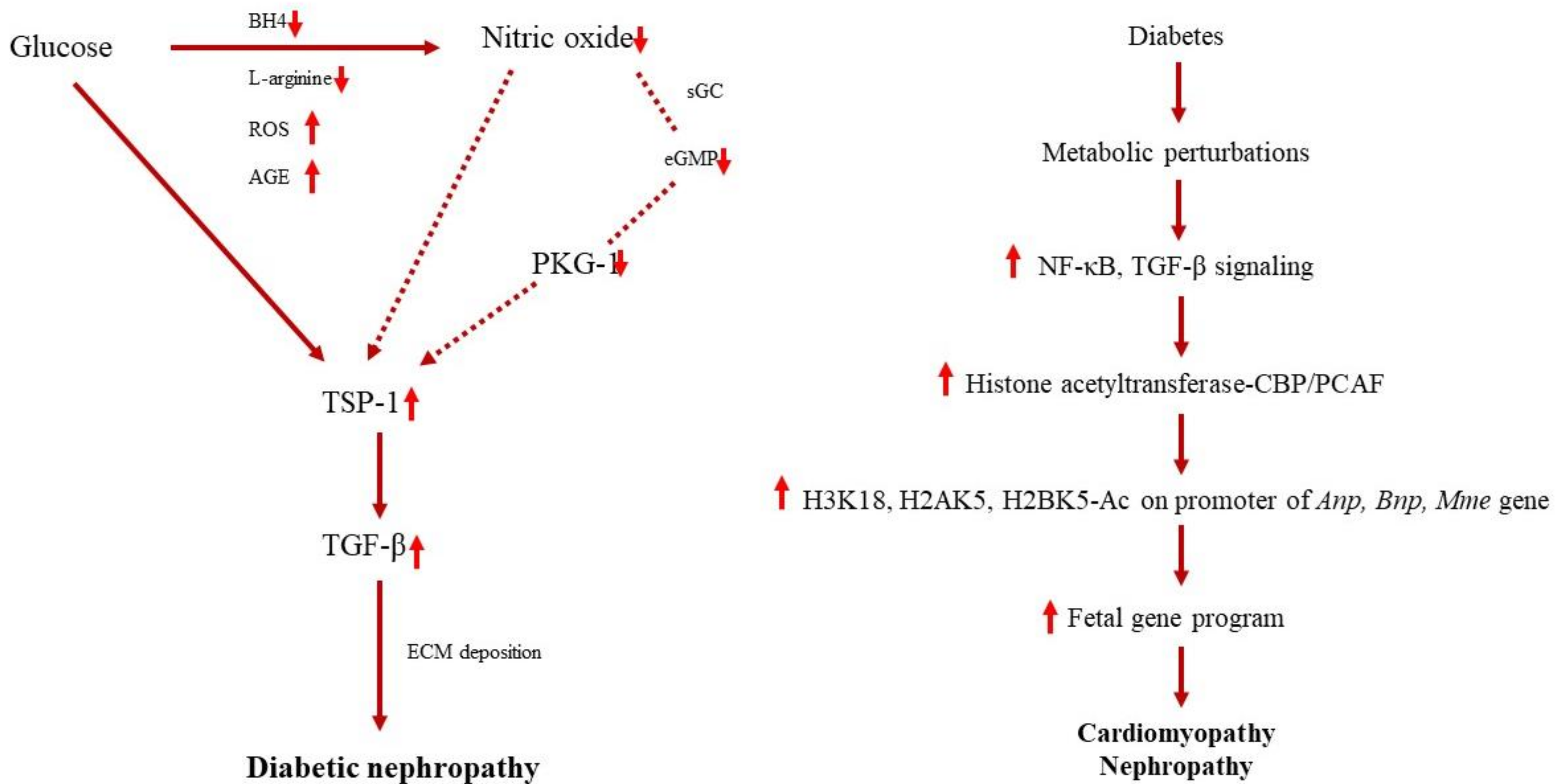
- Vajir, M.; Nisha, S.; Anil Bhanudas, G. Histone Acetylation Regulates Natriuretic Peptides and Neprilysin Gene Expressions in Diabetic Cardiomyopathy and Nephropathy. Curr. Mol. Pharmacol. 2019, 12, 61–71. [Google Scholar] [CrossRef]
- Wang, S.; Shiva, S.; Poczatek, M.H.; Darley-Usmar, V.; Murphy-Ullrich, J.E. Nitric oxide and cGMP-dependent protein kinase regulation of glucose-mediated thrombospondin 1-dependent transforming growth factor-beta activation in mesangial cells. J. Biol. Chem. 2002, 277, 9880–9888. [Google Scholar] [CrossRef] [Green Version]
- Mu, J.; Zhang, D.; Tian, Y.; Xie, Z.; Zou, M.H. BRD4 inhibition by JQ1 prevents high-fat diet-induced diabetic cardiomyopathy by activating PINK1/Parkin-mediated mitophagy in vivo. J. Mol. Cell. Cardiol. 2020, 149, 1–14. [Google Scholar] [CrossRef]
- Chen, W.; Zhang, Y.; Liu, J. Chinese herbal medicine for diabetic peripheral neuropathy. Cochrane Database Syst. Rev. 2011, 10, CD007796. [Google Scholar] [CrossRef] [Green Version]
- Galuppo, M.; Giacoppo, S.; Bramanti, P.; Mazzon, E. Use of natural compounds in the management of diabetic peripheral neuropathy. Molecules 2014, 19, 2877–2895. [Google Scholar] [CrossRef] [Green Version]
- LeBrasseur, N.K.; Fielding, R.A. Exercise and Diabetic Neuropathy. In Clinical Management of Diabetic Neuropathy; Veves, A., Ed.; Humana Press: Totowa, NJ, USA, 1998; pp. 257–271. [Google Scholar]
- Ninomiya, K.; Matsuda, H.; Shimoda, H.; Nishida, N.; Kasajima, N.; Yoshino, T.; Morikawa, T.; Yoshikawa, M. Carnosic acid, a new class of lipid absorption inhibitor from sage. Bioorg. Med. Chem. Lett. 2004, 14, 1943–1946. [Google Scholar] [CrossRef]
- Rau, O.; Wurglics, M.; Paulke, A.; Zitzkowski, J.; Meindl, N.; Bock, A.; Dingermann, T.; Abdel-Tawab, M.; Schubert-Zsilavecz, M. Carnosic acid and carnosol, phenolic diterpene compounds of the labiate herbs rosemary and sage, are activators of the human peroxisome proliferator-activated receptor gamma. Planta Med. 2006, 72, 881–887. [Google Scholar] [CrossRef] [PubMed]
- Kianbakht, S.; Abasi, B.; Perham, M.; Hashem Dabaghian, F. Antihyperlipidemic effects of Salvia officinalis L. leaf extract in patients with hyperlipidemia: A randomized double-blind placebo-controlled clinical trial. Phytother. Res. 2011, 25, 1849–1853. [Google Scholar] [CrossRef]
- Kianbakht, S.; Dabaghian, F.H. Improved glycemic control and lipid profile in hyperlipidemic type 2 diabetic patients consuming Salvia officinalis L. leaf extract: A randomized placebo. Controlled clinical trial. Complement. Ther. Med. 2013, 21, 441–446. [Google Scholar] [CrossRef] [PubMed]
- Nunthaboot, N.; Lugsanangarm, K.; Kokpol, S.; Abd-Elazem, I.S. Binding mode prediction of biologically active compounds from plant Salvia Miltiorrhiza as integrase inhibitor. Bioinformation 2013, 9, 426–431. [Google Scholar] [CrossRef] [PubMed]
- Oboh, G.; Henle, T. Antioxidant and inhibitory effects of aqueous extracts of Salvia officinalis leaves on pro-oxidant-induced lipid peroxidation in brain and liver in vitro. J. Med. Food 2009, 12, 77–84. [Google Scholar] [CrossRef] [PubMed]
- Oniga, I.; Pârvu, A.E.; Toiu, A.; Benedec, D. Effects of Salvia officinalis L. extract on experimental acute inflammation. Rev. Med. Chir. Soc. Med. Nat. Iasi 2007, 111, 290–294. [Google Scholar] [PubMed]
- Sá, C.M.; Ramos, A.A.; Azevedo, M.F.; Lima, C.F.; Fernandes-Ferreira, M.; Pereira-Wilson, C. Sage tea drinking improves lipid profile and antioxidant defences in humans. Int. J. Mol. Sci. 2009, 10, 3937–3950. [Google Scholar] [CrossRef] [Green Version]


| Ref. | Objective | Methods | Findings | Conclusion |
|---|---|---|---|---|
| [31] | To identify the antidiabetic compounds and α-amylase inhibitors from S. virgata |
|
|
|
| [32] | To explore the phytochemical composition, inhibitory enzyme potential, and antioxidant activities of S. viridis ethanolic root extracts obtained by different extraction methods |
|
|
|
| [11] | To investigate the volatile oil composition of the leaves and flowers and to evaluate the antidiabetic enzyme inhibitory assays of the oils and several extracts of S. urmiensis |
|
|
|
| [33] | To evaluate the antidiabetic activities of S. syriaca |
|
|
|
| [34] | To evaluate the chemical composition and enzyme inhibitory activity of S. nemorosa |
|
|
|
| [35] | To evaluate simultaneous determination and quantification of ursolic acid and oleanolic acid in 14 Salvia sp. and to reveal antidiabetic activities (α-glucosidase) along with correlations between antidiabetic activities and both acid contents |
|
|
|
| [36] | To evaluate the effect of SalA on diabetic vascular endothelial dysfunction (VED) |
|
|
|
| [37] | To investigate the protective effects of SalA on the peripheral nerve in diabetic rats |
|
|
|
| [38] | To investigate the in vivo and in vitro antidiabetic effect of SalA and the underlying mechanisms |
|
|
|
| [39] | To evaluate the antidiabetic effect of SalB in multiple low-dose STZ-induced diabetes in rat |
|
|
|
| [40] | To investigate the effects of SalB on diabetes-related metabolic changes in a spontaneous model of T2DM, as well as its potential molecular mechanism |
|
|
|
| [41] | To investigate the effects of SalB on glycometabolism, lipid metabolism, insulin resistance, oxidative stress, and glycogen synthesis in the T2DM rat model |
|
|
|
| [42] | To characterize the effects of S. fruticosa tea (SFT) treatment and of its main phenolic constituent RA on the levels and localization of the intestinal Na+/glucose cotransporter-1 (SGLT1), the facilitative GLUT2 and glucagon-like peptide-1 (GLP-1) |
|
|
|
| [43] | To investigate that eucalyptol inhibited tubular epithelial cell disjunction and tubulointerstitial fibrosis stimulated by glucose |
|
|
|
| [44] | To investigate the phytochemical composition of S. sclarea EO (SSEO) and to explore their acute and subchronic antidiabetic potentials |
|
|
|
| [45] | To examine the effects of lithospermic acid B (LAB) on the prevention of diabetic retinopathy in Otsuka Long-Evans Tokushima fatty (OLETF) rats, an animal model of T2DM |
|
|
|
| [46] | To investigate the effect of cryptotanshinone on myocardial fibrosis in diabetic rats |
|
|
|
| [47] | To screen and explore bioactive constituents from the root of S. miltiorrhiza Bunge acting on renin activity and evaluates its osteoprotective efficacy in diabetic mice |
|
|
|
| [48] | To investigate the potential therapeutic function of TSIIA on diabetic cardiomyopathy in an experimental diabetic rat model |
|
|
|
| [49] | To evaluate the therapeutic potential of the polyphenolic acids fraction (PAF) from S. miltiorrhiza Bunge in the T2DM rats model |
|
|
|
| [50] | To investigate the effects and possible mechanisms of the pharmacodynamic interaction between paeonol (Pae) and danshensu (DSS) on cerebrovascular malfunctioning in diabetes |
|
|
|
| [51] | To investigate the role of AGE-mediated neuroinflammation in learning and memory deficits and the effect of DSS on the cognitive decline in diabetic mice |
|
|
|
| [52] | To examine the protective efficacy of Jiangtang decoction (JTD) in diabetic nephropathy (DN) and elucidate the underlying molecular mechanisms |
|
|
|
| [53] | To evaluate the effect and potential mechanism of Huangqi-danshen decoction (HDD) in the treatment of DN in a T2DM animal model, db/db mice |
|
|
|
| [54] | To assess the effectiveness and safety of compound danshen dripping pill (CDDP) in treating patients with nonproliferative diabetic retinopathy (NPDR) |
|
|
|
| [55] | To determine the effectiveness and safety of Tricardin along with physical rehabilitation in diabetic patients with polyneuropathies and to observe the effect of this drug on the quality of life of these patients |
|
|
|
| [56] | To investigate the involvement of collagen IV (ColIV) and fibronectin (FN) in the occurrence and development of DN and the effects of telmisartan and S. miltiorrhiza injection in the treatment of the patients |
|
|
|
| [57] | To evaluate the efficacy and safety of S. officinalis combined with statin in dyslipidemic T2DM |
|
|
|
Publisher’s Note: MDPI stays neutral with regard to jurisdictional claims in published maps and institutional affiliations. |
© 2021 by the authors. Licensee MDPI, Basel, Switzerland. This article is an open access article distributed under the terms and conditions of the Creative Commons Attribution (CC BY) license (https://creativecommons.org/licenses/by/4.0/).
Share and Cite
Abd Rashed, A.; Rathi, D.-N.G. Bioactive Components of Salvia and Their Potential Antidiabetic Properties: A Review. Molecules 2021, 26, 3042. https://doi.org/10.3390/molecules26103042
Abd Rashed A, Rathi D-NG. Bioactive Components of Salvia and Their Potential Antidiabetic Properties: A Review. Molecules. 2021; 26(10):3042. https://doi.org/10.3390/molecules26103042
Chicago/Turabian StyleAbd Rashed, Aswir, and Devi-Nair Gunasegavan Rathi. 2021. "Bioactive Components of Salvia and Their Potential Antidiabetic Properties: A Review" Molecules 26, no. 10: 3042. https://doi.org/10.3390/molecules26103042