Radiomics in Abdominal Diseases
Share This Topical Collection
Editors
 Dr. Vittorio Miele
Dr. Vittorio Miele
E-Mail
Website
Collection Editor
Department of Radiology, Azienda Ospedaliero Universitaria Careggi, L.go G. A. Brambilla 3, 50134 Florence, Italy
Interests: emergency radiology; abdominal radiology; thoracic imaging; diagnostic imaging in oncology; breast imaging
Special Issues, Collections and Topics in MDPI journals
Dr. Eleonora Bicci
Dr. Eleonora Bicci
E-Mail
Website
Collection Editor
Department of Radiology, Azienda Ospedaliero Universitaria Careggi, L.go G. A. Brambilla 3, 50134 Florence, Italy
Interests: diagnostic imaging in oncology; abdominal imaging; radiomics; magnetic resonance imaging; computed tomography; texture analysis; emergency radiology
Topical Collection Information
Dear Colleagues,
Recently, radiomics has gained attention as a method for computerized image analyses. Radiomics can perform diagnostic or predictive tasks using high-dimensional image-derived features, used to measure tissue heterogeneity based on the distribution of pixel values, not affected by human vision and subjective analyses. Radiomics has the potential to expand the capabilities of abdominal imaging beyond the scope of traditional visual image analyses, with broad application prospects as a promising tool in tumor assessment and as a predictor of prognoses and treatment responses in the oncologic field. This collection aims to report the technical principles of radiomics and its application status with the perspective of abdominal diseases.
Dr. Vittorio Miele
Dr. Eleonora Bicci
Collection Editors
Manuscript Submission Information
Manuscripts should be submitted online at www.mdpi.com by registering and logging in to this website. Once you are registered, click here to go to the submission form. Manuscripts can be submitted until the deadline. All submissions that pass pre-check are peer-reviewed. Accepted papers will be published continuously in the journal (as soon as accepted) and will be listed together on the collection website. Research articles, review articles as well as short communications are invited. For planned papers, a title and short abstract (about 100 words) can be sent to the Editorial Office for announcement on this website.
Submitted manuscripts should not have been published previously, nor be under consideration for publication elsewhere (except conference proceedings papers). All manuscripts are thoroughly refereed through a single-blind peer-review process. A guide for authors and other relevant information for submission of manuscripts is available on the Instructions for Authors page. Diagnostics is an international peer-reviewed open access semimonthly journal published by MDPI.
Please visit the Instructions for Authors page before submitting a manuscript.
The Article Processing Charge (APC) for publication in this open access journal is 2600 CHF (Swiss Francs).
Submitted papers should be well formatted and use good English. Authors may use MDPI's
English editing service prior to publication or during author revisions.
Keywords
- radiomics
- artificial intelligence
- oncology imaging
- abdominal imaging
- magnetic resonance imaging
- computed tomography
Published Papers (2 papers)
2023
Open AccessReview
Prognostic Assessment of Gastropancreatic Neuroendocrine Neoplasm: Prospects and Limits of Radiomics
by
Federica De Muzio, Fabio Pellegrino, Roberta Fusco, Salvatore Tafuto, Mariano Scaglione, Alessandro Ottaiano, Antonella Petrillo, Francesco Izzo and Vincenza Granata
Viewed by 924
Abstract
Neuroendocrine neoplasms (NENs) are a group of lesions originating from cells of the diffuse neuroendocrine system. NENs may involve different sites, including the gastrointestinal tract (GEP-NENs). The incidence and prevalence of GEP-NENs has been constantly rising thanks to the increased diagnostic power of
[...] Read more.
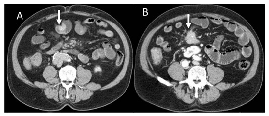
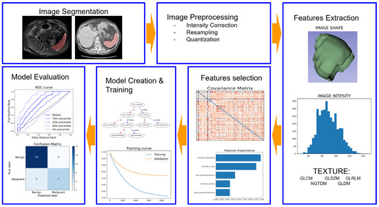
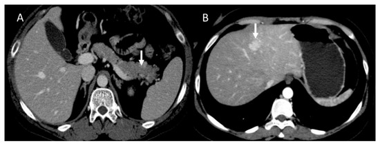
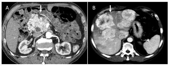
Neuroendocrine neoplasms (NENs) are a group of lesions originating from cells of the diffuse neuroendocrine system. NENs may involve different sites, including the gastrointestinal tract (GEP-NENs). The incidence and prevalence of GEP-NENs has been constantly rising thanks to the increased diagnostic power of imaging and immuno–histochemistry. Despite the plethora of biochemical markers and imaging techniques, the prognosis and therapeutic choice in GEP-NENs still represents a challenge, mainly due to the great heterogeneity in terms of tumor lesions and clinical behavior. The concept that biomedical images contain information about tissue heterogeneity and pathological processes invisible to the human eye is now well established. From this substrate comes the idea of radiomics. Computational analysis has achieved promising results in several oncological settings, and the use of radiomics in different types of GEP-NENs is growing in the field of research, yet with conflicting results. The aim of this narrative review is to provide a comprehensive update on the role of radiomics on GEP-NEN management, focusing on the main clinical aspects analyzed by most existing reports: predicting tumor grade, distinguishing NET from other tumors, and prognosis assessment.
Full article
►▼
Show Figures
Open AccessReview
Radiomics Applications in Spleen Imaging: A Systematic Review and Methodological Quality Assessment
by
Salvatore Claudio Fanni, Maria Febi, Roberto Francischello, Francesca Pia Caputo, Ilaria Ambrosini, Giacomo Sica, Lorenzo Faggioni, Salvatore Masala, Michele Tonerini, Mariano Scaglione, Dania Cioni and Emanuele Neri
Cited by 2 | Viewed by 1091
Abstract
The spleen, often referred to as the “forgotten organ”, plays numerous important roles in various diseases. Recently, there has been an increased interest in the application of radiomics in different areas of medical imaging. This systematic review aims to assess the current state
[...] Read more.
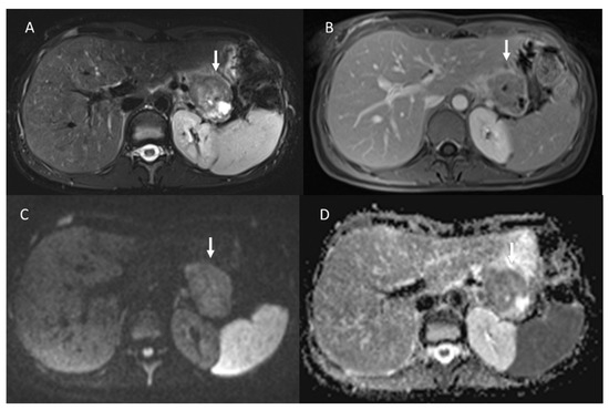
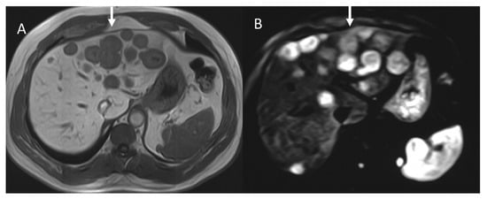
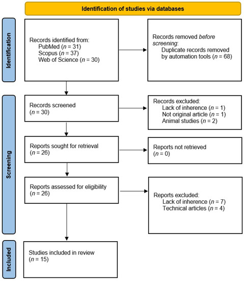
The spleen, often referred to as the “forgotten organ”, plays numerous important roles in various diseases. Recently, there has been an increased interest in the application of radiomics in different areas of medical imaging. This systematic review aims to assess the current state of the art and evaluate the methodological quality of radiomics applications in spleen imaging. A systematic search was conducted on PubMed, Scopus, and Web of Science. All the studies were analyzed, and several characteristics, such as year of publication, research objectives, and number of patients, were collected. The methodological quality was evaluated using the radiomics quality score (RQS). Fourteen articles were ultimately included in this review. The majority of these articles were published in non-radiological journals (78%), utilized computed tomography (CT) for extracting radiomic features (71%), and involved not only the spleen but also other organs for feature extraction (71%). Overall, the included papers achieved an average RQS total score of 9.71 ± 6.37, corresponding to an RQS percentage of 27.77 ± 16.04. In conclusion, radiomics applications in spleen imaging demonstrate promising results in various clinical scenarios. However, despite all the included papers reporting positive outcomes, there is a lack of consistency in the methodological approaches employed.
Full article
►▼
Show Figures


