
Radiomics Applications in Spleen Imaging: A Systematic Review and Methodological Quality Assessment

 , , , and
Abstract
:1. Introduction
2. Materials and Methods
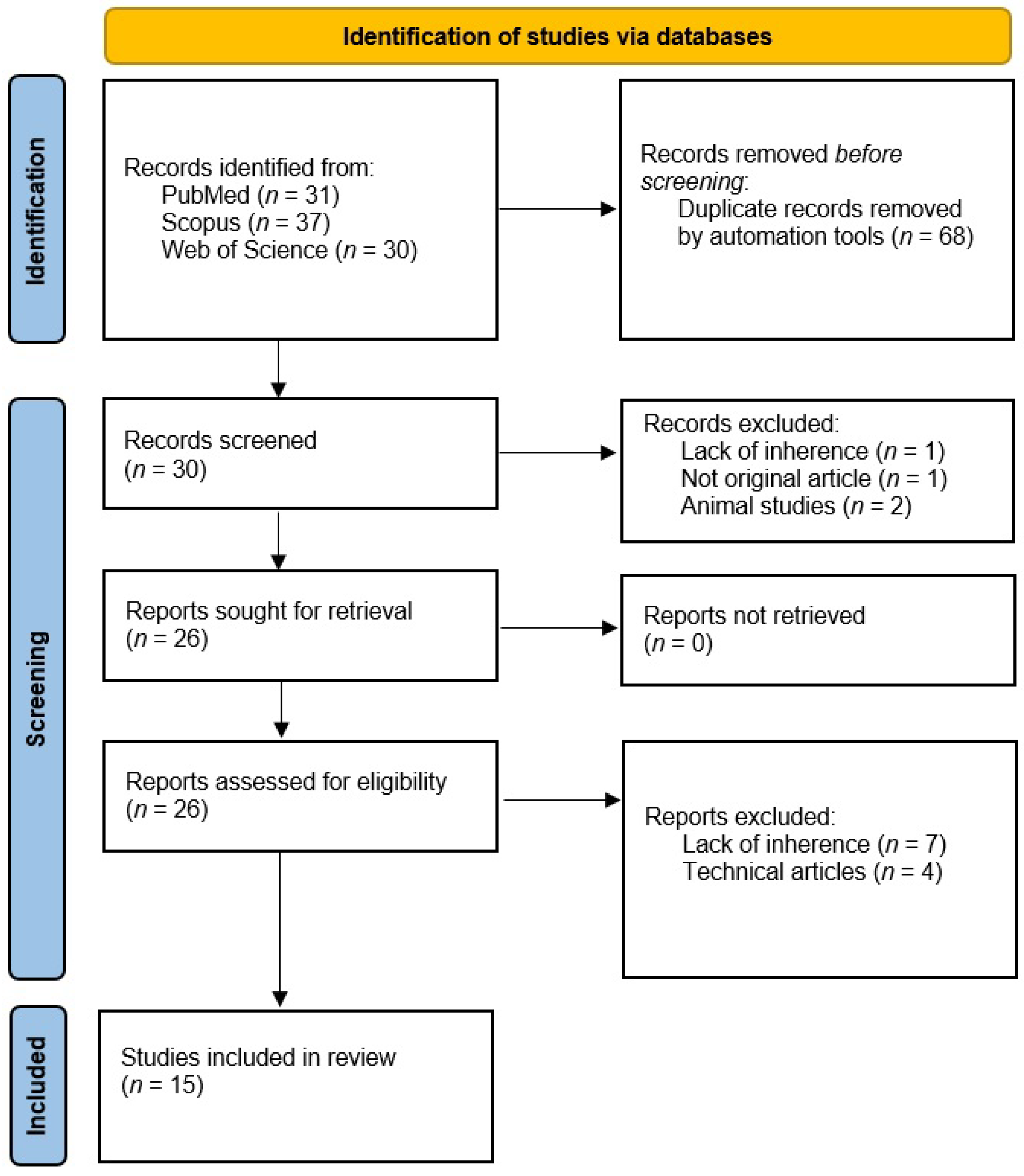
2.1. Literature Search
2.2. Study Evaluation
3. Results
4. Discussion
5. Conclusions
Author Contributions
Funding
Institutional Review Board Statement
Informed Consent Statement
Data Availability Statement
Conflicts of Interest
References
- Vancauwenberghe, T.; Snoeckx, A.; Vanbeckevoort, D.; Dymarkowski, S.; Vanhoenacker, F.M. Imaging of the spleen: What the clinician needs to know. Singap. Med. J. 2015, 56, 133–144. [Google Scholar] [CrossRef] [Green Version]
- Cesta, M.F. Normal Structure, Function, and Histology of the Spleen. Toxicol. Pathol. 2006, 34, 455–465. [Google Scholar] [CrossRef]
- Rotbain, E.C.; Hansen, D.L.; De Muckadell, O.S.; Wibrand, F.; Lund, A.M.; Frederiksen, H. Splenomegaly—Diagnostic validity, work-up, and underlying causes. PLoS ONE 2017, 12, e0186674. [Google Scholar] [CrossRef] [Green Version]
- Mebius, R.E.; Kraal, G. Structure and function of the spleen. Nat. Rev. Immunol. 2005, 5, 606–616. [Google Scholar] [CrossRef] [PubMed]
- Li, L.; Duan, M.; Chen, W.; Li, X.; Yang, J.; Li, Z. The spleen in liver cirrhosis: Revisiting an old enemy with novel targets. J. Transl. Med. 2017, 15, 111. [Google Scholar] [CrossRef] [PubMed] [Green Version]
- Fotiadis, C.; Georgopoulos, I.; Stoidis, C.; Patapis, P. Primary Tumors of the Spleen. Int. J. Biomed. Sci. 2009, 5, 85–91. [Google Scholar]
- Gillies, R.J.; Kinahan, P.E.; Hricak, H. Radiomics: Images are more than pictures, they are data. Radiology 2016, 278, 563–577. [Google Scholar] [CrossRef] [PubMed] [Green Version]
- Lambin, P.; Rios-Velazquez, E.; Leijenaar, R.; Carvalho, S.; van Stiphout, R.G.P.M.; Granton, P.; Zegers, C.M.L.; Gillies, R.; Boellard, R.; Dekker, A.; et al. Radiomics: Extracting more information from medical images using advanced feature analysis. Eur. J. Cancer 2012, 48, 441–446. [Google Scholar] [CrossRef] [PubMed] [Green Version]
- Lambin, P.; Leijenaar, R.T.H.; Deist, T.M.; Peerlings, J.; de Jong, E.E.C.; van Timmeren, J.; Sanduleanu, S.; Larue, R.T.H.M.; Even, A.J.G.; Jochems, A.; et al. Radiomics: The bridge between medical imaging and personalized medicine. Nat. Rev. Clin. Oncol. 2017, 14, 749–762. [Google Scholar] [CrossRef] [PubMed] [Green Version]
- Aringhieri, G.; Fanni, S.C.; Febi, M.; Colligiani, L.; Cioni, D.; Neri, E. The Role of Radiomics in Salivary Gland Imaging: A Systematic Review and Radiomics Quality Assessment. Diagnostics 2022, 12, 3002. [Google Scholar] [CrossRef]
- Candita, G.; Rossi, S.; Cwiklinska, K.; Fanni, S.C.; Cioni, D.; Lencioni, R.; Neri, E. Imaging Diagnosis of Hepatocellular Carcinoma: A State-of-the-Art Review. Diagnostics 2023, 13, 625. [Google Scholar] [CrossRef]
- Aghakhanyan, G.; Di Salle, G.; Fanni, S.C.; Francischello, R.; Cioni, D.; Cosottini, M.; Volterrani, D.; Neri, E. Radiomics insight into the neurodegenerative ‘hot’ brain: A narrative review from the nuclear medicine perspective. Front. Nucl. Med. 2023, 3, 1143256. [Google Scholar] [CrossRef]
- Fanni, S.C.; Febi, M.; Colligiani, L.; Volpi, F.; Ambrosini, I.; Tumminello, L.; Aghakhanyan, G.; Aringhieri, G.; Cioni, D.; Neri, E. A first look into radiomics application in testicular imaging: A systematic review. Front. Radiol. 2023, 3, 1141499. [Google Scholar] [CrossRef] [PubMed]
- Cannella, R.; Vernuccio, F.; Klontzas, M.E.; Ponsiglione, A.; Petrash, E.; Ugga, L.; Santos, D.P.D.; Cuocolo, R. Systematic review with radiomics quality score of cholangiocarcinoma: An EuSoMII Radiomics Auditing Group Initiative. Insights Imaging 2023, 14, 21. [Google Scholar] [CrossRef] [PubMed]
- Ponsiglione, A.; Stanzione, A.A.; Spadarella, G.; Baran, A.; Cappellini, L.A.; Lipman, K.G.; Van Ooijen, P.; Cuocolo, R. Ovarian imaging radiomics quality score assessment: An EuSoMII radiomics auditing group initiative. Eur. Radiol. 2022, 33, 2239–2247. [Google Scholar] [CrossRef] [PubMed]
- Yip, S.S.F.; Aerts, H.J.W.L. Applications and limitations of radiomics. Phys. Med. Biol. 2016, 61, R150–R166. [Google Scholar] [CrossRef] [Green Version]
- Koçak, B.; Cuocolo, R.; Santos, D.P.D.; Stanzione, A.; Ugga, L. Must-have Qualities of Clinical Research on Artificial Intelligence and Machine Learning. Balkan Med. J. 2023, 40, 3–12. [Google Scholar] [CrossRef]
- Page, M.J.; McKenzie, J.E.; Bossuyt, P.M.; Boutron, I.; Hoffmann, T.C.; Mulrow, C.D.; Shamseer, L.; Tetzlaff, J.M.; Akl, E.A.; Brennan, S.E.; et al. The PRISMA 2020 statement: An updated guideline for reporting systematic reviews. BMJ 2021, 372, n71. [Google Scholar] [CrossRef]
- Ouzzani, M.; Hammady, H.; Fedorowicz, Z.; Elmagarmid, A. Rayyan-a web and mobile app for systematic reviews. Syst. Rev. 2016, 5, 210. [Google Scholar] [CrossRef] [Green Version]
- Batur, A.; Kilinçer, A.; Ateş, F.; Demir, N.A.; Ergün, R. Evaluation of systemic involvement of coronavirus disease 2019 through spleen; size and texture analysis. Turk. J. Med. Sci. 2021, 51, 972–980. [Google Scholar] [CrossRef]
- Enke, J.S.; Moltz, J.H.; D’Anastasi, M.; Kunz, W.G.; Schmidt, C.; Maurus, S.; Mühlberg, A.; Katzmann, A.; Sühling, M.; Hahn, H.; et al. Radiomics Features of the Spleen as Surrogates for CT-Based Lymphoma Diagnosis and Subtype Differentiation. Cancers 2022, 14, 713. [Google Scholar] [CrossRef]
- Li, L.; Lin, Y.; Yu, D.; Liu, Z.; Gao, Y.; Qiao, J. A Multi-Organ Fusion and LightGBM Based Radiomics Algorithm for High-Risk Esophageal Varices Prediction in Cirrhotic Patients. IEEE Access 2021, 9, 15041–15052. [Google Scholar] [CrossRef]
- Li, P.; Wu, L.; Li, Z.; Li, J.; Ye, W.; Shi, Z.; Xu, Z.; Zhu, C.; Ye, H.; Liu, Z.; et al. Spleen Radiomics Signature: A Potential Biomarker for Prediction of Early and Late Recurrences of Hepatocellular Carcinoma After Resection. Front. Oncol. 2021, 11, 716849. [Google Scholar] [CrossRef] [PubMed]
- Luo, R.; Gao, J.; Gan, W.; Xie, W.B. Clinical-radiomics nomogram for predicting esophagogastric variceal bleeding risk noninvasively in patients with cirrhosis. World J. Gastroenterol. 2023, 29, 1076–1089. [Google Scholar] [CrossRef]
- Meng, D.; Wei, Y.; Feng, X.; Kang, B.; Wang, X.; Qi, J.; Zhao, Q.X.; Zhu, D. CT-Based Radiomics Score Can Accurately Predict Esophageal Variceal Rebleeding in Cirrhotic Patients. Front. Med. 2021, 8, 745931. [Google Scholar] [CrossRef]
- Nitsch, J.; Sack, J.; Halle, M.W.; Moltz, J.H.; Wall, A.; Rutherford, A.E.; Kikinis, R.; Meine, H. MRI-based radiomic feature analysis of end-stage liver disease for severity stratification. Int. J. Comput. Assist. Radiol. Surg. 2021, 16, 457–466. [Google Scholar] [CrossRef] [PubMed]
- Pan, B.; Zhang, W.; Chen, W.; Zheng, J.; Yang, X.; Sun, J.; Sun, X.; Chen, X. Establishment of the Radiologic Tumor Invasion Index Based on Radiomics Splenic Features and Clinical Factors to Predict Serous Invasion of Gastric Cancer. Front. Oncol. 2021, 11, 682456. [Google Scholar] [CrossRef] [PubMed]
- Sack, J.; Nitsch, J.; Meine, H.; Kikinis, R.; Halle, M.; Rutherford, A. Quantitative Analysis of Liver Disease Using MRI-Based Radiomic Features of the Liver and Spleen. J. Imaging 2022, 8, 277. [Google Scholar] [CrossRef]
- Tseng, Y.; Ma, L.; Li, S.; Luo, T.; Luo, J.; Zhang, W.; Wang, J.; Chen, S. Application of CT-based radiomics in predicting portal pressure and patient outcome in portal hypertension. Eur. J. Radiol. 2020, 126, 108927. [Google Scholar] [CrossRef]
- Wang, X.; Sun, J.; Zhang, W.; Yang, X.; Zhu, C.; Pan, B.; Zeng, Y.; Xu, J.; Chen, X.; Shen, X. Use of radiomics to extract splenic features to predict prognosis of patients with gastric cancer. Eur. J. Surg. Oncol. 2020, 46, 1932–1940. [Google Scholar] [CrossRef]
- Yan, Y.; Li, Y.; Fan, C.; Zhang, Y.; Zhang, S.; Wang, Z.; Huang, T.; Ding, Z.; Hu, K.; Li, L.; et al. A novel machine learning-based radiomic model for diagnosing high bleeding risk esophagea varices in cirrhotic patients. Hepatol. Int. 2022, 16, 423–432. [Google Scholar] [CrossRef] [PubMed]
- Yang, X.; Liu, J.; Lu, X.; Kan, Y.; Wang, W.; Zhang, S.; Liu, L.; Zhang, H.; Li, J.; Yang, J. Development and Validation of a Nomogram Based on 18F-FDG PET/CT Radiomics to Predict the Overall Survival in Adult Hemophagocytic Lymphohistiocytosis. Front. Med. 2021, 8, 792677. [Google Scholar] [CrossRef] [PubMed]
- Yin, Y.; Yakar, D.; Dierckx, R.A.J.O.; Mouridsen, K.B.; Kwee, T.C.; de Haas, R.J. Combining Hepatic and Splenic CT Radiomic Features Improves Radiomic Analysis Performance for Liver Fibrosis Staging. Diagnostics 2022, 12, 550. [Google Scholar] [CrossRef]
- Lyu, D.; Liang, P.; Huang, C.; Chen, X.; Cheng, M.; Zhu, B.; Liu, M.; Yue, S.; Gao, J. Are Radiomic Spleen Features Useful Assess. Differ. Status Adv. Gastric Cancer? Front. Oncol. 2023, 13, 1167602. [Google Scholar] [CrossRef]
- Ponnatt, T.S.; Lilley, C.M.; Mirza, K.M. Hemophagocytic Lymphohistiocytosis. Arch. Pathol. Lab. Med. 2022, 146, 507–519. [Google Scholar] [CrossRef]
- Ye, F.; Zhai, M.; Long, J.; Gong, Y.; Ren, C.; Zhang, D.; Lin, X.; Liu, S. The Burden of Liver Cirrhosis in Mortality: Results from the Global Burden of Disease Study. Front. Public Heal. 2022, 10, 909455. [Google Scholar] [CrossRef]
- Wei, J.; Jiang, H.; Gu, D.; Niu, M.; Fu, F.; Han, Y.; Song, B.; Tian, J. Radiomics in liver diseases: Current progress and future opportunities. Liver Int. 2020, 40, 2050–2063. [Google Scholar] [CrossRef]
- Mohammed, S.E.A.; Abdo, A.E.; Mudawi, H.M.Y. Mortality and Rebleeding Following Variceal Haemorrhage in Liver Cirrhosis and Periportal Fibrosis. World J. Hepatol. 2016, 8, 1336–1342. [Google Scholar] [CrossRef]
- Scapicchio, C.; Chincarini, A.; Ballante, E.; Berta, L.; Bicci, E.; Bortolotto, C.; Brero, F.; Cabini, R.F.; Cristofalo, G.; Fanni, S.C.; et al. A multicenter evaluation of a deep learning software (LungQuant) for lung parenchyma characterization in COVID-19 pneumonia. Eur. Radiol. Exp. 2023, 7, 18. [Google Scholar] [CrossRef]
- Romei, C.; Falaschi, Z.; Danna, P.S.C.; Airoldi, C.; Tonerini, M.; Rocchi, E.; Fanni, S.C.; D’Amelio, C.; Barbieri, G.; Tiseo, G.; et al. Lung vessel volume evaluated with CALIPER software is an independent predictor of mortality in COVID-19 patients: A multicentric retrospective analysis. Eur. Radiol. 2022, 32, 4314–4323. [Google Scholar] [CrossRef]
- Shiri, I.; Salimi, Y.; Pakbin, M.; Hajianfar, G.; Avval, A.H.; Sanaat, A.; Mostafaei, S.; Akhavanallaf, A.; Saberi, A.; Mansouri, Z.; et al. COVID-19 prognostic modeling using CT radiomic features and machine learning algorithms: Analysis of a multi-institutional dataset of 14,339 patients: COVID-19 prognostic modeling using CT radiomics and machine learning. Comput. Biol. Med. 2022, 145, 105467. [Google Scholar] [CrossRef] [PubMed]
- Boraschi, P.; Donati, F.; Ambrosini, I.; Bruni, L.; Mazzeo, M.L.; Tintori, R.; Tonerini, M.; Neri, E. Diagnostic and Therapeutic Radiology of the GI Tract, Liver, and Pancreas in Patients with COVID. Gastroenterol. Clin. North Am. 2023, 52, 185–200. [Google Scholar] [CrossRef] [PubMed]
- Spadarella, G.; Stanzione, A.; Akinci D’Antonoli, T.; Andreychenko, A.; Fanni, S.C.; Ugga, L.; Kotter, E.; Cuocolo, R. Systematic review of the radiomics quality score applications: An EuSoMII Radiomics Auditing Group Initiative. Eur. Radiol. 2023, 33, 1884–1894. [Google Scholar] [CrossRef]


| First Author | Years | Aim | Imaging Technique | Number of Patients | Disease | Targeted Organs | Journal Topic | Feature Order | Study Design |
|---|---|---|---|---|---|---|---|---|---|
| Batur [20] | 2021 | To study changes in the spleen size and textural features of patients with COVID-19 | CT | 91 | Not primarily splenic | Spleen | Not Radiological | First and Higher order | Retrospective |
| Enke [21] | 2022 | To investigate spleen radiomic features’ role in differentiating lymphoma subtypes and non-lymphoma | CT | 326 | Primarily splenic | Spleen | Not Radiological | First and Higher order | Retrospective |
| Li L. [22] | 2021 | To identify high- and low-risk EV patients using liver, spleen, and esophagus CT radiomic features | CT | 188 | Not primarily splenic | Spleen, Liver, and Esophagus | Not Radiological | First and Higher order | Retrospective |
| Li P. [23] | 2021 | To explore the usefulness of spleen radiomic features in predicting early and late recurrences of HCC after curative resection | CT | 237 | Not primarily splenic | Spleen and Liver | Not Radiological | First and Higher order | Retrospective |
| Luo [24] | 2023 | To develop a nomogram based on clinical variables and radiomics to predict esophagogastric variceal bleeding in cirrhotic patients | CT | 211 | Not primarily splenic | Spleen and Liver | Not Radiological | First and Higher order | Retrospective |
| Meng [25] | 2021 | To develop a Rad-score from liver and spleen CT images in cirrhotic patients to predict esophageal variceal rebleeding | CT | 173 | Not primarily splenic | Spleen and Liver | Not Radiological | First and Higher order | Retrospective |
| Nitsch [26] | 2021 | To predict disease severity for cirrhosis using liver and spleen MRI radiomic features compared to MELD score and clinical decompensation | MRI | 90 | Not primarily splenic | Spleen and Liver | Not radiological | First and Higher order | Retrospective |
| Pan [27] | 2021 | To develop a radiomic nomogram for preoperative identification of serosal invasion of gastric cancer | CT | 315 | Not primarily splenic | Spleen | Not Radiological | First and Higher order | Retrospective |
| Sack [28] | 2022 | To implement MR radiomic features from liver and spleen to detect liver cirrhosis | MRI | 167 | Not primarily splenic | Spleen and Liver | Radiological | First and Higher order | Retrospective |
| Tseng [29] | 2020 | To propose a noninvasive predictive model of portal hypertension values based on CT radiomic features | CT | 169 | Not primarily splenic | Spleen and Liver | Radiological | First and Higher order | Retrospective |
| Wang [30] | 2020 | To predict gastric cancer prognosis using splenic features | CT | 243 | Not primarily splenic | Spleen | Not Radiological | First and Higher order | Retrospective |
| Yan [31] | 2022 | To develop a radiomic model for diagnosing high bleeding risk esophageal varices in patients with cirrhosis | CT | 796 | Not primarily splenic | Spleen, Liver, and Esophagus | Not Radiological | First and Higher order | Retrospective |
| Yang [32] | 2021 | To predict survival of patients with Adult Hemophagocytic Lymphohistiocytosis by using 18F-FDG PET/CT- radiomic features | 18F-FDG PET/CT | 70 | Primarily splenic | Spleen and Liver | Radiological | First and Higher order | Retrospective |
| Yin [33] | 2022 | To combine hepatic and splenic CT radiomic features for liver fibrosis staging | CT | 252 | Not primarily splenic | Spleen and Liver | Not Radiological | First and Higher order | Retrospective |
| Lyu [34] | 2023 | To determine whether radiomic spleen features can be used to distinguish advanced gastric cancer with varying states of differentiation | CT | 147 | Not primarily splenic | Spleen and Stomach | Not Radiological | First and Higher order | Retrospective |
| First Author | Item 1 | Item 2 | Item 3 | Item 4 | Item 5 | Item 6 | Item 7 | Item 8 | Item 9 | Item 10 | Item 11 | Item 12 | Item 13 | Item 14 | Item 15 | Item 16 | RQS (Total) | RQS (%) |
|---|---|---|---|---|---|---|---|---|---|---|---|---|---|---|---|---|---|---|
| Batur [20] | 1 | 1 | 0 | 0 | 3 | 1 | 1 | 0 | 0 | 0 | 0 | −5 | 0 | 0 | 0 | 0 | 2 | 5.56 |
| Enke [21] | 0 | 0 | 0 | 0 | 3 | 0 | 1 | 0 | 2 | 0 | 0 | −5 | 2 | 0 | 0 | 0 | 3 | 8.33 |
| Li L. [22] | 1 | 0 | 0 | 0 | 3 | 0 | 1 | 1 | 1 | 0 | 0 | 3 | 2 | 0 | 0 | 0 | 12 | 33.33 |
| Li P. [23] | 1 | 1 | 0 | 0 | 3 | 1 | 1 | 1 | 1 | 1 | 0 | 2 | 2 | 2 | 0 | 0 | 16 | 44.44 |
| Luo [24] | 1 | 1 | 0 | 0 | 3 | 1 | 1 | 1 | 1 | 1 | 0 | 2 | 2 | 2 | 0 | 0 | 16 | 44.44 |
| Meng [25] | 1 | 1 | 0 | 0 | 3 | 1 | 1 | 1 | 1 | 1 | 0 | 2 | 2 | 2 | 0 | 0 | 16 | 44.44 |
| Nitsch [26] | 1 | 0 | 0 | 0 | 3 | 0 | 1 | 1 | 2 | 0 | 0 | −5 | 2 | 0 | 0 | 0 | 5 | 13.89 |
| Pan [27] | 0 | 0 | 0 | 0 | 3 | 1 | 1 | 1 | 1 | 1 | 0 | 2 | 2 | 2 | 0 | 0 | 14 | 38.89 |
| Sack [28] | 0 | 0 | 0 | 0 | −3 | 0 | 1 | 0 | 1 | 0 | 0 | −5 | 2 | 0 | 0 | 0 | −4 | 0.00 |
| Tseng [29] | 0 | 0 | 0 | 0 | 3 | 1 | 1 | 1 | 1 | 0 | 0 | 2 | 2 | 0 | 0 | 0 | 11 | 30.56 |
| Wang [30] | 0 | 0 | 0 | 0 | 3 | 1 | 1 | 1 | 1 | 0 | 0 | 2 | 2 | 0 | 0 | 0 | 11 | 30.56 |
| Yan [31] | 1 | 0 | 0 | 0 | 3 | 1 | 1 | 0 | 1 | 1 | 0 | 2 | 2 | 2 | 0 | 0 | 14 | 38.89 |
| Yang [32] | 1 | 0 | 0 | 0 | 3 | 1 | 1 | 1 | 1 | 1 | 0 | 2 | 2 | 2 | 0 | 0 | 15 | 41.67 |
| Yin [33] | 1 | 0 | 0 | 0 | 3 | 1 | 1 | 0 | 2 | 0 | 0 | −5 | 2 | 0 | 0 | 0 | 5 | 13.89 |
| Lyu [34] | 1 | 0 | 0 | 0 | 3 | 1 | 1 | 1 | 1 | 1 | 0 | 2 | 2 | 2 | 0 | 0 | 15 | 41.67 |
Disclaimer/Publisher’s Note: The statements, opinions and data contained in all publications are solely those of the individual author(s) and contributor(s) and not of MDPI and/or the editor(s). MDPI and/or the editor(s) disclaim responsibility for any injury to people or property resulting from any ideas, methods, instructions or products referred to in the content. |
© 2023 by the authors. Licensee MDPI, Basel, Switzerland. This article is an open access article distributed under the terms and conditions of the Creative Commons Attribution (CC BY) license (https://creativecommons.org/licenses/by/4.0/).
Share and Cite
Fanni, S.C.; Febi, M.; Francischello, R.; Caputo, F.P.; Ambrosini, I.; Sica, G.; Faggioni, L.; Masala, S.; Tonerini, M.; Scaglione, M.; et al. Radiomics Applications in Spleen Imaging: A Systematic Review and Methodological Quality Assessment. Diagnostics 2023, 13, 2623. https://doi.org/10.3390/diagnostics13162623
Fanni SC, Febi M, Francischello R, Caputo FP, Ambrosini I, Sica G, Faggioni L, Masala S, Tonerini M, Scaglione M, et al. Radiomics Applications in Spleen Imaging: A Systematic Review and Methodological Quality Assessment. Diagnostics. 2023; 13(16):2623. https://doi.org/10.3390/diagnostics13162623
Chicago/Turabian StyleFanni, Salvatore Claudio, Maria Febi, Roberto Francischello, Francesca Pia Caputo, Ilaria Ambrosini, Giacomo Sica, Lorenzo Faggioni, Salvatore Masala, Michele Tonerini, Mariano Scaglione, and et al. 2023. "Radiomics Applications in Spleen Imaging: A Systematic Review and Methodological Quality Assessment" Diagnostics 13, no. 16: 2623. https://doi.org/10.3390/diagnostics13162623