Single Crystals for Biomedical Applications

A special issue of Crystals (ISSN 2073-4352).

Deadline for manuscript submissions: closed (30 June 2019) | Viewed by 12252

Special Issue Editor


E-Mail Website
Guest Editor
University of West Attica, Athens, Greece
Interests: medical imaging; nuclear medicine; detectors; scintillators; single crystals

Special Issue Information

Dear colleagues,

Single crystals, either organic or inorganic, have already been used in a wide range of biomedical applications (transducers, sensors, scintillators, wavelength shifters, etc.). Their unique properties have been applied or suggested for use in a huge range of medical modalities. Depending on the application, their size can vary from nanometres to few centymetres and they can be cut or molded into any shape to fit the biomedical application. Their electrical characteristics, light yield and density also make them interesting for applications involving imaging and therapy, including ionizing and non-ionizing radiation medical applications (e.g. ultrasonography, X-ray imaging, radiotherapy, etc.). Current trends in multimodality imaging detectors indicate the exploitation of single crystal scintillators in a wider range of energies, covering CT/PET and portal imaging. New single crystal materials with unique electrical and optical properties are fabricated to cover almost any biomedical application.

Due to the variety of research fields in biomedicine where the application of single crystals can improve or even create new applications, the Editorial Board of Crystals has decided to devote a Special Issue of the journal to the analysis of “Single Crystals for Biomedical Applications”.

Being honored to serve as a Guest Editor, I hereby invite all colleagues who work on “Single Crystals for Biomedical Applications” to contribute to this issue. Topics relating to issues such as the efficiency of single crystals, new crystalline materials for biomedical ionizing and non-ionizing radiation applications, under energies covering X-rays, nuclear medicine and radiation therapy, integration of single crystals into medical devices, theoretical calculations, etc., are welcome.

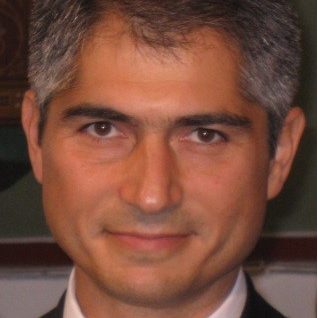
Prof. Dr. Ioannis G. Valais
Guest Editor

Manuscript Submission Information

Manuscripts should be submitted online at www.mdpi.com by registering and logging in to this website. Once you are registered, click here to go to the submission form. Manuscripts can be submitted until the deadline. All submissions that pass pre-check are peer-reviewed. Accepted papers will be published continuously in the journal (as soon as accepted) and will be listed together on the special issue website. Research articles, review articles as well as short communications are invited. For planned papers, a title and short abstract (about 100 words) can be sent to the Editorial Office for announcement on this website.

Submitted manuscripts should not have been published previously, nor be under consideration for publication elsewhere (except conference proceedings papers). All manuscripts are thoroughly refereed through a single-blind peer-review process. A guide for authors and other relevant information for submission of manuscripts is available on the Instructions for Authors page. Crystals is an international peer-reviewed open access monthly journal published by MDPI.

Please visit the Instructions for Authors page before submitting a manuscript. The Article Processing Charge (APC) for publication in this open access journal is 2600 CHF (Swiss Francs). Submitted papers should be well formatted and use good English. Authors may use MDPI's English editing service prior to publication or during author revisions.

Keywords

  • single crystals
  • biomedical applications
  • inorganic–organic single crystals
  • scintillators
  • tranducers
  • nanocrystals

Published Papers (3 papers)

Order results
Result details
Select all
Export citation of selected articles as:

Research

11 pages, 2190 KiB  
Article
Optical Characteristics of ZnCuInS/ZnS (Core/Shell) Nanocrystal Flexible Films Under X-Ray Excitation
by George Saatsakis, Nektarios Kalyvas, Christos Michail, Konstantinos Ninos, Athanasios Bakas, Christina Fountzoula, Ioannis Sianoudis, George E. Karpetas, George Fountos, Ioannis Kandarakis, Ioannis Valais and George Panayiotakis
Crystals 2019, 9(7), 343; https://doi.org/10.3390/cryst9070343 - 04 Jul 2019
Cited by 18 | Viewed by 2867
Abstract
The aim of this article is to evaluate optical characteristics, such as the intrinsic conversion efficiency and the inherent light propagation efficiency of three polymethyl methacrylate (PMMA)/methyl methacrylate (MMA) composite ZnCuInS/ZnS (core/shell) nanocrystal flexible films. The concentrations of these were 100 mg/mL, 150 [...] Read more.
The aim of this article is to evaluate optical characteristics, such as the intrinsic conversion efficiency and the inherent light propagation efficiency of three polymethyl methacrylate (PMMA)/methyl methacrylate (MMA) composite ZnCuInS/ZnS (core/shell) nanocrystal flexible films. The concentrations of these were 100 mg/mL, 150 mg/mL, and 250 mg/mL, respectively. Composite films were prepared by homogeneously diluting dry powder quantum dot (QD) samples in toluene and subsequently mixing these with a PMMA/MMA polymer solution. The absolute luminescence efficiency (AE) of the films was measured using X-ray excitation. A theoretical model describing the optical photon propagation in scintillator materials was used to calculate the fraction of the generated optical photons passed through the different material layers. Finally, the intrinsic conversion efficiency was calculated by considering the QD quantum yield and the optical photon emission spectrum. Full article
(This article belongs to the Special Issue Single Crystals for Biomedical Applications)
Show Figures

Figure 1

12 pages, 2588 KiB  
Article
Absolute Luminescence Efficiency of Europium-Doped Calcium Fluoride (CaF2:Eu) Single Crystals under X-ray Excitation
by Christos Michail, Nektarios Kalyvas, Athanasios Bakas, Konstantinos Ninos, Ioannis Sianoudis, George Fountos, Ioannis Kandarakis, George Panayiotakis and Ioannis Valais
Crystals 2019, 9(5), 234; https://doi.org/10.3390/cryst9050234 - 01 May 2019
Cited by 23 | Viewed by 4056
Abstract
The absolute luminescence efficiency (AE) of a calcium fluoride (CaF2:Eu) single crystal doped with europium was studied using X-ray energies met in general radiography. A CaF2:Eu single crystal with dimensions of 10 × 10 × 10 mm3 was [...] Read more.
The absolute luminescence efficiency (AE) of a calcium fluoride (CaF2:Eu) single crystal doped with europium was studied using X-ray energies met in general radiography. A CaF2:Eu single crystal with dimensions of 10 × 10 × 10 mm3 was irradiated by X-rays. The emission light photon intensity of the CaF2:Eu sample was evaluated by measuring AE within the X-ray range from 50 to 130 kV. The results of this work were compared with data obtained under similar conditions for the commercially employed medical imaging modalities, Bi4Ge3O12 and Lu2SiO5:Ce single crystals. The compatibility of the light emitted by the CaF2:Eu crystal, with the sensitivity of optical sensors, was also examined. The AE of the 10 × 10 × 10 mm3 CaF2:Eu crystal peaked in the range from 70 to 90 kV (22.22 efficiency units; E.U). The light emitted from CaF2:Eu is compatible with photocathodes, charge coupled devices (CCD), and silicon photomultipliers, which are used as radiation sensors in medical imaging systems. Considering the AE results in the examined energies, as well as the spectral compatibility with various photodetectors, a CaF2:Eu single crystal could be considered for radiographic applications, including the detection of charged particles and soft gamma rays. Full article
(This article belongs to the Special Issue Single Crystals for Biomedical Applications)
Show Figures

Figure 1

13 pages, 1160 KiB  
Article
Information Capacity of Positron Emission Tomography Scanners
by Christos Michail, George Karpetas, Nektarios Kalyvas, Ioannis Valais, Ioannis Kandarakis, Kyriakos Agavanakis, George Panayiotakis and George Fountos
Crystals 2018, 8(12), 459; https://doi.org/10.3390/cryst8120459 - 09 Dec 2018
Cited by 14 | Viewed by 3479
Abstract
Background: The aim of the present study was to assess the upper information content bound of positron emission tomography (PET) images, by means of the information capacity (IC). Methods: The Geant4 Application for the Tomographic Emission (GATE) Monte Carlo (MC) package was used, [...] Read more.
Background: The aim of the present study was to assess the upper information content bound of positron emission tomography (PET) images, by means of the information capacity (IC). Methods: The Geant4 Application for the Tomographic Emission (GATE) Monte Carlo (MC) package was used, and reconstructed images were obtained by using the software for tomographic image reconstruction (STIR). The case study for the assessment of the information content was the General Electric (GE) Discovery-ST PET scanner. A thin-film plane source aluminum (Al) foil, coated with a thin layer of silica and with a 18F-fludeoxyglucose (FDG) bath distribution of 1 MBq was used. The influence of the (a) maximum likelihood estimation-ordered subsets-maximum a posteriori probability-one step late (MLE-OS-MAP-OSL) algorithm, using various subsets (1 to 21) and iterations (1 to 20) and (b) different scintillating crystals on PET scanner’s performance, was examined. The study was focused on the noise equivalent quanta (NEQ) and on the single index IC. Images of configurations by using different crystals were obtained after the commonly used 2-dimensional filtered back projection (FBP2D), 3-dimensional filtered back projection re-projection (FPB3DRP) and the (MLE)-OS-MAP-OSL algorithms. Results: Results shown that the images obtained with one subset and various iterations provided maximum NEQ values, however with a steep drop-off after 0.045 cycles/mm. The single index IC data were maximized for the range of 8–20 iterations and three subsets. The PET scanner configuration incorporating lutetium orthoaluminate perovskite (LuAP) crystals provided the highest NEQ values in 2D FBP for spatial frequencies higher than 0.028 cycles/mm. Bismuth germanium oxide (BGO) shows clear dominance against all other examined crystals across the spatial frequency range, in both 3D FBP and OS-MAP-OSL. The particular PET scanner provided optimum IC values using FBP3DRP and BGO crystals (2.4829 bits/mm2). Conclusions: The upper bound of the image information content of PET scanners can be fully characterized and further improved by investigating the imaging chain components through MC methods. Full article
(This article belongs to the Special Issue Single Crystals for Biomedical Applications)
Show Figures

Figure 1

Back to TopTop