Molecular Mechanisms in Human Diseases
Share This Topical Collection
Editor
 Dr. Roberto Campagna
Dr. Roberto Campagna
E-Mail
Website
Guest Editor
Department of Clinical, Specialistic and Dental Sciences, Marche Polytechnic University, 60126 Ancona, Italy
Interests: cell proliferation; chemotherapeutic drugs; cancer; oxidative stress; paraoxonase-2; nicotinamide n-methyltransferase; metastasis
Special Issues, Collections and Topics in MDPI journals
Topical Collection Information
Dear Colleagues,
This topical collection focuses on molecular basis and molecular interactions involved in the development and progression of human diseases, as well as novel molecular biomarkers which could be suitable for diagnosis, prognosis and targeted therapies. Original research articles, reviews and short communications which fit the scope of the Topical Collection are welcome.
Identifying novel molecular mechanisms might help to implement cost effective early prevention and treatment. In the last decades, the identification of molecular mechanisms associated with specific human diseases led to the development of a number of novel drugs characterized by reduced side effects.
In this topical collection, we expect papers on the molecular mechanisms and interactions that comprehensively regulate cellular functions in human diseases, including but not limited to cell proliferation, growth, survival, death, inflammation and signaling observed in human pathological conditions. Studies on the molecular and cellular therapy of human diseases are also welcome.
Dr. Roberto Campagna
Guest Editor
Manuscript Submission Information
Manuscripts should be submitted online at www.mdpi.com by registering and logging in to this website. Once you are registered, click here to go to the submission form. Manuscripts can be submitted until the deadline. All submissions that pass pre-check are peer-reviewed. Accepted papers will be published continuously in the journal (as soon as accepted) and will be listed together on the collection website. Research articles, review articles as well as short communications are invited. For planned papers, a title and short abstract (about 100 words) can be sent to the Editorial Office for announcement on this website.
Submitted manuscripts should not have been published previously, nor be under consideration for publication elsewhere (except conference proceedings papers). All manuscripts are thoroughly refereed through a single-blind peer-review process. A guide for authors and other relevant information for submission of manuscripts is available on the Instructions for Authors page. Current Issues in Molecular Biology is an international peer-reviewed open access monthly journal published by MDPI.
Please visit the Instructions for Authors page before submitting a manuscript.
The Article Processing Charge (APC) for publication in this open access journal is 2200 CHF (Swiss Francs).
Submitted papers should be well formatted and use good English. Authors may use MDPI's
English editing service prior to publication or during author revisions.
Keywords
- molecular biomarker
- pathogenesis
- targeted therapy
- immunotherapy
- inflammation
- genomics
- signaling pathways
- cancer treatment
Published Papers (10 papers)
Open AccessBrief Report
Dual Immunoglobulin Domain-Containing Cell Adhesion Molecule Increases Early in Renal Tubular Cell Injury and Plays Anti-Inflammatory Role
by
Jin Han, Ju-Min Yook, Se-Hyun Oh, Yu Kyung Chung, Hee-Yeon Jung, Ji-Young Choi, Jang-Hee Cho, Sun-Hee Park, Chan-Duck Kim, Yong-Lim Kim, Seungwoo Han and Jeong-Hoon Lim
Viewed by 690
Abstract
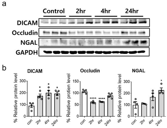
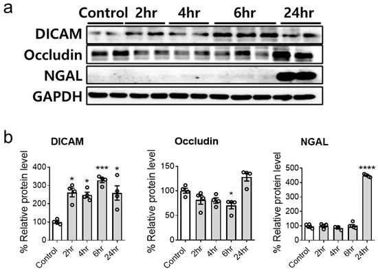
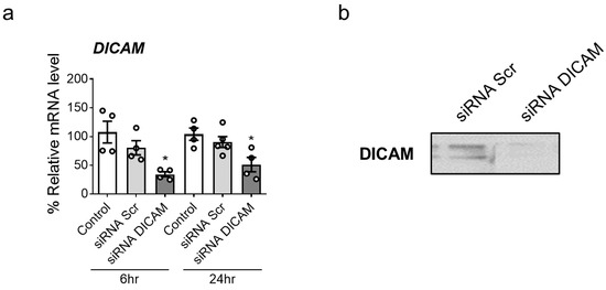
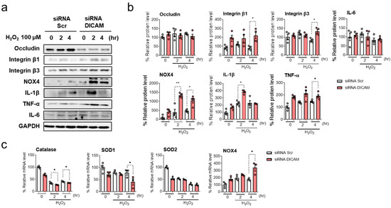
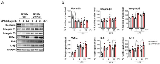
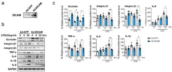
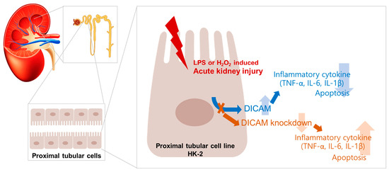
Dual immunoglobulin domain-containing cell adhesion molecule (DICAM) is a type I transmembrane protein that presents in various cells including renal tubular cells. This study evaluated the expression and protective role of DICAM in renal tubular cell injury. HK-2 cells were incubated and treated
[...] Read more.
Dual immunoglobulin domain-containing cell adhesion molecule (DICAM) is a type I transmembrane protein that presents in various cells including renal tubular cells. This study evaluated the expression and protective role of DICAM in renal tubular cell injury. HK-2 cells were incubated and treated with lipopolysaccharide (LPS, 30 μg/mL) or hydrogen peroxide (H
2O
2, 100 μM) for 24 h. To investigate the effect of the gene silencing of DICAM, small interfering RNA of DICAM was used. Additionally, to explain its role in cellular response to injury, DICAM was overexpressed using an adenoviral vector. DICAM protein expression levels significantly increased following treatment with LPS or H
2O
2 in HK-2 cells. In response to oxidative stress, DICAM showed an earlier increase (2–4 h following treatment) than neutrophil gelatinase-associated lipocalin (NGAL) (24 h following treatment). DICAM gene silencing increased the protein expression of inflammation-related markers, including IL-1β, TNF-α, NOX4, integrin β1, and integrin β3, in H
2O
2-induced HK-2 cell injury. Likewise, in the LPS-induced HK-2 cell injury, DICAM knockdown led to a decrease in occludin levels and an increase in integrin β3, IL-1β, and IL-6 levels. Furthermore, DICAM overexpression followed by LPS-induced HK-2 cell injury resulted in an increase in occludin levels and a decrease in integrin β1, integrin β3, TNF-α, IL-1β, and IL-6 levels, suggesting an alleviating effect on inflammatory responses. DICAM was elevated in the early stage of regular tubular cell injury and may protect against renal tubular injury through its anti-inflammatory properties. DICAM has a potential as an early diagnostic marker and therapeutic target for renal cell injury.
Full article
►▼
Show Figures
Open AccessArticle
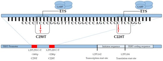
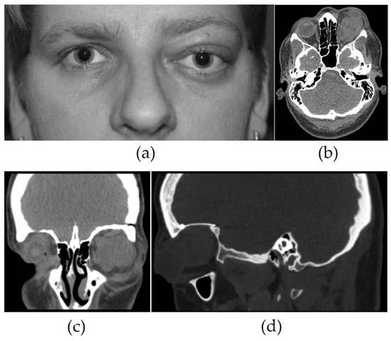
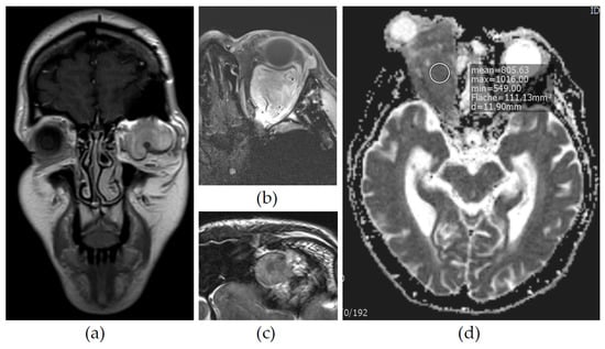
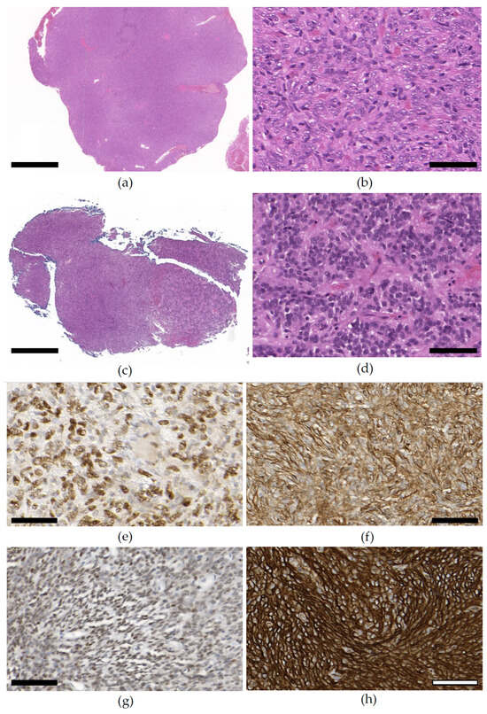
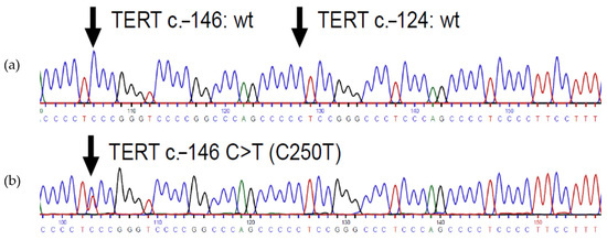
Prevalence of TERT Promoter Mutations in Orbital Solitary Fibrous Tumors
by
David Sinan Koca, Vladimir Kolpakov, Jana Ihlow, Maximilian von Laffert, Katharina Erb-Eigner, Hermann Herbst, Karen Kriese, Leonille Schweizer and Eckart Bertelmann
Viewed by 686
Abstract
The orbital manifestation of a solitary fibrous tumor (SFT) is exceptionally rare and poses specific challenges in diagnosis and treatment. Its rather exceptional behavior among all SFTs comprises a high tendency towards local recurrence, but it rarely culminates in metastatic disease. This raises
[...] Read more.
The orbital manifestation of a solitary fibrous tumor (SFT) is exceptionally rare and poses specific challenges in diagnosis and treatment. Its rather exceptional behavior among all SFTs comprises a high tendency towards local recurrence, but it rarely culminates in metastatic disease. This raises the question of prognostic factors in orbital SFTs (oSFTs). Telomerase reverse transcriptase (
TERT)-promoter mutations have previously been linked to an unfavorable prognosis in SFTs of other locations. We analyzed the prevalence of
TERT promoter mutations of SFTs in the orbital compartment. We performed a retrospective, descriptive clinico-histopathological analysis of nine cases of oSFTs between the years of 2017 and 2021. A
TERT promoter mutation was present in one case, which was classified with intermediate metastatic risk. Local recurrence or progress occurred in six cases after primary resection; no distant metastases were reported. Multimodal imaging repeatedly showed particular morphologic patterns, including tubular vascular structures and ADC reduction. The prevalence of the
TERT promoter mutation in oSFT was 11%, which is similar to the prevalence of extra-meningeal SFTs of the head and neck and lower than that in other extra-meningeal compartments. In the present study, the
TERT promoter mutation in oSFT manifested in a case with an unfavorable prognosis, comprising aggressive local tumor growth, local recurrence, and eye loss.
Full article
►▼
Show Figures
Open AccessArticle
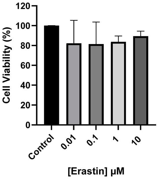
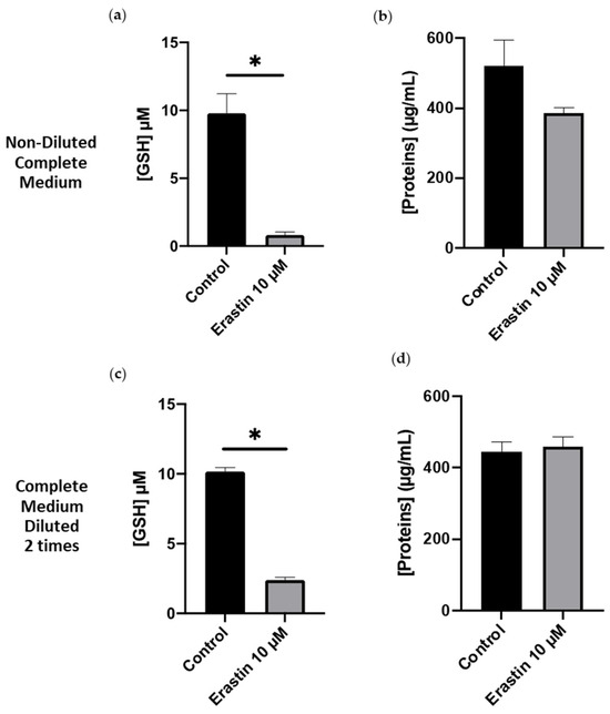
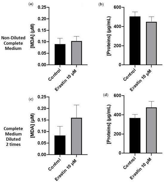
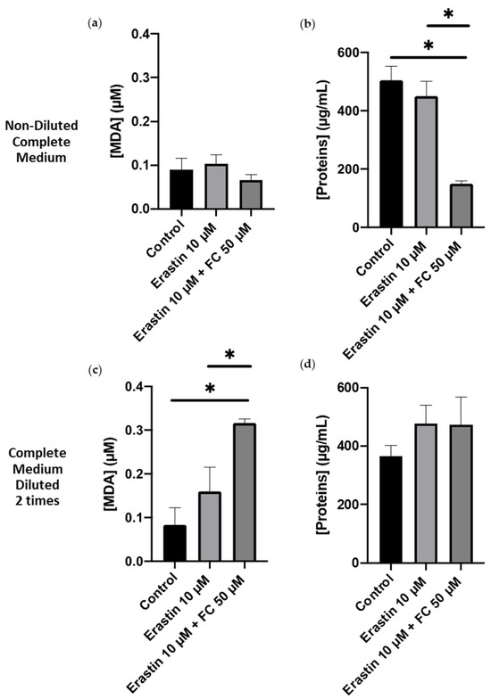
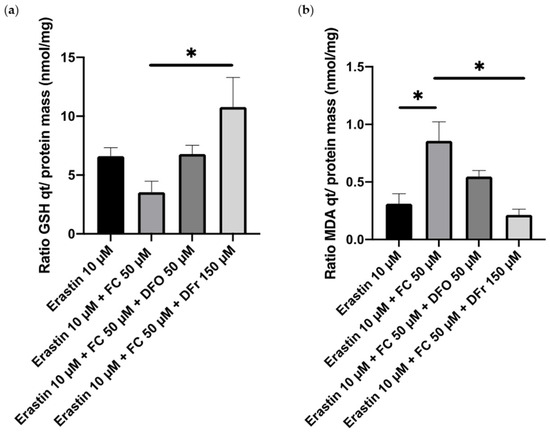
A Smooth Muscle Cell-Based Ferroptosis Model to Evaluate Iron-Chelating Molecules for Cardiovascular Disease Treatment
by
Sarah El Hajj, Laetitia Canabady-Rochelle, Isabelle Fries-Raeth and Caroline Gaucher
Viewed by 745
Abstract
Dysregulation of iron homeostasis causes iron-mediated cell death, recently described as ferroptosis. Ferroptosis is reported in many chronic diseases, such as hepatic cancer, renal, and cardiovascular diseases (heart failure, atherosclerosis). However, there is a notable scarcity of research studies in the existing literature
[...] Read more.
Dysregulation of iron homeostasis causes iron-mediated cell death, recently described as ferroptosis. Ferroptosis is reported in many chronic diseases, such as hepatic cancer, renal, and cardiovascular diseases (heart failure, atherosclerosis). However, there is a notable scarcity of research studies in the existing literature that explore treatments capable of preventing ferroptosis. Additionally, as far as the author is aware, there is currently no established model for studying ferroptosis within cardiovascular cells, which would be essential for assessing metal-chelating molecules with the potential ability to inhibit ferroptosis and their application in the treatment of cardiovascular diseases. In this study, a smooth muscle cell-based ferroptosis model is developed upon the inhibition of the system X
c− transporter by erastin associated or not with Fe(III) overload, and its rescue upon the introduction of well-known iron chelators, deferoxamine and deferiprone. We showed that erastin alone decreased the intracellular concentration of glutathione (GSH) without affecting peroxidized lipid concentrations. Erastin with ferric citrate was able to decrease intracellular GSH and induce lipid peroxidation after overnight incubation. Only deferiprone was able to rescue the cells from ferroptosis by decreasing lipid peroxidation via iron ion chelation in a 3:1 molar ratio.
Full article
►▼
Show Figures
Open AccessArticle
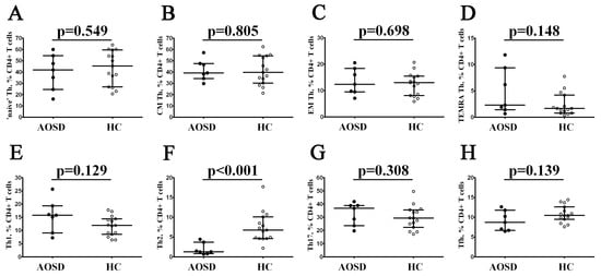
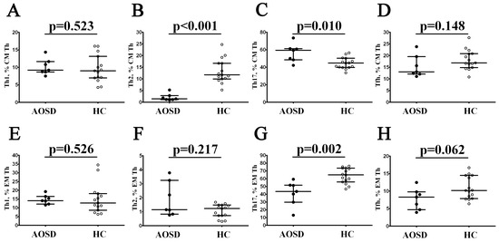
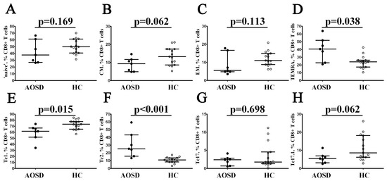
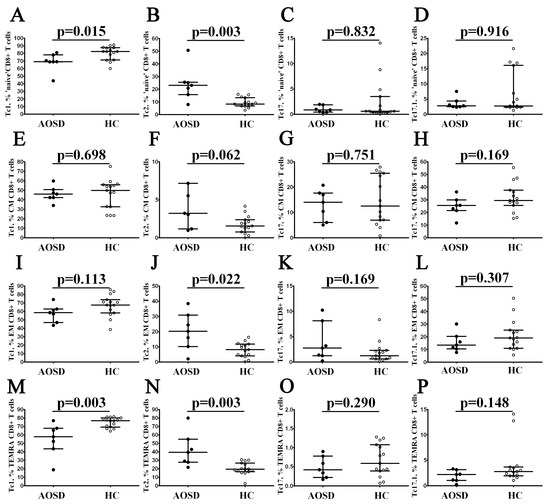
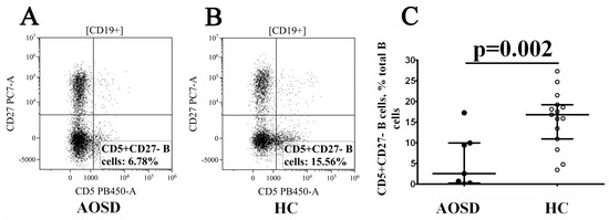
Deep Immunophenotyping of Circulating T and B Cells in Relapsing Adult-Onset Still’s Disease
by
Valentina Myachikova, Igor Kudryavtsev, Artem Rubinstein, Arthur Aquino, Dmitry Isakov, Alexey Golovkin and Alexey Maslyanskiy
Viewed by 762
Abstract
Adult-onset Still’s disease (AOSD) is a complex systemic inflammatory disorder, categorized as an ‘IL-1 driven’ inflammasomapathy. Despite this, the interaction between T and B cells remains poorly understood. We conducted a study, enrolling 7 patients with relapsing AOSD and 15 healthy control subjects,
[...] Read more.
Adult-onset Still’s disease (AOSD) is a complex systemic inflammatory disorder, categorized as an ‘IL-1 driven’ inflammasomapathy. Despite this, the interaction between T and B cells remains poorly understood. We conducted a study, enrolling 7 patients with relapsing AOSD and 15 healthy control subjects, utilizing deep flow cytometry analysis to examine peripheral blood T- and B-cell subsets. T-cell and B-cell subsets were significantly altered in patients with AOSD. Within CD4+ T cells, Th2 cells were decreased. Additionally, Th17 cell and follicular Th cell subsets were altered within CD45RA–CD62L+ and CD45RA–CD62L– Th cells in patients with AOSD compared to healthy controls. We identified changes in CD8+ T cell maturation and ‘polarization’ in AOSD patients, with an elevated presence of the TEMRA CD8+ T cell subset. Furthermore, the percentage of Tc1 cells was decreased, while the frequency of CCR6–CXCR3– Tc2 cells was elevated. Finally, we determined that the frequency of CD5+CD27– B cells was dramatically decreased in patients with AOSD compared to healthy controls. Further investigations on a large group of patients with AOSD are required to evaluate these adaptive immunity cells in the disease pathogenesis.
Full article
►▼
Show Figures
Open AccessBrief Report
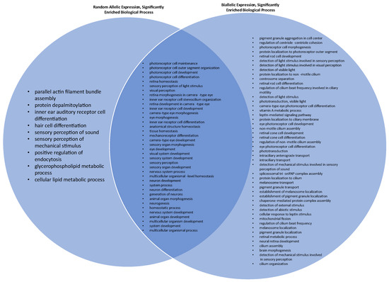
Random Allelic Expression in Inherited Retinal Disease Genes
by
Collin J. Richards and Jose S. Pulido
Viewed by 880
Abstract
Inherited retinal diseases (IRDs) are a significant contributor to visual loss in children and young adults, falling second only to diabetic retinopathy. Understanding the pathogenic mechanisms of IRDs remains paramount. Some autosomal genes exhibit random allelic expression (RAE), similar to X-chromosome inactivation. This
[...] Read more.
Inherited retinal diseases (IRDs) are a significant contributor to visual loss in children and young adults, falling second only to diabetic retinopathy. Understanding the pathogenic mechanisms of IRDs remains paramount. Some autosomal genes exhibit random allelic expression (RAE), similar to X-chromosome inactivation. This study identifies RAE genes in IRDs. Genes in the Retinal Information Network were cross-referenced with the recent literature to identify expression profiles, RAE, or biallelic expression (BAE). Loss-of-function intolerance (LOFI) was determined by cross-referencing the existing literature. Molecular and biological pathways that are significantly enriched were evaluated using gene ontology. A total of 184 IRD-causing genes were evaluated. Of these, 31 (16.8%) genes exhibited RAE. LOFI was exhibited in 6/31 (19.4%) of the RAE genes and 18/153 (11.8%) of the BAE genes. Brain tissue exhibited BAE in 107/128 (83.6%) genes for both sexes. The molecular pathways significantly enriched among BAE genes were photoreceptor activity, tubulin binding, and nucleotide/ribonucleotide binding. The biologic pathways significantly enriched for RAE genes were equilibrioception, parallel actin filament bundle assembly, photoreceptor cell outer segment organization, and protein depalmitoylation. Allele-specific expression may be a mechanism underlying IRD phenotypic variability, with clonal populations of embryologic precursor cells exhibiting RAE. Brain tissue preferentially exhibited BAE, possibly due to selective pressures against RAE. Pathways critical for cellular and visual function were enriched in BAE, which may offer a survival benefit.
Full article
►▼
Show Figures
Open AccessReview
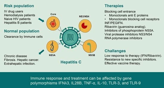
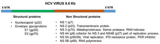
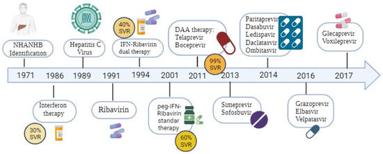
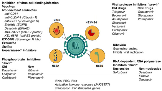
A Synopsis of Hepatitis C Virus Treatments and Future Perspectives
by
Christian Medina, Alexis Hipólito García, Francis Isamarg Crespo, Félix Isidro Toro, Soriuska José Mayora and Juan Bautista De Sanctis
Cited by 1 | Viewed by 2318
Abstract
Hepatitis C virus (HCV) infection is a worldwide public health problem. Chronic infection with HCV can lead to liver cirrhosis or cancer. Although some immune-competent individuals can clear the virus, others develop chronic HCV disease due to viral mutations or an impaired immune
[...] Read more.
Hepatitis C virus (HCV) infection is a worldwide public health problem. Chronic infection with HCV can lead to liver cirrhosis or cancer. Although some immune-competent individuals can clear the virus, others develop chronic HCV disease due to viral mutations or an impaired immune response. IFNs type I and III and the signal transduction induced by them are essential for a proper antiviral effect. Research on the viral cycle and immune escape mechanisms has formed the basis of therapeutic strategies to achieve a sustained virological response (SVR). The first therapies were based on IFNα; then, IFNα plus ribavirin (IFN–RBV); and then, pegylated-IFNα-RBV (PEGIFNα-RIV) to improve cytokine pharmacokinetics. However, the maximum SVR was 60%, and several significant side effects were observed, decreasing patients’ treatment adherence. The development of direct-acting antivirals (DAAs) significantly enhanced the SVR (>90%), and the compounds were able to inhibit HCV replication without significant side effects, even in paediatric populations. The management of coinfected HBV–HCV and HCV–HIV patients has also improved based on DAA and PEG-IFNα-RBV (HBV–HCV). CD4 cells are crucial for an effective antiviral response. The IFNλ3, IL28B, TNF-α, IL-10, TLR-3, and TLR-9 gene polymorphisms are involved in viral clearance, therapeutic responses, and hepatic pathologies. Future research should focus on searching for strategies to circumvent resistance-associated substitution (RAS) to DAAs, develop new therapeutic schemes for different medical conditions, including organ transplant, and develop vaccines for long-lasting cellular and humoral responses with cross-protection against different HCV genotypes. The goal is to minimise the probability of HCV infection, HCV chronicity and hepatic carcinoma.
Full article
►▼
Show Figures
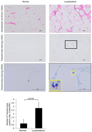
Open AccessArticle
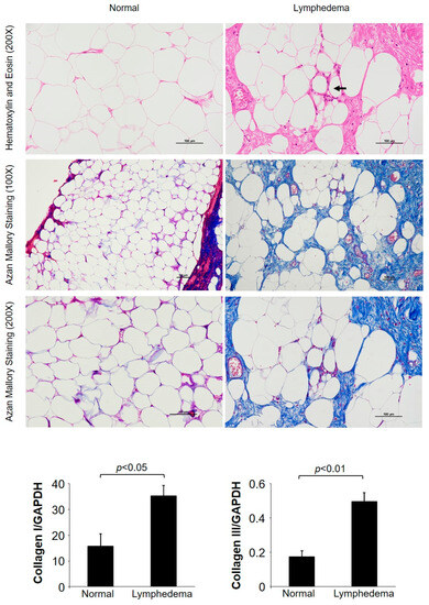
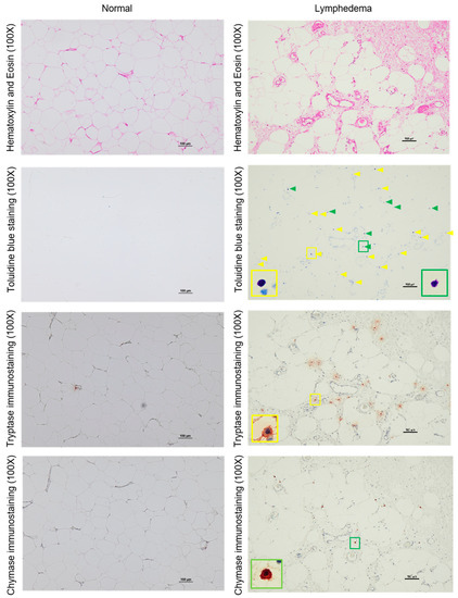
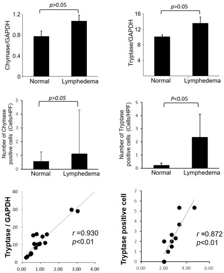
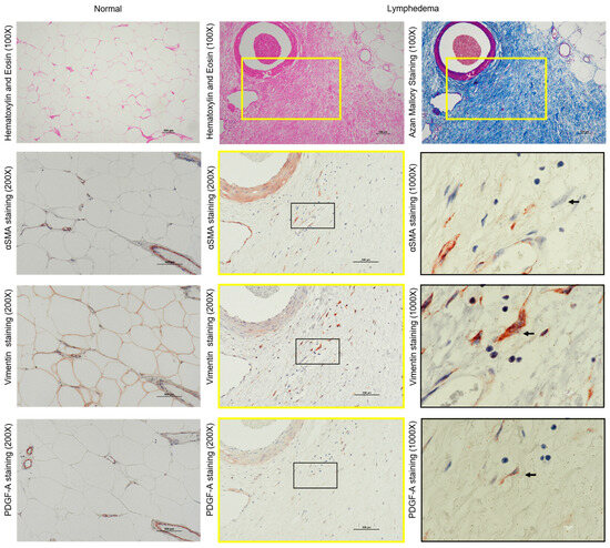
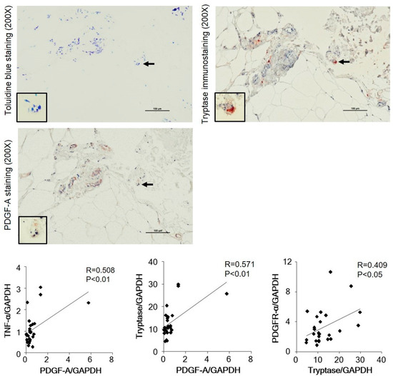
Tryptase-Positive Mast Cells Promote Adipose Fibrosis in Secondary Lymphedema through PDGF
by
Takashi Nuri, Denan Jin, Shinji Takai and Koichi Ueda
Viewed by 881
Abstract
Lymphedema is a chronic and progressive condition that causes physical disfigurement and psychological trauma due to the accumulation of lymphatic fluid in the interstitial space. Once it develops, lymphedema is difficult to treat because it leads to the fibrosis of adipose tissue. However,
[...] Read more.
Lymphedema is a chronic and progressive condition that causes physical disfigurement and psychological trauma due to the accumulation of lymphatic fluid in the interstitial space. Once it develops, lymphedema is difficult to treat because it leads to the fibrosis of adipose tissue. However, the mechanism behind this remains unclear. The purpose of this study was to investigate the involvement of mast cells (MCs) in the adipose tissues of patients with lymphedema. We found that fibrosis spread through blood vessels in the adipose tissues of lymphedema patients, and the expression of the collagen I and III genes was significantly increased compared to that of those in normal adipose tissue. Immunostaining of vimentin and α-smooth muscle actin showed that fibroblasts were the main cellular components in severely fibrotic regions. Toluidine blue staining confirmed a significant increase in the number of MCs in the adipose tissues of lymphedema patients, and immunostaining of serial sections of adipose tissue showed a significant increase in the number of tryptase-positive cells in lymphedema tissues compared with those in normal adipose tissues. Linear regression analyses revealed significant positive correlations between tryptase and the expressions of the TNF-α, platelet-derived growth factor (PDGF)-A, and PDGFR-α genes. PDGF-A–positive staining was observed in both fibroblasts and granules of tryptase-positive MCs. These results suggest that MC-derived tryptase plays a role in the fibrosis of adipose tissue due to lymphedema directly or in cooperation with other mediators.
Full article
►▼
Show Figures
Open AccessArticle
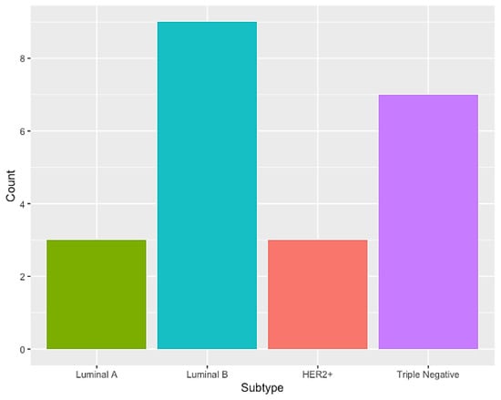
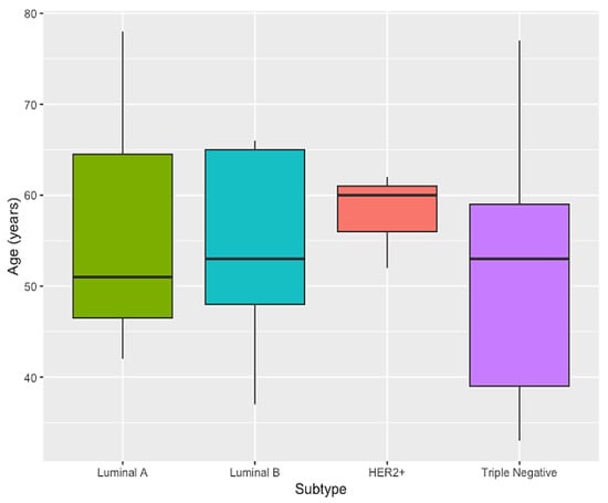
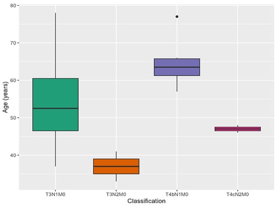
Investigation of Genetic Factors and Clinical Data in Breast Cancer Highlights the Importance of Breastfeeding and Cancer History
by
Amanda Mercês, Rebecca da-Silva-Cruz, Caio S. Silva, Rommel Burbano, Ândrea Ribeiro-dos-Santos and Giovanna C. Cavalcante
Viewed by 786
Abstract
Breast cancer (BC) is the type of neoplasm that most affects women worldwide. It is known that one of the hallmarks of cancer is the resistance to cell death with the evasion of apoptosis. Considering the relevance of
TP53,
BCL2,
CASP3,
[...] Read more.
Breast cancer (BC) is the type of neoplasm that most affects women worldwide. It is known that one of the hallmarks of cancer is the resistance to cell death with the evasion of apoptosis. Considering the relevance of
TP53,
BCL2,
CASP3, and
CASP9 genes for the occurrence of the intrinsic apoptosis, this study investigated the distribution of the genetic variants rs17880560 (
TP53), rs11269260 (
BCL2), rs4647655 (
CASP3), rs4645982, and rs61079693 (
CASP9), as well as genetic ancestry and clinical data, in a BC cohort from the Brazilian Amazon that other variants in these genes might play a role in this process. In the present study, 22 breast cancer tissues and 10 non-cancerous tissues were used, therefore, 32 samples from different patients were subjected to genotyping. We observed that breastfeeding and cancer history were factors that need to be considered for BC (
p = 0.022). Therefore, this study contributed to a greater understanding of intrinsic apoptosis in BC, reinforcing previous data that suggest that the history of cancer might be a condition that affects the development of BC and that breastfeeding may act as a protective factor for this type of cancer. We recommend more studies on the genetic factors investigated here, aiming at a future with tools that can help in the early diagnosis.
Full article
►▼
Show Figures
Open AccessReview
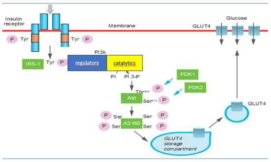
Gender Differences in Insulin Resistance: New Knowledge and Perspectives
by
Tiziana Ciarambino, Pietro Crispino, Gloria Guarisco and Mauro Giordano
Cited by 7 | Viewed by 2349
Abstract
Insulin resistance is the main mechanism in a whole series of pathological conditions, which are not only of metabolic interest but also of a systemic type. This phenomenon means that the body’s cells become less sensitive to the hormone insulin, leading to higher
[...] Read more.
Insulin resistance is the main mechanism in a whole series of pathological conditions, which are not only of metabolic interest but also of a systemic type. This phenomenon means that the body’s cells become less sensitive to the hormone insulin, leading to higher levels of insulin in the blood. Insulin resistance is a phenomenon that can be found in both men and women and in particular, in the latter, it is found mainly after menopause. Premenopause, hormonal fluctuations during the menstrual cycle, and the presence of estrogen can affect insulin sensitivity. Androgens, such as testosterone, are typically higher in men and can contribute to insulin resistance. In both sexes, different human body types affect the distribution and location of body fat, also influencing the development of diabetes and cardiovascular disease. Insulin resistance is also associated with some neurological and neurogenerative disorders, polycystic ovary syndrome, atherosclerosis, and some of the main neoplastic pathologies. A healthy lifestyle, including regular physical activity, a balanced diet, and self-maintenance, can help to prevent the onset of insulin resistance, regardless of gender, although the different habits between men and women greatly affect the implementation of preventative guidelines that help in fighting the manifestations of this metabolic disorder. This review may help to shed light on gender differences in metabolic diseases by placing a necessary focus on personalized medical management and by inspiring differentiated therapeutic approaches.
Full article
►▼
Show Figures
Open AccessArticle
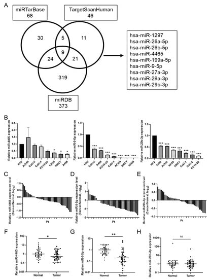
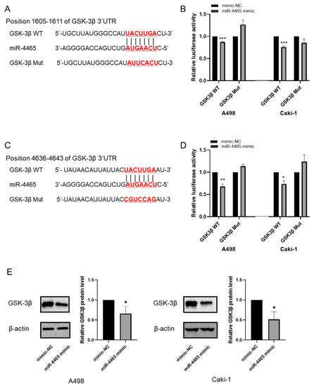
MicroRNAs as Potential Regulators of GSK-3β in Renal Cell Carcinoma
by
Masaki Murata, Vladimir Bilim, Yuko Shirono, Akira Kazama, Kaede Hiruma, Masayuki Tasaki and Yoshihiko Tomita
Viewed by 1116
Abstract
The prognosis of patients with advanced renal cell carcinoma (RCC) has improved with newer therapies, including molecular-targeted therapies and immuno-oncology agents. Despite these therapeutic advances, many patients with metastatic disease remain uncured. Inhibition of glycogen synthase kinase-3β (GSK-3β) is a promising new therapeutic
[...] Read more.
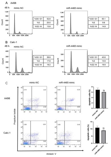
The prognosis of patients with advanced renal cell carcinoma (RCC) has improved with newer therapies, including molecular-targeted therapies and immuno-oncology agents. Despite these therapeutic advances, many patients with metastatic disease remain uncured. Inhibition of glycogen synthase kinase-3β (GSK-3β) is a promising new therapeutic strategy for RCC; however, the precise regulatory mechanism has not yet been fully elucidated. MicroRNAs (miRNAs) act as post-translational regulators of target genes, and we investigated the potential regulation of miRNAs on GSK-3β in RCC. We selected nine candidate miRNAs from three databases that could potentially regulate GSK-3β. Among these, hsa-miR-4465 (miR-4465) was downregulated in RCC cell lines and renal cancer tissues. Furthermore, luciferase assays revealed that miR-4465 directly interacted with the 3′ untranslated region of GSK-3β, and Western blot analysis showed that overexpression of miR-4465 significantly decreased GSK-3β protein expression. Functional assays showed that miR-4465 overexpression significantly suppressed cell invasion of A498 and Caki-1 cells; however, cell proliferation and migration were suppressed only in Caki-1 and A498 cells, respectively, with no effect on cell cycle and apoptosis. In conclusion, miR-4465 regulates GSK-3β expression but does not consistently affect RCC cell function as a single molecule. Further comprehensive investigation of regulatory networks is required in this field.
Full article
►▼
Show Figures


