Cell Biology of Cancer Invasion

A special issue of Cancers (ISSN 2072-6694). This special issue belongs to the section "Molecular Cancer Biology".

Deadline for manuscript submissions: 30 June 2024 | Viewed by 4278

Special Issue Editor


E-Mail Website
Guest Editor
Sasaki Institute, Tokyo, Japan
Interests: invadopodia; cell biology; cancer biology; cell culture; cell signaling; apoptosis; cell proliferation; western blot analysis; immunohistochemistry; flow cytometry; molecular cell biology

Special Issue Information

Dear Colleagues,

The invasion of tumor cells is a prerequisite for metastasis, the most dreadful aspect of cancer. Cancer cells detach from the primary tumor and migrate through the tumor stroma to reach blood vessels, where they intravasate into the bloodstream. After cancer cells arrive at the distant organ, they extravasate and colonize to form secondary tumors. A dense extracellular matrix that acts as a physical barrier exists within the basement membrane, tumor stroma, and blood vessel walls. Therefore, the invasive activity of tumor cells, i.e., degradation of the extracellular matrix and coordinated migration, is necessary for cancer metastasis.

The acquisition of invasive phenotypes is induced by the activation of oncogenic signaling pathways and epithelial–mesenchymal transition. The invasion of tumor cells is mediated by the formation of cellular structures called invadopodia, dynamic reorganization of the cytoskeleton, secretion and focalization of matrix metalloproteinases, and remodeling of the extracellular matrix. Moreover, tumor microenvironments, such as fibrotic stroma and tumor stromal cells, including cancer-associated fibroblasts (CAF) and tumor-associated macrophages (TAM), are known to promote the invasion of tumor cells. Recent advances including genome sequencing, multi-omics analyses, gene-editing technology, and state-of-the-art microscopies have provided new insights into the mechanisms of cancer invasion.

This Special Issue invites original research articles and reviews that are related to cell biological aspects of cancer invasion, such as the topics described above.

We look forward to receiving your contributions.

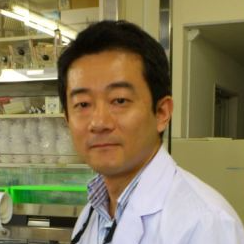
Dr. Hideki Yamaguchi
Guest Editor

Manuscript Submission Information

Manuscripts should be submitted online at www.mdpi.com by registering and logging in to this website. Once you are registered, click here to go to the submission form. Manuscripts can be submitted until the deadline. All submissions that pass pre-check are peer-reviewed. Accepted papers will be published continuously in the journal (as soon as accepted) and will be listed together on the special issue website. Research articles, review articles as well as short communications are invited. For planned papers, a title and short abstract (about 100 words) can be sent to the Editorial Office for announcement on this website.

Submitted manuscripts should not have been published previously, nor be under consideration for publication elsewhere (except conference proceedings papers). All manuscripts are thoroughly refereed through a single-blind peer-review process. A guide for authors and other relevant information for submission of manuscripts is available on the Instructions for Authors page. Cancers is an international peer-reviewed open access semimonthly journal published by MDPI.

Please visit the Instructions for Authors page before submitting a manuscript. The Article Processing Charge (APC) for publication in this open access journal is 2900 CHF (Swiss Francs). Submitted papers should be well formatted and use good English. Authors may use MDPI's English editing service prior to publication or during author revisions.

Keywords

  • invasion
  • metastasis
  • intravasation
  • extravasation
  • invadopodia
  • tumor microenvironment
  • epithelial–mesenchymal transition
  • cell migration
  • cytoskeleton
  • cell adhesion
  • extracellular matrix
  • matrix metalloprotease
  • cancer-associated fibroblast
  • tumor-associated macrophage

Published Papers (5 papers)

Order results
Result details
Select all
Export citation of selected articles as:

Research

Jump to: Review, Other

17 pages, 5166 KiB  
Article
KLC1-ROS1 Fusion Exerts Oncogenic Properties of Glioma Cells via Specific Activation of JAK-STAT Pathway
by Takashi Fujii, Yoshiko Nakano, Daichi Hagita, Nobuyuki Onishi, Arumu Endo, Masaya Nakagawa, Toru Yoshiura, Yohei Otsuka, Satoru Takeuchi, Mario Suzuki, Yuzaburo Shimizu, Terushige Toyooka, Yuko Matsushita, Yuko Hibiya, Satoshi Tomura, Akihide Kondo, Kojiro Wada, Koichi Ichimura and Arata Tomiyama
Cancers 2024, 16(1), 9; https://doi.org/10.3390/cancers16010009 - 19 Dec 2023
Viewed by 757
Abstract
Here, we investigated the detailed molecular oncogenic mechanisms of a novel receptor tyrosine kinase (RTK) fusion, KLC1-ROS1, with an adapter molecule, KLC1, and an RTK, ROS1, discovered in pediatric glioma, and we explored a novel therapeutic target for glioma that possesses oncogenic [...] Read more.
Here, we investigated the detailed molecular oncogenic mechanisms of a novel receptor tyrosine kinase (RTK) fusion, KLC1-ROS1, with an adapter molecule, KLC1, and an RTK, ROS1, discovered in pediatric glioma, and we explored a novel therapeutic target for glioma that possesses oncogenic RTK fusion. When wild-type ROS1 and KLC1-ROS1 fusions were stably expressed in the human glioma cell lines A172 and U343MG, immunoblotting revealed that KLC1-ROS1 fusion specifically activated the JAK2-STAT3 pathway, a major RTK downstream signaling pathway, when compared with wild-type ROS1. Immunoprecipitation of the fractionated cell lysates revealed a more abundant association of the KLC1-ROS1 fusion with JAK2 than that observed for wild-type ROS1 in the cytosolic fraction. A mutagenesis study of the KLC1-ROS1 fusion protein demonstrated the fundamental roles of both the KLC1 and ROS1 domains in the constitutive activation of KLC1-ROS1 fusion. Additionally, in vitro assays demonstrated that KLC1-ROS1 fusion upregulated cell proliferation, invasion, and chemoresistance when compared to wild-type ROS1. Combination treatment with the chemotherapeutic agent temozolomide and an inhibitor of ROS1, JAK2, or a downstream target of STAT3, demonstrated antitumor effects against KLC1-ROS1 fusion-expressing glioma cells. Our results demonstrate that KLC1-ROS1 fusion exerts oncogenic activity through serum-independent constitutive activation, resulting in specific activation of the JAK-STAT pathway. Our data suggested that molecules other than RTKs may serve as novel therapeutic targets for RTK fusion in gliomas. Full article
(This article belongs to the Special Issue Cell Biology of Cancer Invasion)
Show Figures

Figure 1

Review

Jump to: Research, Other

13 pages, 618 KiB  
Review
A Review of the Paradigmatic Role of Adipose Tissue in Renal Cancer: Fat Measurement and Tumor Behavior Features
by Eliodoro Faiella, Elva Vergantino, Federica Vaccarino, Amalia Bruno, Gloria Perillo, Rosario Francesco Grasso, Bruno Beomonte Zobel and Domiziana Santucci
Cancers 2024, 16(9), 1697; https://doi.org/10.3390/cancers16091697 (registering DOI) - 27 Apr 2024
Viewed by 277
Abstract
(1) Background: Renal-cell carcinoma (RCC) incidence has been steadily rising, with obesity identified as a potential risk factor. However, the relationship between obesity and RCC prognosis remains unclear. This systematic review aims to investigate the impact of different adipose tissue measurements on RCC [...] Read more.
(1) Background: Renal-cell carcinoma (RCC) incidence has been steadily rising, with obesity identified as a potential risk factor. However, the relationship between obesity and RCC prognosis remains unclear. This systematic review aims to investigate the impact of different adipose tissue measurements on RCC behavior and prognosis. (2) Methods: A search of MEDLINE databases identified 20 eligible studies focusing on various fat measurements, including visceral adipose tissue (VAT), subcutaneous adipose tissue (SAT), perirenal adipose tissue (PRAT), and the Mayo adhesive probability (MAP) score. (3) Results: The review revealed conflicting findings regarding the association between adipose tissue measurements and RCC outcomes. While some studies suggested a protective role of certain fat deposits, particularly VAT, against disease progression and mortality, others reported contradictory results across different adipose metrics and RCC subtypes. (4) Conclusions: Methodological variations and limitations, such as retrospective designs and sample size constraints, pose challenges to standardization and generalizability. Further research is needed to understand these associations better and establish standardized approaches for adiposity assessment in RCC patients, which could inform clinical practice and therapeutic decision-making. Full article
(This article belongs to the Special Issue Cell Biology of Cancer Invasion)
Show Figures

Figure 1

12 pages, 2652 KiB  
Review
Heterocellular Adhesion in Cancer Invasion and Metastasis: Interactions between Cancer Cells and Cancer-Associated Fibroblasts
by Hideki Yamaguchi and Makoto Miyazaki
Cancers 2024, 16(9), 1636; https://doi.org/10.3390/cancers16091636 - 24 Apr 2024
Viewed by 252
Abstract
Cancer invasion is a requisite for the most malignant progression of cancer, that is, metastasis. The mechanisms of cancer invasion were originally studied using in vitro cell culture systems, in which cancer cells were cultured using artificial extracellular matrices (ECMs). However, conventional culture [...] Read more.
Cancer invasion is a requisite for the most malignant progression of cancer, that is, metastasis. The mechanisms of cancer invasion were originally studied using in vitro cell culture systems, in which cancer cells were cultured using artificial extracellular matrices (ECMs). However, conventional culture systems do not precisely recapitulate in vivo cancer invasion because the phenotypes of cancer cells in tumor tissues are strongly affected by the tumor microenvironment (TME). Cancer-associated fibroblasts (CAFs) are the most abundant cell type in the TME and accelerate cancer progression through invasion, metastasis, therapy resistance, and immune suppression. Thus, the reciprocal interactions between CAFs and cancer cells have been extensively studied, leading to the identification of factors that mediate cellular interactions, such as growth factors, cytokines, and extracellular vesicles. In addition, the importance of direct heterocellular adhesion between cancer cells and CAFs in cancer progression has recently been elucidated. In particular, CAFs are directly associated with cancer cells, allowing them to invade the ECM and metastasize to distant organs. In this review, we summarize the recent progress in understanding the molecular and cellular mechanisms of the direct heterocellular interaction in CAF-led cancer invasion and metastasis, with an emphasis on gastric cancer. Full article
(This article belongs to the Special Issue Cell Biology of Cancer Invasion)
Show Figures

Figure 1

30 pages, 1874 KiB  
Review
Molecular Insight into Gastric Cancer Invasion—Current Status and Future Directions
by Tasuku Matsuoka and Masakazu Yashiro
Cancers 2024, 16(1), 54; https://doi.org/10.3390/cancers16010054 - 21 Dec 2023
Cited by 2 | Viewed by 1151
Abstract
Gastric cancer (GC) is one of the most common malignancies worldwide. There has been no efficient therapy for stage IV GC patients due to this disease’s heterogeneity and dissemination ability. Despite the rapid advancement of molecular targeted therapies, such as HER2 and immune [...] Read more.
Gastric cancer (GC) is one of the most common malignancies worldwide. There has been no efficient therapy for stage IV GC patients due to this disease’s heterogeneity and dissemination ability. Despite the rapid advancement of molecular targeted therapies, such as HER2 and immune checkpoint inhibitors, survival of GC patients is still unsatisfactory because the understanding of the mechanism of GC progression is still incomplete. Invasion is the most important feature of GC metastasis, which causes poor mortality in patients. Recently, genomic research has critically deepened our knowledge of which gene products are dysregulated in invasive GC. Furthermore, the study of the interaction of GC cells with the tumor microenvironment has emerged as a principal subject in driving invasion and metastasis. These results are expected to provide a profound knowledge of how biological molecules are implicated in GC development. This review summarizes the advances in our current understanding of the molecular mechanism of GC invasion. We also highlight the future directions of the invasion therapeutics of GC. Compared to conventional therapy using protease or molecular inhibitors alone, multi-therapy targeting invasion plasticity may seem to be an assuring direction for the progression of novel strategies. Full article
(This article belongs to the Special Issue Cell Biology of Cancer Invasion)
Show Figures

Figure 1

Other

Jump to: Research, Review

11 pages, 2766 KiB  
Commentary
Extracellular Vesicles: Biological Packages That Modulate Tumor Cell Invasion
by Madison Schmidtmann and Crislyn D’Souza-Schorey
Cancers 2023, 15(23), 5617; https://doi.org/10.3390/cancers15235617 - 28 Nov 2023
Viewed by 826
Abstract
Tumor progression, from early-stage invasion to the formation of distal metastases, relies on the capacity of tumor cells to modify the extracellular matrix (ECM) and communicate with the surrounding stroma. Extracellular vesicles (EVs) provide an important means to regulate cell invasion due to [...] Read more.
Tumor progression, from early-stage invasion to the formation of distal metastases, relies on the capacity of tumor cells to modify the extracellular matrix (ECM) and communicate with the surrounding stroma. Extracellular vesicles (EVs) provide an important means to regulate cell invasion due to the selective inclusion of cargoes such as proteases and matrix proteins into EVs that can degrade or modify the ECM. EVs have also been shown to facilitate intercellular communication in the tumor microenvironment through paracrine signaling, which can impact ECM invasion by cancer cells. Here, we describe the current knowledge of EVs as facilitators of tumor invasion by virtue of their effects on proteolytic degradation and modification of the ECM, their ability to educate the stromal cells in the tumor microenvironment, and their role as mediators of long-range communication aiding in cell invasion and matrix remodeling at secondary sites. Full article
(This article belongs to the Special Issue Cell Biology of Cancer Invasion)
Show Figures

Figure 1

Back to TopTop