Radiation Therapy for Brain Tumors

A special issue of Cancers (ISSN 2072-6694). This special issue belongs to the section "Cancer Therapy".

Deadline for manuscript submissions: 31 October 2024 | Viewed by 4574

Special Issue Editor

Department of Radiation Oncology, Winship Cancer Institute of Emory University, Atlanta, GA 30322, USA
Interests: advanced imaging; proton therapy, image-guided radiation therapy, gliomas, radiosurgery, hypofractionated radiotherapy

Special Issue Information

Dear Colleagues,

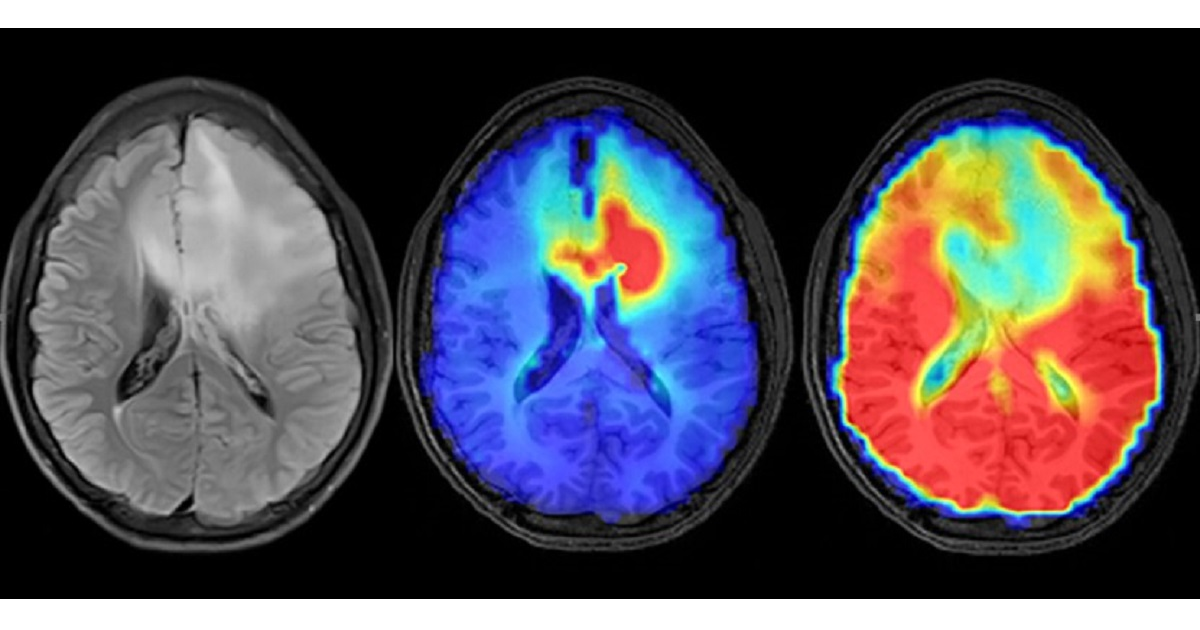
Radiation therapy plays a crucial role in managing both benign and malignant brain tumors. Recent advances in imaging and radiation delivery have expanded the use of radiation therapy and significantly improved patient outcomes. Advanced imaging techniques such as magnetic resonance imaging (MRI) utilizing three-dimensional spectroscopy and perfusion have enhanced the delineation of gliomas, allowing for more precise treatment planning and assessment of the treatment response. Furthermore, new radiation delivery techniques such as IMRT, stereotactic radiosurgery, and proton therapy have enabled more accurate targeting of tumors while minimizing damage to surrounding healthy tissue. These advancements have resulted in improved treatment outcomes and enhanced quality of life for patients with brain and spine tumors.

This series of articles will focus on recent breakthroughs in tumor imaging and radiation therapy for brain and spine tumors. The articles will cover various topics, including the role of functional imaging in treatment planning, the effectiveness of immunotherapy in combination with radiation therapy, and the potential of radiomics in predicting the treatment response. These articles will provide insights into the current state-of-the-art in radiation therapy for brain and spine tumors and offer valuable information for clinicians involved in managing these patients.

Dr. Jim Zhong
Guest Editor

Manuscript Submission Information

Manuscripts should be submitted online at www.mdpi.com by registering and logging in to this website. Once you are registered, click here to go to the submission form. Manuscripts can be submitted until the deadline. All submissions that pass pre-check are peer-reviewed. Accepted papers will be published continuously in the journal (as soon as accepted) and will be listed together on the special issue website. Research articles, review articles as well as short communications are invited. For planned papers, a title and short abstract (about 100 words) can be sent to the Editorial Office for announcement on this website.

Submitted manuscripts should not have been published previously, nor be under consideration for publication elsewhere (except conference proceedings papers). All manuscripts are thoroughly refereed through a single-blind peer-review process. A guide for authors and other relevant information for submission of manuscripts is available on the Instructions for Authors page. Cancers is an international peer-reviewed open access semimonthly journal published by MDPI.

Please visit the Instructions for Authors page before submitting a manuscript. The Article Processing Charge (APC) for publication in this open access journal is 2900 CHF (Swiss Francs). Submitted papers should be well formatted and use good English. Authors may use MDPI's English editing service prior to publication or during author revisions.

Keywords

  • radiosurgery
  • hypofractionated radiosurgery
  • spectroscopic MRI
  • proton radiation
  • image-guided radiotherapy
  • radiotherapy for benign tumors
  • radiotherapy for gliomas
  • gamma knife

Published Papers (3 papers)

Order results
Result details
Select all
Export citation of selected articles as:

Research

Jump to: Review

12 pages, 1374 KiB  
Article
A Deep Learning Approach for Automatic Segmentation during Daily MRI-Linac Radiotherapy of Glioblastoma
by Adrian L. Breto, Kaylie Cullison, Evangelia I. Zacharaki, Veronica Wallaengen, Danilo Maziero, Kolton Jones, Alessandro Valderrama, Macarena I. de la Fuente, Jessica Meshman, Gregory A. Azzam, John C. Ford, Radka Stoyanova and Eric A. Mellon
Cancers 2023, 15(21), 5241; https://doi.org/10.3390/cancers15215241 - 31 Oct 2023
Viewed by 1563
Abstract
Glioblastoma changes during chemoradiotherapy are inferred from high-field MRI before and after treatment but are rarely investigated during radiotherapy. The purpose of this study was to develop a deep learning network to automatically segment glioblastoma tumors on daily treatment set-up scans from the [...] Read more.
Glioblastoma changes during chemoradiotherapy are inferred from high-field MRI before and after treatment but are rarely investigated during radiotherapy. The purpose of this study was to develop a deep learning network to automatically segment glioblastoma tumors on daily treatment set-up scans from the first glioblastoma patients treated on MRI-linac. Glioblastoma patients were prospectively imaged daily during chemoradiotherapy on 0.35T MRI-linac. Tumor and edema (tumor lesion) and resection cavity kinetics throughout the treatment were manually segmented on these daily MRI. Utilizing a convolutional neural network, an automatic segmentation deep learning network was built. A nine-fold cross-validation schema was used to train the network using 80:10:10 for training, validation, and testing. Thirty-six glioblastoma patients were imaged pre-treatment and 30 times during radiotherapy (n = 31 volumes, total of 930 MRIs). The average tumor lesion and resection cavity volumes were 94.56 ± 64.68 cc and 72.44 ± 35.08 cc, respectively. The average Dice similarity coefficient between manual and auto-segmentation for tumor lesion and resection cavity across all patients was 0.67 and 0.84, respectively. This is the first brain lesion segmentation network developed for MRI-linac. The network performed comparably to the only other published network for auto-segmentation of post-operative glioblastoma lesions. Segmented volumes can be utilized for adaptive radiotherapy and propagated across multiple MRI contrasts to create a prognostic model for glioblastoma based on multiparametric MRI. Full article
(This article belongs to the Special Issue Radiation Therapy for Brain Tumors)
Show Figures

Figure 1

17 pages, 1676 KiB  
Article
Examining the Effect of ALK and EGFR Mutations on Survival Outcomes in Surgical Lung Brain Metastasis Patients
by Sneha Sai Mannam, David P. Bray, Chibueze D. Nwagwu, Jim Zhong, Hui-Kuo Shu, Bree Eaton, Lisa Sudmeier, Subir Goyal, Christopher Deibert, Edjah K. Nduom, Jeffrey Olson and Kimberly B. Hoang
Cancers 2023, 15(19), 4773; https://doi.org/10.3390/cancers15194773 - 28 Sep 2023
Viewed by 928
Abstract
In the context of the post-genomic era, where targeted oncological therapies like monoclonal antibodies (mAbs) and tyrosine-kinase inhibitors (TKIs) are gaining prominence, this study investigates whether these therapies can enhance survival for lung carcinoma patients with specific genetic mutations—EGFR-amplified and ALK-rearranged mutations. Prior [...] Read more.
In the context of the post-genomic era, where targeted oncological therapies like monoclonal antibodies (mAbs) and tyrosine-kinase inhibitors (TKIs) are gaining prominence, this study investigates whether these therapies can enhance survival for lung carcinoma patients with specific genetic mutations—EGFR-amplified and ALK-rearranged mutations. Prior to this study, no research series had explored how these mutations influence patient survival in cases of surgical lung brain metastases (BMs). Through a multi-site retrospective analysis, the study examined patients who underwent surgical resection for BM arising from primary lung cancer at Emory University Hospital from January 2012 to May 2022. The mutational statuses were determined from brain tissue biopsies, and survival analyses were conducted. Results from 95 patients (average age: 65.8 ± 10.6) showed that while 6.3% had anaplastic lymphoma kinase (ALK)-rearranged mutations and 20.0% had epidermal growth factor receptor (EGFR)-amplified mutations—with 9.5% receiving second-line therapies—these mutations did not significantly correlate with overall survival. Although the sample size of patients receiving targeted therapies was limited, the study highlighted improved overall survival and progression-free survival rates compared to earlier trials, suggesting advancements in systemic lung metastasis treatment. The study suggests that as more targeted therapies emerge, the prospects for increased overall survival and progression-free survival in lung brain metastasis patients will likely improve. Full article
(This article belongs to the Special Issue Radiation Therapy for Brain Tumors)
Show Figures

Figure 1

Review

Jump to: Research

19 pages, 1000 KiB  
Review
Treatment of Central Nervous System Tumors on Combination MR-Linear Accelerators: Review of Current Practice and Future Directions
by John Michael Bryant, Ajay Doniparthi, Joseph Weygand, Ruben Cruz-Chamorro, Ibrahim M. Oraiqat, Jacqueline Andreozzi, Jasmine Graham, Gage Redler, Kujtim Latifi, Vladimir Feygelman, Stephen A. Rosenberg, Hsiang-Hsuan Michael Yu and Daniel E. Oliver
Cancers 2023, 15(21), 5200; https://doi.org/10.3390/cancers15215200 - 29 Oct 2023
Cited by 1 | Viewed by 1625
Abstract
Magnetic resonance imaging (MRI) provides excellent visualization of central nervous system (CNS) tumors due to its superior soft tissue contrast. Magnetic resonance-guided radiotherapy (MRgRT) has historically been limited to use in the initial treatment planning stage due to cost and feasibility. MRI-guided linear [...] Read more.
Magnetic resonance imaging (MRI) provides excellent visualization of central nervous system (CNS) tumors due to its superior soft tissue contrast. Magnetic resonance-guided radiotherapy (MRgRT) has historically been limited to use in the initial treatment planning stage due to cost and feasibility. MRI-guided linear accelerators (MRLs) allow clinicians to visualize tumors and organs at risk (OARs) directly before and during treatment, a process known as online MRgRT. This novel system permits adaptive treatment planning based on anatomical changes to ensure accurate dose delivery to the tumor while minimizing unnecessary toxicity to healthy tissue. These advancements are critical to treatment adaptation in the brain and spinal cord, where both preliminary MRI and daily CT guidance have typically had limited benefit. In this narrative review, we investigate the application of online MRgRT in the treatment of various CNS malignancies and any relevant ongoing clinical trials. Imaging of glioblastoma patients has shown significant changes in the gross tumor volume over a standard course of chemoradiotherapy. The use of adaptive online MRgRT in these patients demonstrated reduced target volumes with cavity shrinkage and a resulting reduction in radiation dose to uninvolved tissue. Dosimetric feasibility studies have shown MRL-guided stereotactic radiotherapy (SRT) for intracranial and spine tumors to have potential dosimetric advantages and reduced morbidity compared with conventional linear accelerators. Similarly, dosimetric feasibility studies have shown promise in hippocampal avoidance whole brain radiotherapy (HA-WBRT). Next, we explore the potential of MRL-based multiparametric MRI (mpMRI) and genomically informed radiotherapy to treat CNS disease with cutting-edge precision. Lastly, we explore the challenges of treating CNS malignancies and special limitations MRL systems face. Full article
(This article belongs to the Special Issue Radiation Therapy for Brain Tumors)
Show Figures

Figure 1

Back to TopTop