Article

Correlative Light and Electron Microscopy (CLEM): A Multifaceted Tool for the Study of Geological Specimens

1
Department of Basic and Applied Sciences for Engineering (SBAI), University of Rome La Sapienza, Via Antonio Scarpa 14, 00161 Rome, Italy
2
National Institute of Geophysics and Volcanology (INGV)—The Etna Observatory, Piazza Roma 2, 95125 Catania, Italy
3
Research Microscopy Solutions (RMS), Carl Zeiss S.p.A., Via Varesina 162, 20156 Milan, Italy
*
Author to whom correspondence should be addressed.
J. Exp. Theor. Anal. 2023, 1(2), 74-85; https://doi.org/10.3390/jeta1020006
Submission received: 4 October 2023 / Revised: 11 November 2023 / Accepted: 21 November 2023 / Published: 27 November 2023

Abstract

:
Correlative light and electron microscopy (CLEM) is an advanced imaging approach that faces critical challenges in the analysis of both materials and biological specimens. CLEM integrates the strengths of both light and electron microscopy, in a hardware and software correlative environment, to produce a composite image that combines the high resolution of the electron microscope with the large field of view of the light microscope. It enables a more comprehensive understanding of a sample’s microstructure, texture, morphology, and elemental distribution, thereby facilitating the interpretation of its properties and characteristics. CLEM has diverse applications in the geoscience field, including mineralogy, petrography, and geochemistry. Despite its many advantages, CLEM has some limitations that need to be considered. One of its major limitations is the complexity of the imaging process. CLEM requires specialized equipment and expertise, and it can be challenging to obtain high-quality images that are suitable for analysis. In this study, we present a CLEM workflow based on an innovative sample holder design specially dedicated to the examination of thin sections and three-dimensional samples, with a particular emphasis on geosciences.

Graphical Abstract

1. Introduction

Correlative light and electron microscopy (CLEM) is an imaging approach that combines the strengths of light microscopy (LM) and electron microscopy (EM) to provide crucial information in the analysis of both materials and biological specimens [1,2]. This approach allows researchers to visualize the same sample using both LM and EM, thereby enabling the identification and localization of specific regions of interest (ROIs) and features to be characterized in terms of morphology, function, and chemistry [3,4]. CLEM has been used in a wide range of biological studies to investigate transient intracellular features of interest [5,6,7] and cell biology [4,8,9], to target and characterize sites within multicellular organisms [10,11], and to understand the dynamics of cellular trafficking [12,13], and has been used in neuroscience research [14,15,16]. One of the most common applications of CLEM is the analysis of the cellular structure and function, which can help researchers to understand the cellular mechanisms underlying various biological processes [17,18].
CLEM can be combined with complementary destructive and non-destructive characterization techniques such as X-ray microscopy (XRM) [19,20,21,22,23], X-ray diffraction (XRD) [24,25], Raman spectroscopy [26,27], electron backscatter diffraction (EBSD) [28,29], and focused ion beam scanning electron microscopy (FIB-SEM) tomography [30].
In material sciences, CLEM has been used to study the structure and properties of various materials using a multiscale and multimodal approach. For example, CLEM, coupled with XRM and EBSD, was employed to investigate the microstructure of AlSi10Mg components fabricated via Powder Bed Fusion—Laser Beam (PBF-LB), a promising additive manufacturing technique in the aerospace industry. The study aimed to provide a comprehensive understanding of the relationship between the fabrication process and the effective microstructure of the components using a multimodal and multiscale correlative microscopy approach [28]. In Cultural Heritage research and conservation strategies, CLEM can be coupled with non-destructive techniques such as XRM and Raman spectroscopy for the study of ancient metallic artifacts, to reveal the original microstructures and corrosion patterns that threaten these unique objects. This approach unlocked a quantitative analysis of the 3D distribution and the orientation of fractures, as well as uncorroded metal particles within a wrought iron javelin unearthed at the Phoenician–Punic site of Motya, Italy [31]. Researchers also proposed an integrated workflow to explore corrosion mechanisms in ancient metallic artifacts based on CLEM, XRM, and Raman spectroscopy, which permits the extraction of the maximum information with the minimum sampling. The results revealed the internal structure of the artefact and the structural discontinuities which lead to the corrosion [32].
CLEM is particularly useful in cases where LM or EM alone cannot provide sufficient information about the sample. For example, LM can provide detailed information about the localization of fluorescently labeled proteins, but it lacks the resolution needed to identify the fine details of cellular structures. On the other hand, EM can provide high-resolution images of cellular structures, but it cannot identify specific proteins or structures in the same way that LM can. By combining LM and EM, CLEM overcomes the limitations of both techniques and provides a more complete picture of the sample. Despite its many advantages, CLEM has some limitations that need to be considered. One of the major limitations of CLEM is the complexity of the imaging process. CLEM requires specialized equipment and expertise, and it can be challenging to obtain high-quality images that are suitable for analysis. Additionally, the preparation of samples for CLEM can be time-consuming and labor-intensive, which can limit the throughput of the technique.
Despite the plethora of standard hardware and software solutions available on the market, specific applications remain unexplored or under-optimized from a design perspective. The novelty of our study lies in the development of a tailored and optimized workflow, with a particular emphasis on applications within the geosciences, which addresses previously unmet requirements through the innovative sample holder design, called GTSx6. Furthermore, this scientific contribution underscores the remarkable versatility of CLEM, where established software tools, such as ZEN Connect, can serve as open development platforms and are compatible with innovative hardware solutions that address pertinent demands, exemplified by the GTSx6 sample holder. Additionally, our work furnishes comprehensive guidelines pertaining to the fundamental attributes of a new sample holder design for correlative microscopy, thus serving as a potential reference manual for those engaged in the design of novel products.
In this paper, we present a CLEM workflow based on an innovative sample holder design, called GTSx6, specially dedicated to the examination of thin sections and three-dimensional samples, with a particular emphasis on geosciences. The sample holder is designed to provide accuracy and efficiency for CLEM experiments and is compatible with both light and electron microscopes.

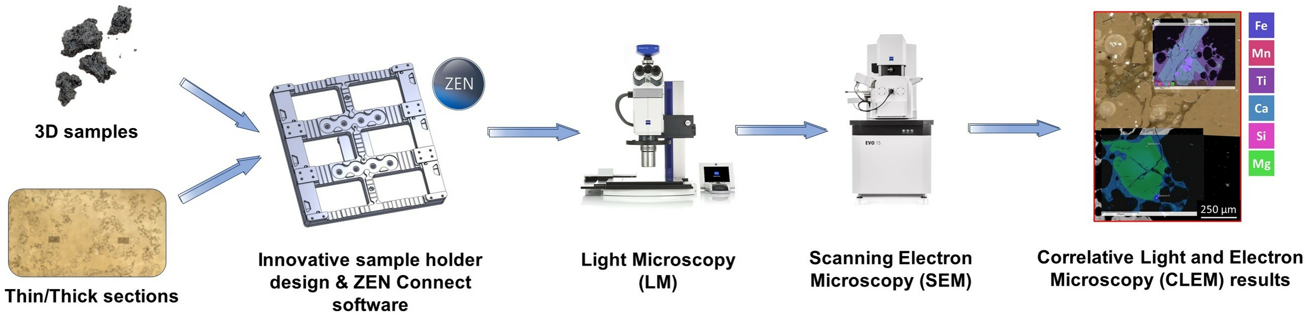
2. Materials and Methods

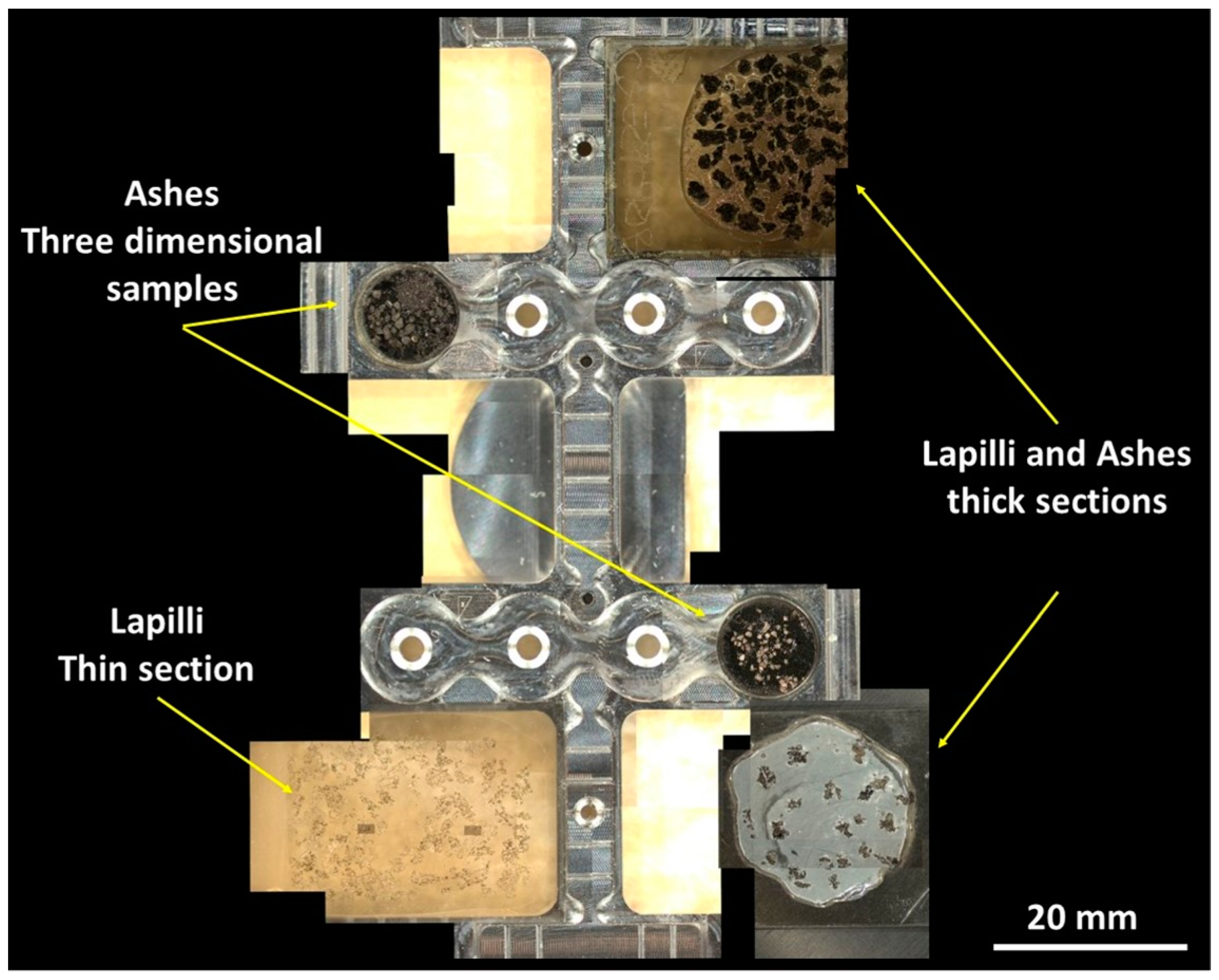
2.1. Sample Preparation

After initial LM observations, the samples were selected and glued on a glass slide using crystalbond inert balm, for compositional analyses, or on a stub using a disc of carbon tape, for morphoscopic observations. The slide samples were successively smoothed to obtain a flat and shiny surface. For the progressive grinding of the samples, abrasive sheets and pastes ranging from 30 to 3 μm were used, while for polishing, those with 1 μm were employed. Samples were coated with a 15 nm carbon layer using graphite braids mounted in a Quorum 150 R ES Plus from Quorum Technologies (Lewes, UK). The thick sections and the three-dimensional samples were prepared in the National Institute of Geophysics and Volcanology (INGV), The Etna Observatory (OE, Catania Section) laboratories, while the transparent thin sections were prepared in external laboratories.

2.2. Light Microscopy (LM)

The initial investigation was performed using a ZEISS Axio Zoom V16 (Carl Zeiss, Oberkochen, Germany) [33]. The microscope was equipped with a non-motorized planar stage where samples were accommodated.

2.3. Scanning Electron Microscopy (SEM) and Energy Dispersive X-ray Spectroscopy (EDX)

After the initial survey that was performed using LM, samples were investigated using a ZEISS EVO 15 [34] scanning electron microscope equipped with an EDX probe Ultim Max 65 mm2 from Oxford Instruments (Abingdon, UK) [35]. Before conducting the analyses, limits of the stage movements in x, y, and z rotation and tilt were imposed using SmartSEM (Version 6.09, Carl Zeiss, Oberkochen, Germany) software to prevent collisions between the sample holder and the inner walls of the SEM chamber during the analysis session. As additional settings, the scan rotation was deactivated and so was the beam shift option for centering points and features. The latter was set to use only the movement of the stage—and not the shifting of the beam—for reaching the regions of interest and navigating the sample. This was crucial for the accuracy of the entire correlative workflow. Imaging and EDX analyses were performed using the following SEM parameters and beam conditions: acceleration voltage: 20 kV, probe current: 300 pA, beam current: 30 µA, WD: 13.5 mm, and XPP data reduction routine.

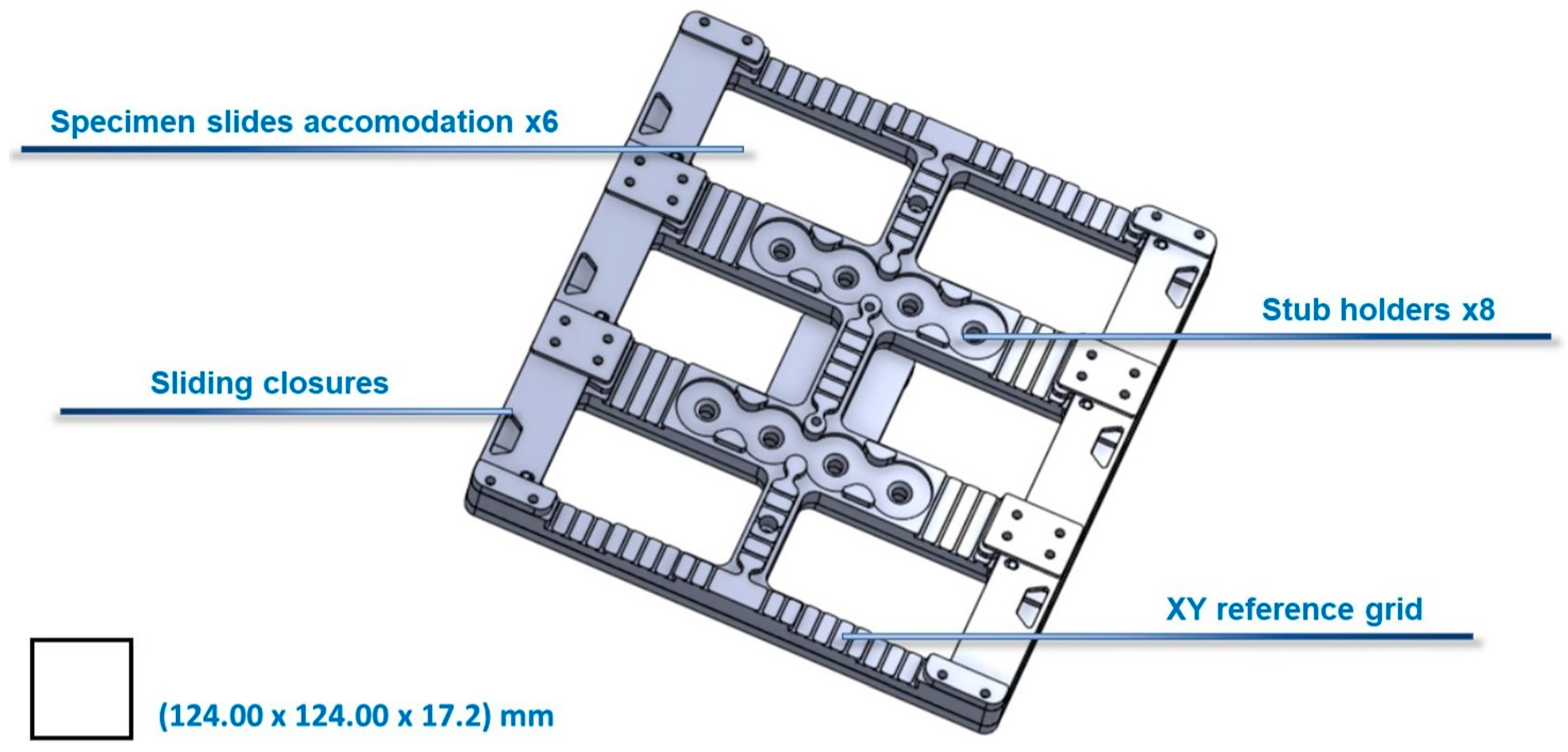
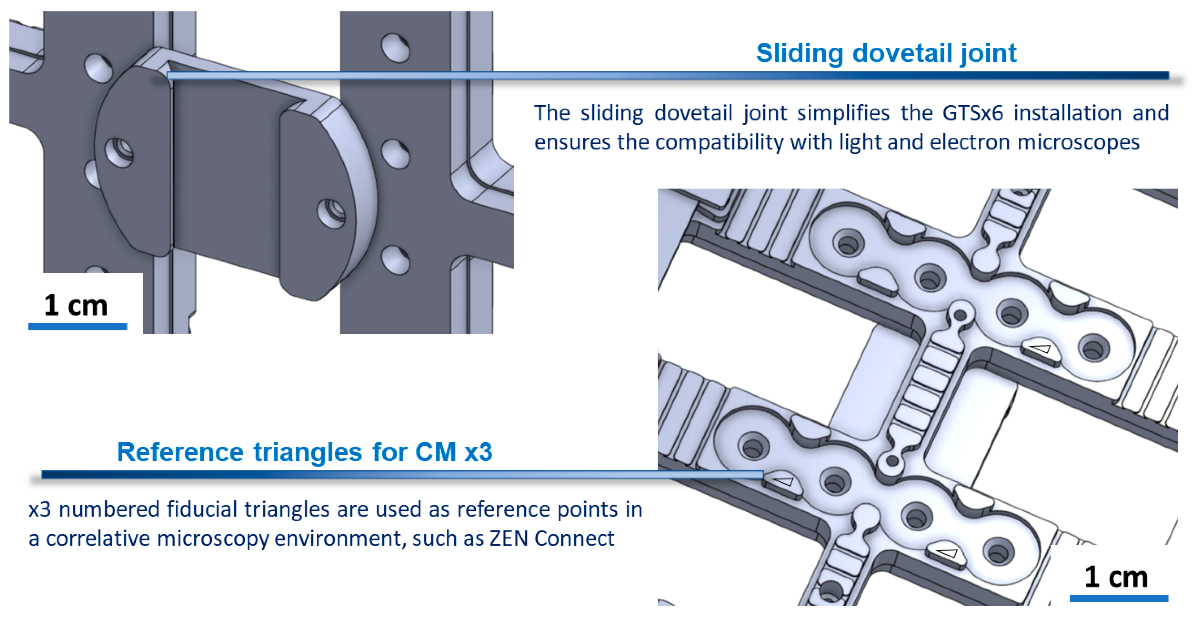
2.4. Description of the Innovative Sample Holder (GTSx6)

The design of the innovative sample holder came from collaboration between Sapienza University of Rome, INGV-OE laboratories, and Carl Zeiss—Research Microscopy Solutions. The idea stemmed from the need to develop state-of-the-art solutions tailored to specific research needs and capable of enhancing scientific research activities.
The innovative GTSx6 sample holder for CLEM was specifically designed to investigate thick/thin sections and three-dimensional samples in a correlative microscopy environment. GTSx6 is equipped with a range of features that make it a valuable solution for correlative microscopy research needs. With the capability to accommodate up to six specimen thick/thin sections, it makes it possible to analyze multiple samples with ease, maximizing the throughput of the experiments.
The six sliding closures ensure that samples are stabilized during analysis, avoiding motion artifacts and alignment problems. The eight stub holders provide precise positioning for standards used in chemical analysis or can be used as additional sample accommodations. The sample holder features an XY reference grid to easily identify and relocate the same regions of interest (ROIs) when moving from one characterization platform to the other, simplifying the correlative microscopy workflow. The presence of three fiducial markers (reference triangles) allows the calibration and alignment of the holder within the microscope, which can be performed using SmartSEM and ZEN Connect software (Version 3.2, Carl Zeiss, Oberkochen, Germany) (see Figure 1).
Fiducial markers are essential as they enable the creation of a consistently valid sample holder’s reference model, regardless of the type, geometry, and morphology of the specimens mounted on it. During calibration, the vertices associated with the right angles of each triangle were taken as reference points. Although it is challenging to assign a numerical value to calibration accuracy, it is possible to assess the quality of the calibration: ZEN Connect allows for the visualization of the acquired image in a global reference system and the option to translate/rotate the image to align it perfectly with other acquired images. In the following analyses, we observed a perfect alignment that did not require manual intervention by the operator. The sliding dovetail joint ensures compatibility with ZEISS light and electron microscopes and simplifies the installation process (see Figure 2). These features make the product an ideal solution for research needs, providing the accuracy and efficiency needed to get the most out of CLEM experiments.

2.5. ZEISS ZEN Connect Software

LM, SEM, and EDX data were integrated in a correlative microscopy environment provided by ZEISS ZEN Connect [36] software (Version 3.2). This software allows the integration and overlapping of different microscopy images and data from the same sample on different-length scales. ZEISS ZEN Connect software was also used at the beginning of the SEM investigation to calibrate the sample holder using three fiducial markers and to upload its digital model.

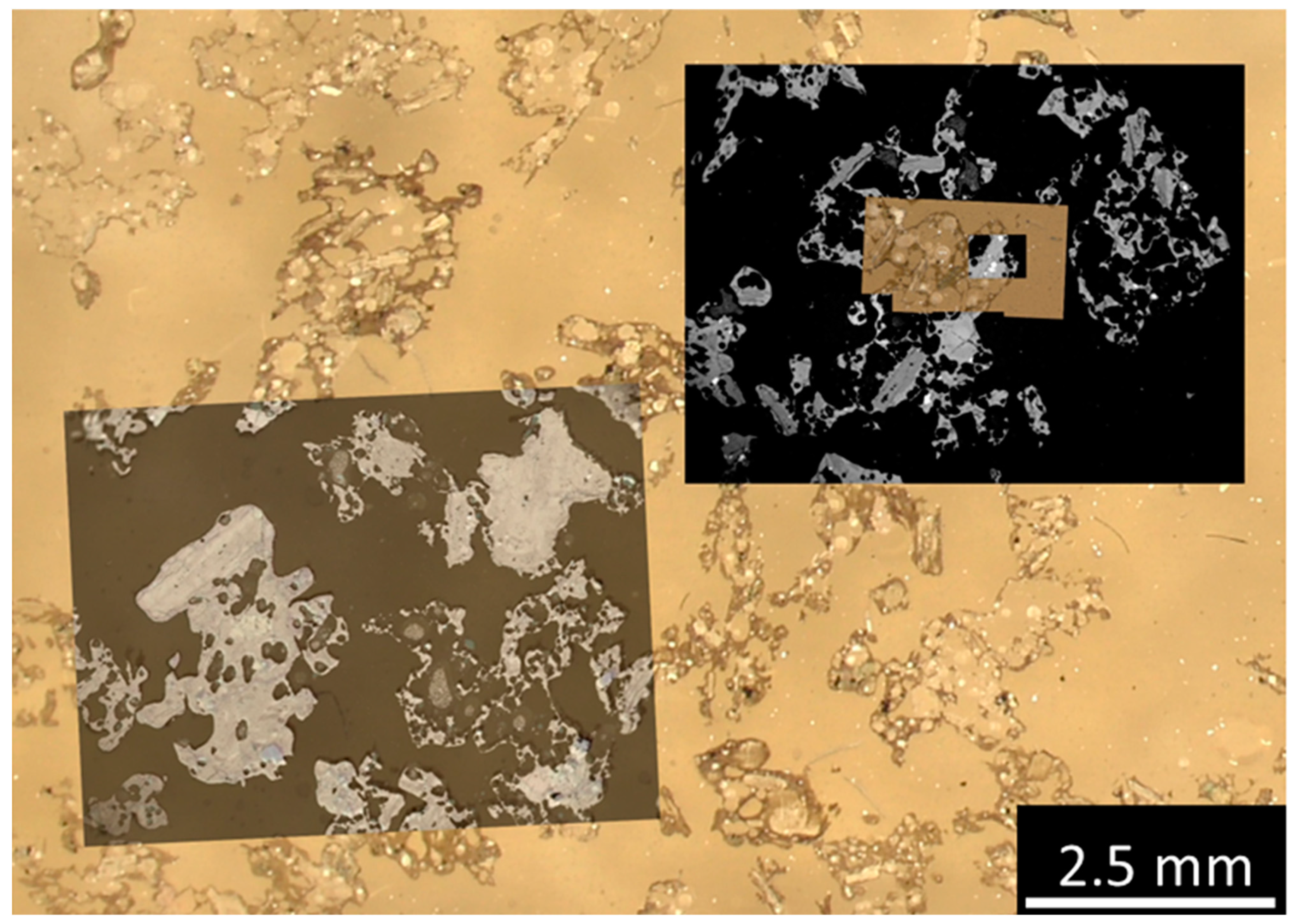
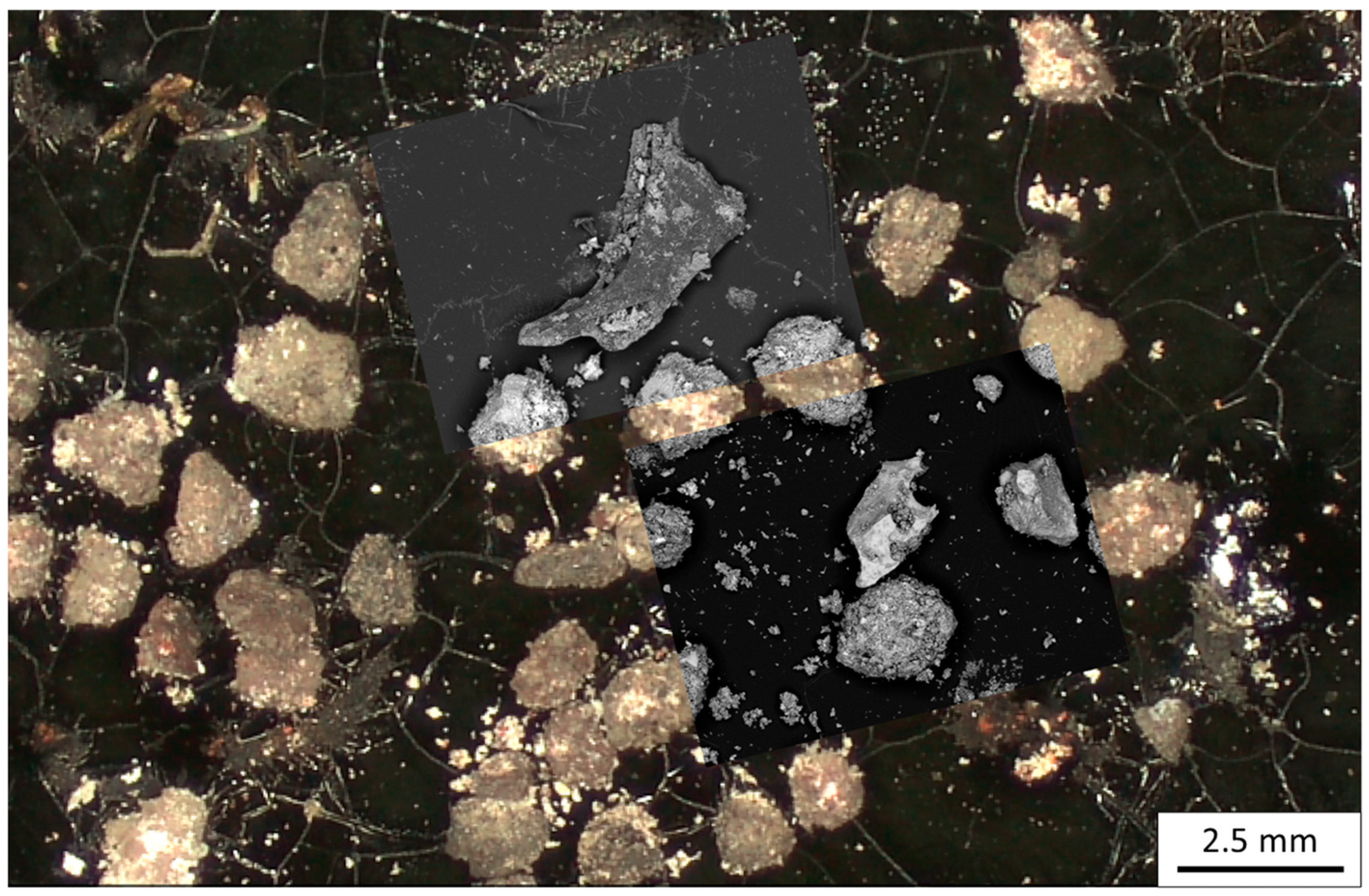
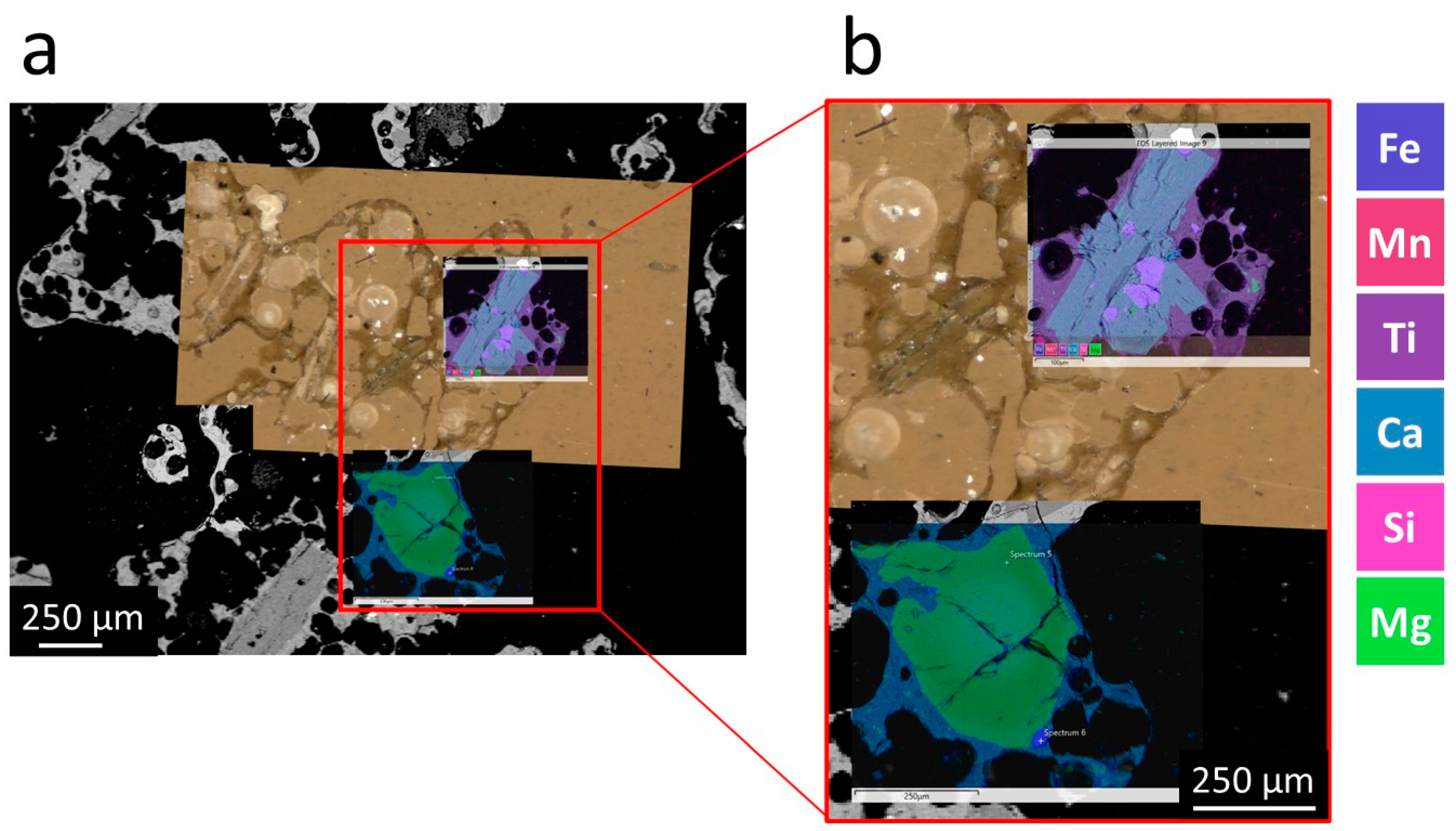
3. Results and Discussion

In this paper, we present a CLEM workflow based on the innovative GTSx6 sample holder that enables the convenient and simultaneous mounting of several types of samples such as three-dimensional samples on stubs and thick/thin sections, with a particular focus on applications in the field of geosciences. Our study involves the characterization of thick/thin sections and three-dimensional samples of ashes and lapilli from the volcanic activity of Mount Etna and Stromboli (Sicily, Italy), as depicted in Figure 3, from both morphological and compositional perspectives. When it comes to preparing three-dimensional samples, the process is streamlined and efficient. These samples are swiftly affixed to stubs using adhesive graphite discs. Such prepared samples facilitate morphoscopic studies and qualitative analysis, allowing researchers to examine their shape characteristics. Thin sections can either be transparent (thin), making them visible under a light microscope with both reflected and transmitted light, or they can be non-transparent (thick), which are suitable for quantitative analysis utilizing scanning electron microscopy (SEM).
Three-dimensional and two-dimensional observations together with quantitative analyses allowed us to obtain information on the processes that triggered the eruption and were involved during the ascent of the magma.
The first step of the CLEM workflow involves creating a reference model of the sample holder being used. This procedure is carried out using the fiducial markers on the GTSx6 sample holder, specifically selecting the vertices associated with the right angle of each of the reference triangles. Since the marker traces have finite dimensions, it is good practice to capture high-magnification images with the marker within the microscope’s field of view (FOV) to minimize uncertainty in the selection of the reference point. Fiducial markers are essential as they enable the creation of a consistently valid sample holder’s reference model, regardless of the type, geometry, and morphology of the specimens mounted on it. Performing calibration with a robust and precise procedure greatly facilitates the alignment of images acquired using various instruments. Then, multiple reference images of the GTSx6 sample holder with the specimens mounted on it should be acquired using LM to obtain a general overview and drive the further workflow’s steps. Additionally, a multiscale approach should also be employed during LM acquisition to achieve a comparable FOV to that of the SEM, as demonstrated in Figure 3. This enabled us to obtain specimen images where features were represented with similar resolution, maximizing the information obtained from CLEM.
It should be emphasized that during the acquisition using LM, the sample holder needs to be moved to capture a multiscale reference model of the entire holder and specimens. While a motorized microscope stage would facilitate and streamline this operation, in our demonstration, we employed a manually movable table to showcase the ease with which this can be achieved.
Once LM acquisition is complete, the GTSx6 sample holder can be transferred and positioned within the SEM chamber.
The following Figure 4 showcases lapilli captured using LM on a transparent section, where images acquired through SEM were overlaid. This example serves as a comparison between two distinct observations and analytical systems that enable the investigation on different-length scales. The key advantage lies in the correlation between these images, as it enables a rapid and precise identification of the observed features during the previous LM investigation.
By superimposing the SEM images onto the LM results of the transparent section containing lapilli, researchers gain valuable insights into the sample’s structure and composition. The light microscope provides a larger FOV, allowing for an initial examination of the lapilli’s overall morphology and spatial arrangement. This is particularly important when dealing with samples with large regions of interest, such as thin sections of rocks, which can be easily navigated and reached thanks to the overview image provided by LM. On the other hand, SEM provides high-resolution details, unveiling intricate surface characteristics and facilitating the analysis of fine structures.
The combination of both observation techniques offers a comprehensive approach to the study of specimens. Researchers can quickly pinpoint specific ROIs using the LM image as a reference and then delve into detailed analyses with the SEM images.
This integrated approach enhances the accuracy and efficiency of the investigation, ensuring a thorough understanding of the observed features aiding in the characterization, classification, and further scientific investigations related to these intriguing geological formations. LM in transmitted light is not only valuable as a tool for connecting the microscopic to the nanoscopic investigation of the sample. It is also an important technique for the identification and characterization of the mineral species in the thin section. By observing the sample in plane and crossed polarized light, researchers can gain unique diagnostic features of the samples that cannot be achieved with SEM-EDX alone, for example, colors, angle of extinction, birefringence, grain size and grain orientation [37], and the internal structure of lapilli [38].
The same procedure was adopted to investigate three-dimensional ashes samples, as reported in Figure 5. By combining the capabilities of both LM and SEM, researchers can delve into a multifaceted examination of ash particles. The stereo-like LM images offer a macroscopic 3D perspective, allowing for initial observations of the overall color, size, shape, and distribution patterns of the particles. The details of the surface structure, such as fractures, smoothness, and vesicularity, can be firstly assessed on unpolished samples [39]. On the other hand, the SEM images provide high-resolution details, revealing complex surface features, topography, and compositional information.
There is numerous additional information coming from the observation of volcanic ashes, which can be correlated with electron microscopy, that was not included in the workflow presented in this paper. For example, stereomicroscopes can provide a quantitative characterization of the texture of ash particles [40] and an estimation of the percentages of abundance of different ash components [41]. The color of ash particles can also be measured and correlated with the chemical composition, to gain insights into the processes that occur in the conduit just before a volcanic eruption [42].
The results shown in Figure 6 showcase lapilli, captured through both SEM and an LM (see Figure 6a). Near to the areas of particular interest, EDX maps were generated, focusing on the distribution of two specific mineral types: plagioclase in the upper section and olivine in the lower section, which can be also appreciated in the enlarged view of Figure 6b.
The SEM image offers a high-resolution view of the lapilli’s surface, revealing intricate details and textural features. The combination of SEM images and EDX maps in a correlative microscopy environment allows researchers to examine the fine structure and morphology coupled with the elemental composition of the minerals within the sample at the same time.
These maps highlight the distribution and abundance of plagioclase minerals in the upper section and olivine minerals in the lower section. By analyzing these maps, researchers can gain insights into the mineralogy of the lapilli, helping to identify specific mineral phases and understand their spatial relationships within the sample. A multiscale and multimodal overview of the investigated specimens is reported in Video S1.

4. Conclusions

Correlative light and electron microscopy (CLEM) is an imaging approach that combines the strengths of light microscopy (LM) and electron microscopy (EM) to provide crucial information in the analysis of both materials and biological specimens. CLEM has diverse applications in the geoscience field, including mineralogy, petrography, and geochemistry. Despite its many advantages, CLEM has some limitations that need to be considered. One of the major limitations of CLEM is the complexity of the imaging process. CLEM requires specialized equipment and expertise, and it can be challenging to obtain high-quality images that are suitable for analysis. In this work, we have presented a CLEM workflow applied to geosciences, focusing on the use of an innovative sample holder called GTSx6 which is designed to improve and ease data correlation with an enhanced throughput. We demonstrated the correlative microscopy approach to be invaluable in the investigation of lapilli and ash particles from volcanic activity. By correlating LM, SEM, and EDX analysis, we gained insights into the morphology, composition, and distribution of minerals within the samples. The ability to analyze multiple samples simultaneously and efficiently align the holder between different microscopes significantly improved the accuracy and efficiency of the investigation. We also proved that the GTSx6 sample holder streamlines the CLEM workflow, allowing for the simultaneous analysis of multiple samples, simplifying calibration and alignment. The application of the holder in the study of lapilli and ash particles demonstrates its effectiveness in geosciences, enabling researchers to gain insights into the samples’ characteristics and mineral distribution. Finally, this scientific contribution underscores the remarkable versatility of CLEM, where established software tools, such as ZEN Connect, can serve as open development platforms and are compatible with innovative hardware solutions that address pertinent demands, exemplified by the GTSx6 sample holder.

Supplementary Materials

The following supporting information can be downloaded at https://www.mdpi.com/article/10.3390/jeta1020006/s1, Video S1: CLEM sample navigation using ZEN Connect.

Author Contributions

Conceptualization, F.C., L.M., F.B. and M.R.; Data curation, F.C., L.M. and S.C.; Funding acquisition, L.M., F.B. and M.R.; Investigation, F.C., L.M. and S.C.; Methodology, F.C., L.M., S.C. and F.B.; Resources, L.M., F.B. and M.R.; Software, F.C., L.M. and S.C.; Supervision, F.B. and M.R.; Validation, F.B. and M.R.; Visualization, F.C., L.M. and S.C.; Writing—original draft, F.C., L.M., S.C., F.B. and M.R.; Writing—review and editing, F.C., L.M., S.C., F.B. and M.R. All authors have read and agreed to the published version of the manuscript.

Funding

This work was co-funded by “Advanced Tomography and Microscopies” (ATOM) Project, granted by Lazio Region (Prot. #173-2017-17395 L.R. 13/2008), Regional call “Open Infrastructures for Research”, and by Piano Nazionale di Ripresa e Resilienza (PNRR)—Research Infrastructure Project iENTRANCE@ENL (www.ientrance.eu, accessed on 16 September 2023) “Infrastructure for Energy Transition and Circular Economy @ EuroNanoLab”—granted by the Italian Ministry of University and Research (MUR), (Prot #IR0000027, call 3264, 28 December 2021). Coordinator of both projects: Prof. Marco Rossi.

Data Availability Statement

The data presented in this study are available upon request from the corresponding author.

Acknowledgments

The authors thank Carl Zeiss SpA—Research Microscopy Solutions—for co-funding the R & D activities and fabrication of the innovative GTSx6 sample holder design presented in this work.

Conflicts of Interest

The authors declare no conflict of interest.

References

  1. Carl Zeiss—Research Microscopy Solutions. Correlative Microscopy in Materials Science. 2017. Available online: www.essentialknowledgebriefings.com (accessed on 25 August 2023).
  2. Timmermans, F.J.; Otto, C. Contributed Review: Review of integrated correlative light and electron microscopy. Rev. Sci. Instrum. 2015, 86, 011501. [Google Scholar] [CrossRef]
  3. Schorb, M.; Briggs, J.A.G. Correlated cryo-fluorescence and cryo-electron microscopy with high spatial precision and improved sensitivity. Ultramicroscopy 2014, 143, 24–32. [Google Scholar] [CrossRef]
  4. Giepmans, B.N.G.; Adams, S.R.; Ellisman, M.H.; Tsien, R.Y. The fluorescent toolbox for assessing protein location and function. Science 2006, 312, 217–224. [Google Scholar] [CrossRef]
  5. van Rijnsoever, C.; Oorschot, V.; Klumperman, J. Correlative light-electron microscopy (CLEM) combining live-cell imaging and immunolabeling of ultrathin cryosections. Nat. Methods 2008, 5, 973–980. [Google Scholar] [CrossRef]
  6. Polishchuk, R.S.; Polishchuk, E.V.; Marra, P.; Alberti, S.; Buccione, R.; Luini, A.; Mironov, A.A. Correlative Light-Electron Microscopy Reveals the Tubular-Saccular Ultrastructure of Carriers Operating between Golgi Apparatus and Plasma Membrane. J. Cell Biol. 2000, 148, 45–58. [Google Scholar] [CrossRef]
  7. Spiegelhalter, C.; Tosch, V.; Hentsch, D.; Koch, M.; Kessler, P.; Schwab, Y.; Laporte, J. From Dynamic Live Cell Imaging to 3D Ultrastructure: Novel Integrated Methods for High Pressure Freezing and Correlative Light-Electron Microscopy. PLoS ONE 2010, 5. [Google Scholar] [CrossRef]
  8. Meißlitzer-Ruppitsch, C.; Röhrl, C.; Neumüller, J.; Pavelka, M.; Ellinger, A. Photooxidation technology for correlated light and electron microscopy. J. Microsc. 2009, 235, 322–335. [Google Scholar] [CrossRef]
  9. Shaner, N.C.; Patterson, G.H.; Davidson, M.W. Advances in fluorescent protein technology. J. Cell Sci. 2007, 120, 4247–4260. [Google Scholar] [CrossRef]
  10. Müller-Reichert, T.; Srayko, M.; Hyman, A.; O’Toole, E.T.; McDonald, K. Correlative Light and Electron Microscopy of Early Caenorhabditis elegans Embryos in Mitosis. Methods Cell Biol. 2007, 79, 101–119. [Google Scholar] [CrossRef]
  11. Kolotuev, I.; Bumbarger, D.J.; Labouesse, M.; Schwab, Y. Targeted Ultramicrotomy: A Valuable Tool for Correlated Light and Electron Microscopy of Small Model Organisms. Methods Cell Biol. 2012, 111, 203–222. [Google Scholar] [CrossRef]
  12. Brown, E.; Verkade, P. The use of markers for correlative light electron microscopy. Protoplasma 2010, 244, 91–97. [Google Scholar] [CrossRef]
  13. Han, S.; Raabe, M.; Hodgson, L.; Mantell, J.; Verkade, P.; Lasser, T.; Landfester, K.; Weil, T.; Lieberwirth, I. High-Contrast Imaging of Nanodiamonds in Cells by Energy Filtered and Correlative Light-Electron Microscopy: Toward a Quantitative Nanoparticle-Cell Analysis. Nano Lett. 2019, 19, 2178–2185. [Google Scholar] [CrossRef]
  14. Begemann, I.; Galic, M. Correlative light electron microscopy: Connecting synaptic structure and function. Front. Synaptic Neurosci. 2016, 8. [Google Scholar] [CrossRef]
  15. Hayashi, S.; Ohno, N.; Knott, G.; Molnár, Z. Correlative light and volume electron microscopy to study brain development. Microscopy 2023, 72, 279–286. [Google Scholar] [CrossRef]
  16. Booth, D.G.; Beckett, A.J.; Prior, I.A.; Meijer, D. SuperClem: An accessible correlative light and electron microscopy approach for investigation of neurons and glia in vitro. Biol. Open 2019, 8, bio042085. [Google Scholar] [CrossRef]
  17. Russell, M.R.G.; Lerner, T.R.; Burden, J.J.; Nkwe, D.O.; Pelchen-Matthews, A.; Domart, M.-C.; Durgan, J.; Weston, A.; Jones, M.L.; Peddie, C.J.; et al. 3D correlative light and electron microscopy of cultured cells using serial blockface scanning electron microscopy. J. Cell Sci. 2017, 130, 278–291. [Google Scholar] [CrossRef]
  18. Murphy, G.E.; Narayan, K.; Lowekamp, B.C.; Hartnell, L.M.; Heymann, J.A.; Fu, J.; Subramaniam, S. Correlative 3D imaging of whole mammalian cells with light and electron microscopy. J. Struct. Biol. 2011, 176, 268–278. [Google Scholar] [CrossRef]
  19. Dini, D.; Cognigni, F.; Passeri, D.; Scaramuzzo, F.A.; Pasquali, M.; Rossi, M. Review—Multiscale Characterization of Li-Ion Batteries through the Combined Use of Atomic Force Microscopy and X-ray Microscopy and Considerations for a Correlative Analysis of the Reviewed Data. J. Electrochem. Soc. 2021, 168. [Google Scholar] [CrossRef]
  20. Cognigni, F.; Dinarelli, S.; Girasole, M.; Longo, G.; Fabi, G.; Rossi, M. 3D X-ray Microscopy (XRM) investigation of exogenous materials inside mussels’ organs. In IOP Conference Series: Materials Science and Engineering; IOP Publishing: Bristol, UK, 2022; p. 012012. [Google Scholar] [CrossRef]
  21. Cognigni, F.; Pasquali, M.; Prosini, P.P.; Paoletti, C.; Aurora, A.; Scaramuzzo, F.A.; Rossi, M. X-ray Microscopy: A Non-Destructive Multi-Scale Imaging to Study the Inner Workings of Batteries. ChemElectroChem 2023, 10. [Google Scholar] [CrossRef]
  22. Carmignato, S.; Dewulf, W.; Leach, R. Industrial X-ray Computed Tomography; Springer International Publishing: Cham, Switzerland, 2018. [Google Scholar] [CrossRef]
  23. Bernabale, M.; Cognigni, F.; Mura, F.; Nigro, L.; Montanari, D.; Rossi, M.; De Vito, C. 3D imaging of micro-segregation and corrosion behavior of alloying elements in archaeological artefacts from Motya (Sicily, Italy). Corros. Sci. 2023, 211, 110900. [Google Scholar] [CrossRef]
  24. Whittig, L.D.; Allardice, W.R. X-ray Diffraction Techniques. In Methods of Soil Analysis; Klute, A., Ed.; American Society of Agronomy, Inc.: Madison, WI, USA, 1986. [Google Scholar] [CrossRef]
  25. Bunaciu, A.A.; Udriştioiu, E.G.; Aboul-Enein, H.Y. X-ray Diffraction: Instrumentation and Applications. Crit. Rev. Anal. Chem. 2015, 45, 289–299. [Google Scholar] [CrossRef]
  26. Mulvaney, S.P.; Keating, C.D. Raman Spectroscopy. Anal. Chem. 2000, 72, 145–158. [Google Scholar] [CrossRef]
  27. Hanlon, E.B.; Manoharan, R.; Koo, T.-W.; Shafer, K.E.; Motz, J.T.; Fitzmaurice, M.; Kramer, J.R.; Itzkan, I.; Dasari, R.R.; Feld, M.S. Prospects for in vivo Raman spectroscopy. Phys. Med. Biol. 2000, 45. [Google Scholar] [CrossRef]
  28. Cognigni, F.; Sgambetterra, M.; Zucca, G.; Gentile, D.; Ricci, S.; Testa, G.; Rizzi, G.; Rossi, M. Multimodal and multiscale investigation for the optimization of AlSi10Mg components made by powder bed fusion-laser beam. Discov. Mater. 2023, 3, 21. [Google Scholar] [CrossRef]
  29. Schwartz, A.J.; Kumar, M.; Adams, B.L.; Field, D.P. Electron Backscatter Diffraction in Materials Science; Springer: New York, NY, USA, 2009. [Google Scholar] [CrossRef]
  30. Mura, F.; Cognigni, F.; Ferroni, M.; Morandi, V.; Rossi, M. Advances in Focused Ion Beam Tomography for Three-Dimensional Characterization in Materials Science. Materials 2023, 16, 5808. [Google Scholar] [CrossRef]
  31. Bernabale, M.; Cognigni, F.; Mancini, C.; Proietti, A.; Mura, F.; Montanari, D.; Nigro, L.; Rossi, M.; De Vito, C. 3D fractures analysis and conservation assessment of wrought iron javelin through advanced non-invasive techniques. Sci. Rep. 2023, 13, 10142. [Google Scholar] [CrossRef]
  32. Bernabale, M.; Cognigni, F.; Nigro, L.; Rossi, M.; de Caro, T.; De Vito, C. A comprehensive strategy for exploring corrosion in iron-based artefacts through advanced Multiscale X-ray Microscopy. Sci. Rep. 2022, 12, 6125. [Google Scholar] [CrossRef]
  33. Carl Zeiss—Research Microscopy Solutions. ZEISS Axio Zoom V16 for Materials. Available online: https://www.zeiss.com/microscopy/en/products/light-microscopes/stereo-and-zoom-microscopes/axio-zoom-v16-for-materials.html (accessed on 25 August 2023).
  34. Carl Zeiss—Research Microscopy Solutions. ZEISS EVO Scanning Electron Microscope. Available online: https://www.zeiss.com/microscopy/en/products/sem-fib-sem/sem/evo.html (accessed on 25 August 2023).
  35. Oxford Instruments. Introducing the Ultim® Max Range: A New Suite of High Performance SEM-EDS Detectors. Available online: https://www.oxinst.com/news/introducing-the-ultim-max-range-a-new-suite-of-high-performance-sem-eds-detectors/ (accessed on 5 September 2023).
  36. Carl Zeiss—Research Microscopy Solutions. Visualize Images and Data in Context—ZEISS ZEN Connect. Available online: https://www.zeiss.com/microscopy/en/resources/insights-hub/manufacturing-assembly/visualize-images-and-data-in-context---zeiss-zen-connect.html (accessed on 25 August 2023).
  37. Nesse, W.D. Introduction to Mineralogy, 3rd ed.; Oxford University Press: Oxford, UK, 2016. [Google Scholar]
  38. Schumacher, R.; Schmincke, H.U. Internal structure and occurrence of accretionary lapilli—A case study at Laacher See Volcano. Bull. Volcanol. 1991, 53, 612–634. [Google Scholar] [CrossRef]
  39. Taddeucci, J.; Pompilio, M.; Scarlato, P. Monitoring the explosive activity of the July–August 2001 eruption of Mt. Etna (Italy) by ash characterization. Geophys. Res. Lett. 2002, 29, 71-1–71-4. [Google Scholar] [CrossRef]
  40. Miwa, T.; Shimano, T.; Nishimura, T. Characterization of the luminance and shape of ash particles at Sakurajima volcano, Japan, using CCD camera images. Bull. Volcanol. 2015, 77, 5. [Google Scholar] [CrossRef]
  41. Lücke, O.H.; Calderón, A. Characterization Of The Ashes From The 2014-2015 Turrialba Volcano Eruptions By Means Of Scanning Electron Microscopy And Energy Dispersive X-ray Spectroscopy. Rev. Geog. Am. Cent. 2016, 54, 109–123. [Google Scholar] [CrossRef]
  42. Yamanoi, Y.; Takeuchi, S.; Okumura, S.; Nakashima, S.; Yokoyama, T. Color measurements of volcanic ash deposits from three different styles of summit activity at Sakurajima volcano, Japan: Conduit processes recorded in color of volcanic ash. J. Volcanol. Geotherm. 2008, 178, 81–93. [Google Scholar] [CrossRef]
Figure 1. Innovative sample holder design features. GTSx6 sample holder accommodates six specimen slides and features six sliding closures for optimal sample stabilization. The holder is provided with eight stub holders for positioning additional samples and/or standards used in chemical analysis. GTSx6 also includes an XY reference grid and three fiducial markers for easy calibration and alignment using ZEN Connect software.
Jeta 01 00006 g001
Figure 2. Innovative sample holder design compatibility and calibration. The holder features three numbered fiducial markers (reference triangles) for ZEN Connect software alignment and a sliding dovetail joint for easy installation and microscope compatibility.
Jeta 01 00006 g002
Figure 3. Acquisition of reference images for the application of the CLEM workflow. Multiple reference images of the GTSx6 sample holder with the specimens mounted on it were acquired using LM. Lapilli and ashes thick/thin sections, as well as three-dimensional samples, were investigated.
Jeta 01 00006 g003
Figure 4. CLEM images of a lapilli thin section. Images of lapilli acquired using LM on a thin section where SEM images were superimposed. This example compares two different observation and analytical systems that operate at different magnifications. Thanks to the correlation between the images, it is possible to quickly identify and accurately analyze what was observed during the LM investigation using the higher SEM resolution.
Jeta 01 00006 g004
Figure 5. CLEM images of a three-dimensional sample of ashes. Multi-scale and multi-technique investigation of ash particles captured using LM, with their corresponding images acquired through SEM (superimposed).
Jeta 01 00006 g005
Figure 6. CLEM images with compositional maps. (a) Combination of LM and SEM images coupled with the generation of EDX maps where (b) an enlarged view highlighting the chemical elements composing the sample is reported.
Jeta 01 00006 g006
Disclaimer/Publisher’s Note: The statements, opinions and data contained in all publications are solely those of the individual author(s) and contributor(s) and not of MDPI and/or the editor(s). MDPI and/or the editor(s) disclaim responsibility for any injury to people or property resulting from any ideas, methods, instructions or products referred to in the content.

