Review

Regulation of Ras-GTPase Signaling and Localization by Post-Translational Modifications

National Centre for Cell Science, Ganeshkhind, Pune 411007, India
*
Authors to whom correspondence should be addressed.
Kinases Phosphatases 2023, 1(2), 97-116; https://doi.org/10.3390/kinasesphosphatases1020007
Submission received: 31 January 2023 / Revised: 18 March 2023 / Accepted: 4 April 2023 / Published: 21 April 2023

Abstract

:
Ras, a GTP-GDP binary switch protein, transduces signals from diverse receptors to regulate various signaling networks. Three Ras genes encode for protein isoforms, namely, Harvey Ras (H-Ras), Kirsten Ras (K-Ras, with two splice variants, K-Ras4A and K-Ras4B), and Neuroblastoma Ras (N-Ras). The isoforms undergo a series of post-translational modifications that enable their membrane attachment and biological activity. The activation of Ras isoforms is tightly regulated, and any dysregulation affects cellular processes, such as cell division, apoptosis, differentiation, cell migration, etc. The Ras gene is highly prone to mutation, and ~30% of cancers carry somatic mutations in Ras, whereas germline mutations clinically manifest as various rasopathies. In addition to regulation by the Guanine nucleotide exchange factors and the GTPase activation proteins, Ras signaling, and localization are also regulated by phosphorylation-dephosphorylation, ubiquitination, nitrosylation, and acetylation. Herein, we review the regulation of Ras signaling and localization by various regulatory enzymes in depth and assess the current status of Ras drug discovery targeting these regulatory enzymes.

1. Introduction

Ras GTPases, the cell’s molecular switches, regulate signal relays from diverse receptors. The canonical Ras gene encodes four protein isoforms: Harvey Ras (H-Ras), Kirsten Ras (K-Ras, with two splice variants, 4A, and 4B), and Neuroblastoma Ras (N-Ras). The nascent protein is synthesized in the cytosol by free polysomes [1]. This globular hydrophilic protein undergoes a series of post-translational modifications (PTMs) in the cytosol and the endoplasmic reticulum (ER). H-Ras and N-Ras and K-Ras4A further undergo modification in the Golgi complex (GC) to be targeted to the inner leaflet of the plasma membrane (PM), the primary platform for Ras signaling. Ras is primarily regulated by the guanine nucleotide exchange factors (GEFs) that enable Ras to switch between a GDP-complexed to a GTP-complexed state. The inactivation of Ras is affected by GTPase-activating proteins (GAPs) that catalyze the reverse reaction. Additionally, various kinases and phosphatases also regulate Ras signaling and localization.

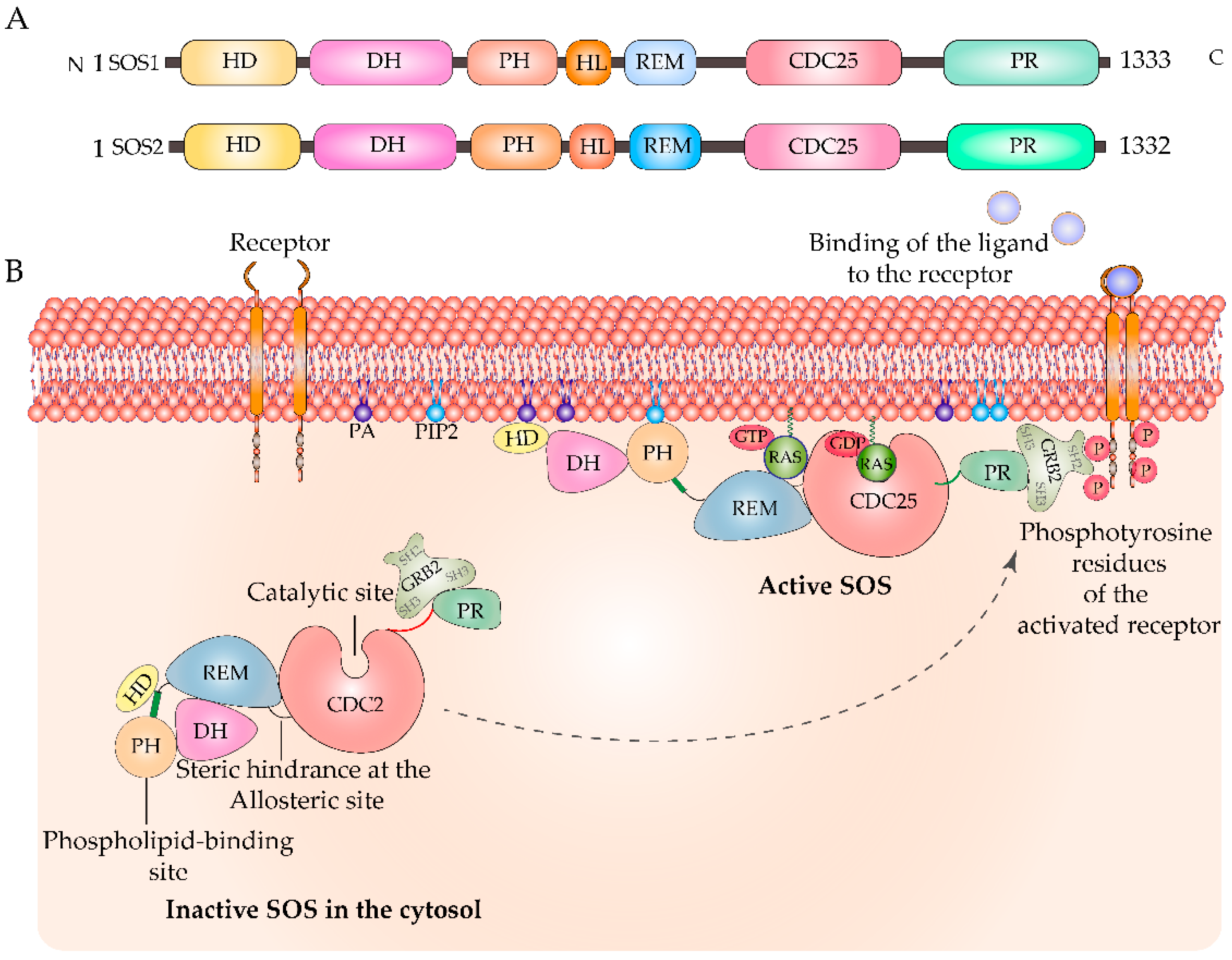
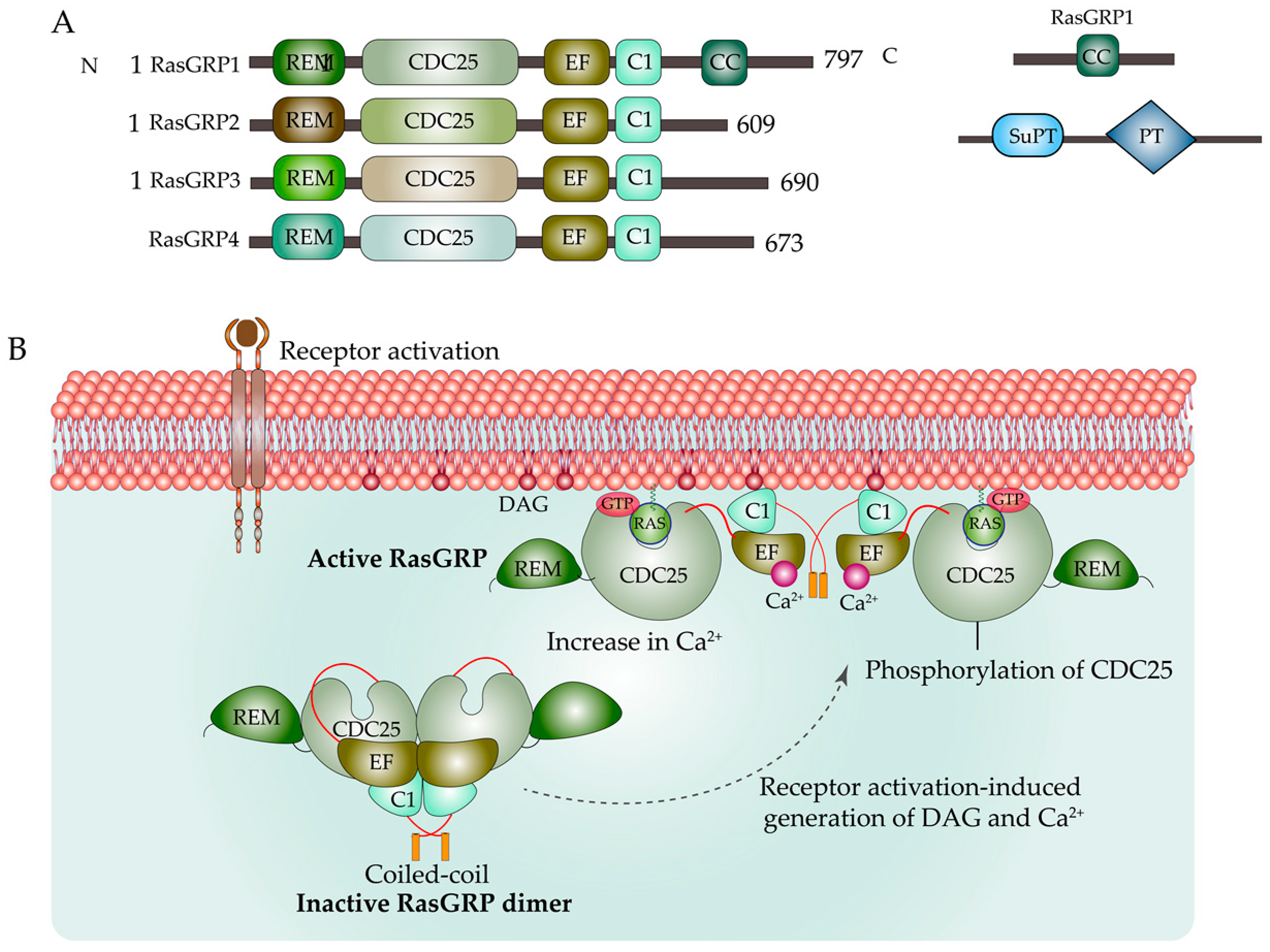
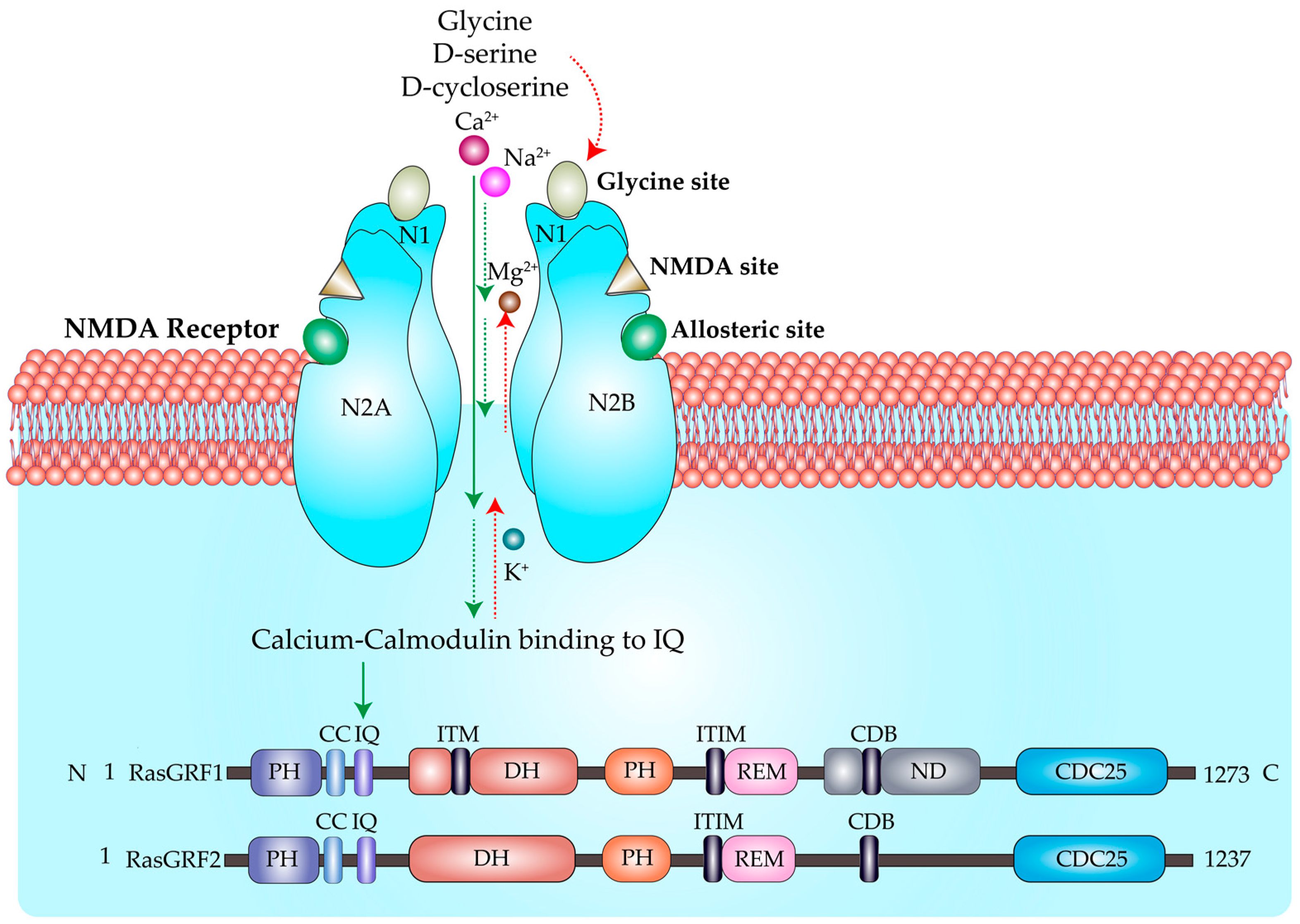
2. Regulation of Ras by GEFs and GAPs

Ras cycles between an inactive GDP-bound state and an active GTP-bound state and this guanine nucleotide exchange is reciprocally catalyzed by GEFs and GAPs. The main RasGEF proteins are Son of Sevenless (SOS), Ras guanyl nucleotide-releasing proteins (RasGRPs), and RAS Protein-Specific Guanine Nucleotide-Releasing Factors (RasGRFs). Ras GEFs play specialized roles in developmental stages and cell-specific contexts, and their deletion results in developmental defects [2,3]. In quiescent cells, the Ras GEFs remain inactive or autoinhibited, but on receptor activation, these GEFs and different adaptor proteins are recruited to the phosphotyrosine residues of the receptor. The kinetics of recruitment of adaptors, lipid second messengers, and Ca2+ also play a pivotal role in the activation of RasGEFs [4].
Son of Sevenless: Mammalian SOS consists of the ubiquitously expressed homologs SOS1 and SOS2, which share 70% sequence similarity. SOS1 is composed of six domains, namely, the N-terminus histone-domain (HD), followed by the Dbl homology domain (DH), the Pleckstrin homology (PH) domain, the Ras exchange motif (REM), the Cell division cycle 25 (CDC25) homology domain, and the proline-rich (PR) domain. In quiescent cells, C-terminal proline-rich (PXXP) remains bound to Src Homology 3 (SH3) domain of the adaptor protein Growth factor receptor bound 2 (Grb2). DH and PH autoinhibit SOS activation by inducing steric hindrance at the allosteric site. HD further stabilizes this autoinhibited state by docking to a helical linker between PH and REM domains [5]. On receptor stimulation, the SOS1—Grb2 complex is recruited to the PM, where it binds to the phosphotyrosine residues of the activated receptor. The catalytic core, also known as SOScat, is the center portion of SOS1 and contains the REM and CDC25 domains. SOS is activated on the binding of Ras-GTP to an allosteric site located between REM and CDC25, which opens the active sites of SOS, and further stimulation from CDC25 leads to nucleotide exchange. Thus, Ras acts as an allosteric modulator of SOS activation by inducing positive feedback [6]. PH and HD domains also engage in interactions with membrane lipids, such as phosphatidylinositol-4,5-bisphosphate (PIP2), to further release this inhibition [7] (Figure 1). There are multiple phosphorylation sites on SOS, and a study identified five MAP kinase sites—Ser1137, Ser1167, Ser1178, Ser1193, and Ser1197—on human SOS1 (hSOS) and only one phosphorylation site on hSOS2 that regulate Grb2-SOS association [8]. Ras activation by SOS predominantly activates signaling from the PM; however, the phospholipase D2-controlled localization of Grb2—SOS—Ras to the GC also suggests its role in activating Golgi-Ras [9]. SOS1 could also stimulate nucleotide exchange in the ER pool of H-Ras [10].
Ras guanyl nucleotide-releasing proteins: There are four RasGRP proteins: RasGRP1 through to RasGRP4. Like SOS, RasGRP has a catalytic segment composed of REM and CDC25; additionally, it has a calcium-binding helix-loop-helix EF-hand and C1 domain which is similar to the Diacylglycerol (DAG)-binding domain of PKC, with the exception of RasGRP2 [11]. Hence, RasGRP1 and RasGRP3 function in a DAG-dependent manner, whereas calcium is the primary trigger for RasGRP2 activation. Divergence in the sequence of the EF-hand(s) in different RasGRPs suggests that their binding to Ca2+ and hence their regulation might be different [12]. The C-terminus of RasGRP1 has ~140 residues coiled-coil (CC). This region was later identified as the suppressor of PT (SuPT) and the plasma membrane-targeting (PT) domain in RasGRP1. As a dimer stabilized by the C1-dimer interface, in which the allosteric site of CDC25 is occluded, RasGRP remains inactive in an autoinhibited form. This dimer state is further stabilized by the PT domain of RasGRP1. Receptor activation induces an increase in DAG and Ca2+, which regulate RasGRP’s activation. The RasGRP dimer is disrupted by the C1 domain’s binding to DAG and the PT domain’s binding to phosphoinositides. In addition, the inhibitory linker is released and the catalytic site is unblocked by the phosphorylation of CDC25 and the binding of EF1 to calcium [13] (Figure 2).
DAG recruits RasGRP1/3/4 to the PM via its C1 domain, where it activates Ras. In TCR-stimulated cells, DAG mediates PKC-dependent phosphorylation of Tyr184 on RasGRP1, [14] whereas PKC phosphorylates Thr133 on RasGRP3 in BCR-stimulated cells [15]. PKA phosphorylates Ser116/117, Ser528, and Ser587 on RasGRP2, regulating its GTP exchange activity on Rap1 [16,17]. RasGRP2 is also phosphorylated on Ser394, which impairs its nucleotide exchange activity [18]. Hence, a key step in the RasGRP-induced activation of GTPase signaling is phosphorylation. RasGRP is the primary GEF responsible for the preferential Ras activation in the Golgi in T-cells. High-grade TCR stimulation using 5 μg/mL of anti- CD3 plus anti-CD28 antibodies activates H-Ras and N-Ras on the Golgi but only ~20% of K-Ras on the PM. The preferential activation of H-Ras on the Golgi is due to the Src-dependent phosphoactivation of PLC-γ at Tyr771, 783, and 1254 [19], which activates RasGRP1 via DAG in the Golgi. By Ca2+-dependent activation of the Ras GTPase-activating protein CAPRI, H-Ras is simultaneously deactivated at the PM [20]. In HEK293 cells, it has also been demonstrated that RasGRP3 is redistributed to the ER in response to the diacylglycerol analogue 1,2-dioctanoyl-sn-glycerol and the phorbol 12-myristate 13-acetate (PMA) [21].
RAS Protein-Specific Guanine Nucleotide-Releasing Factor: RasGRF has a REM, CDC25, and two PH domains (PH1 and PH2) and is closely related to SOS. In response to receptor stimulation, PH1 interacts with membrane lipids, enhances membrane localization, and binds to G-protein β/γ subunits, whereas PH2 is necessary for calcium-mediated activation. Protein oligomerization is mediated by the CC domain. The activation of Ras and Ras-independent ionomycin—ERK activation is both regulated by the DH domain [22]. RasGRF1 has two Immunoreceptor Tyrosine-based Inhibition Motifs (ITIMs) (306 IFYQGL and 639 IRYASV), and RasGRF2 contains one ITIM (639 IRYASV). These may play a role in T-Cell Receptor—RasGRF signaling as well as be the target residues for Src homology region 2 domain-containing (SHP) phosphatase [23]. The Cyclin Destruction Box (CBD) motif, which is involved in ubiquitination and proteolytic degradation, is located between the REM and the catalytic CDC25. The Proline-Glutamic acid-Serine-Threonine (PEST) region is the target for calpain-type protease degradation, and PKA, CDK5 phosphorylation, which regulates RasGRF’s GEF activity [24]. RasGRF1(p140) and RasGRF2 (p135) expression are primarily restricted to neuronal cells, and RasGRF2 is also expressed in embryonic stem cells. RasGRF1 and GRF2 can activate both Ras and Rac owing to their two GEF domains. RasGRF1 was first implicated in its ability to activate Ras in response to calcium fluxes and G protein-coupled receptors signals. The ilimaquinone (IQ) domain of RasGRF mediates calcium/calmodulin binding [25], which enables calcium sensing and signaling through N-methyl-D-aspartate (NMDA)-type receptor (NMDAR) and α-amino-3-hydroxy-5-methyl-4-isoxazolepropionic acid (AMPA)-type glutamate receptors (Figure 3). RasGRF1 but not RasGRF2 contains a neuronal binding domain that binds to the NR2B subunit of NMDAR [26]. A study has shown that RasGRF1 and RasGRF2 mediate opposing synaptic plasticity and long-term potentiation (LTP) in the mouse hippocampus by preferential activation of Ras/ERK and Rac/p38, respectively [27]. In addition to the IQ domain, the CC, PH, and DH domains are required for calcium ionophore-induced MAPK activation. RasGRF is expressed at high levels in post-natal neurons of the central nervous system; hence, RasGRF plays a pivotal role in the activation of neuronal Ras [28]. The phosphorylation of Ras GRF1 at Ser916 is crucial for its physiological functions [29]. RasGRF1 and RasGRF2 preferentially activate PM and ER-associated H-Ras but not GC-associated H-Ras [10]. Moreover, RasGRF demonstrates preferential activation of H-Ras over K-Ras and N-Ras in vivo, primarily owing to the different C-terminal HVR regions and partially to the palmitoylation [30]. Carbachol, a Muscarinic receptor agonist, specifically increases the serine phosphorylation and the activity of RasGRF in the brain, which was reversed by protein phosphatase 1 [31].
Ras has an intrinsic GTPase activity which is accelerated by GAPs. The conserved arginine finger of GAP moves the catalytic Gln61 closer to the γ-phosphate of GTP, stabilizes it, and neutralizes the negative charge on the phosphates. Thus, it stimulates the hydrolysis of GTP [32]. In addition to the two well-characterized GAPs neurofibromin (NF1) and Ras P21 Protein Activator 1 (p120GAP/RASA1), additional Ras GAPs have been identified, such as IQGAP1, PLEXIN-B1, RASAL1, RASAL2, etc. C2GAP2 has been identified as a Ras GAP that negatively regulates folic acid-mediated chemotaxis, phagocytosis, and micropinocytosis [33]. The GAP NF1 has two domains—a cysteine-serine-rich domain (CSRD) and a C-terminal domain (CTD)—both of which are phosphorylated by PKA. CSRD also gets phosphorylated by both PKC, which enhances its GAP activity, and association with actin [34]. Phosphorylation of the CTD domain at Ser2576, 2578, 2580, and 2813 and Thr2556 is essential for its interaction with 14-3-3 protein, which negatively regulates its GAP activity [35]. p120GAP is also subjected to phosphoregulation by a kinase, p210bcr/abl. p120GAP associates with two proteins—p190 and p62—which are tyrosine phosphorylated by p210bcr/abl, and elevated levels of Tyr phosphorylated p190 complexed with p120GAP inhibit the GTPase-promoting activity of p120GAP [36]. Erythropoietin also induces tyrosine phosphorylation of p120GAP and inhibition of its activity [37].
Proteins with Ras-binding domains and Ras association domains have been defined as Ras effectors. Ras engages in two major downstream pathways—the Ras-Raf-MEK1/2-ERK1/2 and Ras-PI3K-Akt-mTOR pathways. Other Ras effectors include Tiam1, Ral-GDS, AF6, PLC-ε, RIN, Nore1, etc. GTP-bound Ras drives the relocalization of cytosolic Raf to the PM. This exposes the inhibitory sites of Raf to phosphatase PP1 or PP2 for dephosphorylation. Ras binding also promotes the phosphorylation of activating sites on Raf kinase, which is a prerequisite for Raf dimerization. Phosphorylation of Ser338 and Tyr341 synergizes Raf1 activation. The residues Thr491 and Ser494 also get phosphorylated and cooperate in the activation loop of Raf1 [38]. PKA negatively regulates Raf1 by phosphorylating residues Ser43 [39], Ser259 [40], and Ser621 [41]. Another class of kinase regulatory proteins is the MOBs (monopolar spindle-one-binder proteins), which are crucial signal adaptors of the NDR (nuclear Dbf2-related)/LATS (large tumor suppressor)—hippo pathway. The Hippo pathway includes key signaling cascades which regulate cell proliferation, differentiation, and death, thus maintaining homeostasis. Dysregulation of the hippo pathway leads to diseases such as cancers. Unlike MOB1A/B proteins, MOB3A inhibits the Hippo pathway, thus permitting proliferation and suppressing Ras—BRaf -driven senescence in cancer cells [42]. PI3K is recruited to the PM in response to the phosphorylation of tyrosine residues of the receptor. As mentioned in an earlier section, active PI3K catalyzes the conversion of PIP2 to PIP3. PIP3 further recruits effectors, such as Akt, serine-threonine kinase PDK1, etc. PDK1 selectively phosphorylates Thr308 on Akt, thus activating it [43]. Akt is also activated by phosphorylation at Ser437 by mTORC-2 [44]. Phosphatases PP2A and PH domain leucine-rich repeat protein phosphatases 1 and 2 dephosphorylate Akt at Thr308 [45] and Ser473 [46], respectively, thus negatively regulating PI3K—Akt signaling. A recent study generated a Ras-effector network by identifying PPIs (protein–protein interaction) downstream of Ras up to three layers (L) and their subcellular localization. The authors identified 2290 proteins, of which 43 were high-confidence Ras effectors. These proteins interact via 19,080 binary protein–protein interactions that are maximally distributed in the order of layer 3 > layer 2 > layer 1. Moreover, the majority of the L1 proteins were in the extracellular environment, while the L2 proteins were in the cytosol and the L3 proteins were in the nucleus [47].

3. Ras Post-Translational Modifications

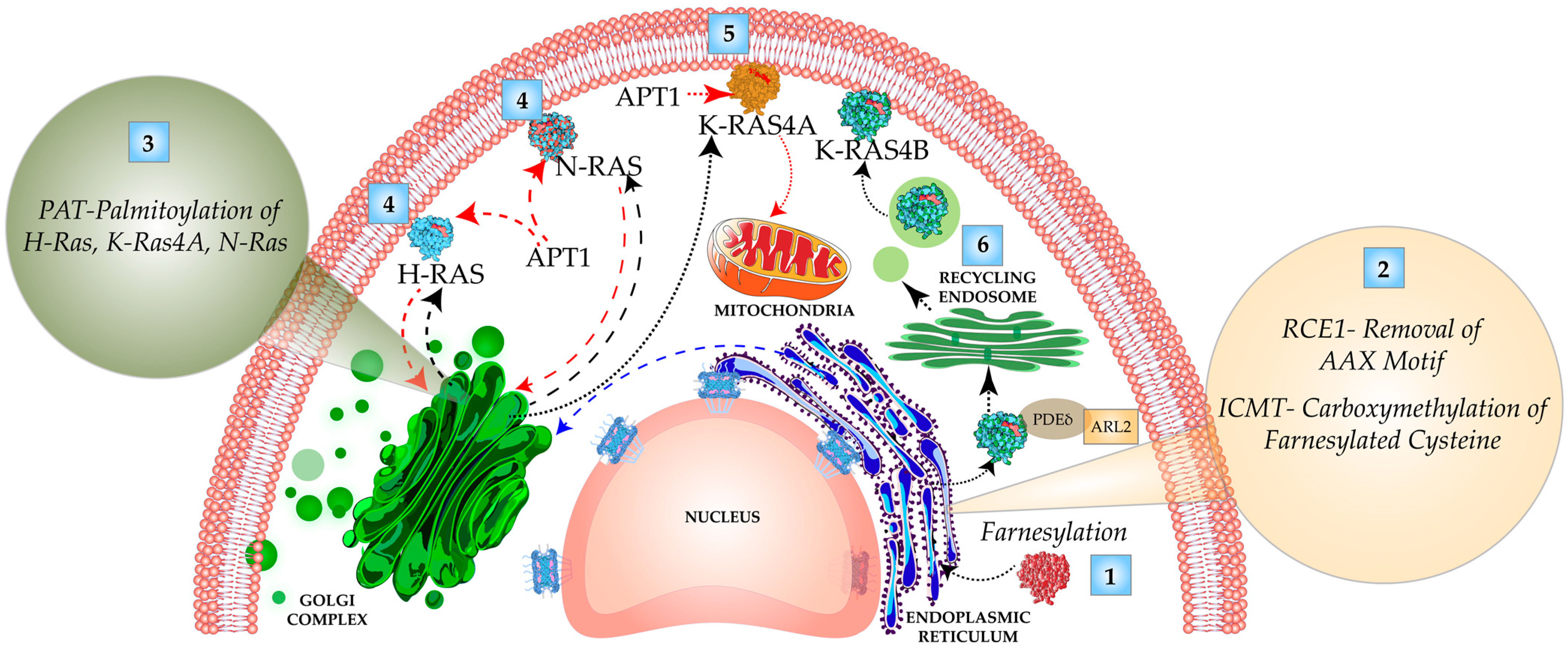
3.1. C-Terminal Carboxy-Terminal Modifications and Membrane Targeting of Ras Isoforms

The biologically inactive pro-Ras polypeptide has a C-terminus CAAX motif. C represents an invariant cysteine; AA represents two aliphatic amino acids. The amino acid residue at the X position determines the substrate specificity for farnesyl transferase versus geranyl geranyl transferase 1. If X is methionine or serine, it undergoes farnesylation—the addition of a 15-carbon farnesyl isoprenoid group from farnesyl diphosphate by an enzyme, farnesyl transferase (FTase), by a stable thioether linkage. If the amino acid residue at position X is leucine, it undergoes prenylation by geranyl geranyl transferase 1 (GGTase). In the case of blocking the FT using a farnesyl transferase inhibitor (FTI), K-Ras and N-Ras undergo alternative prenylation by GGTase [48]. Following prenylation in the cytoplasm, the insertion of Ras into the membrane of the ER takes place, wherein the AAX motif is cleaved off by the Ras converting endopeptidases (RCE1). The farnesylated cysteine further gets carboxymethylated by the enzyme isoprenylcysteine carboxyl methyltransferase (ICMT). H-Ras, N-Ras, and K-Ras 4A are further modified in the GC by the addition of a 16-carbon palmitoyl group to cysteine by palmitoyl acyltransferase (PAT) by a labile thioester bond. H-Ras has dual palmitoylation on residues 181 and 184, N-Ras on 181, and K-Ras 4A on 180. Palmitoylation imparts hydrophobicity to H-Ras, N-Ras, and K-Ras4A for their membrane attachment. However, K-Ras 4B has a stretch of eight lysine residues that gives it a strong positive charge, allowing its membrane interaction with the negatively charged polar head of the PM phospholipids. Palmitoylation is a reversible process catalyzed by thioesterase acyl protein thioesterase1 (APT1). The cyclic palmitoylation and depalmitoylation shuttles H- and N-Ras between the PM and subcellular compartments [49] (Figure 4).

3.2. Microregulation of Ras Isoforms by Ubiquitination, Acetylation, and Nitrosylation

Furthermore, ubiquitination, acetylation, and nitrosylation have a role in the microregulation of Ras isoform activation and subcellular localization. The dynamic reversible process of ubiquitination regulates the Ras isoform’s localization and signaling properties. Rabex-5 acts as Rab5 GEF or an A20-like E3 ubiquitin ligase for Ras. Washington et al. identified that the phosphorylation of Tyr4 is indispensable for the ubiquitination of Ras and Phe substitution at this site, rendering Ras insensitive to Rabex5 ubiquitination [50]. Rabex5/RabGEF1 promotes mono- and diubiquitination of H-Ras and N-Ras [51]. H-Ras is modified at Lys63, which stabilizes its association with the endosome and modulates downstream signaling potential. Ubiquitination preferentially localizes H-Ras to endocytic compartments and impairs its ability to recruit Raf1 and thus signaling to MAPK [52]. Monoubiquitination regulates H-Ras and K-Ras differently because ubiquitination of H-Ras at Lys117 improves nucleotide exchange and GTP loading and its activation, but ubiquitination of K-Ras at Lys147 reduces GAP-mediated hydrolysis [53]. Baieitti et al. showed that OTUB1 deubiquitinates Ras, which results in the sequestration of Ras on the PM, hence promoting Ras activation and tumorigenesis. The suppression of the deubiquitinase OTUB1 increases the mono- and diubiquitination of N-Ras [54]. The ubiquitination of K-Ras at Lys147 enhances GDP release, thus activating Ras and enhancing its interaction with effectors, such as Raf, PI3K, RalGDS, etc. [55]. The inactivation of the cullin-3 adapter protein encoding leucine zipper-like transcription regulator 1 (LZTR1) decreases the ubiquitination of K-Ras and enhances its PM localization [56]. Acetylation of K-Ras at Lys104 affects the conformational stability of the α2 helix of switch II and displaces Tyr64, which is crucial for K-Ras’s interaction with GEF [57]. This acetylation is primarily regulated by the class I/II histone deacetylases, HDAC6, and the class III sirtuin deacetylase family SIRT2 [58]. The NKCD motif (Asn116-Lys 117-Cys118-Asp119) makes Ras redox-sensitive to ROS and RNS. Nitric oxide synthase and inducible NO synthase (NOS2) nitrosylate Ras at Cys118, activate the ERK/MAPK pathway, and induce neural stem cell proliferation [59], promoting the formation of pancreatic tumors and promote an aggressive tumor phenotype in estrogen receptor-negative breast cancer [60]. In T-cells, upon TCR engagement, N and H-Ras, but not K-Ras, are selectively activated at the Golgi by nitrosylation of Cys118 located in the G4 motif by endothelium-derived nitric oxide (eNOS). Positive selection of T-cells is directed by N-Ras activation at the GC, whereas negative selection of T-cells is induced by N-Ras activation at the PM [61].

3.3. Regulation of Ras Signaling and Localization by Phosphorylation-Dephosphorylation

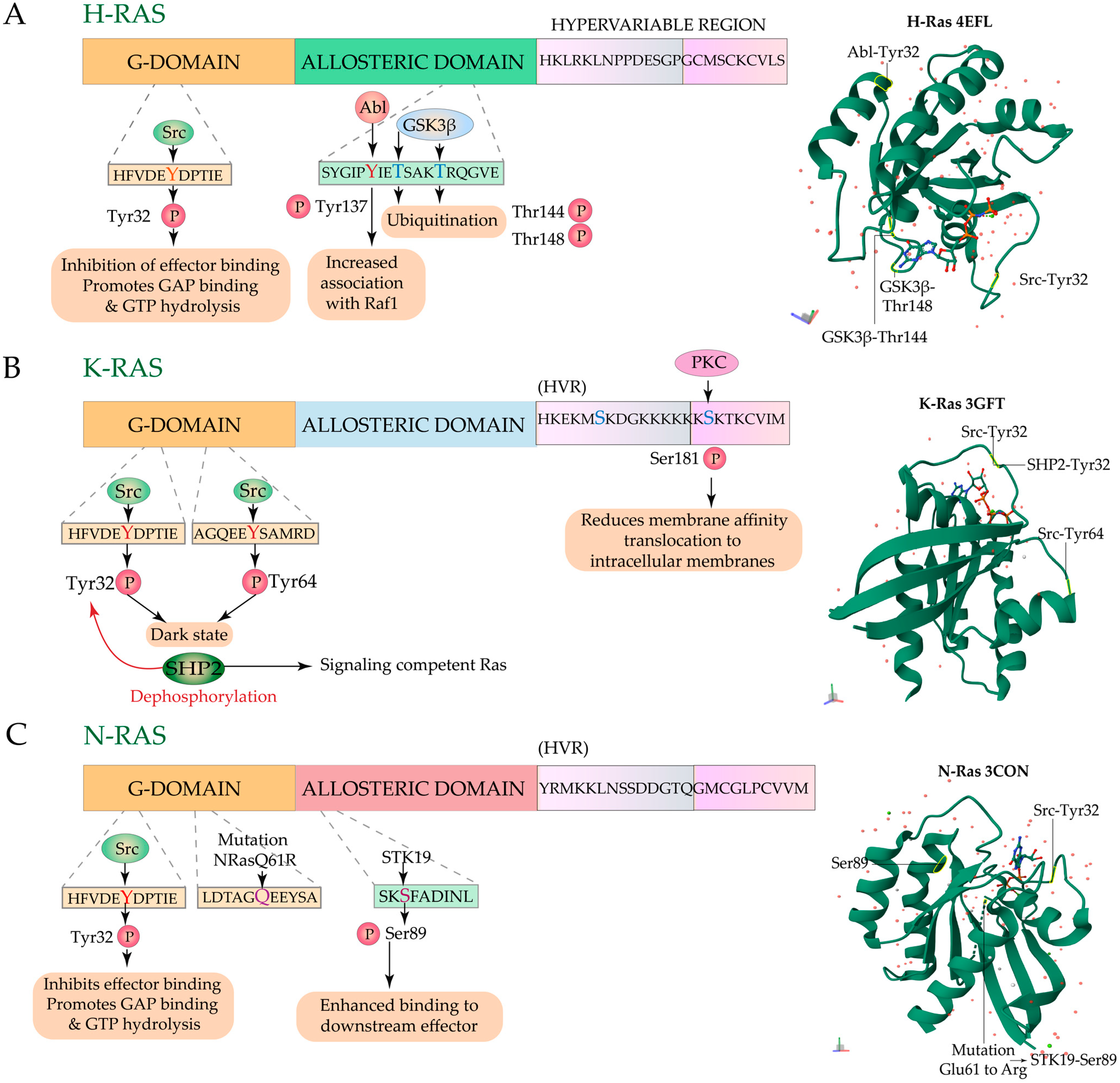
3.3.1. Regulation by Phosphorylation

The primary regulation of Ras activation is by the GEFs and GAPs. Upon EGFR stimulation, a complex is formed between the EGFR-phosphorylated Shc—Grb2—SOS that activates Ras isoforms [62]. Src protein-tyrosine kinase coordinately phosphorylates Tyr239 and Tyr240 residues of the adaptor protein Shc, which enables its association with Grb2 [63]. Grb2 is phosphorylated at Tyr209 by the Bcr/Abl oncoprotein and EGF receptor, which impairs the binding of Grb2 to SOS [64]. Src can also directly bind to and phosphorylate GTP-bound H/N-Ras on Tyr32 within the switch I region. Tyr32 phosphorylation on Ras reduces its affinity for its effector Raf but increases its binding to the GAPs and GTP hydrolysis [65]. Further studies have shown that the Src-mediated phosphorylation of K-Ras on Tyr32 and Tyr64 leads to conformational changes in the switch I (aa 30–38) and switch II (aa 59–76) of K-Ras, which attenuate its sensitivities towards GEF, GAP, and binding to Raf. This results in the accumulation of phosphorylated-K-Ras-GTP, which is insensitive to further activation or activating downstream Raf and is termed the ‘dark state’ [66]. In 1989, it was first reported that purified insulin receptor kinase phosphorylates H-Ras in the presence of poly (L-lysine) and K-Ras even in the absence of a basic protein [67]. The positively charged polylysine residues in the HVR of K-Ras4B interact with the phospholipid bilayer’s anionic headgroup [68]. Ballester et al. showed that PKC-mediated phosphorylation at Ser181 of K-Ras4B neutralizes its charges, lowering its membrane affinity [69]. This induces a reversible translocation of K-Ras4B from the PM to the intracellular membranes of organelles, such as the GC, ER, and mitochondria. Additionally, Ser171 may also act as a phosphate acceptor in K-Ras4B. At the outer mitochondrial membrane, K-Ras-GTP associates with Bcl-XL in a PKC-dependent manner to induce apoptosis [70]. Ras can also be phosphorylated by Abelson tyrosine-protein kinase (ABL). RIN1 (Ras and Rab interactor-1) is an activator of ABL tyrosine kinases. In a ternary complex, RIN1 also facilitates Ras Tyr137 phosphorylation by ABL. The phosphorylation of Ras on Tyr137 allosterically alters the protein conformation and effector binding [71]. ABL phosphorylates H-Ras at Tyr137, which leads to a conformational change, increases the association of phosphorylated H-Ras with Raf1, and enhances H-Ras’s signaling capacity. This phosphorylation was found to be independent of the nucleotide-bound state of H-Ras but dependent on the palmitoylation state [71]. Yin et al. identified an array of 12 kinases whose knockdown led to 50% inhibition of N-Ras activity and 6 kinases that led to up to two folds of upregulation of N-Ras activity. The Ser/Thr-protein kinase 19 (STK19) preferentially phosphorylates oncogenic N-RasQ61R at Ser89, which enhances its binding with downstream effectors, and STK knockdown inhibits the activity of N-Ras by 35% [72]. The Wnt/ β-catenin pathway stabilizes Ras activation, which leads to ERK signaling cellular proliferation and tumorigenesis. Glycogen synthase kinase 3 beta (GSK3β), a Ser-Thr kinase and a negative regulator of the Wnt/ β-catenin pathway, phosphorylates H-Ras at Thr144 and Thr148. This recruits β-transducin repeat-containing proteins (β-TrCP), an E3-ubiquitin ligase substrate recognition protein, and marks H-Ras for polyubiquitination and degradation [73]. Inhibition of H-Ras degradation by an aberration in the Wnt/β-catenin pathway leads to Ras-induced transformation in colorectal tumorigenesis. Contrarily, GSK3β is required for the proliferation and survival of pancreatic cancers with K-Ras mutation [74].

3.3.2. Regulation by Dephosphorylation

As mentioned earlier, Tyr32 is a phosphoregulatory residue in Ras. Phospho-Tyr32 can be dephosphorylated by SHP2 (SH2-containing protein tyrosine phosphatase-2 phosphatase), which activates H/N-Ras and reinstates Ras downstream signaling by binding to Raf [75]. SHP2 reverses the Src phosphorylation, thus unleashing K-Ras from its ‘dark state’ and making it signaling competent (Figure 5).
Phosphorylation also alters the localization of proteins, such as the Kinase Suppressor of Ras (KSR), which acts as a molecular scaffold for the Raf/MEK/ERK kinase cascade. In quiescent cells, KSR is present in the cytosol associated with MEK [76], CDC25-associated kinase 1 (C-TAK1) [77], and 14-3-3 [78]. 14-3-3 ensures the retainment of KSR in the cytoplasm by phosphorylating Ser297 and Ser392. Upon Ras activation, Raf is phosphorylated at Ser338 and Tyr341 and the serine-threonine phosphatase Protein Phosphatase 2A (PP2A) dephosphorylates KSR1 at Ser392 and Raf at Ser259 [79], which causes KSR1 and Raf to move to the PM [77]. KSR1 further acts as a scaffold to facilitate the phosphorylation of MEK by Raf. Hence, PP2A positively regulates Ras signaling. Protein tyrosine phosphatase (PTP) 1B has also been implicated in the regulation of Ras. The loss of PTP1B results in the attenuation of Ras activity by an increase in the expression of the GAP p120Ras. p62dok, a 62 kDa protein downstream of tyrosine kinase, binds to p120GAP and is a potential substrate of PTP1B [80]. Overexpression of p62dok inhibits the activity of Ras, suggesting that p62dok is a negative regulator of Ras [81]. Mutational studies have shown that tyrosines 296, 315, 362, 398, and 409 are crucial for the binding of p62dok to GAP [82].

4. Therapeutic Approaches Targeting Enzymatic Regulation of Ras

Targeting of RasGEFs: Various therapeutic approaches have been designed to target the Ras activators and post-translational regulatory enzymes. The depletion of SOS1 leads to a decrease in the survival of tumors harboring K-Ras mutations but not wild-type K-Ras. Further, this effect was not rescued by catalytically impaired (SOS1F929A) or allosterically impaired (SOS1L687E/R688A) SOS1 mutants, demonstrating the possibility of targeting both for therapeutic interventions [5]. A small molecule inhibitor, BI-3406, selectively binds to the catalytic site of SOS1 and prevents SOS1-K-Ras interaction and hence GTP binding to K-Ras. BI-3406 administration inhibited SOS1-catalyzed K-Ras-GTP with an IC50 = 83–231 nm and reduced cellular proliferation and anchorage-dependent 3D growth in K-Ras mutant cell lines and xenograft models. In addition to blocking the feedback reactivation observed in the combination suppression of SOS1 and MEK, BI-3406 targets SOS1 specifically but not SOS2. Moreover, cells carrying NF1 and EGFR mutations were also sensitive to BI-3406 treatment [83]. To find fragments and inhibitors binding to SOS1 or K-Ras to disrupt the enzymatic activity of SOS1, Bayer and Boehringer Ingelheim used fragment screening and high-throughput screening. The GDP-K-Ras: SOS1 PPI was blocked by identical selective, cell-active quinazoline SOS1 inhibitors, Bayer Hit 7 and Boehringer Ingelheim Hit 8, with IC50 values of 320 nm and 1 μM, respectively. By combining the activator fragment F1, which stabilizes the KRasG12C–SOS1cat complex with the quinazoline inhibitor 7, Bayer generated an inhibitor called BAY 293 with an IC50 = 21 nm [84]. In mutant cell lines, a combinatorial treatment strategy combining BAY293 with ARS-853, a covalent inhibitor of K-Ras G12C, showed an antiproliferative impact. In 2021, Boehringer Ingelheim in collaboration with Amgen announced the clinical trial of BI 1701963, which is closely related to BI-3406 and LUMAKRAS (sotorasib), in adult patients with locally advanced or metastatic non-small cell lung cancer. Currently, BI 1701963 is in clinical trials to determine its efficacy against K-Ras mutated cancers alone or in combination with K-Ras inhibitors, a topoisomerase I inhibitor Irinotecan, or trametinib, an MEK inhibitor.
The therapeutic targeting of RasGRP is still in a nascent stage. Indirect inhibition of RasGRP by means of PKC inhibitors shows inhibition of RasGRP3′s phosphorylation and attenuation of activation of Ras. RasGRP3 is phosphorylated in vitro by PKCθ and PKC-β2 [85]. Bryostatin 1 (Bryo1), a potent activator of PKC, in combination with PMA suppressed the PMA-induced activation of RasGRP3 in a prostate cancer cell line. Bryo1 has divergent effects and selectively induces a few subsets of responses induced by PMA and antagonizes other responses, e.g., Bryo1 downregulated PKCα, caused a biphasic downregulation of PKC δ, and suppressed the phosphorylation of PKC and PKD in an LNCaP cell line [86]. The limitation of this approach is exemplified by the fact that redundant PKCs may regulate different RasGRPs and that PKCs have diverse cellular targets. As mentioned earlier, DAG can regulate the activity of RasGRP both directly and indirectly by membrane recruitment and PKC-mediated phosphorylation, respectively. DAG kinases (DGKs) regulate the concentration of DAG available for signaling by metabolizing it to phosphatidic acid. DGKζ significantly eliminated RasGRP-induced activation of Ras [87]. However, DAG may recruit several proteins to the membrane surface; moreover, DGK subtype-specific activators and inhibitors and their effects on all mammalian DGKs have yet to be fully characterized.
As with RasGRP, attempts to target RasGRF have been very limited. As mentioned earlier, the Ser916 phosphorylation on RasGRF1 represents a readout of its activation. NMDAR-dependent induction of LTP requires Calmodulin-Dependent Kinase Kinase/Calmodulin Kinase I (CaMKI), which activates the Ras-ERK pathway. Schmitt et al. showed that STO-609, a pharmacological inhibitor of CaMKK, and dominant-negative CaMKI blocked the phosphorylation of RasGRF1 and hence NMDAR-dependent ERK activation in hippocampal neurons [88]. Additionally, further research into the potential therapeutic applications of PKA [27] and other tyrosine kinases, such as Src [89] and Ack1 [90], in the phosphoregulation of RasGRF1 is possible. As previously mentioned, there are multiple Ras effectors which function in a context-/tissue-/cell-type-/condition-specific manner which limits their therapeutic targeting.
Targeting of post-translational modifications of Ras: With the failure of FTIs, such as lonafarnib and tipifarnib, in advanced clinical trials owing to their inability to block the prenylation of N-Ras and K-Ras, dual inhibition of FFTase and GGTase was attempted. However, this caused extensive toxicity. The limitations of RCE1 or ICMT inhibitors to be translated to the clinic led to the next strategy, i.e., targeting Ras’s membrane association using Farnesylcystein mimetics, namely, S-trans, trans farnesyl thiosalicylic acid (FTS). FTS competes with Ras isoforms for membrane targeting by binding to the chaperone proteins, and this leads to improper membrane trafficking and dislodging of Ras from the membrane. Unlike FTIs, FTS affects all the isoforms, as shown by the dislodging of H-Ras, K-Ras, and N-Ras from their membrane anchoring sites and blockade of Ras activation in FTS-treated Ras-transformed rodent fibroblasts [91]. FTS Salirasib disrupts the spatiotemporal localization of all Ras isoforms [92,93] and has shown anti-tumor efficacy in several cell line and xenograft models, such as pancreatic cancer [94], glioblastoma [95], and hepatocarcinoma [96]. However, in clinical trials of K-Ras mutated lung adenocarcinoma [97] or refractory hematologic malignancies [98], the results obtained were not promising, albeit the life expectancy of patients with pancreatic cancer was doubled in phase I/II clinical study with co-treatment of Salirasib and Gemcitabine [99].
In addition to Farnesylation, H-Ras, N-Ras, and K-Ras 4A undergo palmitoylation. Although non-palmitoylated K-Ras 4B is the predominantly mutated Ras isoform (~85%), emerging studies on the role of K-Ras4A in colorectal adenocarcinoma [100] present PAT as an attractive anti-cancer drug target. Palmitoylation-defective mutants of N-Ras show defective activation of PI3K and ERK [101]. The lipid-based PAT inhibitors, such as 2-bromopalmitate (2BP), tunicamycin, and cerulenin analogs, are non-specific and their modes of action are not well understood. Since the molecular identities, functioning, and localization of ~23 mammalian ZDHHC PAT family members are not well characterized, specific small molecule inhibitors against these have not yet been developed. Huntingtin interacting protein 14 (HIP14) or DHHC17 palmitoylates H-Ras [102] and the specificity of the DHHC9/GPC16 complex as the PAT for H-Ras and N-Ras further suggest the possibility of specific PAT targeting. Acyl protein thioesterase (APT), like APT1, APT2, APTL1, and Palmitoyl-protein thioesterase-1 (PPT1), reverses the process of palmitoylation. A chiral acyl-β-lactone-Palmostatin B was one of the first APT inhibitors discovered by screening a series of β-lactone derivatives against APT1 (APT1 IC50 = 5.4 nM). Further optimization yielded a more soluble and effective Palmostatin M (APT1; IC50 = 2.5 nM) [103,104]. By screening a library of natural products, Zimmerman et al. developed boron-based inhibitors that show APT isoform selectivity and low toxicity [105]. Using fluopol-activity-based protein profiling (ABPP), Bachovchin et al. further developed a piperazine amide motif containing lead molecules inhibitor 21 and inhibitor 1 which block APT1 and APT2, respectively [106]. Whether these inhibitors could be converted to pharmacological leads remains to be validated.
As mentioned earlier, acetylation of Lys104 attenuates the transforming potential of K-Ras. Yang et al. demonstrated that inhibition of HDAC6, using a specific inhibitor Tubastatin A, and knockdown of SIRT2 resulted in increased K-Ras acetylation. This study opens up the future possibility of generating specific inhibitors for targeting HDAC6 and SIRT2 in K-Ras mutant cancers [58]. Further characterization of the regulators of Ras ubiquitination-deubiquitination regulators, such as LZTR1 and OTUB1, could also possibly open new avenues of therapeutic targeting. Although NOS inhibition and antioxidant treatment showed little success in the treatment of neurodegenerative disorders, strategies for specific targeting of Cys118 S-nitrosylation may be adopted for blocking Ras activation. Another therapeutic target specific for pancreatic cancer patients with K-Ras mutation is Glycogen synthase kinase 3α/β. Inhibition of GSK3 was observed to reduce the growth of primary and metastatic patient-derived xenografts in pancreatic cancer patients with the K-Ras mutations KRasG12D, G12V, and G12C [74].
Targeting the phosphorylation-dephosphorylation of Ras: An emerging strategy to combat Ras-driven malignancies is to target Ras’s phosphorylation, whereby either inhibitors/ligands can target the kinases or phosphatases, or the phosphorylation sites on Ras. Although Src is conventionally considered a proto-oncogene, its role in the phosphoregulation of K-Ras represents its tumor-suppressive functions, opening new avenues for targeting Src kinase for Ras-cancer therapies. Reports of clinical trials using SHP inhibitors have shown that therapeutic targeting of kinases has had some encouraging results. SHP2 dephosphorylates the phosphoregulatory Tyr32, thus activating Ras signaling, and SHP2 inhibitors are forerunning candidates for clinical trials. A combination therapy of RMC-4630, an SHP2 inhibitor, and LY3214996, an ERK inhibitor, are currently in phase 1 of a clinical trial in patients with K-Ras mutant cancers, colorectal cancer (CRC), non-small cell lung cancer (NSCLC), and pancreatic ductal adenocarcinoma (PDAC) [107]. Treatments consisting of the SHP2 inhibitor BBP-398 in combination with Nivolumab [108] and Sotorasib [109] are in phase 1 clinical trials for NSCLCs and advanced solid tumors with K-Ras mutations, respectively. The SHP2 inhibitor TNO155 alone and in combination with EGF816 (nazartinib) is also in phase 1 of a clinical trial for patients with K-RasG12-mutant NSCLC [110]. The first-in-human (phase 1/1b) study of the SHP2 Inhibitor BBP-398 in patients with MAPK pathway- or RTK-driven advanced solid tumors was initiated in 2020 [111]. In addition, emerging small molecular inhibitors targeting the orthosteric or allosteric sites of SHP2 include II-B0862, 11a-147, GS49364, and SHP09947. These SHP2 inhibitors work by preventing dynamic cycling and stalling phosphorylated K-Ras-GTP in the ‘dark state’ [66].
Phosphorylation of oncogenic N-RasQ61R at Ser89 by STK 19 enhances its downstream signaling. Ser89 is conserved across all Ras isoforms, suggesting that other mutant Ras isoforms may also be preferentially phosphorylated, making STK a potential drug target. Qian et al. identified chelidonine as a selective inhibitor of STK 19 (IC50 = 125.5 ± 19.3 mol/L), which inhibited Ser89 phosphorylation of Ras in both in vitro and in vivo models [112]. The targeting of Ser89 phosphorylation provides a novel avenue in Ras cancer therapy; however, the intrinsic kinase activity of STK 19 [113] and the efficacy of STK19 in clinical trials still need to be confirmed.
ABL kinase inhibitors can constrain the tyr137 phosphorylation of Ras, which would lead to a conformational shift of Ras to the GDP-bound state. Imatinib and dasatinib, two ABL kinase or BCR-ABL fusion protein inhibitors, are being evaluated in a clinical trial to treat CML. These inhibitors must be evaluated in terms of their efficacy regarding Ras-mutational status. As GSK-3β is a potentially important therapeutic target in human malignancies, the GSK-3β inhibitor 9-ING-41, alone or in combination with gemcitabine, doxorubicin, lomustine, carboplatin, paclitaxel, irinotecan [114], etc., is currently being assessed for its safety and efficacy in patients with refractory cancers. The specific role of GSK-3β in the phosphorylation of H-Ras may be further evaluated for its potential to be translated into a Ras-isoform-specific therapy. Another drug that targets Ras membrane interaction is deltarasin, which blocks K-Ras-PDE6δ interaction and has been shown to attenuate K-Ras mutation-harboring tumors [115]. The lack of understanding of the specificity of PDE6δ for various farnesylated proteins and the evitable dependence of K-Ras on PDE6 δ are the drawbacks of drugs that target chaperone proteins. The PKC agonist bryostatin reduced the growth of oncogenic K-Ras4B tumors but not that of K-Ras4B phosphorylation-deficient tumors in mice [70]. As PKC mediates the activation of diverse signaling molecules and bryostatin has divergent effects, the use of bryostatins and analogs as anticancer drugs pose the problem of specific targeting of K-Ras4B phosphorylation without hampering other PKC-dependent signaling molecules. Bryostatin alone or in combination with other drugs, such as cisplatin, paclitaxel, vincristine, cytarabine, cladribine, etc., is in various phases of clinical trials. Table 1 lists various drugs in different phases of clinical trial.

5. Conclusions

Ras’s membrane targeting and functionality depend on post-translational modification, which has led to the therapeutic targeting of the enzymes implicated in these processes. Unfortunately, the application of FTIs and palmitoylation inhibitors in anti-cancer therapy has been constrained by their poor selectivity, low specificity, and severe cellular toxicity. The lack of hydrophobic druggable pockets in Ras has remained a long-standing obstacle to the effective targeting of Ras. Since phosphoregulation has been identified as the regulatory process in the signaling of not only wild-Ras isoforms but also oncogenic Ras, a better understanding of this process may circumvent the druggability issue. In-depth research on how these post-translational modifications influence Ras’s interaction with other proteins is necessary for the controlled targeting of Ras’s nitrosylated, acetylated, ubiquitinated, and phosphorylated residues. In this review, we have discussed how the GEFs—SOS, RasGRP, and RasGRF—control Ras activation and how the microregulation mechanisms of acetylation, nitrosylation, and ubiquitination control Ras signaling and localization. Based on the approach of focusing on Ras’s PTMs, we have also summarized the numerous medications and therapeutic approaches in various phases of clinical trials. Ras isoforms are differentially involved in various cancers, mouse embryonic development, and infection [116], and their signaling specificities [117] and localization are not yet fully deciphered. A comprehensive study of all the post-translationally regulated residues in Ras isoforms with a special focus on phosphoregulatory residues and the isoform-specific differences in the regulation of these sites in wild type and various mutants may offer a new prospect in anti-Ras isoform-specific cancer therapy.

Author Contributions

A.N. and B.S. conceptualized the manuscript. A.N. contributed to researching the content for the review, discussion of the content, writing of the review, and editing before submission. A.N. perceived and designed the figures. B.S. critically reviewed the manuscript. All authors have read and agreed to the published version of the manuscript.

Funding

This research received no external funding.

Institutional Review Board Statement

Not applicable.

Informed Consent Statement

Not applicable.

Data Availability Statement

Not applicable.

Conflicts of Interest

The authors declare no conflict of interest.

References

  1. Ahearn, I.M.; Haigis, K.; Bar-Sagi, D.; Philips, M.R. Regulating the Regulator: Post-Translational Modification of RAS. Nat. Rev. Mol. Cell Biol. 2012, 13, 39–51. [Google Scholar] [CrossRef] [PubMed]
  2. Dower, N.A.; Stang, S.L.; Bottorff, D.A.; Ebinu, J.O.; Dickie, P.; Ostergaard, H.L.; Stone, J.C. RasGRP Is Essential for Mouse Thymocyte Differentiation and TCR Signaling. Nat. Immunol. 2000, 1, 317–321. [Google Scholar] [CrossRef] [PubMed]
  3. Kortum, R.L.; Sommers, C.L.; Alexander, C.P.; Pinski, J.M.; Li, W.; Grinberg, A.; Lee, J.; Love, P.E.; Samelson, L.E. Targeted Sos1 Deletion Reveals Its Critical Role in Early T-Cell Development. Proc. Natl. Acad. Sci. USA 2011, 108, 12407–12412. [Google Scholar] [CrossRef] [PubMed]
  4. Jun, J.E.; Rubio, I.; Roose, J.P. Regulation of Ras Exchange Factors and Cellular Localization of Ras Activation by Lipid Messengers in T Cells. Front. Immunol. 2013, 4, 239. [Google Scholar] [CrossRef]
  5. Sondermann, H.; Soisson, S.M.; Boykevisch, S.; Yang, S.-S.; Bar-Sagi, D.; Kuriyan, J. Structural Analysis of Autoinhibition in the Ras Activator Son of Sevenless. Cell 2004, 119, 393–405. [Google Scholar] [CrossRef]
  6. Groves, J.T.; Kuriyan, J. Molecular Mechanisms in Signal Transduction at the Membrane. Nat. Struct. Mol. Biol. 2010, 17, 659–665. [Google Scholar] [CrossRef]
  7. Gureasko, J.; Kuchment, O.; Makino, D.L.; Sondermann, H.; Bar-Sagi, D.; Kuriyan, J. Role of the Histone Domain in the Autoinhibition and Activation of the Ras Activator Son of Sevenless. Proc. Natl. Acad. Sci. USA 2010, 107, 3430–3435. [Google Scholar] [CrossRef]
  8. Corbalan-Garcia, S.; Yang, S.S.; Degenhardt, K.R.; Bar-Sagi, D. Identification of the Mitogen-Activated Protein Kinase Phosphorylation Sites on Human Sos1 That Regulate Interaction with Grb2. Mol. Cell. Biol. 1996, 16, 5674–5682. [Google Scholar] [CrossRef]
  9. Baschieri, F.; Farhan, H. Crosstalk of Small GTPases at the Golgi Apparatus. Small GTPases 2012, 3, 80–90. [Google Scholar] [CrossRef]
  10. Arozarena, I.; Matallanas, D.; Berciano, M.T.; Sanz-Moreno, V.; Calvo, F.; Muñoz, M.T.; Egea, G.; Lafarga, M.; Crespo, P. Activation of H-Ras in the Endoplasmic Reticulum by the RasGRF Family Guanine Nucleotide Exchange Factors. Mol. Cell. Biol. 2004, 24, 1516–1530. [Google Scholar] [CrossRef]
  11. Irie, K.; Masuda, A.; Shindo, M.; Nakagawa, Y.; Ohigashi, H. Tumor Promoter Binding of the Protein Kinase C C1 Homology Domain Peptides of RasGRPs, Chimaerins, and Unc13s. Bioorganic. Med. Chem. 2004, 12, 4575–4583. [Google Scholar] [CrossRef] [PubMed]
  12. Ebinu, J.O.; Bottorff, D.A.; Chan, E.Y.; Stang, S.L.; Dunn, R.J.; Stone, J.C. RasGRP, a Ras guanyl nucleotide-releasing protein with calcium-and diacylglycerol-binding motifs. Science 1998, 280, 1082–1086. [Google Scholar] [CrossRef] [PubMed]
  13. Iwig, J.S.; Vercoulen, Y.; Das, R.; Barros, T.; Limnander, A.; Che, Y.; Pelton, J.G.; Wemmer, D.E.; Roose, J.P.; Kuriyan, J. Structural Analysis of Autoinhibition in the Ras-Specific Exchange Factor RasGRP1. eLife 2013, 2, e00813. [Google Scholar] [CrossRef] [PubMed]
  14. Roose, J.P.; Mollenauer, M.; Gupta, V.A.; Stone, J.; Weiss, A. A Diacylglycerol-Protein Kinase C-RasGRP1 Pathway Directs Ras Activation upon Antigen Receptor Stimulation of T Cells. Mol. Cell. Biol. 2005, 25, 4426–4441. [Google Scholar] [CrossRef]
  15. Zheng, Y.; Liu, H.; Coughlin, J.; Zheng, J.; Li, L.; Stone, J.C. Phosphorylation of RasGRP3 on Threonine 133 Provides a Mechanistic Link between PKC and Ras Signaling Systems in B Cells. Blood 2005, 105, 3648–3654. [Google Scholar] [CrossRef] [PubMed]
  16. Guidetti, G.F.; Manganaro, D.; Consonni, A.; Canobbio, I.; Balduini, C.; Torti, M. Phosphorylation of the Guanine-Nucleotide-Exchange Factor CalDAG-GEFI by Protein Kinase A Regulates Ca2+-Dependent Activation of Platelet Rap1b GTPase. Biochem. J. 2013, 453, 115–123. [Google Scholar] [CrossRef]
  17. Nagai, T.; Nakamuta, S.; Kuroda, K.; Nakauchi, S.; Nishioka, T.; Takano, T.; Zhang, X.; Tsuboi, D.; Funahashi, Y.; Nakano, T. Phosphoproteomics of the Dopamine Pathway Enables Discovery of Rap1 Activation as a Reward Signal in Vivo. Neuron 2016, 89, 550–565. [Google Scholar] [CrossRef]
  18. Ren, J.; Cook, A.A.; Bergmeier, W.; Sondek, J. A Negative-Feedback Loop Regulating ERK1/2 Activation and Mediated by RasGPR2 Phosphorylation. Biochem. Biophys. Res. Commun. 2016, 474, 193–198. [Google Scholar] [CrossRef]
  19. Liao, F.; Shin, H.S.; Rhee, S.G. In Vitro Tyrosine Phosphorylation of PLC-Γ1 and PLC-Γ2 by Src-Family Protein Tyrosine Kinases. Biochem. Biophys. Res. Commun. 1993, 191, 1028–1033. [Google Scholar] [CrossRef]
  20. Bivona, T.G.; Pérez de Castro, I.; Ahearn, I.M.; Grana, T.M.; Chiu, V.K.; Lockyer, P.J.; Cullen, P.J.; Pellicer, A.; Cox, A.D.; Philips, M.R. Phospholipase Cγ Activates Ras on the Golgi Apparatus by Means of RasGRP1. Nature 2003, 424, 694–698. [Google Scholar] [CrossRef]
  21. Lorenzo, P.S.; Kung, J.W.; Bottorff, D.A.; Garfield, S.H.; Stone, J.C.; Blumberg, P.M. Phorbol Esters Modulate the Ras Exchange Factor RasGRP3. Cancer Res. 2001, 61, 943–949. [Google Scholar] [PubMed]
  22. Freshney, N.W.; Goonesekera, S.D.; Feig, L.A. Activation of the Exchange Factor Ras-GRF by Calcium Requires an Intact Dbl Homology Domain. FEBS Lett. 1997, 407, 111–115. [Google Scholar] [CrossRef] [PubMed]
  23. Sinclair, N.R.S. Immunoreceptor Tyrosine-Based Inhibitory Motifs on Activating Molecules. Crit. Rev. Immunol. 2000, 20, 89–102. [Google Scholar] [CrossRef] [PubMed]
  24. Kesavapany, S.; Pareek, T.K.; Zheng, Y.-L.; Amin, N.; Gutkind, J.S.; Ma, W.; Kulkarni, A.B.; Grant, P.; Pant, H.C. Neuronal Nuclear Organization Is Controlled by Cyclin-Dependent Kinase 5 Phosphorylation of Ras Guanine Nucleotide Releasing Factor-1. Neurosignals 2006, 15, 157–173. [Google Scholar] [CrossRef]
  25. Bähler, M.; Rhoads, A. Calmodulin Signaling via the IQ Motif. FEBS Lett. 2002, 513, 107–113. [Google Scholar] [CrossRef]
  26. Krapivinsky, G.; Krapivinsky, L.; Manasian, Y.; Ivanov, A.; Tyzio, R.; Pellegrino, C.; Ben-Ari, Y.; Clapham, D.E.; Medina, I. The NMDA Receptor Is Coupled to the ERK Pathway by a Direct Interaction between NR2B and RasGRF1. Neuron 2003, 40, 775–784. [Google Scholar] [CrossRef]
  27. Feig, L.A. Regulation of Neuronal Function by Ras-GRF Exchange Factors. Genes Cancer 2011, 2, 306–319. [Google Scholar] [CrossRef]
  28. Wei, W.; Schreiber, S.S.; Baudry, M.; Tocco, G.; Broek, D. Localization of the Cellular Expression Pattern of Cdc25NEF and Ras in the Juvenile Rat Brain. Mol. Brain Res. 1993, 19, 339–344. [Google Scholar] [CrossRef]
  29. Yang, H.; Cooley, D.; Legakis, J.E.; Ge, Q.; Andrade, R.; Mattingly, R.R. Phosphorylation of the Ras-GRF1 Exchange Factor at Ser916/898 Reveals Activation of Ras Signaling in the Cerebral Cortex. J. Biol. Chem. 2003, 278, 13278–13285. [Google Scholar] [CrossRef]
  30. Jones, M.K.; Jackson, J.H. Ras-Grf Activates Ha-Ras, but Not n-Ras or k-Ras 4b, Protein in Vivo. J. Biol. Chem. 1998, 273, 1782–1787. [Google Scholar] [CrossRef]
  31. Mattingly, R.R.; Saini, V.; Macara, I.G. Activation of the Ras-GRF/CDC25Mm Exchange Factor by Lysophosphatidic Acid. Cell. Signal. 1999, 11, 603–610. [Google Scholar] [CrossRef] [PubMed]
  32. Resat, H.; Straatsma, T.P.; Dixon, D.A.; Miller, J.H. The Arginine Finger of RasGAP Helps Gln-61 Align the Nucleophilic Water in GAP-Stimulated Hydrolysis of GTP. Proc. Natl. Acad. Sci. USA 2001, 98, 6033–6038. [Google Scholar] [CrossRef] [PubMed]
  33. Xu, X.; Pots, H.; Gilsbach, B.K.; Parsons, D.; Veltman, D.M.; Ramachandra, S.G.; Li, H.; Kortholt, A.; Jin, T. C2GAP2 Is a Common Regulator of Ras Signaling for Chemotaxis, Phagocytosis, and Macropinocytosis. Front. Immunol. 2022, 13, 1075386. [Google Scholar] [CrossRef] [PubMed]
  34. Mangoura, D.; Sun, Y.; Li, C.; Singh, D.; Gutmann, D.H.; Flores, A.; Ahmed, M.; Vallianatos, G. Phosphorylation of Neurofibromin by PKC Is a Possible Molecular Switch in EGF Receptor Signaling in Neural Cells. Oncogene 2006, 25, 735–745. [Google Scholar] [CrossRef]
  35. Feng, L.; Yunoue, S.; Tokuo, H.; Ozawa, T.; Zhang, D.; Patrakitkomjorn, S.; Ichimura, T.; Saya, H.; Araki, N. PKA Phosphorylation and 14-3-3 Interaction Regulate the Function of Neurofibromatosis Type I Tumor Suppressor, Neurofibromin. FEBS Lett. 2004, 557, 275–282. [Google Scholar] [CrossRef]
  36. Skorski, T.; Kanakaraj, P.; Ku, D.-H.; Nieborowska-Skorska, M.; Canaani, E.; Zon, G.; Perussia, B.; Calabretta, B. Negative Regulation of P120GAP GTPase Promoting Activity by P210bcr/Abl: Implication for RAS-Dependent Philadelphia Chromosome Positive Cell Growth. J. Exp. Med. 1994, 179, 1855–1865. [Google Scholar] [CrossRef]
  37. Torti, M.; Marti, K.B.; Altschuler, D.; Yamamoto, K.; Lapetina, E.G. Erythropoietin Induces P21ras Activation and P120GAP Tyrosine Phosphorylation in Human Erythroleukemia Cells. J. Biol. Chem. 1992, 267, 8293–8298. [Google Scholar] [CrossRef]
  38. Chong, H.; Lee, J.; Guan, K.-L. Positive and Negative Regulation of Raf Kinase Activity and Function by Phosphorylation. EMBO J. 2001, 20, 3716–3727. [Google Scholar] [CrossRef]
  39. Wu, J.; Dent, P.; Jelinek, T.; Wolfman, A.; Weber, M.J.; Sturgill, T.W. Inhibition of the EGF-Activated MAP Kinase Signaling Pathway by Adenosine 3′, 5′-Monophosphate. Science 1993, 262, 1065–1069. [Google Scholar] [CrossRef]
  40. Dhillon, A.S.; Pollock, C.; Steen, H.; Shaw, P.E.; Mischak, H.; Kolch, W. Cyclic AMP-Dependent Kinase Regulates Raf-1 Kinase Mainly by Phosphorylation of Serine 259. Mol. Cell. Biol. 2002, 22, 3237–3246. [Google Scholar] [CrossRef]
  41. Mischak, H.; Seitz, T.; Janosch, P.; Eulitz, M.; Steen, H.; Schellerer, M.; Philipp, A.; Kolch, W. Negative Regulation of Raf-1 by Phosphorylation of Serine 621. Mol. Cell. Biol. 1996, 16, 5409–5418. [Google Scholar] [CrossRef] [PubMed]
  42. Dutchak, K.; Garnett, S.; Nicoll, M.; de Bruyns, A.; Dankort, D. MOB3A Bypasses BRAF and RAS Oncogene-Induced Senescence by Engaging the Hippo Pathway. Mol. Cancer Res. 2022, 20, 770–781. [Google Scholar] [CrossRef] [PubMed]
  43. Dangelmaier, C.; Manne, B.K.; Liverani, E.; Jin, J.; Bray, P.; Kunapuli, S.P. PDK1 Selectively Phosphorylates Thr (308) on Akt and Contributes to Human Platelet Functional Responses. Thromb. Haemost. 2014, 112, 508–517. [Google Scholar] [CrossRef] [PubMed]
  44. Sarbassov, D.D.; Guertin, D.A.; Ali, S.M.; Sabatini, D.M. Phosphorylation and Regulation of Akt/PKB by the Rictor-MTOR Complex. Science 2005, 307, 1098–1101. [Google Scholar] [CrossRef]
  45. Ugi, S.; Imamura, T.; Maegawa, H.; Egawa, K.; Yoshizaki, T.; Shi, K.; Obata, T.; Ebina, Y.; Kashiwagi, A.; Olefsky, J.M. Protein Phosphatase 2A Negatively Regulates Insulin’s Metabolic Signaling Pathway by Inhibiting Akt (Protein Kinase B) Activity in 3T3-L1 Adipocytes. Mol. Cell. Biol. 2004, 24, 8778–8789. [Google Scholar] [CrossRef]
  46. Brognard, J.; Sierecki, E.; Gao, T.; Newton, A.C. PHLPP and a Second Isoform, PHLPP2, Differentially Attenuate the Amplitude of Akt Signaling by Regulating Distinct Akt Isoforms. Mol. Cell 2007, 25, 917–931. [Google Scholar] [CrossRef]
  47. Catozzi, S.; Ternet, C.; Gourrege, A.; Wynne, K.; Oliviero, G.; Kiel, C. Reconstruction and Analysis of a Large-Scale Binary Ras-Effector Signaling Network. Cell Commun. Signal. 2022, 20, 24. [Google Scholar] [CrossRef]
  48. Whyte, D.B.; Kirschmeier, P.; Hockenberry, T.N.; Nunez-Oliva, I.; James, L.; Catino, J.J.; Bishop, W.R.; Pai, J.-K. K-and N-Ras Are Geranylgeranylated in Cells Treated with Farnesyl Protein Transferase Inhibitors. J. Biol. Chem. 1997, 272, 14459–14464. [Google Scholar] [CrossRef]
  49. Nair, A.; Kubatzky, K.F.; Saha, B. Ras Isoforms from Lab Benches to Lives—What Are We Missing and How Far Are We? Int. J. Mol. Sci. 2021, 22, 6508. [Google Scholar] [CrossRef]
  50. Washington, C.; Chernet, R.; Gokhale, R.H.; Martino-Cortez, Y.; Liu, H.-Y.; Rosenberg, A.M.; Shahar, S.; Pfleger, C.M. A Conserved, N-Terminal Tyrosine Signal Directs Ras for Inhibition by Rabex-5. PLoS Genet. 2020, 16, e1008715. [Google Scholar] [CrossRef]
  51. Xu, L.; Lubkov, V.; Taylor, L.J.; Bar-Sagi, D. Feedback Regulation of Ras Signaling by Rabex-5-Mediated Ubiquitination. Curr. Biol. 2010, 20, 1372–1377. [Google Scholar] [CrossRef] [PubMed]
  52. Jura, N.; Scotto-Lavino, E.; Sobczyk, A.; Bar-Sagi, D. Differential Modification of Ras Proteins by Ubiquitination. Molecular Cell 2006, 21, 679–687. [Google Scholar] [CrossRef]
  53. Baker, R.; Wilkerson, E.M.; Sumita, K.; Isom, D.G.; Sasaki, A.T.; Dohlman, H.G.; Campbell, S.L. Differences in the Regulation of K-Ras and H-Ras Isoforms by Monoubiquitination. J. Biol. Chem. 2013, 288, 36856–36862. [Google Scholar] [CrossRef] [PubMed]
  54. Baietti, M.F.; Simicek, M.; Abbasi Asbagh, L.; Radaelli, E.; Lievens, S.; Crowther, J.; Steklov, M.; Aushev, V.N.; Martínez García, D.; Tavernier, J. OTUB 1 Triggers Lung Cancer Development by Inhibiting RAS Monoubiquitination. EMBO Mol. Med. 2016, 8, 288–303. [Google Scholar] [CrossRef]
  55. Sasaki, A.T.; Carracedo, A.; Locasale, J.W.; Anastasiou, D.; Takeuchi, K.; Kahoud, E.R.; Haviv, S.; Asara, J.M.; Pandolfi, P.P.; Cantley, L.C. Ubiquitination of K-Ras Enhances Activation and Facilitates Binding to Select Downstream Effectors. Sci. Signal. 2011, 4, ra13. [Google Scholar] [CrossRef]
  56. Bigenzahn, J.W.; Collu, G.M.; Kartnig, F.; Pieraks, M.; Vladimer, G.I.; Heinz, L.X.; Sedlyarov, V.; Schischlik, F.; Fauster, A.; Rebsamen, M. LZTR1 Is a Regulator of RAS Ubiquitination and Signaling. Science 2018, 362, 1171–1177. [Google Scholar] [CrossRef]
  57. Yang, M.H.; Nickerson, S.; Kim, E.T.; Liot, C.; Laurent, G.; Spang, R.; Philips, M.R.; Shan, Y.; Shaw, D.E.; Bar-Sagi, D. Regulation of RAS Oncogenicity by Acetylation. Proc. Natl. Acad. Sci. USA 2012, 109, 10843–10848. [Google Scholar] [CrossRef] [PubMed]
  58. Yang, M.H.; Laurent, G.; Bause, A.S.; Spang, R.; German, N.; Haigis, M.C.; Haigis, K.M. HDAC6 and SIRT2 Regulate the Acetylation State and Oncogenic Activity of Mutant K-RASRegulation of K-RAS Acetylation. Mol. Cancer Res. 2013, 11, 1072–1077. [Google Scholar] [CrossRef]
  59. Santos, A.I.; Carreira, B.P.; Izquierdo-Alvarez, A.; Ramos, E.; Lourenço, A.S.; Filipa Santos, D.; Morte, M.I.; Ribeiro, L.F.; Marreiros, A.; Sánchez-López, N. S-Nitrosylation of Ras Mediates Nitric Oxide-Dependent Post-Injury Neurogenesis in a Seizure Model. Antioxid. Redox Signal. 2018, 28, 15–30. [Google Scholar] [CrossRef]
  60. Marshall, H.E.; Foster, M.W. S-Nitrosylation of Ras in Breast Cancer. Breast Cancer Res. 2012, 14, 1–2. [Google Scholar] [CrossRef]
  61. Ibiza, S.; Pérez-Rodríguez, A.; Ortega, Á.; Martínez-Ruiz, A.; Barreiro, O.; García-Domínguez, C.A.; Víctor, V.M.; Esplugues, J.V.; Rojas, J.M.; Sánchez-Madrid, F. Endothelial Nitric Oxide Synthase Regulates N-Ras Activation on the Golgi Complex of Antigen-Stimulated T Cells. Proc. Natl. Acad. Sci. USA 2008, 105, 10507–10512. [Google Scholar] [CrossRef] [PubMed]
  62. Basu, T.; Warne, P.H.; Downward, J. Role of Shc in the Activation of Ras in Response to Epidermal Growth Factor and Nerve Growth Factor. Oncogene 1994, 9, 3483–3491. [Google Scholar] [PubMed]
  63. van der Geer, P.; Wiley, S.; Gish, G.D.; Pawson, T. The Shc Adaptor Protein Is Highly Phosphorylated at Conserved, Twin Tyrosine Residues (Y239/240) That Mediate Protein–Protein Interactions. Curr. Biol. 1996, 6, 1435–1444. [Google Scholar] [CrossRef] [PubMed]
  64. Li, S.; Couvillon, A.D.; Brasher, B.B.; Van Etten, R.A. Tyrosine Phosphorylation of Grb2 by Bcr/Abl and Epidermal Growth Factor Receptor: A Novel Regulatory Mechanism for Tyrosine Kinase Signaling. EMBO J. 2001, 20, 6793–6804. [Google Scholar] [CrossRef]
  65. Bunda, S.; Heir, P.; Srikumar, T.; Cook, J.D.; Burrell, K.; Kano, Y.; Lee, J.E.; Zadeh, G.; Raught, B.; Ohh, M. Src Promotes GTPase Activity of Ras via Tyrosine 32 Phosphorylation. Proc. Natl. Acad. Sci. USA 2014, 111, E3785–E3794. [Google Scholar] [CrossRef]
  66. Kano, Y.; Gebregiworgis, T.; Marshall, C.B.; Radulovich, N.; Poon, B.P.; St-Germain, J.; Cook, J.D.; Valencia-Sama, I.; Grant, B.M.; Herrera, S.G. Tyrosyl Phosphorylation of KRAS Stalls GTPase Cycle via Alteration of Switch I and II Conformation. Nat. Commun. 2019, 10, 224–237. [Google Scholar] [CrossRef] [PubMed]
  67. Fujita-Yamaguchi, Y.; Kathuria, S.; Xu, Q.-Y.; McDonald, J.M.; Nakano, H.; Kamata, T. In Vitro Tyrosine Phosphorylation Studies on RAS Proteins and Calmodulin Suggest That Polylysine-like Basic Peptides or Domains May Be Involved in Interactions between Insulin Receptor Kinase and Its Substrate. Proc. Natl. Acad. Sci. USA 1989, 86, 7306–7310. [Google Scholar] [CrossRef]
  68. Leventis, R.; Silvius, J.R. Lipid-Binding Characteristics of the Polybasic Carboxy-Terminal Sequence of K-Ras 4B. Biochemistry 1998, 37, 7640–7648. [Google Scholar] [CrossRef]
  69. Ballester, R.; Furth, M.E.; Rosen, O.M. Phorbol Ester-and Protein Kinase C-Mediated Phosphorylation of the Cellular Kirsten Ras Gene Product. J. Biol. Chem. 1987, 262, 2688–2695. [Google Scholar] [CrossRef]
  70. Bivona, T.G.; Quatela, S.E.; Bodemann, B.O.; Ahearn, I.M.; Soskis, M.J.; Mor, A.; Miura, J.; Wiener, H.H.; Wright, L.; Saba, S.G. PKC Regulates a Farnesyl-Electrostatic Switch on K-Ras That Promotes Its Association with Bcl-XL on Mitochondria and Induces Apoptosis. Molecular Cell 2006, 21, 481–493. [Google Scholar] [CrossRef]
  71. Ting, P.Y.; Johnson, C.W.; Fang, C.; Cao, X.; Graeber, T.G.; Mattos, C.; Colicelli, J. Tyrosine Phosphorylation of RAS by ABL Allosterically Enhances Effector Binding. FASEB J. 2015, 29, 3750–3761. [Google Scholar] [CrossRef]
  72. Yin, C.; Zhu, B.; Zhang, T.; Liu, T.; Chen, S.; Liu, Y.; Li, X.; Miao, X.; Li, S.; Mi, X. Pharmacological Targeting of STK19 Inhibits Oncogenic NRAS-Driven Melanomagenesis. Cell 2019, 176, 1113–1127.e16. [Google Scholar] [CrossRef] [PubMed]
  73. Jeong, W.-J.; Yoon, J.; Park, J.-C.; Lee, S.-H.; Lee, S.-H.; Kaduwal, S.; Kim, H.; Yoon, J.-B.; Choi, K.-Y. Ras Stabilization through Aberrant Activation of Wnt/β-Catenin Signaling Promotes Intestinal Tumorigenesis. Sci. Signal. 2012, 5, ra30. [Google Scholar] [CrossRef] [PubMed]
  74. Kazi, A.; Xiang, S.; Yang, H.; Delitto, D.; Trevino, J.; Jiang, R.H.; Ayaz, M.; Lawrence, H.R.; Kennedy, P.; Sebti, S.M. GSK3 Suppression Upregulates β-Catenin and c-Myc to Abrogate KRas-Dependent Tumors. Nat. Commun. 2018, 9, 5154–5162. [Google Scholar] [CrossRef] [PubMed]
  75. Bunda, S.; Burrell, K.; Heir, P.; Zeng, L.; Alamsahebpour, A.; Kano, Y.; Raught, B.; Zhang, Z.-Y.; Zadeh, G.; Ohh, M. Inhibition of SHP2-Mediated Dephosphorylation of Ras Suppresses Oncogenesis. Nat. Commun. 2015, 6, 8859–8870. [Google Scholar] [CrossRef] [PubMed]
  76. Ritt, D.A.; Daar, I.O.; Morrison, D.K. KSR Regulation of the Raf-MEK-ERK Cascade. Methods Enzymol. 2006, 407, 224–237. [Google Scholar] [PubMed]
  77. Müller, J.; Ory, S.; Copeland, T.; Piwnica-Worms, H.; Morrison, D.K. C-TAK1 Regulates Ras Signaling by Phosphorylating the MAPK Scaffold, KSR1. Molecular Cell 2001, 8, 983–993. [Google Scholar] [CrossRef] [PubMed]
  78. Cacace, A.M.; Michaud, N.R.; Therrien, M.; Mathes, K.; Copeland, T.; Rubin, G.M.; Morrison, D.K. Identification of Constitutive and Ras-Inducible Phosphorylation Sites of KSR: Implications for 14-3-3 Binding, Mitogen-Activated Protein Kinase Binding, and KSR Overexpression. Mol. Cell. Biol. 1999, 19, 229–240. [Google Scholar] [CrossRef]
  79. Abraham, D.; Podar, K.; Pacher, M.; Kubicek, M.; Welzel, N.; Hemmings, B.A.; Dilworth, S.M.; Mischak, H.; Kolch, W.; Baccarini, M. Raf-1-Associated Protein Phosphatase 2A as a Positive Regulator of Kinase Activation. J. Biol. Chem. 2000, 275, 22300–22304. [Google Scholar] [CrossRef] [PubMed]
  80. Dubé, N.; Cheng, A.; Tremblay, M.L. The Role of Protein Tyrosine Phosphatase 1B in Ras Signaling. Proc. Natl. Acad. Sci. USA 2004, 101, 1834–1839. [Google Scholar] [CrossRef]
  81. Yoshida, K.; Yamashita, Y.; Miyazato, A.; Ohya, K.; Kitanaka, A.; Ikeda, U.; Shimada, K.; Yamanaka, T.; Ozawa, K.; Mano, H. Mediation by the Protein-Tyrosine Kinase Tec of Signaling between the B Cell Antigen Receptor and Dok-1. J. Biol. Chem. 2000, 275, 24945–24952. [Google Scholar] [CrossRef]
  82. Kashige, N.; Carpino, N.; Kobayashi, R. Tyrosine Phosphorylation of P62dok by P210bcr-Abl Inhibits RasGAP Activity. Proc. Natl. Acad. Sci. USA 2000, 97, 2093–2098. [Google Scholar] [CrossRef] [PubMed]
  83. Hofmann, M.H.; Gmachl, M.; Ramharter, J.; Savarese, F.; Gerlach, D.; Marszalek, J.R.; Sanderson, M.P.; Kessler, D.; Trapani, F.; Arnhof, H. BI-3406, a Potent and Selective SOS1–KRAS Interaction Inhibitor, Is Effective in KRAS-Driven Cancers through Combined MEK InhibitionPan-KRAS SOS1 Protein–Protein Interaction Inhibitor BI-3406. Cancer Discov. 2021, 11, 142–157. [Google Scholar] [CrossRef] [PubMed]
  84. Hillig, R.C.; Sautier, B.; Schroeder, J.; Moosmayer, D.; Hilpmann, A.; Stegmann, C.M.; Werbeck, N.D.; Briem, H.; Boemer, U.; Weiske, J. Discovery of Potent SOS1 Inhibitors That Block RAS Activation via Disruption of the RAS–SOS1 Interaction. Proc. Natl. Acad. Sci. USA 2019, 116, 2551–2560. [Google Scholar] [CrossRef] [PubMed]
  85. Teixeira, C.; Stang, S.L.; Zheng, Y.; Beswick, N.S.; Stone, J.C. Integration of DAG Signaling Systems Mediated by PKC-Dependent Phosphorylation of RasGRP3. Blood 2003, 102, 1414–1420. [Google Scholar] [CrossRef]
  86. Kedei, N.; Yang, D.; Blumberg, P. Bryostatin 1 Suppresses Induction by Protein Kinase C of the Ras Activator RasGRP3 in LNCaP Prostrate Cancer Cells. Cancer Res. 2007, 67, 3954. [Google Scholar]
  87. Stone, J.C. Regulation and Function of the RasGRP Family of Ras Activators in Blood Cells. Genes Cancer 2011, 2, 320–334. [Google Scholar] [CrossRef]
  88. Schmitt, J.M.; Guire, E.S.; Saneyoshi, T.; Soderling, T.R. Calmodulin-Dependent Kinase Kinase/Calmodulin Kinase I Activity Gates Extracellular-Regulated Kinase-Dependent Long-Term Potentiation. J. Neurosci. 2005, 25, 1281–1290. [Google Scholar] [CrossRef]
  89. Giglione, C.; Gonfloni, S.; Parmeggiani, A. Differential Actions of P60c-Src and Lck Kinases on the Ras Regulators P120-GAP and GDP/GTP Exchange Factor CDC25Mm. Eur. J. Biochem. 2001, 268, 3275–3283. [Google Scholar] [CrossRef]
  90. Kiyono, M.; Kato, J.; Kataoka, T.; Kaziro, Y.; Satoh, T. Stimulation of Ras Guanine Nucleotide Exchange Activity of Ras-GRF1/CDC25Mm upon Tyrosine Phosphorylation by the Cdc42-Regulated Kinase ACK1. J. Biol. Chem. 2000, 275, 29788–29793. [Google Scholar] [CrossRef]
  91. Marom, M.; Haklai, R.; Ben-Baruch, G.; Marciano, D.; Egozi, Y.; Kloog, Y. Selective Inhibition of Ras-Dependent Cell Growth by Farnesylthiosalisylic Acid (∗). J. Biol. Chem. 1995, 270, 22263–22270. [Google Scholar] [CrossRef] [PubMed]
  92. Elad, G.; Paz, A.; Haklai, R.; Marciano, D.; Cox, A.; Kloog, Y. Targeting of K-Ras 4B by S-Trans, Trans-Farnesyl Thiosalicylic Acid. Biochim. Biophys. Acta (BBA)-Mol. Cell Res. 1999, 1452, 228–242. [Google Scholar] [CrossRef]
  93. Weisz, B.; Giehl, K.; Gana-Weisz, M.; Egozi, Y.; Ben-Baruch, G.; Marciano, D.; Gierschik, P.; Kloog, Y. A New Functional Ras Antagonist Inhibits Human Pancreatic Tumor Growth in Nude Mice. Oncogene 1999, 18, 2579–2588. [Google Scholar] [CrossRef] [PubMed]
  94. Goldberg, L.; Israeli, R.; Kloog, Y. FTS and 2-DG Induce Pancreatic Cancer Cell Death and Tumor Shrinkage in Mice. Cell Death Dis. 2012, 3, e284. [Google Scholar] [CrossRef] [PubMed]
  95. Goldberg, L.; Ocherashvilli, A.; Daniels, D.; Last, D.; Cohen, Z.R.; Tamar, G.; Kloog, Y.; Mardor, Y. Salirasib (Farnesyl Thiosalicylic Acid) for Brain Tumor Treatment: A Convection-Enhanced Drug Delivery Study in Rats. Mol. Cancer Ther. 2008, 7, 3609–3616. [Google Scholar] [CrossRef]
  96. Charette, N.; De Saeger, C.; Lannoy, V.; Horsmans, Y.; Leclercq, I.; Stärkel, P. Salirasib Inhibits the Growth of Hepatocarcinoma Cell Lines in Vitro and Tumor Growth in Vivo through Ras and MTOR Inhibition. Molecular Cancer 2010, 9, 256–269. [Google Scholar] [CrossRef]
  97. Riely, G.J.; Johnson, M.L.; Medina, C.; Rizvi, N.A.; Miller, V.A.; Kris, M.G.; Pietanza, M.C.; Azzoli, C.G.; Krug, L.M.; Pao, W. A Phase II Trial of Salirasib in Patients with Lung Adenocarcinomas with KRAS Mutations. J. Thorac. Oncol. 2011, 6, 1435–1437. [Google Scholar] [CrossRef]
  98. Badar, T.; Cortes, J.E.; Ravandi, F.; O’Brien, S.; Verstovsek, S.; Garcia-Manero, G.; Kantarjian, H.; Borthakur, G. Phase I Study of S-Trans, Trans-Farnesylthiosalicylic Acid (Salirasib), a Novel Oral RAS Inhibitor in Patients with Refractory Hematologic Malignancies. Clin. Lymphoma Myeloma Leuk. 2015, 15, 433–438.e2. [Google Scholar] [CrossRef]
  99. Laheru, D.; Rudek, M.; Taylor, G.; Goldsweig, H.; Rajeshkumar, N.V.; Linden, S.; Angenendt, M.; Le, D.; Donehower, R.; Jimeno, A. Integrated Development of S-Trans, Trans-Farnesylthiosalicyclic Acid (FTS, Salirasib) in Advanced Pancreatic Cancer. J. Clin. Oncol. 2009, 27, 4529. [Google Scholar] [CrossRef]
  100. Tsai, F.D.; Lopes, M.S.; Zhou, M.; Court, H.; Ponce, O.; Fiordalisi, J.J.; Gierut, J.J.; Cox, A.D.; Haigis, K.M.; Philips, M.R. K-Ras4A Splice Variant Is Widely Expressed in Cancer and Uses a Hybrid Membrane-Targeting Motif. Proc. Natl. Acad. Sci. USA 2015, 112, 779–784. [Google Scholar] [CrossRef]
  101. Cuiffo, B.; Ren, R. Palmitoylation of Oncogenic NRAS Is Essential for Leukemogenesis. Blood J. Am. Soc. Hematol. 2010, 115, 3598–3605. [Google Scholar] [CrossRef]
  102. Ducker, C.E.; Stettler, E.M.; French, K.J.; Upson, J.J.; Smith, C.D. Huntingtin Interacting Protein 14 Is an Oncogenic Human Protein: Palmitoyl Acyltransferase. Oncogene 2004, 23, 9230–9237. [Google Scholar] [CrossRef] [PubMed]
  103. Dekker, F.J.; Rocks, O.; Vartak, N.; Menninger, S.; Hedberg, C.; Balamurugan, R.; Wetzel, S.; Renner, S.; Gerauer, M.; Schölermann, B. Small-Molecule Inhibition of APT1 Affects Ras Localization and Signaling. Nat. Chem. Biol. 2010, 6, 449–456. [Google Scholar] [CrossRef]
  104. Hedberg, C.; Dekker, F.J.; Rusch, M.; Renner, S.; Wetzel, S.; Vartak, N.; Gerding-Reimers, C.; Bon, R.S.; Bastiaens, P.I.; Waldmann, H. Development of Highly Potent Inhibitors of the Ras-targeting Human Acyl Protein Thioesterases Based on Substrate Similarity Design. Angew. Chem. Int. Ed. 2011, 50, 9832–9837. [Google Scholar] [CrossRef]
  105. Zimmermann, T.J.; Bürger, M.; Tashiro, E.; Kondoh, Y.; Martinez, N.E.; Görmer, K.; Rosin-Steiner, S.; Shimizu, T.; Ozaki, S.; Mikoshiba, K. Boron-Based Inhibitors of Acyl Protein Thioesterases 1 and 2. Chembiochem 2013, 14, 115–122. [Google Scholar] [CrossRef] [PubMed]
  106. Bachovchin, D.A.; Brown, S.J.; Rosen, H.; Cravatt, B.F. Identification of Selective Inhibitors of Uncharacterized Enzymes by High-Throughput Screening with Fluorescent Activity-Based Probes. Nat. Biotechnol. 2009, 27, 387–394. [Google Scholar] [CrossRef] [PubMed]
  107. Frans, L.; Opdam, E.V. Combination Therapy of RMC-4630 and LY3214996 in Metastatic KRAS Mutant Cancers (SHERPA); National Library of Medicine (U.S.): Bethesda, MD, USA, 2022.
  108. Lauren Wood, S.W. SHP2 Inhibitor BBP-398 in Combination with Nivolumab in Patients with Advanced Non-Small Cell Lung Cancer With a KRAS Mutation; National Library of Medicine (U.S.): Bethesda, MD, USA, 2022.
  109. Susanna Wen, L.W. SHP2 Inhibitor BBP-398 in Combination with Sotorasib in Patients with Advanced Solid Tumors and a KRAS-G12C Mutation (Argonaut); National Library of Medicine (U.S.): Bethesda, MD, USA, 2022.
  110. Novartis Pharmaceuticals. Dose Finding Study of TNO155 in Adult Patients with Advanced Solid Tumors; National Library of Medicine (U.S.): Bethesda, MD, USA, 2022.
  111. Susanna Wen, L.W. First-in-Human Study of the SHP2 Inhibitor BBP-398 in Patients with Advanced Solid Tumors; National Library of Medicine (U.S.): Bethesda, MD, USA, 2022.
  112. Qian, L.; Chen, K.; Wang, C.; Chen, Z.; Meng, Z.; Wang, P. Targeting NRAS-Mutant Cancers with the Selective STK19 Kinase Inhibitor ChelidonineChelidonine for NRAS-Mutant Cancer Treatment. Clin. Cancer Res. 2020, 26, 3408–3419. [Google Scholar] [CrossRef]
  113. Rodríguez-Martínez, M.; Boissiére, T.; Gonzalez, M.N.; Litchfield, K.; Mitter, R.; Walker, J.; Kjœr, S.; Ismail, M.; Downward, J.; Swanton, C. Evidence That STK19 Is Not an NRAS-Dependent Melanoma Driver. Cell 2020, 181, 1395–1405.e11. [Google Scholar] [CrossRef]
  114. Dan, M.; Schmitt, A.P.M. 9-ING-41 in Patients with Advanced Cancers; National Library of Medicine (U.S.): Bethesda, MD, USA, 2022.
  115. Zimmermann, G.; Papke, B.; Ismail, S.; Vartak, N.; Chandra, A.; Hoffmann, M.; Hahn, S.A.; Triola, G.; Wittinghofer, A.; Bastiaens, P.I. Small Molecule Inhibition of the KRAS–PDEδ Interaction Impairs Oncogenic KRAS Signalling. Nature 2013, 497, 638–642. [Google Scholar] [CrossRef]
  116. Chakraborty, S.; Srivastava, A.; Jha, M.K.; Nair, A.; Pandey, S.P.; Srivastava, N.; Kumari, S.; Singh, S.; Krishnasastry, M.V.; Saha, B. Inhibition of CD40-Induced N-Ras Activation Reduces Leishmania Major Infection. J. Immunol. 2015, 194, 3852–3860. [Google Scholar] [CrossRef]
  117. Nair, A.; Chakraborty, S.; Banerji, L.A.; Srivastava, A.; Navare, C.; Saha, B. Ras Isoforms: Signaling Specificities in CD40 Pathway. Cell Commun. Signal. 2020, 18, 3–14. [Google Scholar] [CrossRef] [PubMed]
Figure 1. (A) Domain organization of the SOS isoforms SOS1 and SOS2 is similar. It comprises an N-terminal Histone Fold domain (HD), the Pleckstrin homology (PH) domain, the Dbl homology (DH) domain, the catalytic core ~500-residue comprise of the Ras Exchanger Motif (REM), the cell division cycle 25 (CDC25) domain, and the disordered carboxy-terminal Proline-Rich (PR domain). (B) In the cytosol of quiescent cells, SOS remains inactive in a specific conformation bound to the Src Homology 3 (SH3) domain of the adapter protein Growth factor receptor bound 2 (Grb2). SOS is autoinhibited by steric hindrance induced by DH and PH at the allosteric site in the catalytic domain. The helical linker between the PH and REM domains is docked by the HD domain, further stabilizing this conformation. On receptor stimulation, SOS-Grb2 is recruited to the PM, where Grb2 binds via its SH2 domain to the activated receptor at its phosphotyrosine residues. The binding of HD and PH to the membrane phospholipids Phosphatidic acid (PA) and Phosphatidylinositol 4,5-bisphosphate (PIP2) releases the autoinhibited state of SOS. The binding of Ras-GTP at the membrane to the allosteric site located between REM and CDC25 opens the catalytic site on CDC25, enabling nucleotide exchange.
Kinasesphosphatases 01 00007 g001
Figure 2. (A) RasGRP domain organization comprises the catalytic Ras Exchanger Motif (REM), the cell division cycle 25 (CDC25) domain, followed by regulatory calcium-binding EF-hand, a lipid second messenger binding C1 domain which binds to the membrane, and a C-terminal Coiled-coil domain (CC) comprising the SuPT (suppressor of PT) and PT (plasma membrane-targeting) domains. (B) In quiescent cells, a RasGRP dimer is stabilized by a C1-dimer interface which blocks the allosteric sites of CDC25. PT also facilitates this dimerized conformation of RasGRP. The SuPT domain of RasGRP1 prevents the membrane-targeting activity of the PT domain. On receptor stimulation (primarily lymphocyte receptors), the lipid second messenger Diacylglycerol (DAG) and calcium are induced. RasGRP1, 3, and 4 are DAG-dependent, and the binding of the C1 domain to DAG or phorbol esters and the PT domain to phosphoinositides disrupt the dimer. DAG binding also releases the suppressive effect of SuPT on PT. The binding of EF to calcium and the phosphorylation of CDC25 unblocks the catalytic site to initiate nucleotide exchange.
Kinasesphosphatases 01 00007 g002
Figure 3. RasGRF1 and RasGRF2 have two pleckstrin homology PH domains, wherein PH1 binds to phospholipids and PH2 is required for calcium-mediated activation of RasGRF. PH1 along with the ilimaquinone domain (IQ) and the coiled-coil (CC) domain is involved in interaction with various scaffold proteins. The immunoreceptor tyrosine-based inhibition motif (ITIM), with a possible role in ITAM-based activation and phosphatase-mediated regulation, is followed by the regulatory Dbl homology (DH) domain. Flanked by the REM and CDC25 is the ubiquitinating Cyclin Destruction Box (CDB) domain, the IQ motifs enable calcium sensing and NDMAR signaling via binding to calcium and calmodulin.
Kinasesphosphatases 01 00007 g003
Figure 4. Post-translational modifications of Ras isoforms and their subcellular localization. 1. Synthesized on the polyribosome, the nascent Ras protein undergoes the addition of a 15-carbon farnesyl moiety at its CAAX motif followed by insertion into the endoplasmic reticulum (ER) membrane. 2. The AAX motif is cleaved by ER resident Ras converting enzyme (RCE1). The farnesylated Cysteine is further carboxymethylated by the enzyme Isoprenylcysteine carboxyl methyltransferase (ICMT). 3. From the ER, H-Ras, K-Ras4A, and N-Ras are translocated to the Golgi complex (GC), where H-Ras undergo the addition of a 16-C palmitic acid on residues 181 and 184, K-Ras4A on 180 and N-Ras on 181. H-Ras, K-Ras4A, and N-Ras are targeted to the plasma membrane (PM). 4. At the membrane, the cytosolic enzyme Acyl-protein thioesterase (APT) cleaves off the palmitate residue and redistributes Ras from the PM to the endomembranes. 5. On depalmitoylation, K-Ras4A loses its affinity to the PM and translocates to an endomembrane, such as the outer mitochondrial membrane (OMM). 6. PDEδ and ARL2 mediate the transport of K-Ras4B from the ER to the recycling endosomes, from where it is targeted to the membrane. The targeting of K-Ras to the PM occurs via spatial cycles of solubilization, trapping, and vesicular transport.
Kinasesphosphatases 01 00007 g004
Figure 5. The various phosphoregulatory residues on H-Ras, K-Ras, and N-Ras and the effect of phosphorylation-dephosphorylation on these residues (A) the H-Ras human Fasta sequence (left) H-Ras human-PDB-ID 4EFL (Web*GL view), (B) the K-Ras4B human Fasta sequence (left) K-Ras human-PDB-ID 3GFT (Web*GL view), and (C) the N-Ras human Fasta sequence (left) N-Ras human-PDB-ID 3CON (Web*GL view). (PDB structures: H-Ras [76]; K-Ras [77]; N-Ras [78]; NGL (WebGL) viewer.) The structures are adapted for visualization purposes only.
Kinasesphosphatases 01 00007 g005
Table 1. A list of the drugs under clinical trial compiled using data from the NIH US Library of Medicine—ClinicalTrials.gov (www.clinicaltrials.gov; accessed on 20 January 2023).
S. No.Condition or Disease Intervention/Treatment Clinical Trial Status
1Solid Tumors, KRAS Mutation; SOS1Drug: BI 1701963
Drug: Trametinib
Phase 1
2Non-Small-Cell Lung Carcinoma (NSCLC)Drug: SalirasibPhase 2
3Pancreatic Cancer
Unresectable Pancreatic Cancer
Metastatic Pancreatic Cancer
KRAS P.G12C
Drug: Sotorasib
Drug: Liposomal Irinotecan (nal-IRI)
Drug: 5 Fluorouracil (5FU)
Drug: Leucovorin (LV)
Drug: Gemcitabine (GEM)
Drug: Nab paclitaxel
Phase 1
Phase 2
4Pancreatic Cancer, Colorectal Cancer
Non-Small Cell Lung Cancer
KRAS Mutation-Related Tumors
Drug: RMC-4630
Drug: LY3214996
Phase 1
Advanced EGFR mutant NSCLC, KRAS G12-mutant NSCLC, Esophageal Squamous Cell Cancer (SCC), Head/Neck SCC, MelanomaDrug: TNO155
Drug: TNO155 in combination with EGF816 (nazartinib)
Phase 1
5Tumor, SolidDrug: BBP-398 (formerly known as IACS-15509)Phase 1
6Solid Tumor, Adult
Metastatic Solid Tumor
Metastatic NSCLC
Non-Small Cell Lung Cancer
Drug: BBP-398
Drug: sotorasib
Phase 1
7Non-Small Cell Lung Cancer
Solid Tumor
Drug: BBP-398 with nivolumabPhase 1
8Cancer
Pancreatic Cancer
Sarcoma
Renal Cancer
Refractory Cancer
Refractory Neoplasm
Refractory Non-Hodgkin Lymphoma
Pancreatic Adenocarcinoma
Resistant Cancer
Neoplasm Metastasis
Neoplasm of Bone
Neoplasm, Breast
Neoplasm of Lung
Neoplasms, Colorectal
Neoplasms, Pancreatic
Malignant Glioma
Malignancies
Malignancies, Multiple
Bone Metastases
Bone Neoplasm
Bone Cancer
Pancreatic Cancer
Pancreatic Neoplasms
Breast Neoplasms
Acute T Cell Leukemia Lymphoma
Drug: 9-ING-41(GSK 3 β inhibitor)
Drug: Gemcitabine—21-day cycle
Drug: Doxorubicin
Drug: Lomustine
Drug: Carboplatin
Drug: Nab paclitaxel
Drug: Paclitaxel
Drug: Gemcitabine—28-day cycle
Drug: Irinotecan
Phase 2
Disclaimer/Publisher’s Note: The statements, opinions and data contained in all publications are solely those of the individual author(s) and contributor(s) and not of MDPI and/or the editor(s). MDPI and/or the editor(s) disclaim responsibility for any injury to people or property resulting from any ideas, methods, instructions or products referred to in the content.

