Review

When Vessels and Sarcomas Combine: A Review of the Inferior Vena Cava Leiomyosarcoma

by
João Martins Gama
1,2,
Rui Almeida
1,
Rui Caetano Oliveira
3,4,5,6,7,* and
José Casanova
4,5,6,7,8
1
Pathology Department, Centro Hospitalar e Universitário de Coimbra, Hospitais Universitários de Coimbra, 3000-075 Coimbra, Portugal
2
Doctoral Programme in Molecular Pathology and Genetics, Institute of Biomedical Sciences Abel Salazar, University of Porto (ICBAS-UP), 4050-313 Porto, Portugal
3
Centro de Diagnóstico Anatomopatológico Germano de Sousa, 3000-377 Coimbra, Portugal
4
Institute for Clinical and Biomedical Research (iCBR), Faculty of Medicine, University of Coimbra, 3000-548 Coimbra, Portugal
5
Centre of Investigation on Genetics and Oncobiology (CIMAGO), Faculty of Medicine, University of Coimbra, 3000-548 Coimbra, Portugal
6
Clinical and Academic Center of Coimbra (CACC), 3000-075 Coimbra, Portugal
7
Faculty of Medicine, University of Coimbra, 3000-075 Coimbra, Portugal
8
Tumor Unit of the Locomotor Apparatus (UTAL), University Clinic of Orthopedics, Orthopedics Service, Coimbra Hospital and University Centre (CHUC), 3000-075 Coimbra, Portugal
*
Author to whom correspondence should be addressed.
J. Vasc. Dis. 2024, 3(1), 34-48; https://doi.org/10.3390/jvd3010003
Submission received: 7 August 2023 / Revised: 7 November 2023 / Accepted: 2 January 2024 / Published: 8 January 2024
(This article belongs to the Section Peripheral Vascular Diseases)

Abstract

:
Leiomyosarcomas (LMSs) are malignant neoplasms of soft muscle differentiation that can be classified into five distinct groups according to site-related origin: intra-abdominal, subcutaneous or deep soft tissue of the limbs, cutaneous, external genitalia, and vascular. This distinction reflects different biological behaviors as well as molecular changes, thus reflecting different prognoses and therapeutic options. Vascular LMSs are the least frequent, arising from the walls of the blood vessels, most commonly from the inferior vena cava. Due to its deep location, symptoms are non-specific, and the disease presents at an advanced stage, sometimes with metastases. Surgery is the treatment of choice, associated with chemo- and radiotherapy. Due to its rarity, most departments have minimal experience handling this disease. This article reviews the current knowledge on vascular leiomyosarcomas, particularly the inferior vena cava leiomyosarcoma.

1. Introduction

Leiomyosarcoma (LMS) is a malignant neoplasm of soft muscle differentiation [1]. Although uncommon, it is one of the most common sarcomas. LMSs of the soft tissue can be grouped into five distinctive categories according to site-related origin: intra-abdominal, subcutaneous or deep soft tissue of the limbs, cutaneous, external genitalia, and vascular. The distinction is important since there are clinical differences between these subgroups [2]. Intra-abdominal and deep soft tissue are the most common subgroups, and the least common is the vascular LMS group. Although rare, LMS is the most common malignant tumor involving the vascular system [3]. Vascular LMS originates in the walls of medium-sized or large blood vessels, usually from the inferior vena cava. This distinction is fundamental since it reflects distinct biological origins, molecular changes, and prognoses.
Inferior vena cava LMS was first described by Leopold Perl and Rudolph Virchow in 1871 [4]. Since then, many discoveries have been made and a clearer image of this interesting entity has emerged.
This article provides an overview of the current knowledge on vascular leiomyosarcomas, beginning with epidemiological studies and their clinical implications, followed by a discussion of differential diagnosis through imaging and histology, highlighting recently discovered genes involved in the prognosis. We explore the connection between these new findings and how they can be applied in diagnostic, prognostic, and treatment approaches.
Due to its rarity, limited and sometimes conflicting information exists. When there is no specific published literature about vascular LMSs, knowledge from leiomyosarcomas in other locations is used.
The review also includes an overview of the treatment, featuring ongoing clinical trials and future directions in leiomyosarcomas.

2. Epidemiology

It is estimated that LMS of the soft tissue represents up to 15% of all soft tissue sarcomas [5]. LMS of vascular origin is rare, and most of the knowledge gathered so far comes from isolated case reports or small series.
It is estimated that vascular LMS represents 5% of soft tissue LMS, and half arise from the inferior vena cava or veins of the lower limbs. Leiomyosarcomas are five times more common in veins than arteries [6]. Autopsy-based studies estimated an incidence of 1/7000-34,000 autopsies [7]. Currently, less than 400 cases are described in the literature.
Older adults are usually the most affected, with a median age at diagnosis of 56 years (range: 34–75) [8], but interestingly, more than three-quarters of all vena cava leiomyosarcomas occur in women [8,9]. The hormonal influence on growth and proliferation of smooth muscle tissue might explain this. LMS is the most common malignancy in the vascular system in adults, and albeit rare, some cases have been reported in the pediatric age [10].
The distribution of vascular leiomyosarcomas is grossly inversely proportional to the pressure in the vascular bed [7,11]. Vascular leiomyosarcomas are more common in large veins, where the pressure is lower [12]; the most common location is in the inferior vena cava, followed by other large veins. Less commonly, vascular LMSs can arise in the pulmonary artery and in large systemic arteries [6,11]. In 1973, Kevorkian and Cento [11] conducted an extensive review of all of the cases of vascular leiomyosarcomas published in the literature, and out of a total of 86 patients, 33 were found in the inferior vena cava, 35 in other large veins, 10 in the pulmonary artery, and 8 cases in large systemic arteries. LMS arising in an arteriovenous fistula has also been reported [13].
Different authors have reported that between 24 to 90% of LMSs of the soft tissue arise from blood vessels, so the true incidence of vascular LMS is probably underestimated [14,15,16]. Consequently, true clinical behavior is not completely understood [17]. In this review, vascular LMSs are considered as tumors arising from a major vessel.

3. Clinical Presentation

The clinical symptoms are diverse, non-specific, dependent on the location of the tumor, the growth rate, and the development of collateral blood flow [6]. When leiomyosarcomas develop in the superior vena cava segment, symptoms manifest as Budd–Chiari syndrome with hepatomegaly, jaundice, ascites, and nausea. In the middle segment, the symptoms are associated with abdominal discomfort and sometimes related to vascular compromise (e.g., edema of the lower limbs) [14]. In the inferior segment, the symptoms appear relatively late (e.g., edema, nausea, and back pain) [18,19]. Rarer presenting symptoms are recurrent pulmonary embolisms and metastases. LMSs may remain asymptomatic and are often incidentally diagnosed [11].

4. Etiology and Pathogenesis

Not much is known about the predisposing factors and etiology of vascular LMS, and the experience gathered from leiomyosarcomas from other sites is not always directly applicable to the soft tissue and vascular counterpart [7].
Leiomyosarcomas have a complex karyotype with complex numeric and structural anomalies, and multiple genes have been implicated in its pathogenesis [20], with significant mutational heterogeneity and frequent copy number variations with no characteristic chromosomal rearrangements [21]. In a study by Chudasama et al., chromothripsis was reported in 35% of LMSs. Losses of chromosome regions encoding for tumor suppressor genes such as TP53, PTEN, CDH1, and MYOCD have been reported [21,22], as well as genes involved in DNA homologous recombination repair (BRCA2 and ATM are a frequent target of deletions). Other recurrently mutated genes are chromatin modifiers (RBL2, DNMT3A, and KAT6B), cytokine receptors (ALK, FGFR2, FLT3, and LIFR), and transcriptional regulators (PAX3, FOXO1, CDX2, and SUFU) [21].
Alternative telomere lengthening, with alterations in telomere maintenance genes such as ATRX, RBL2, and SP100, were identified in 78% of leiomyosarcomas [21]. In the same study, anomalies in the Retinoblastoma–cyclin D1 pathway and TP53 were found in almost every single case of LMS.

5. Imaging Diagnosis

Radiology is essential for the diagnosis, and since the symptoms are rather unspecific, the clinical differential diagnosis is broad, encompassing entities such as liposarcomas and lymphomas [23]. Invariably, the clinician will order an imaging exam such as ultrasonography (US), computerized tomography (CT), magnetic resonance imaging (MRI), or positron emission tomography (PET). US is rather unspecific [24], so CT is usually the method of choice for the first approach, enabling imaging-guided biopsy [25,26].
MRI has no specific signal; it is usually hypointense in T1 and intermediate in T2 but may be helpful in assessing necrosis [27]. MRI has a higher soft tissue resolution, allowing for a better definition of the vascular structures involved, and does not expose the patients to radiation [28]. MRI can also be combined with angiography for better local tumor evaluation. Contrast-enhanced cavography MRI can be used to differentiate an intraluminal mass from a thrombus and will also allow for the determination of the degree of obstruction and the development of collateral circulation [29].
PET has been proposed as a valuable indicator of tumor grade. A study by Punt et al. has demonstrated a correlation between a higher standard uptake value (SUVmax) and higher histological grade and tumor size [30]. In addition, it is a useful tool for assessing distant metastases [28]. Therefore, PET is valuable for a pre-operative decision.
Despite the value of radiology, pathology is fundamental and the gold standard in assessing LMS [31].

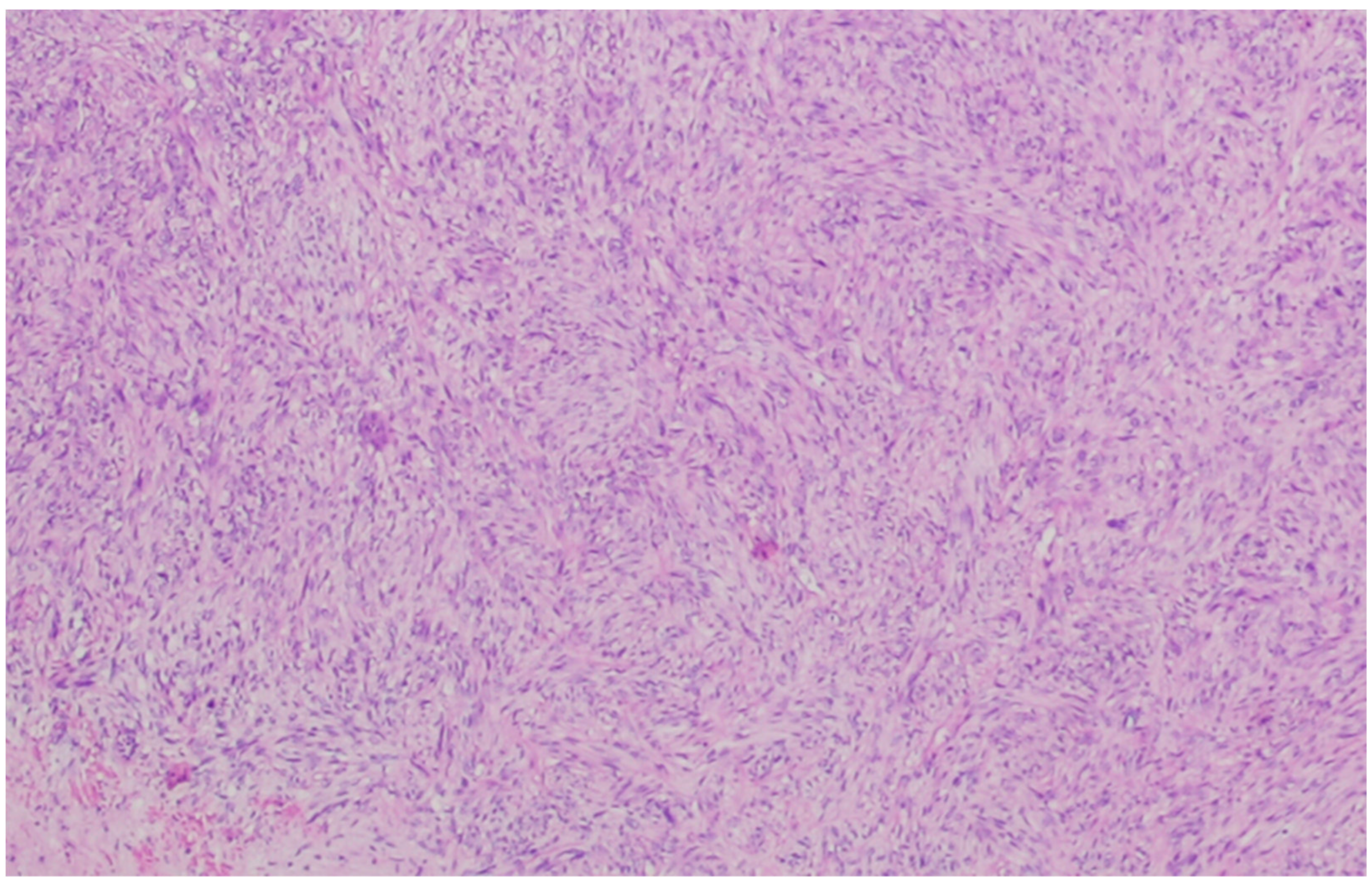
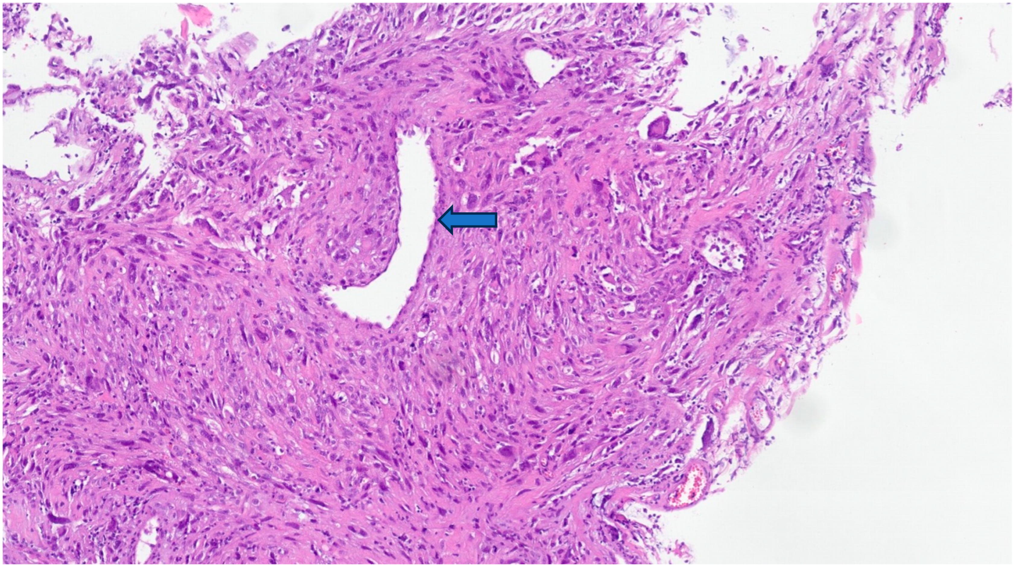
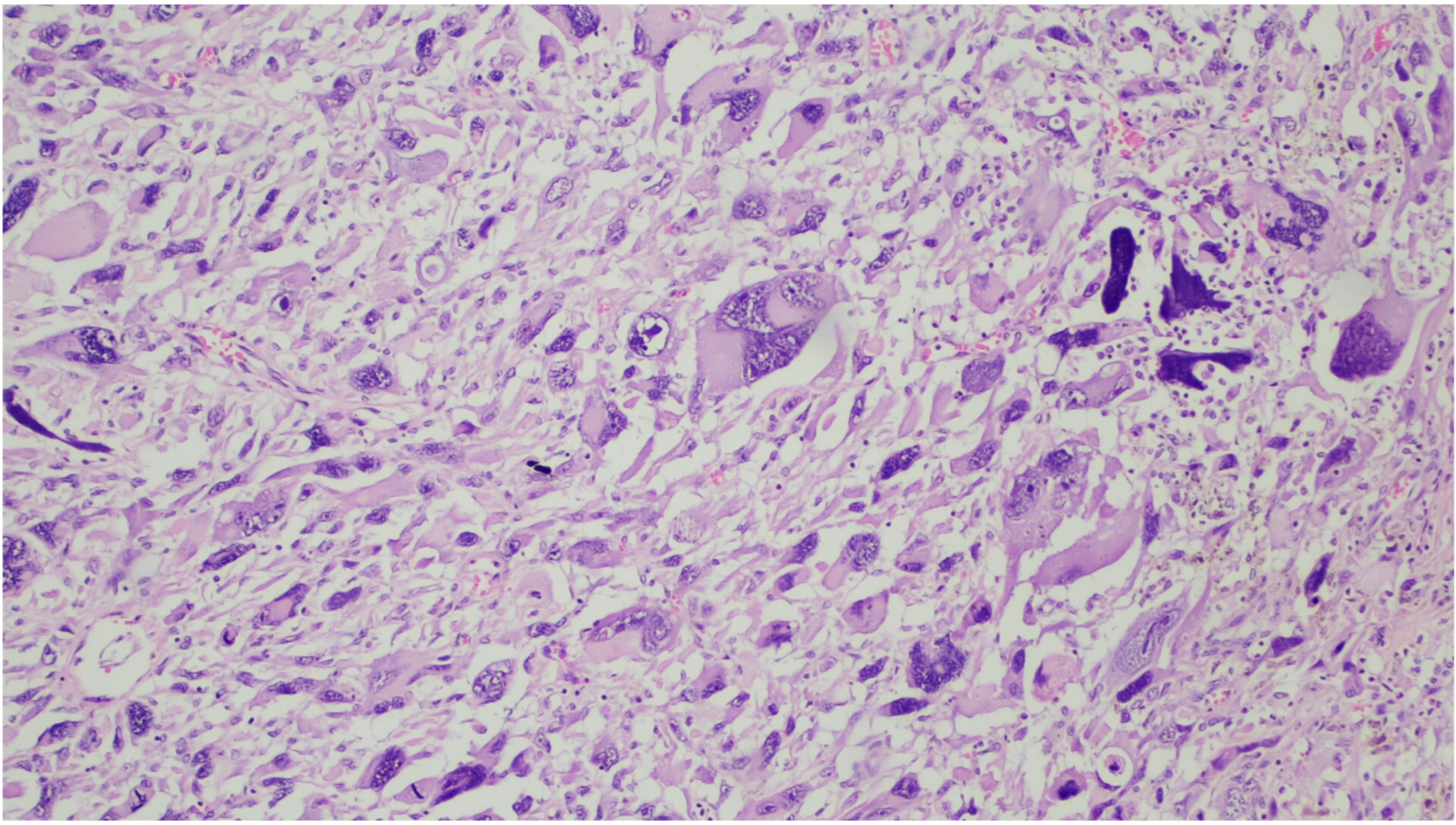
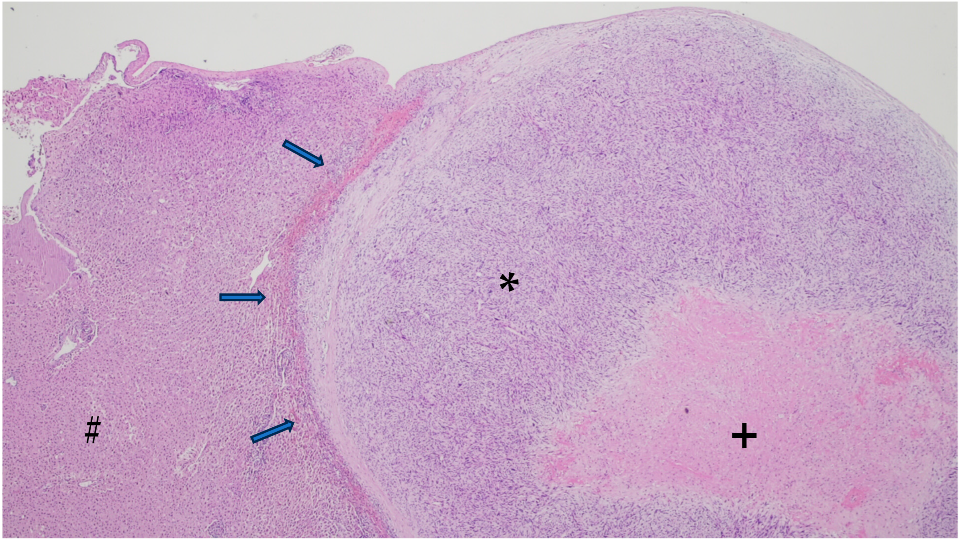
6. Gross and Histologic Features

On gross examination, the majority are extraluminal (76%), with minimal or even absent luminal growth. Tumor size can range from 2 to 30 cm [8,9].
Histologically, the cells are elongated in shape, with abundant and eosinophilic cytoplasm, centrally placed nuclei, and blunt-ended edges, sometimes called “cigar-shaped”. Architecturally, the cells are arranged in fascicles [1]. Cytoplasmatic and perinuclear haloes are frequently observed [15]. An example can be seen in Figure 1.
At higher magnification, longitudinally oriented myofibrils are a common characteristic. They can clump, giving a clotted appearance of the cytoplasm [7].
As leiomyosarcomas become less differentiated, they lose the resemblance to the normal counterpart. In well-differentiated tumors, the cells are arranged in fascicles, there is mild nuclear polymorphism, and nuclei are centrally located and have a low mitotic rate. In moderately differentiated tumors, the mitotic rate, nuclear polymorphism, and nuclear hyperchromasia are increased. In higher grade tumors, the nuclei often lose the central location, and a fascicular architecture and multinucleated cells are common [8].
This kind of cellular changes can be seen in Figure 2 and Figure 3.
Morphologically, there seems to be no histologic difference between vascular LMS and other types of soft tissue LMS [15]. The intimal layer is usually intact, but protrusions into the lumen can also be seen [15].
Immunohistochemistry is helpful in diagnosing leiomyosarcoma and is based on demonstrating myoid differentiation with muscle markers. α-smooth muscle actin (α-SMA), desmin, heavy chain muscle actin, and h-caldesmon, which are myoid markers, are expressed in leiomyosarcomas [32]. α-SMA and muscle-specific actin are considered the most sensitive markers [33]. Desmin is expressed in around 70% of leiomyosarcomas [34,35,36]. It is important to take into consideration not only the expression but also the type of expression of these antibodies: diffuse expression of desmin is indicative of myoid differentiation, but the focal expression of actin or desmin can also be found in myofibroblasts [37,38]. An example of the immunostaining is represented in Figure 4.
There are interesting differences in the immunostaining characteristics of leiomyosarcomas arising from vascular smooth muscle compared to leiomyosarcomas from different locations. Vascular leiomyosarcomas are more often desmin negative and h-caldesmon positive than leiomyosarcomas arising from soft tissue [38,39].
The expression of keratins, which is usually regarded as evidence of epithelial differentiation, can be detected in leiomyosarcomas [39]. A cross-reaction with the antibodies used to detect cytokeratins was first suspected to be the cause of immunoreactivity, but it has been demonstrated that cytokeratins are present both in non-neoplastic smooth muscle and in smooth muscle tumors [40,41]. The expression of cytokeratins is limited to low molecular weight keratins [42]. Epithelial membrane antigen (EMA) immunoreactivity can occur in up to 45–60% of cases. When present, staining for cytokeratins is usually focal, but diffuse expression was seen in 11% of cytokeratins and 6% with EMA [39,42]. A dot-like pattern of cytokeratin expression has been described, and it is associated, at least focally, with a diffuse or a fibrillary pattern [39]. There is no correlation between the expression of cytokeratins and the location, sex, age, histological grade, and histological features of LMS, but EMA expression is more common in vascular LMS when compared to LMS of the soft tissue, skin, and uterus [39].
CD34 [43], S100 [44], and HMB45 [45] can also be expressed. Estrogen and progesterone receptors were previously suggested as adjunct (and helpful) markers to distinguish between retroperitoneal leiomyosarcomas and leiomiomas [46], but it was found that they can be expressed in both uterine and extrauterine LMS [47,48], therefore not being useful in this distinction.
Histologically, the differential diagnosis for leiomyosarcoma can be extensive, including other spindle cell lesions such as malignant peripheral nerve sheath tumors, synovial sarcomas, leiomyomas, schwannomas, benign cellular myofibroblastic tumors, gastrointestinal stromal tumor, synovial sarcoma, and inflammatory myofibroblastic tumors, as well as other high-grade malignancies, in poorly differentiated cases.

7. Staging

Appropriate criteria for malignancy in soft tissue muscle tumors is challenging as these tumors have a biologic continuum [49,50]. Several characteristics such as necrosis, the degree of differentiation, size, cellularity, and atypia help in the diagnosis of leiomyosarcomas versus leiomyomas [51]. Paal and Miettinen [46], in a series of retroperitoneal leiomyosarcomas, demonstrated that tumors with less than three mitosis per high-power field (HPF) with no mitotic atypia and coagulative tumor necrosis have a benign behavior. On the other hand, the finding of atypia, even if focal is concerning, should prompt the pathologist to evaluate more carefully the mitotic activity [49], as the mitotic activity seems to be one of the most reliable parameters for malignancy. Interestingly, low mitotic activity (<1/10 HPF) with significant atypia is enough evidence of malignancy [7].
Histological scores used in grading are the main backbone for therapy in soft tissue sarcomas [31]. LMS can be graded according to the Fédération Nationale des Centres de Lutte Contre le Cancer (FNCLCC) [52] three-tiered system. This grading system takes into account de-differentiation (score 1 to 3), mitoses (score from 1 to 3), and necrosis (score from 0 to 2) [1].
The TNM staging system of the American Joint Committee on Cancer/Union for International Cancer Control is based on tumor size, nodal involvement, and the presence of metastases and is used widely as a staging tool [53]. It has several important shortcomings, namely, in the soft tissue neoplasms, because of the wide variety of entities and prognostic behaviors. To overcome these limitations, newer tools, such as normograms, are being developed [54].
The International Registry of Inferior Vena Cava Leiomyosarcoma (IRIVCL) was established to study the natural history of the inferior vena cava LMS, and with a total of 218 patients, it is the largest case series published. In this registry, 21.6% of patients had a well-differentiated tumor, 25.2% were moderately differentiated, and they were poorly differentiated in 23.8%; grading was not available in 29.4% of cases [9].
Vascular LMS metastasizes primarily through a hematogenous spread. Metastasis can occur in bones, skin, kidney, brain, especially in the liver and lungs [12,55,56], and less frequently in regional lymph nodes. The rate at which the tumors metastasize depends on the location of the tumor in relation to the vessel wall. Intimal sarcomas are more prone to metastasize in comparison with intramural ones [55].
An example of a metastatic LMS can be seen in Figure 5.

8. Prognosis

The true incidence of vascular LMS is most probably underestimated, and thus, the true prognosis is not known [17]. This may be the reason why the prognostic figures vary so much among different publications. Some studies state that inferior vena cava LMS has a worse prognosis than retroperitoneal LMS, while other studies suggest that the prognosis is similar to retroperitoneal LMS and uterine LMS [12,15,57], and others even suggest that the prognosis is better than uterine LMS [3]. A common consensus is that LMSs are characterized to have an aggressive clinical course [58].
In one study, the 3-year survival rate was reported to be 100% [59]. The 5-year survival rate ranged between 31 and 62% [3,18,60], and the 10-year survival in a study was 29.5%, but some studies reported survival rates as low as 22% [61]. The overall survival of patients with metastatic vascular LMS is similar to that observed in metastatic LMS of other locations [12].
In the biggest study to date, the IRIVCL [18], the prognosis was referred to as dismal. Of 218 patients, follow-up was available in 180 patients. In the population of patients not submitted to surgery, all died of disease progression after a median of 2 months (mean 4.7 ± 1.1 months), and patients submitted to palliative resection survived 19 ± 3.9 months. The median survival of patients submitted to radical resection was 36 months (mean 42.3 ± 3.9 months).
In the same study, several factors were associated with prognosis. To study the prognosis, a division based on location (upper, middle, and lower third) was made, and this division is important as a different surgical approach based on topography [62].
Radical tumor resection, the presence of abdominal pain at presentation, and tumor location in the middle segment of the vena cava were good prognostic factors. Inferior vena cava occlusion, lower limb edema, and tumor location in the upper segment of the inferior vena cava were associated with a worse prognosis.
The authors propose one explanation for a worse survival of tumors arising in the upper segment, which is the technical difficulty of complete excision. The middle segment location of the LMS and its better survival is associated with earlier symptoms and consequently faster recognition of the disease and therapy.
In most cases, the outcome of LMS is associated with the extent of the disease, as concluded in a review of Borghi et al. [63]. Patients with positive margins have a worse prognosis [8]. Different studies have confirmed that intraluminal growth is related to a worse prognosis [61]; one possible explanation might be the vascular spread and development of distant metastasis. The metastatic risk was the major factor of poor outcomes; however, long-term survivors in the metastatic setting were also recorded [63]. Moderate to poor tumor differentiation is also associated with a worse prognosis, but surprisingly, FNCLCC grading was not found to affect prognosis [11,61].
Vascular LMSs often recur at distant sites with a metastatic rate of 50% [9]. In a paper by Roland et al., from a total of 63 patients, there were local and/or distant recurrences in 42, with a median time to recurrence of 33 months [3].
Intimal sarcomas have a worse prognosis in comparison with intramural tumors as they have a bigger metastatic potential, metastasize earlier, and a good percentage of cases are already disseminated at the time of diagnosis [55]. Two studies reported that LMSs arising from arteries have a more aggressive behavior in comparison with venous LMSs [12,56].
Poorly differentiated LMSs frequently lose one or more smooth muscle markers [7,64]. Co-expression of desmin, h-caldesmon, α-SMA, and smooth muscle myosin or the expression of three of the more recently described markers ACTG2, CFL2, CASQ2, MYLK2, and SLMAP are associated with improved prognosis. The progressive loss of myogenic differentiation has a negative prognostic value. The loss of conventional smooth muscle markers and SLMAP, ACTG2, and MYLK is associated with a worse prognosis [64]. In the same study, the expression of desmin and CFL2 in the primary tumor is associated with improved prognosis.
Other studies reported diffuse expression of α-SMA in poorly differentiated tumors, with a loss of expression of either desmin or h-caldesmon [38,65]. A study by Carvalho et al. found no association between grade and expression of myoid markers [33].
High expression of cytoplasmatic β-catenin by immunohistochemistry was associated with a higher rate of distant recurrence and reduced disease-specific survival [3]. Elevated levels of IGF-1R were associated with a reduced time to recurrence and disease-specific survival [3]. Inactivation of RASSF1A and p16INK4 have been associated with a dismal prognosis, and decreased levels of p16 by immunohistochemistry were shown to be correlated with a poorer prognosis [66].

9. Treatment

Due to its complexity, a multidisciplinary approach for neoplasms with vena cava involvement is required, since multiple strategies and surgical approaches are demanded to achieve a good outcome [67].
The optimal treatment for inferior vena cava LMS has been challenging to establish because of the limited knowledge, and so far, no standard approach has been established [8].
Complete surgical resection when feasible is the cornerstone of treatment [9,12,68]. Palliative resection results only in an improvement in symptoms [6].
The improvement in surgical techniques has expanded the possibilities of resection in inferior vena cava LMS, with good outcomes even with extensive resection and resorting to vascular grafts and reconstructions, allowing for complete removal [69]. Reconstruction has been discussed in several papers, comparing the different methodologies such as a prosthetic or autologous patch, direct suture, or simple ligation without IVC reconstruction [23,70,71,72,73]. In some cases, vein reconstruction was performed using a peritoneal patch [74].
The need of vascular reconstruction is not always mandatory, especially if venous collaterality is well established [75]; if necessary, the prosthetic polytetrafluoroethylene graft seems the best option [75]. This decision must be performed in high-volume centers with a multidisciplinary team of sarcoma surgeons.
LMSs have a poor response to chemotherapy and radiotherapy [76], and the role of chemotherapy and radiotherapy has yet to be established [12].
As recurrences are associated with a dismal prognosis [64], when radical excision is not possible, neoadjuvant or adjuvant radiation may be used for local control and to reduce local recurrence [77]. However, Ito et al. did not find a decrease in the rate of local recurrence in patients who were submitted to perioperative radiotherapy [78]. Thus, radiotherapy has a limited role in the management of patients with vascular LMS.
Chemotherapy may be considered as an adjuvant treatment [79], but it has not been shown to improve survival [9], although this might be related to the lack of standardization of the chemotherapy regimens used. In patients with high-risk tumors or high metastatic burden, neoadjuvant chemotherapy may have a role by selecting patients with a more biologically responsive disease [80].
In a systematic review of 118 cases performed by Saikia J et al., aggressive upfront surgical resection with clear margins was the key to long-term survival (60 months of overall survival). An inferior vena cava graft was performed in 91.8% of patients, which allowed microscopic tumor-free margins in 85.5% of the patients. Regarding chemotherapy, the most used drugs were doxorubicin and ifosfamide. In this study, 88% of the patients were eligible for upfront resection, but it should be stated that 52.8% went to surgery without histological confirmation of disease, a percentage that nowadays would be almost impossible and counter-productive [81].
Italiano et al. [12] described their experience with fourteen patients with vascular LMS. In their study, at diagnosis, nine patients presented with localized disease and five with metastatic disease. In the course of the disease, five patients developed metastasis. Of the patients with localized disease, seven underwent surgery, with complete gross resection in all of them. Five out of seven received postoperative radiotherapy. The two patients with localized disease who did not undergo surgery received doxorubicin-based chemotherapy. The patients with metastatic disease at diagnosis and during the course of the disease received an anthracycline-based regimen as first-line therapy. In the metastatic disease setting, the best response was a partial response in two, four had stable disease, and two had progression of the disease. Interestingly, patients treated with an anthracycline-based chemotherapy had a similar response to LMS at other sites.
In the LMS-04 study, from the French sarcoma group, it was shown that an association of doxorubicin with trabectedin as a first-line therapy was found to increase the progression-free survival in patients with metastatic or unresectable disease in comparison with the standard doxorubicin, although with a higher toxicity [82]. A study from de Graaff et al. has explored a synergistic effect of doxorubicin and ABT-737, with good results, but it was directed to soft tissue LMS. Nevertheless, since the rationale was directed to Bcl-2, Bcl-xL, and Bcl-W expression by immunohistochemistry, it may represent a good therapeutic approach in patients with irresectable inferior vena cava LMS since the protein expression can be assessed in the biopsy [83,84].
There are reports of good results with trabectedin in soft tissue leiomyosarcomas [85,86]. In 2021, the results of the French sarcoma group study T-SAR were published [87]. T-SAR was a randomized phase III trial comparing trabectedin to the best supportive care in patients with advanced soft tissue sarcoma and demonstrated the superiority of trabectedin in disease control (with an objective response rate >20% and a median progression-free survival of 5.1 months) in comparison to the best supportive care in this group of patients.
Recently, new techniques have emerged that can be a valuable aid against refractory LMS. Viral vectors, such as third-generation genetically modified herpes simplex virus type 1, have demonstrated good results in vitro [88]. The major drawback of this technique in vascular LMS is difficult tumor access for viral inoculation. Nevertheless, with imaging guidance, it should be an easy limitation to overcome.
Programmed Death Ligand 1 (PD-L1) has been a major advancement in solid tumors, and there are some phase II studies assessing its antitumoral activity against sarcomas. The PD-1/PD-L1 antagonist activity was shown to be higher in alveolar soft-part sarcoma and undifferentiated pleomorphic sarcoma, with lower response rates in leiomyosarcoma [89].
The keynote 158 study was a major success, demonstrating the efficacy of pembrolizumab in non-colorectal high microsatellite instability [90], thus paving the way for microsatellite instability testing in sarcomas. Albeit rare [91], some soft tissue sarcomas may exhibit this biological propriety, with or without PD-L1 expression, prompting pembrolizumab as an effective treatment; recently, Yon Tay et al. described a case of complete tumoral response in a soft tissue LMS with these features [92].

10. Conclusions

Vascular leiomyosarcomas are uncommon neoplasms, and although many studies have been conducted, there is still limited knowledge on their etiology, pathogenesis, and, consequently, optimal treatment. Multidisciplinary management is key to good clinical results; however, the prognosis remains relatively unfavorable. Surgery is the backbone of treatment and, combined with neoadjuvant therapy and radiotherapy, delivers acceptable results. To advance the knowledge in this field, further studies with national and international collaborations should be conducted, focusing on understanding the etiology and finding actionable mutations that can be used for treatment.

Author Contributions

Conceptualization, J.M.G. and R.C.O.; methodology, J.M.G. and R.C.O.; validation, J.C.; writing—review and editing, J.M.G., R.C.O. and R.A.; supervision, J.C. All authors have read and agreed to the published version of the manuscript.

Funding

This research received no external funding.

Conflicts of Interest

The authors declare no conflict of interest.

Abbreviations

α-SMA—α-Smooth Muscle Actin; CT—computerized tomography; EMA—Epithelial Membrane Antigen; FNCLCC—Fédération Nationale des Centres de Lutte Contre le Cancer; HPF—High-Power Field; IRIVCL—International Registry of Inferior Vena Cava Leiomyosarcoma; LMS—Leiomyosarcoma; MRI—magnetic resonance imaging; US—ultrasonography; PD-L1—Programmed Death Ligand 1; PET—positron emission tomography; SUVmax—Maximum Standard Unit Value.

References

  1. IARC. WHO Classification of Soft Tissue and Bone Tumours. WHO Classification of Tumours Soft Tissue and Bone Tumours. 2020. Available online: https://www.ncbi.nlm.nih.gov/pmc/articles/PMC8167394/ (accessed on 6 August 2023).
  2. Fletcher, C.D.M. Diagnostic Histopathology of Tumors, 5th ed.; Elsevier: Amsterdam, The Netherlands, 2020. [Google Scholar]
  3. Roland, C.L.; Boland, G.M.; Demicco, E.G.; Lusby, K.; Ingram, D.; May, C.D.; Kivlin, C.M.; Watson, K.; Sanaa, G.A.A.; Wang, W.L.; et al. Clinical Observations and Molecular Variables of Primary Vascular Leiomyosarcoma. JAMA Surg. 2016, 151, 347–354. Available online: https://pubmed.ncbi.nlm.nih.gov/26629783/ (accessed on 31 July 2023). [CrossRef] [PubMed]
  4. Perl, L.; Virchow, R. Ein Fall von Sarkom der Vena cava inferior. Arch Pathol. Anat. Physiol. Klin. Med. 1871, 53, 378–383. Available online: https://link.springer.com/article/10.1007/BF01957198 (accessed on 29 July 2023). [CrossRef]
  5. Mastrangelo, G.; Coindre, J.M.; Ducimetière, F.; Dei Tos, A.P.; Fadda, E.; Blay, J.Y.; Buja, A.; Fedeli, U.; Cegolon, L.; Frasson, A.; et al. Incidence of soft tissue sarcoma and beyond. Cancer 2012, 118, 5339–5348. Available online: https://onlinelibrary.wiley.com/doi/full/10.1002/cncr.27555 (accessed on 31 July 2023). [CrossRef] [PubMed]
  6. Müller, A.M.; Chromik, A.M.; Bolik, B.; Müller, K.M.; Mittelkötter, U. Leiomyosarkom der vena cava inferior: Übersicht zu einem seltenen krankheitsbild. Pathologe 2005, 26, 153–158. Available online: https://link.springer.com/article/10.1007/s00292-004-0745-y (accessed on 29 July 2023). [CrossRef] [PubMed]
  7. Goldblum, J.R.; Weiss, S.W.; Folpe, A.L. Enzinger and Weiss’s Soft Tissue Tumors, 7th ed.; Elsevier: Amsterdam, The Netherlands, 2019. [Google Scholar]
  8. Leiomyosarcoma of the Inferior Vena Cava—Hines—1999—Cancer—Wiley Online Library. Available online: https://acsjournals.onlinelibrary.wiley.com/doi/pdf/10.1002/%28SICI%291097-0142%2819990301%2985%3A5%3C1077%3A%3AAID-CNCR10%3E3.0.CO%3B2-0 (accessed on 29 July 2023).
  9. International Registry of Inferior Vena Cava Leiomyosarcoma: Analysis of a World Series on 218 Patients—PubMed. Available online: https://pubmed.ncbi.nlm.nih.gov/8920790/ (accessed on 30 July 2023).
  10. Yoshizawa, K.; Ohno, Y.; Kurata, T.; Takagi, Y.; Kasai, T.; Takizawa, M.; Soyejima, Y. Primary leiomyosarcoma of the inferior vena cava in a pediatric case: A case report and literature review. Surg. Case Rep. 2023, 9, 52. Available online: https://pmc/articles/PMC10079787/ (accessed on 6 April 2023). [CrossRef] [PubMed]
  11. Kevorkian, J.; Cento, D.P. Leiomyosarcoma of large arteries and veins. Surgery 1973, 73, 390–400. Available online: http://www.surgjournal.com/article/0039606073903073/fulltext (accessed on 27 July 2023). [PubMed]
  12. Italiano, A.; Toulmonde, M.; Stoeckle, E.; Kind, M.; Kantor, G.; Coindre, J.M.; Bui, B. Clinical outcome of leiomyosarcomas of vascular origin: Comparison with leiomyosarcomas of other origin. Ann. Oncol. 2010, 21, 1915–1921. Available online: http://www.annalsofoncology.org/article/S0923753419400628/fulltext (accessed on 2 August 2023). [CrossRef]
  13. Weinreb, W.; Steinfeld, A.; Rodil, J.; Esparza, A.; Trebbin, W. Leiomyosarcoma Arising in an Arteriovenous Fistula. Available online: https://pubmed.ncbi.nlm.nih.gov/6861080/ (accessed on 30 July 2023).
  14. Hashimoto, H.; Daimaru, Y.; Tsuneyoshi, M.; Enjoji, M. Leiomyosarcoma of the External Soft Tissues A Clinicopathologic, lmmunohistochemical, and Electron Microscopic Study Materials and Methods. Available online: https://onlinelibrary.wiley.com/terms-and-conditions (accessed on 29 July 2023).
  15. Farshid, G.; Pradhan, M.; Goldblum, J.; Weiss, S.W. Leiomyosarcoma of somatic soft tissues: A tumor of vascular origin with multivariate analysis of outcome in 42 cases. Am. J. Surg. Pathol. 2002, 26, 14–24. Available online: https://pubmed.ncbi.nlm.nih.gov/11756765/ (accessed on 29 July 2023). [CrossRef] [PubMed]
  16. Gustafson, P.; Willkn, H.; Baldetorp, B.; Ferno, M.; Akerman, M.; Rydholm, A. Soft Tissue Leiomyosarcoma A Population-Based Epidemiologic and Prognostic Study of 48 Patients, Including Cellular DNA Content. Cancer 1992, 70, 114–119. [Google Scholar] [CrossRef]
  17. Enzinger, F.M.; Shiraki, M. Extraskeletal myxoid chondrosarcoma. An analysis of 34 cases. Hum. Pathol. 1972, 3, 421–435. [Google Scholar] [CrossRef] [PubMed]
  18. Mingoli, A.; Feldhaus, R.J.; Cavallaro, A.; Stipa, S. Leiomyosarcoma of the inferior vena cava: Analysis and search of world literature on 141 patients and report of three new cases. J. Vasc. Surg. 1991, 14, 688–699. [Google Scholar] [CrossRef] [PubMed]
  19. Gomes, J.F.; Vieira, I.; Mendes, J.; Donaire, D.; Almeida, R. Leiomyosarcoma of Inferior Vena Cava in an Immunocompetent Young-adult Female Patient. J. Coll. Physicians Surg. Pak. 2022, 32, 1353–1355. Available online: https://pubmed.ncbi.nlm.nih.gov/36205287/ (accessed on 23 September 2023). [PubMed]
  20. Nilbert, M.; Mandahl, N.; Heim, S.; Rydholm, A.; Helm, G.; Willén, H.; Baldetorp, B.; Mitelman, F. Complex karyotypic changes, including rearrangements of 12q13 and 14q24, in two leiomyosarcomas. Cancer Genet. Cytogenet. 1990, 48, 217–223. Available online: https://pubmed.ncbi.nlm.nih.gov/2397453/ (accessed on 31 July 2023). [CrossRef] [PubMed]
  21. Chudasama, P.; Mughal, S.S.; Sanders, M.A.; Hübschmann, D.; Chung, I.; Deeg, K.I.; Wong, S.H.; Rabe, S.; Hlenvjak, M.; Zapatka, M.; et al. Integrative genomic and transcriptomic analysis of leiomyosarcoma. Nat. Commun. 2018, 9, 1–15. Available online: https://www.nature.com/articles/s41467-017-02602-0 (accessed on 31 July 2023). [CrossRef] [PubMed]
  22. Gladdy, R.A.; Qin, L.X.; Moraco, N.; Agaram, N.P.; Brennan, M.F.; Singer, S. Predictors of Survival and Recurrence in Primary Leiomyosarcoma. Ann. Surg. Oncol. 2013, 20, 1851. Available online: https://pmc/articles/PMC3657306/ (accessed on 28 August 2023). [CrossRef] [PubMed]
  23. Rusu, C.B.; Gorbatâi, L.; Szatmari, L.; Koren, R.; Bungărdean, C.I.; Feciche, B.O.; Bumbulut, C.; Andras, I.M.; Rahota, R.; Telecan, T.; et al. Leiomyosarcoma of the inferior vena cava. Our experience and a review of the literature. Rom J. Morphol. Embryol. 2020, 61, 227. Available online: https://www.ncbi.nlm.nih.gov/pmc/articles/PMC7728114/ (accessed on 23 September 2023). [CrossRef] [PubMed]
  24. Wu, X.; Zhou, P.; Li, K. Contrast-enhanced ultrasonography of intraluminal inferior vena cava leiomyosarcoma: A case report. J. Clin. Ultrasound. 2020, 48, 357–361. Available online: https://pubmed.ncbi.nlm.nih.gov/32027023/ (accessed on 23 September 2023). [CrossRef] [PubMed]
  25. Webb, E.M.; Wang, Z.J.; Westphalen, A.C.; Nakakura, E.K.; Coakley, F.V.; Yeh, B.M. Can CT features differentiate between inferior vena cava leiomyosarcomas and primary retroperitoneal masses? Am. J. Roentgenol. 2013, 200, 205–209. Available online: https://www.ajronline.org/doi/10.2214/AJR.11.7476 (accessed on 23 September 2023). [CrossRef]
  26. Mastoraki, A.; Leotsakos, G.; Mastoraki, S.; Papanikolaou, I.S.; Danias, N.; Smyrniotis, V.; Arkadapoulos, N. Challenging diagnostic and therapeutic modalities for leiomyosarcoma of inferior vena cava. Int. J. Surg. 2015, 13, 92–95. Available online: https://pubmed.ncbi.nlm.nih.gov/25489949/ (accessed on 23 September 2023). [CrossRef]
  27. Sessa, B.; Iannicelli, E.; Caterino, S.; D’Angelo, F.; Milione, M.; Ziparo, V.; David, V. Imaging of leiomyosarcoma of the inferior vena cava: Comparison of 2 cases and review of the literature. Cancer Imaging 2010, 10, 80. Available online: https://pmc/articles/PMC2842181/ (accessed on 23 September 2023). [CrossRef]
  28. Ronchi, B.; Peña, G.A.; Sacchi, C. PET/MR: Primary inferior vena cava leiomyosarcoma. Eur. J. Hybrid Imaging 2022, 6, 1–8. Available online: https://ejhi.springeropen.com/articles/10.1186/s41824-022-00144-3 (accessed on 23 September 2023). [CrossRef] [PubMed]
  29. Kim, J.T.; Kwon, T.; Cho, Y.; Shin, S.; Lee, S.; Moon, D. Multidisciplinary treatment and long-term outcomes in six patients with leiomyosarcoma of the inferior vena cava. J. Korean Surg. Soc. 2012, 82, 101–109. Available online: https://pubmed.ncbi.nlm.nih.gov/22347712/ (accessed on 23 September 2023). [CrossRef] [PubMed]
  30. Punt, S.E.W.; Eary, J.F.; O’Sullivan, J.; Conrad, E.U. Fluorodeoxyglucose positron emission tomography in leiomyosarcoma: Imaging characteristics. Nucl. Med. Commun. 2009, 30, 546–549. Available online: https://pubmed.ncbi.nlm.nih.gov/19440162/ (accessed on 23 September 2023). [CrossRef] [PubMed]
  31. Gronchi, A.; Miah, A.B.; Dei Tos, A.P.; Abecassis, N.; Bajpai, J.; Bauer, S.; Biagini, R.; Bielak, S.; Blay, J.Y.; Bolle, S.; et al. Soft tissue and visceral sarcomas: ESMO-EURACAN-GENTURIS Clinical Practice Guidelines for diagnosis, treatment and follow-up☆. Ann. Oncol. Off. J. Eur. Soc. Med. Oncol. 2021, 32, 1348–1365. Available online: https://pubmed.ncbi.nlm.nih.gov/34303806/ (accessed on 23 September 2023). [CrossRef] [PubMed]
  32. Tsukada, T.; McNutt, M.A.; Ross, R.; Gown, A.M. HHF35, a muscle actin-specific monoclonal antibody. II. Reactivity in normal, reactive, and neoplastic human tissues. Am. J. Pathol. 1987, 127, 389. Available online: https://pmc/articles/PMC1899748/?report=abstract (accessed on 29 July 2023). [PubMed]
  33. Carvalho, J.C.; Thomas, D.G.; Lucas, D.R. Cluster analysis of immunohistochemical markers in leiomyosarcoma delineates specific anatomic and gender subgroups. Cancer 2009, 115, 4186–4195. Available online: https://pubmed.ncbi.nlm.nih.gov/19626649/ (accessed on 2 August 2023). [CrossRef] [PubMed]
  34. Azumi, N.; Ben-Ezra, J.; Battifora, H. Immunophenotypic diagnosis of leiomyosarcomas and rhabdomyosarcomas with monoclonal antibodies to muscle-specific actin and desmin in formalin-fixed tissue. Mod. Pathol. 1988, 1, 469–474. [Google Scholar] [PubMed]
  35. Pollock, L.; Rampling, D.; Greenwald, S.E.; Malone, M. Desmin expression in rhabdomyosarcoma: Influence of the desmin clone and immunohistochemical method. J. Clin. Pathol. 1995, 48, 535–538. Available online: https://jcp.bmj.com/content/48/6/535 (accessed on 29 July 2023). [CrossRef]
  36. Truong, L.D.; Rangdaeng, S.; Cagle, P.; Ro, J.Y.; Hawkins, H.; Font, R.L. The diagnostic utility of desmin. A study of 584 cases and review of the literature. Am. J. Clin. Pathol. 1990, 93, 305–314. Available online: https://pubmed.ncbi.nlm.nih.gov/1689936/ (accessed on 29 July 2023). [CrossRef]
  37. Dabbs, D.J. Diagnostic Immunohistochemistry, 6th ed.; Elsevier: Amsterdam, The Netherlands, 2022. [Google Scholar]
  38. Matsuyama, A.; Hisaoka, M.; Hashimoto, H. Vascular leiomyosarcoma: Clinicopathology and immunohistochemistry with special reference to a unique smooth muscle phenotype. Pathol. Int. 2010, 60, 212–216. Available online: https://pubmed.ncbi.nlm.nih.gov/20403047/ (accessed on 29 July 2023). [CrossRef]
  39. Iwata, J.; Fletcher, C.D.M. Immunohistochemical detection of cytokeratin and epithelial membrane antigen in leiomyosarcoma: A systematic study of 100 cases. Pathol. Int. 2000, 50, 7–14. Available online: https://onlinelibrary.wiley.com/doi/full/10.1046/j.1440-1827.2000.01001.x (accessed on 29 July 2023). [CrossRef] [PubMed]
  40. Traweek, S.T.; Liu, J.; Battifora, H. Keratin gene expression in non-epithelial tissues. Detection with polymerase chain reaction. Am. J. Pathol. 1993, 142, 1111. Available online: https://pmc/articles/PMC1886881/?report=abstract (accessed on 28 October 2023). [PubMed]
  41. Gown, A.M.; Boyd, H.C.; Chang, Y.; Ferguson, M.; Reichler, B.; Tippens, D. Smooth muscle cells can express cytokeratins of “simple” epithelium. Immunocytochemical and biochemical studies in vitro and in vivo. Am. J. Pathol. 1988, 132, 223. Available online: https://pmc/articles/PMC1880728/?report=abstract (accessed on 28 October 2023). [PubMed]
  42. Immunoreactivity for Cytokeratin and Epithelial Membrane Antigen in Leiomyosarcoma—PubMed. Available online: https://pubmed.ncbi.nlm.nih.gov/2454091/ (accessed on 29 July 2023).
  43. Fisher, C. Immunohistochemistry in diagnosis of soft tissue tumours. Histopathology 2011, 58, 1001–1012. Available online: https://pubmed.ncbi.nlm.nih.gov/21143519/ (accessed on 30 July 2023). [CrossRef] [PubMed]
  44. Kanamori, T.; Takakura, K.; Mandai, M.; Kariya, M.; Fukuhara, K.; Sakaguchi, M.; Huh, N.H.; Saito, K.; Sakurai, T.; Fujita, J.; et al. Increased expression of calcium-binding protein S100 in human uterine smooth muscle tumours. Mol. Hum. Reprod. 2004, 10, 735–742. Available online: https://pubmed.ncbi.nlm.nih.gov/15322223/ (accessed on 30 July 2023). [CrossRef] [PubMed]
  45. Silva, E.G.; Bodurka, D.C.; Scouros, M.A.; Ayala, A. A uterine leiomyosarcoma that became positive for HMB45 in the metastasis. Ann. Diagn. Pathol. 2005, 9, 43–45. [Google Scholar] [CrossRef] [PubMed]
  46. Paal, E.; Miettinen, M. Retroperitoneal Leiomyomas: A Clinicopathologic and Immunohi: The American Journal of Surgical Pathology. Am. J. Surg. Pathol. 2001, 25, 1355–1363. Available online: https://journals.lww.com/ajsp/Abstract/2001/11000/Retroperitoneal_Leiomyomas__A_Clinicopathologic.2.aspx (accessed on 30 July 2023). [CrossRef]
  47. Kelley, T.W.; Borden, E.C.; Goldblum, J.R. Estrogen and progesterone receptor expression in uterine and extrauterine leiomyosarcomas: An immunohistochemical study. Appl. Immunohistochem Mol. Morphol. AIMM 2004, 12, 338–341. Available online: https://pubmed.ncbi.nlm.nih.gov/15536333/ (accessed on 30 July 2023). [CrossRef]
  48. Leiomyosarcoma of the Pulmonary Veins: The American Journal of Surgical Pathology. Available online: https://journals.lww.com/ajsp/Abstract/1999/09000/Leiomyosarcoma_of_the_Pulmonary_Veins.11.aspx (accessed on 30 July 2023).
  49. Miettinen, M.; Fetsch, J.F. Evaluation of biological potential of smooth muscle tumours. Histopathology 2006, 48, 97–105. Available online: https://pubmed.ncbi.nlm.nih.gov/16359541/ (accessed on 30 July 2023). [CrossRef]
  50. The Difficulty in Predicting Behavior of Smooth-Muscle Tumor: The American Journal of Surgical Pathology. Available online: https://journals.lww.com/ajsp/Citation/1995/01000/The_Difficulty_in_Predicting_Behavior_of.15.aspx (accessed on 30 July 2023).
  51. Billings, S.D.; Folpe, A.L.; Weiss, S.W. Do leiomyomas of deep soft tissue exist? An analysis of highly differentiated smooth muscle tumors of deep soft tissue supporting two distinct subtypes. Am. J. Surg. Pathol. 2001, 25, 1134–1142. Available online: https://pubmed.ncbi.nlm.nih.gov/11688572/ (accessed on 30 July 2023). [CrossRef]
  52. Trojani, M.; Contesso, G.; Coindre, J.M.; Rouesse, J.; Bui, N.B.; De Mascarel, A.; Goussot, J.F.; David, M.; Bonichon, F. Lagarde C. Soft-tissue sarcomas of adults; study of pathological prognostic variables and definition of a histopathological grading system. Int. J. Cancer 1984, 33, 37–42. Available online: https://onlinelibrary.wiley.com/doi/full/10.1002/ijc.2910330108 (accessed on 30 July 2023). [CrossRef] [PubMed]
  53. Amin, M.B.; Edge, S.; Greene, F.; Byrd, D.R.; Brookland, R.K.; Washington, M.K.; Gershenwald, J.E.; Compton, C.C.; Hess, K.R.; Sullivan, D.C.; et al. AJCC Cancer Staging Manual, 8th ed.; Springer: Chicago, IL, USA, 2018. [Google Scholar]
  54. Danieli, M.; Gronchi, A. Staging Systems and Nomograms for Soft Tissue Sarcoma. Curr. Oncol. 2023, 30, 3648–3671. Available online: https://www.mdpi.com/1718-7729/30/4/278/htm (accessed on 2 August 2023). [CrossRef] [PubMed]
  55. Székely, E.; Kulka, J.; Miklós, I.; Kaliszky, P. Leiomyosarcomas of great vessels. Pathol. Oncol. Res. 2000, 6, 233–236. Available online: https://pubmed.ncbi.nlm.nih.gov/11033466/ (accessed on 30 July 2023). [CrossRef] [PubMed]
  56. Penel, N.; Taieb, S.; Ceugnart, L.; Dansin, E.; Hoguet, D.; Vanseymortier, L.; Lartigau, E. Report of eight recent cases of locally advanced primary pulmonary artery sarcomas: Failure of Doxorubicin-based chemotherapy. J. Thorac. Oncol. 2008, 3, 907–911. Available online: https://pubmed.ncbi.nlm.nih.gov/18670310/ (accessed on 2 August 2023). [CrossRef] [PubMed]
  57. Burke, A.P.; Virmani, X. Sarcomas of the Great Vessels A Clinicopathologic Study. Available online: https://pubmed.ncbi.nlm.nih.gov/8448740/ (accessed on 30 July 2023).
  58. Coindre, J.M.; Terrier, P.; Guillou, L.; Le Doussal, V.; Oise Collin, F.; Ranchè, D.; Sastre, X.; Vilain, M.O.; Bonichon, F.; N’Guyen Bui, B. Predictive Value of Grade for Metastasis Development in the Main Histologic Types of Adult Soft Tissue Sarcomas A Study of 1240 Patients from the French Federation of Cancer Centers Sarcoma Group. 1999. Available online: https://pubmed.ncbi.nlm.nih.gov/11346874/ (accessed on 2 August 2023).
  59. Teixeira, F.J.R.; Netto SD do, C.; Perina AL de, F.; Torricelli, F.C.M.; Teixeira, L.R.; Zerati, A.E.; Ferreira, F.O.; Akaishi, E.H. Nahas W.C. Utiyama E.M. Leiomyosarcoma of the inferior vena cava: Survival rate following radical resection. Oncol. Lett. 2017, 14, 3909–3916. Available online: https://pubmed.ncbi.nlm.nih.gov/29098019/ (accessed on 23 September 2023). [CrossRef] [PubMed]
  60. Dew, J.; Hansen, K.; Hammon, J.; McCoy, T.; Levine, E.A.; Shen, P. Leiomyosarcoma of the inferior vena cava: Surgical management and clinical results. Am. Surg. 2005, 71, 497–501. Available online: https://pubmed.ncbi.nlm.nih.gov/16044929/ (accessed on 31 July 2023). [CrossRef] [PubMed]
  61. Laskin, W.B.; Fanburg-Smith, J.C.; Burke, A.P.; Kraszewska, E.; Fetsch, J.F.; Miettinen, M. Leiomyosarcoma of the inferior vena cava: Clinicopathologic study of 40 cases. Am. J. Surg. Pathol. 2010, 34, 873–881. Available online: https://pubmed.ncbi.nlm.nih.gov/20463568/ (accessed on 31 July 2023). [CrossRef] [PubMed]
  62. Borghese, O.; Pisani, A.; Dubrez, J.; Di Centa, I. Treatment outlines for the management of primary leiomyosarcoma of the inferior vena cava. J. Med. Vasc. 2022, 47, 65–70. Available online: https://pubmed.ncbi.nlm.nih.gov/35691665/ (accessed on 23 September 2023). [CrossRef]
  63. Borghi, A.; Scotto DIUccio, A.; Gronchi, A. Primary malignancy of the inferior vena cava, a review of surgical treatments and outcomes. J. Cardiovasc. Surg. 2022, 63, 649–663. Available online: https://pubmed.ncbi.nlm.nih.gov/36239928/ (accessed on 23 September 2023). [CrossRef]
  64. Demicco, E.G.; Boland, G.M.; Savannah, K.J.B.; Lusby, K.; Young, E.D.; Ingram, D.; Watson, K.L.; Bailey, M.; Guo, X.; Hornick, J.L.; et al. Progressive loss of myogenic differentiation in leiomyosarcoma has prognostic value. Histopathology 2015, 66, 627–638. Available online: https://pubmed.ncbi.nlm.nih.gov/24889065/ (accessed on 30 July 2023). [CrossRef]
  65. Miettinen, M.; Kopczynski, J.; Makhlouf, H.R.; Sarlomo-Rikala, M.; Gyorffy, H.; Burke, A.; Sobin, L.H.; Lasota, J. Gastrointestinal stromal tumors, intramural leiomyomas, and leiomyosarcomas in the duodenum: A clinicopathologic, immunohistochemical, and molecular genetic study of 167 cases. Am. J. Surg. Pathol. 2003, 27, 625–641. Available online: https://pubmed.ncbi.nlm.nih.gov/12717247/ (accessed on 28 August 2022). [CrossRef] [PubMed]
  66. Kawaguchi, K.I.; Oda, Y.; Saito, T.; Yamamoto, H.; Tamiya, S.; Takahira, T.; Miyajima, K.; Iwamoto, Y.; Tsuneyoshi, M. Mechanisms of inactivation of the p16INK4a gene in leiomyosarcoma of soft tissue: Decreased p16 expression correlates with promoter methylation and poor prognosis. J. Pathol. 2003, 201, 487–495. Available online: https://pubmed.ncbi.nlm.nih.gov/14595762/ (accessed on 31 July 2023). [CrossRef] [PubMed]
  67. Baia, M.; Naumann, D.N.; Wong, C.S.; Mahmood, F.; Parente, A.; Bissacco, D.; Almond, M.; Ford, S.J.; Tirotta, F.; Desai, A. Dealing with malignancy involving the inferior vena cava in the 21st century. J. Cardiovasc. Surg. 2022, 63, 664–673. Available online: https://pubmed.ncbi.nlm.nih.gov/36239927/ (accessed on 23 September 2023). [CrossRef] [PubMed]
  68. Pan, J.; Qiu, C.Y.; He, Y.Y.; Xue, X.; Li, D.L.; Tian, L.; Cheng, F.; Wu, Z.H.; Zhang, H.K. A 10-year experience of leiomyosarcoma of the inferior vena cava. Phlebology 2022, 37, 572–578. Available online: https://pubmed.ncbi.nlm.nih.gov/35570826/ (accessed on 23 September 2023). [CrossRef] [PubMed]
  69. Ferraris, M.; Callegaro, D.; Barretta, F.; Fiore, M.; Radaelli, S.; Stacchiotti, S.; Miceli, R.; Socrate, A.M.; Locati, P.; Gronchi, A. Outcome of iliocaval resection and reconstruction for retroperitoneal sarcoma. J. Vasc. Surg. Venous Lymphat. Disord. 2019, 7, 547–556. Available online: https://pubmed.ncbi.nlm.nih.gov/30792153/ (accessed on 23 September 2023). [CrossRef] [PubMed]
  70. Gaignard, E.; Bergeat, D.; Robin, F.; Corbière, L.; Rayar, M.; Meunier, B. Inferior Vena Cava Leiomyosarcoma: What Method of Reconstruction for Which Type of Resection? World J. Surg. 2020, 44, 3537–3544. Available online: https://pubmed.ncbi.nlm.nih.gov/32445073/ (accessed on 23 September 2023). [CrossRef] [PubMed]
  71. Silva, T.A.; Buonfiglio, V.B.; Sadi, M.V. Leiomyosarcoma of the Inferior Vena Cava and Vascular Reconstruction. Urology 2022, 168, e15–e17. Available online: https://pubmed.ncbi.nlm.nih.gov/35908743/ (accessed on 23 September 2023). [CrossRef] [PubMed]
  72. Goodsell, K.E.; Sharib, J.M.; Pillarisetty, V.G.; Sham, J.G. Leiomyosarcoma of the inferior vena cava: An uncommon malignancy requiring unique reconstructive approaches. Am. J. Surg. 2023, 226, 286–289. Available online: https://pubmed.ncbi.nlm.nih.gov/36959023/ (accessed on 23 September 2023). [CrossRef]
  73. Wachtel, H.; Jackson, B.M.; Bartlett, E.K.; Karakousis, G.C.; Roses, R.E.; Bavaria, J.E.; Fraker, D.L. Resection of primary leiomyosarcoma of the inferior vena cava (IVC) with reconstruction: A case series and review of the literature. J. Surg. Oncol. 2015, 111, 328–333. Available online: https://pubmed.ncbi.nlm.nih.gov/25297650/ (accessed on 23 September 2023). [CrossRef]
  74. Risaliti, M.; Fortuna, L.; Bartolini, I.; Taddei, A.; Muiesan, P. Inferior vena cava resection and reconstruction with a peritoneal patch for a leiomyosarcoma: A case report. Int. J. Surg. Case Rep. 2020, 71, 37–40. [Google Scholar] [CrossRef]
  75. Kunte, A.; Patkar, S.; Goel, M. Inferior Vena Cava (IVC) Resection without Reconstruction for a Large IVC Leiomyosarcoma. J. Gastrointest. Surg. 2022, 26, 2014–2018. Available online: https://pubmed.ncbi.nlm.nih.gov/35581461/ (accessed on 23 September 2023). [CrossRef] [PubMed]
  76. Villar, V.H.; Vögler, O.; Martínez-Serra, J.; Ramos, R.; Calabuig-Fariñas, S.; Gutiérrez, A.; Barceló, F. Martín-Broto J.; Alemany R. Nilotinib counteracts P-glycoprotein-mediated multidrug resistance and synergizes the antitumoral effect of doxorubicin in soft tissue sarcomas. PLoS ONE 2012, 7, e37735. [Google Scholar] [CrossRef] [PubMed]
  77. Hollenbeck, S.T.; Grobmyer, S.R.; Kent, K.C.; Brennan, M.F. Surgical treatment and outcomes of patients with primary inferior vena cava leiomyosarcoma. J. Am. Coll. Surg. 2003, 197, 575–579. Available online: https://pubmed.ncbi.nlm.nih.gov/14522326/ (accessed on 31 July 2023). [CrossRef] [PubMed]
  78. Ito, H.; Hornick, J.L.; Bertagnolli, M.M.; George, S.; Morgan, J.A.; Baldini, E.H.; Wagner, A.J.; Demetri, G.D.; Raut, C.P. Leiomyosarcoma of the inferior vena cava: Survival after aggressive management. Ann. Surg. Oncol. 2007, 14, 3534–3541. Available online: https://pubmed.ncbi.nlm.nih.gov/17896156/ (accessed on 2 August 2023). [CrossRef] [PubMed]
  79. Sakpal, S.V.; Mehta, R.; Babel, N.; Chamberlain, R.S. Peripheral artery leiomyosarcoma. J. Vasc. Surg. 2009, 49, 217–221. Available online: http://www.jvascsurg.org/article/S0741521408011750/fulltext (accessed on 31 July 2023). [CrossRef] [PubMed]
  80. Zhou, M.; Javadi, C.; Charville, G.W.; Bui, N.Q.; Harris, E.J.; Poultsides, G.A.; Norton, J.A.; Visser, B.; Lee, B.; Dua, M.M.; et al. Surgical resection of leiomyosarcoma of the inferior vena cava: A case series and literature review. Surg. Oncol. 2021, 39, 101670. Available online: https://pubmed.ncbi.nlm.nih.gov/34710646/ (accessed on 23 September 2023). [CrossRef]
  81. Saikia, J.; Rastogi, S.; Barwad, A.; Dhamija, E.; Pandey, R.; Bhoriwal, S.; Deo, S.; Kumar, S. A systematic review of the current management approaches in leiomyosarcoma of inferior vena cava-Results from analysis of 118 cases. Asian Cardiovasc. Thorac Ann. 2022, 30, 349–363. Available online: https://pubmed.ncbi.nlm.nih.gov/34672808/ (accessed on 23 September 2023). [CrossRef]
  82. Pautier, P.; Italiano, A.; Piperno-Neumann, S.; Chevreau, C.; Penel, N.; Firmin, N.; Boudou-Rouquette, P.; Bertucci, F.; Balleyguier, C.; Lebrun-Ly, V.; et al. Doxorubicin alone versus doxorubicin with trabectedin followed by trabectedin alone as first-line therapy for metastatic or unresectable leiomyosarcoma (LMS-04): A randomised, multicentre, open-label phase 3 trial. Lancet Oncol. 2022, 23, 1044–1054. Available online: http://www.thelancet.com/article/S1470204522003801/fulltext (accessed on 2 August 2023). [CrossRef]
  83. De Graaff, M.A.; De Rooij, M.A.J.; Van Den Akker, B.E.W.M.; Gelderblom, H.; Chibon, F.; Coindre, J.M.; Marino-Enriquez, A.; Fletcher, J.A.; Cleton-Jensen, A.M.; Bovée, J.V.M.G. Inhibition of Bcl-2 family members sensitises soft tissue leiomyosarcomas to chemotherapy. Br. J. Cancer 2016, 114, 1219. Available online: https://pmc/articles/PMC4891502/ (accessed on 23 September 2023). [CrossRef]
  84. Oliveira, R.C.; Gama, J.; Casanova, J. B-cell lymphoma 2 family members and sarcomas: A promising target in a heterogeneous disease. Explor. Target Anti-Tumor Ther. 2023, 4, 583–599. Available online: https://www.explorationpub.com/Journals/etat/Article/1002154 (accessed on 23 September 2023). [CrossRef]
  85. Nakamura, T.; Sudo, A. The Role of Trabectedin in Soft Tissue Sarcoma. Front. Pharmacol. 2022, 13, 777872. [Google Scholar] [CrossRef] [PubMed]
  86. Cordeiro, M.; Casanova, J.M.; Rodrigues, J.; Freitas, J.; Fonseca, R.; Caetano De Oliveira, R.; Tavares, P.F. Long-Term Response after 94 Cycles of Trabectedin in a Patient with Metastatic Leiomyosarcoma of the Lower Extremity. Case Rep. Oncol. 2020, 13, 113–119. Available online: https://pubmed.ncbi.nlm.nih.gov/32231532/ (accessed on 2 August 2023). [CrossRef] [PubMed]
  87. Le Cesne, A.; Blay, J.Y.; Cupissol, D.; Italiano, A.; Delcambre, C.; Penel, N.; Isambert, N.; Chevreau, C.; Bompas, E.; Bertucci, F.; et al. A randomized phase III trial comparing trabectedin to best supportive care in patients with pre-treated soft tissue sarcoma: T-SAR, a French Sarcoma Group trial. Ann. Oncol. 2021, 32, 1034–1044. Available online: http://www.annalsofoncology.org/article/S0923753421011716/fulltext (accessed on 2 August 2023). [CrossRef] [PubMed]
  88. Hatta, M.; Kaibori, M.; Matsushima, H.; Yoshida, T.; Okumura, T.; Hayashi, M.; Yoshii, K.; Todo, T.; Sekimoto, M. Efficacy of a third-generation oncolytic herpes simplex virus in refractory soft tissue sarcoma xenograft models. Mol. Ther. Oncolytics 2022, 25, 225–235. Available online: https://pubmed.ncbi.nlm.nih.gov/35615265/ (accessed on 2 August 2023). [CrossRef] [PubMed]
  89. Italiano, A.; Bellera, C.; D’Angelo, S. PD1/PD-L1 targeting in advanced soft-tissue sarcomas: A pooled analysis of phase II trials. J. Hematol. Oncol. 2020, 13, 55. Available online: https://pubmed.ncbi.nlm.nih.gov/32430039/ (accessed on 5 August 2023). [CrossRef] [PubMed]
  90. Marabelle, A.; Le, D.T.; Ascierto, P.A.; Di Giacomo, A.M.; de Jesus-Acosta, A.; Delord, J.P.; Geva, R.; Gottfried, M.; Penel, N.; Hansen, A.R.; et al. Efficacy of pembrolizumab in patients with noncolorectal high microsatellite instability/mismatch repair–deficient cancer: Results from the phase II KEYNOTE-158 study. J. Clin. Oncol. 2020, 38, 1–10. [Google Scholar] [CrossRef]
  91. Lam, S.W.; Kostine, M.; de Miranda, N.F.C.C.; Schöffski, P.; Lee, C.J.; Morreau, H.; Bovée, J.V.M.G. Mismatch repair deficiency is rare in bone and soft tissue tumors. Histopathology 2021, 79, 509. Available online: https://pmc/articles/PMC8518745/ (accessed on 5 August 2023). [CrossRef]
  92. Tay, T.K.Y.; Yeong, J.P.S.; Chen, E.X.; Sam, X.X.; Lim, J.X.; Chan, J.Y. Soft Tissue Leiomyosarcoma With Microsatellite Instability, High Tumor Mutational Burden, and Programmed Death Ligand-1 Expression Showing Pathologic Complete Response to Pembrolizumab: A Case Report. JCO Precis. Oncol. 2022, 6, e2200068. Available online: https://pubmed.ncbi.nlm.nih.gov/35939769/ (accessed on 5 August 2023). [CrossRef]
Figure 1. Leiomyosarcoma composed of cells with a fascicular architecture. The tumor cells are elongated with an eosinophilic cytoplasm. The cells, although atypical, resemble normal smooth cells, indicating mild atypia.
Jvd 03 00003 g001
Figure 2. In this leiomyosarcoma, there is a pronounced atypia with pseudoinclusions and pleomorphic, hyperchromatic, and bizarre nuclei. The preserved vascular lumen can be recognized in the central region of the photo (arrow).
Jvd 03 00003 g002
Figure 3. In this tumor, the cells are markedly pleomorphic with multinucleation.
Jvd 03 00003 g003
Figure 4. Diffuse staining for α-SMA (left side) and desmin (right side). Abbreviations: α-SMA—α-smooth muscle actin.
Jvd 03 00003 g004
Figure 5. Metastasis of a leiomyosarcoma to the liver. On the right side of the photo, a leiomyosarcoma can be seen (*); note the expansive pattern of growth (arrows), delimited by a fibrotic band and central necrosis (+). On the left, there is normal liver parenchyma (#). This patient had hepatic metastasis at the time of the presentation.
Jvd 03 00003 g005
Disclaimer/Publisher’s Note: The statements, opinions and data contained in all publications are solely those of the individual author(s) and contributor(s) and not of MDPI and/or the editor(s). MDPI and/or the editor(s) disclaim responsibility for any injury to people or property resulting from any ideas, methods, instructions or products referred to in the content.

