Review

Neurointerventional Treatment of Vein of Galen Malformation (VGM): A Structured Review with a Proposal for the Comparison of Outcome Quality

1
Clinic for Radiology and Neuroradiology, Sana Clinics Duisburg, Zu den Rehwiesen 9-11, 47055 Duisburg, Germany
2
Center for Pediatric Interventional Radiology/Neuroradiology and Interventional Treatment of Vascular Malformations, Sana Clinics Duisburg, Zu den Rehwiesen 9-11, 47055 Duisburg, Germany
3
Department of Diagnostic and Interventional Neuroradiology, Medical School Hannover, Carl-Neuberg-Str. 1, 30625 Hannover, Germany
4
Clinic for Neurosurgery, Sana Clinics Duisburg, Zu den Rehwiesen 9-11, 47055 Duisburg, Germany
5
Department of Diagnostic and Interventional Radiology, University Hospital Frankfurt, Theodor-Stertn-Kai 7, 60597 Frankfurt am Main, Germany
6
Clinic for Pediatrics and Adolescent Medicine, Sana Clinics Duisburg, Zu den Rehwiesen 9-11, 47055 Duisburg, Germany
7
Clinic for Neuroradiology, Stuttgart Clinics, Kriegsbergstrasse 60, 70174 Stuttgart, Germany
8
Clinic for Diagnostic and Interventional Radiology, Heidelberg University Hospital, INF 420, 69120 Heidelberg, Germany
9
Heidelberg Faculty of Medicine, University of Heidelberg, INF 672, 69120 Heidelberg, Germany
*
Authors to whom correspondence should be addressed.
J. Vasc. Dis. 2023, 2(2), 236-258; https://doi.org/10.3390/jvd2020018
Submission received: 9 January 2023 / Revised: 1 May 2023 / Accepted: 5 May 2023 / Published: 1 June 2023
(This article belongs to the Topic Advances in Neurocritical Care)

Abstract

:
Background: Vein of Galen malformation (VGM) is a congenital intracranial vascular anomaly consisting of arteriovenous fistulas and/or malformations between various arterial feeders and the median prosencephalic vein of Markowski (MPV). Despite its rare occurrence, VGM is of particular clinical relevance, as the excessive intracranial shunt volume leads to high mortality without appropriate treatment. Methods: The objective of this article is to review the published data on neurointerventional treatment and compare outcome quality in the included studies. Eight studies were included and synthesized. One study was multicentric and the rest were retrospective monocentric (level 4 evidence studies according to the Oxford Centre for Evidence-based Medicine). Results: The total number of included patients was 480 and patient age ranged from 1 day to 18 years. Mild or severe heart failure, hydrocephalus, and other reasons led to the indication for neurointerventional treatment, which was performed in all studies in the form of embolization. Under consideration of the introduced semiquantitative multidimensional scoring system, the highest total score, i.e., the best outcome quality, was found for the study “Houston” 2002–2018 (19 points) and the study “Duisburg” 2001–2010 (19 points). Conclusions: Neurointerventional treatment represents the essential pillar in the interdisciplinary management of patients with VGM, although standardization is lacking—based on the results of the structured review. As complementary treatments, pediatric critical care is mandatory and includes medical hemodynamic stabilization.

Graphical Abstract

Key Points

  • Vein of Galen Malformation (VGM) is a congenital intracranial vascular anomaly consisting of arteriovenous fistulae and/or malformations.
  • In neonates and infants with VGM, priority is given to the treatment of heart failure caused by excess intracranial shunt volume.
  • A semiquantitative multidimensional scoring system, which allows a more objective comparison of the included studies taking into account six key dimensions, was introduced.
  • The best outcome quality was found for the study “Houston” 2002–2018 (19 points) and the study “Duisburg” 2001–2010 (19 points).
  • Neurointerventional treatment is the essential pillar in the interdisciplinary management of patients with VGM, although standardization is lacking—based on the results of the structured review.
  • As a complementary treatment, pediatric critical care is mandatory and includes pre-, peri-, and post-neurointerventional medical hemodynamic stabilization.
  • Neurosurgery and radiotherapy currently have no role as first-line treatments due to the high morbidity and mortality and/or lack of efficacy associated with the procedures.

1. Introduction

Vein of Galen Malformation (VGM) is a congenital intracranial vascular anomaly [1,2]. It was first described in 1895 by Sigmund Oskar Steinheil in his dissertation paper “Über einen Fall von Varix aneurysmaticums im Bereich der Gehirngefässe” [Study of a case of varix aneurysmaticus in the area of the cerebral vessels] [3]. To date, the treatment of patients with VGM remains challenging, as will be illustrated by this review of published data.

1.1. Nomenclature

During the 20th and 21st centuries, various definitions and re-evaluations of the clinical picture provided by “VGM” were established [2]. However, only the embryonic aspects were considered in the current generally accepted understanding of the complex pathophysiological pattern: Arterial-venous fistulae and/or malformations between various arterial feeders (typically from the anterior choroidal artery, posterior choroidal artery, as well as thalamoperforating arteries) on the one hand and the Median Prosencephalic Vein of Markowski (MPV) on the other [4,5]. Accordingly, the term VGM is misleading, since it is not precisely a vascular anomaly of the Vein of Galen, but rather a vascular anomaly of its embryonic precursor vein, the MPV [6,7].

1.2. Classification

The systematic classification of VGM can be based on angioarchitecture. The Litvak classification from 1960 describes VGM type A (aneurysm of the Vein of Galen), VGM type B (razematous vascular conglomerates deep in the cerebral structures with dilated venous portions), and VGM type C (translational forms of arteriovenous midline shunts) [2]. Due to lack of clinical relevance, the Litvak classification is not discussed further here. The decades-old Yasargil classification distinguishes between VGM types with arteriovenous fistulae only (types I–III) and VGM types of arteriovenous malformations with or without additional arteriovenous fistulae (types IVA–C) [2,8]. The Yasargil classification is a surgical classification and considers the functionally relevant aspect of venous outflow: dilated internal cerebral veins and dilated veins of the mesencephalon in types IVA–C [2,8]. In VGM Yasargil type I, the pericallosal artery and the P3 segment of the posterior cerebral artery define the arterial feeders with (potential) venous outflow via the internal cerebral veins and atrial veins. In VGM Yasargil type II, the P1 segment of the posterior cerebral artery defines the arterial feeder with venous outflow via the dilated MPV and with (potential) venous outflow via the internal cerebral veins and atrial veins. In VGM Yasargil type III, the pericallosal artery, the P3 segment of the posterior cerebral artery, the P1 segment of the posterior cerebral artery, and the thalamoperforating arteries define the arterial feeders with venous outflow via the dilated MPV and with (potential) venous outflow via the internal cerebral veins and atrial veins. In VGM Yasargil type IVa, a thalamic arteriovenous malformation is present. The P1 segment of the posterior cerebral artery and the thalamoperforating arteries define the arterial feeders with venous outflow via the dilated MPV. In VGM Yasargil type IVb, a mesencephalic arteriovenous malformation is present. The posterior communicating artery, the P1 segment of the posterior cerebral artery, as well as the P3 segment of the posterior cerebral artery define the arterial feeders with venous outflow via the dilated MPV and (potential) venous outflow via the internal cerebral veins and atrial veins. In VGM Yasargil type IVc, a mesodiencephalic arteriovenous malformation and an arteriovenous fistula are present. The posterior communicating artery and the P1 segment of the posterior cerebral artery (malformation component), as well as the pericallosal artery and the P3 segment of the posterior cerebral artery (fistula component) define the arterial feeders with venous outflow via the dilated MPV. For a better overview, a tabular listing of the VGM Yasargil types I–IVc is given in Table 1. A neuroradiological classification that has set standards and is still regularly used today is the result of research by Pierre L. Lasjaunias and his team [2,9]. Considering the origin of the arterial inflow, the binary classification is: type “choroidal” versus type “mural”. In the VGM Lasjaunias type 1 “choroidal”, there are numerous diffuse arterial feeders (from the anterior choroidal artery, posterior choroidal artery, anterior cerebral artery, thalamoperforating arteries, and/or collicular and quadrigeminal arteries) and venous outflow is to the ventral portion of the MPV [2,9]. In the VGM Lasjaunias type 2 “mural”, there are circumscribed fistulous arterial feeders (from the collicular or quadrigeminal artery and/or posterior choroidal artery) and venous outflow into the inferior and lateral portions of the MPV [2,9]. For a better overview, a tabular listing of the VGM Lasjaunias types 1 “choroidal” and 2 “mural” is given in Table 2. The VGM Lasjaunias type 1 “choroidal” occurs more frequently and is regularly associated with life-threatening hemodynamic decompensation in neonates and infants due to the excessive intracranial shunt volume [2,10]. The VGM Lasjaunias type 2 “mural” can manifest later in life as hydrocephalus and neurological maldevelopment, e.g., in infants, children, and adolescents [2,10].

1.3. Epidemiology

VGM is a rare disorder and accounts for 30% of all pediatric vascular malformations [11]. Despite its uncommon occurrence—with an estimated true annual incidence rate of 1:58,100 live births—VGM is of particular clinical relevance as, without adequate treatment, the associated excessive intracranial shunt volume leads to high mortality in neonates and infants, as well as neurological maldevelopment in children and adolescents [2,6,7,8,12]. The diagnosis is made prenatally in just under one-third of all patients, in the neonatal period in slightly more than one-third, in infancy and early childhood in one-quarter, and in childhood and adolescence in the remaining patients [6]. Prognosis depends on the age of the patient and the severity of clinical symptoms. In fetuses with prenatal cardiomegaly or neonates with heart failure without adequate treatment, the mortality rate is 100% [2,7,9]. In neonates and infants, causal treatment of heart failure, which is regularly observed in the first hours of life, is a priority [2,8,9].

1.4. Clinical Symptoms and Challenges

In terms of a vicious circle, during the course of physiological postnatal lung maturation (with reduction of pulmonary arterial resistance), there is an increase in cardiac decompensation [2,8,9]. Hepatic and renal failure and eventually multiorgan failure are other consequences caused by the combination of reflex arterial vasoconstriction in the splanchnic area and venous congestion in right heart failure [10]. This vicious cycle can be broken using neurointerventional treatment. After identification of the feeders using high-resolution angiography, superselective embolization represents the classical causal treatment. The Bicêtre Neonatal Evaluation Score for quantifying the severity of illness can be used to make the indication (Table 3). If VGM is diagnosed in childhood or adolescence and treated appropriately, life expectancy increases significantly [13,14,15]. Emphasis is then placed on treatment of hydrocephalus and recurrent seizures and prophylaxis of intracranial hemorrhage, with the goal of allowing normal or quasi-normal neurological development [16,17]. Persistent venous hypertension with malabsorption of cerebral spinal fluid and initiation of intracranial hemorrhage due to secondary changes in venous outflow (including persistence of fetal falciform sinus, lack of development of the rectus sinus, or dilatation of the internal cerebral veins) are pathophysiological explanations for the exacerbation of symptoms [11,18,19]. In affected infants and adolescents, targeted neurointerventional treatment can control and ideally eliminate these symptoms.

1.5. Outcome Scores

Different scores for classifying the outcomes of patients with VGM have been published. Two established scores are the Bicêtre Admission and Outcome Score and the Jones Score. The Bicêtre Admission and Outcome Score classifies outcomes as follows [20]:
5—normal.
4—minimal neurological symptoms (untreated) and/or asymptomatic enlargement of the heart silhouette.
3—temporary neurological symptoms (untreated) and/or asymptomatic heart overload (treated).
2—permanent minor neurological symptoms, mental retardation ≤ 20% (SNS), no permanent neurological symptoms (treated), attending normal school (with support), and/or stable cardiac failure (treated).
1—severe neurological symptoms, mental retardation > 20% (SNS), specialized school, and/or unstable heart failure (although treated).
0—death.
The Jones Score is a simpler 5-point scale defined as [21]:
4—neurologically normal.
3—neurologically mildly impaired.
2—neurologically moderately impaired.
1—neurologically severely impaired.
0—dead.
These two scores can be used, independently of the treatment strategy, to describe the clinical outcome in a reasonably standardized manner.

1.6. Objectives of This Review

The objective of this article is to review published data on neurointerventional treatment and to compare outcome quality among the included studies.

2. Materials and Methods

2.1. Search Strategy, Eligibility Criteria, and Data Collection

Standardized literature searches were performed independently in PubMed by four authors (F.B., R.L., S.S., and F.P.). The term “Vein of Galen malformation” was used in the primary search to identify original studies published between 01/1980 and 07/2022. The titles and abstracts of the collected studies were independently reviewed by the same four authors, and studies that did not contain any specific information on neurointerventional treatment were excluded. For the remaining studies that were not excluded up to this point, or in other words, for studies in which neurointerventional treatment was clearly stated, the full texts were reviewed and cross-references were manually searched for in other relevant studies. Exclusion criteria were defined to identify representative studies: (I) VGM studies containing other forms of cerebral vascular malformations such as dural arteriovenous fistulae, (II) VGM studies in which surgical transtorcular embolization was performed via transosseous access, (III) VGM studies in which ≤10 patients underwent neurointerventional treatment, and (IV) VGM studies with collectives already published. If there were disagreements among the four authors at this stage of the structured literature review, consensus was reached through discussion by all authors.

2.2. Summary Measures and Synthesis

The extracted data were entered and processed in an Excel sheet (Microsoft Excel for Mac, v.15.25, Microsoft, Redmond, WA, USA). Data analysis according to the current PRISMA (Preferred Reporting Items for Systematic Reviews and Meta-Analyses) guidelines requires the presence of studies with a high level of evidence [22]. Because none of the identified studies met these quality criteria, a systematic review or meta-analysis was considered impossible. Instead, extracted and analyzed data from level 4 evidence studies (Oxford Centre for Evidence-based Medicine) were presented qualitatively and quantitatively, either as a summary in the main body or as tables detailing the individual studies [22,23]: clinical background, patient characteristics, indications, procedural aspects, and technical and clinical outcome.

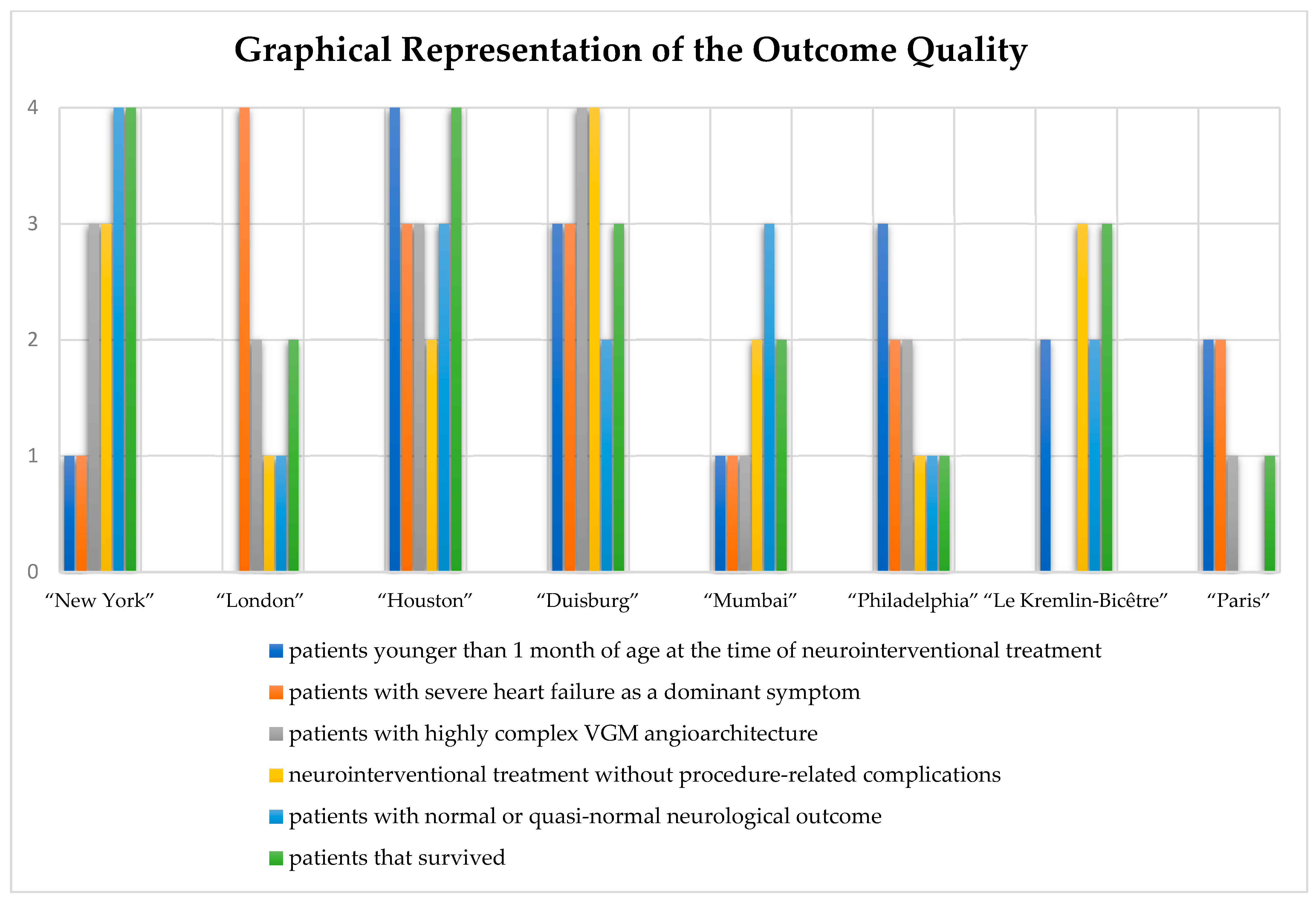
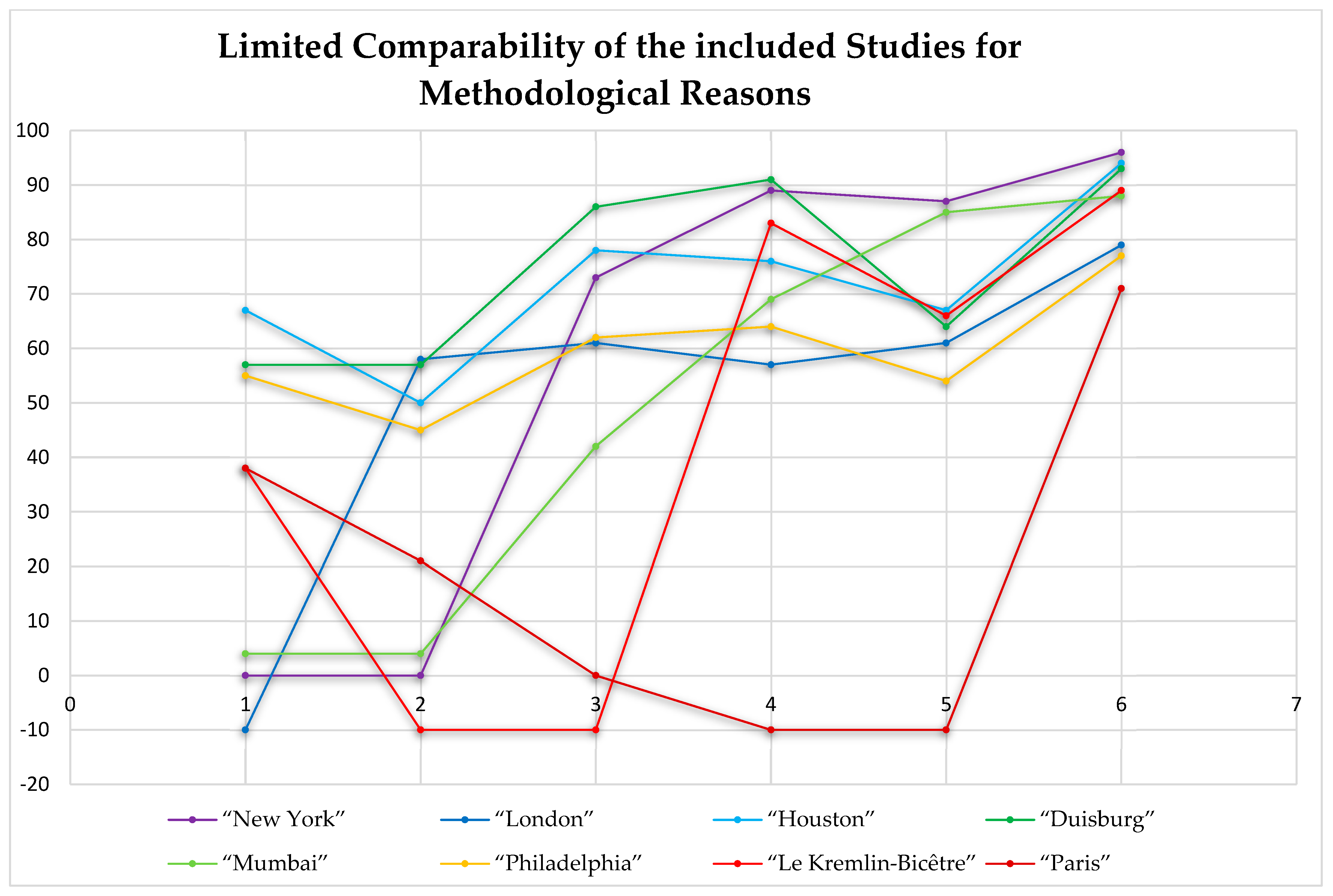
2.3. Comparison of Outcome Quality

As described above, the comparability of the included studies is limited for methodological reasons. Lack of standardization, e.g., in reporting and interpretation of results, or different inclusion criteria and interdisciplinary approaches, exacerbates this. In order to enable the best possible comparison and to meaningfully weight outcome quality by taking into account the specific patient and treatment characteristics, a semiquantitative multidimensional scoring system was introduced. Six different dimensions were covered: (1) patient age, (2) clinical symptoms, (3) VGM angioarchitecture, (4) procedure-related complications, (5) neurological outcome, and (6) overall survival. These dimensions were selected because their impact on treatment outcomes has been described as significant in various publications [2,9,10,15,19,20,24,25,26,27,28,29,30]. To obtain the final study ranking, two sequential transformation steps were required. In the first transformation step, percentage values for the following conditions were determined for each study:
  • Patients younger than 1 month of age at the time of neurointerventional treatment.
  • Patients with severe heart failure as a dominant symptom (e.g., high-output heart failure, severe congestive heart failure, or cyanotic heart failure).
  • Patients with highly complex VGM angioarchitecture (VGM Yasargil types IVa–IVc or VGM Lasjaunias type 1 “choroidal”).
  • Neurointerventional treatment without procedure-related complications.
  • Patients with neurologically normal or quasi-normal outcomes.
  • Patients that survived.
In the second transformation step, the determined percentage values were converted into points according to the following criteria:
0 points—percentage value cannot be derived from the study.
1 point—lowest and second lowest percentage values of all included studies.
2 points—third lowest or fourth lowest percentage value of all included studies.
3 points—fifth lowest or sixth lowest percentage value of all included studies.
4 points—seventh lowest or eighth lowest percentage value of all included studies.
5 points—ninth lowest or tenth lowest percentage value of all included studies.
and so forth.
Finally, the total score for each study was calculated as the sum of the points awarded for each of the six dimensions. Consequently, the theoretical minimum of the total score is 0 points and the theoretical maximum of the total score depends on the number of included studies (e.g., for 8 included studies, the theoretical maximum of the total score is 24). The total score of each study was used to obtain the final study ranking, with a higher total score corresponding to a better outcome quality.

3. Results

After a comprehensive review of the literature, eight studies were included in the synthesis and analysis [11,15,20,29,31,32,33,34]. One study was multicentric and the rest were retrospective monocentric (level 4 evidence studies according to the Oxford Centre for Evidence-based Medicine). The years of publication ranged from 1997 to 2019, with the first patients undergoing treatment in 1981 and the last patients in 2018. The total number of included patients was 480 and patient age ranged from 1 day to 18 years. Neurointerventional treatment was performed in all studies in the form of embolization, although it is worth noting that different embolization techniques were used in some of the studies.

3.1. Summary of Measures and Synthesis

3.1.1. Study “New York” 2004–2015 [35]

Based on the inclusion and exclusion criteria, a total of 45 patients were enrolled in this monocentric study, all of whom underwent neurointerventional treatment. At the time of the first neurointerventional treatment, no patient was younger than one month of age. Mild heart failure, pulmonary arterial hypertension, headache, cognitive decline, and other reasons led to the indication for neurointerventional treatment. VGM Lasjaunias type 1 “choroidal” was present in 33 patients, consistent with a highly complex VGM angioarchitecture in 73% of patients. Neurointerventional treatment was performed preferably as transarterial embolization, and only as a last resort as transvenous embolization (note: the authors reported an increased rate of cerebral hemorrhage after transvenous embolization). Different embolic materials such as glue/iodized oil and EthyleneVinyl Alcohol Copolymer (EVOH) were used regularly, whereas coils were used only in exceptional cases. Staged embolization was performed regularly, with the number of embolizations ranging from 1 to 11 per patient. In six and two patients, respectively, implantation of a ventriculo-peritoneal shunt and ventriculostomy were performed as a local treatment in addition to neurointerventional treatment. The degree of VGM obliteration was complete in 82% and partial in 13% of patients. The procedure-related complication rate was 11%. Complications included intracranial hemorrhage, cerebral ischemia, and subarchnoid hemorrhage. The follow-up period was not specified. The percentage of patients with neurologically normal or quasi-normal outcome was 87%. The percentage of patients with severe neurological impairment was 0%. A total of two patients died.

3.1.2. Study “London” 2003–2008 [34]

Based on the inclusion and exclusion criteria, a total of 33 patients were enrolled in this monocentric study, 28 of whom underwent neurointerventional treatment. In five patients, neurointerventional treatment was not performed because of diffuse ischemic brain injury or a stable clinical condition. The age of the patients at the time of the first neurointerventional treatment was not specified. Severe heart failure, macrocephalus, unnatural gait, and other reasons led to the indication for neurointerventional treatment. VGM Lasjaunias type 1 “choroidal” was present in 20 patients, consistent with a highly complex VGM angioarchitecture in 61% of patients. Neurointerventional treatment was exclusively performed as transarterial embolization. Only glue (presumably together with iodized oil) was used as embolic material. Staged embolization was performed regularly, with the number of embolizations ranging from 1 to 7 per patient. In five, four, and two patients, respectively, ventriculostomy, positioning of an external ventricular drainage, and implantation of a ventriculo-peritoneal shunt were performed as a local treatment in addition to neurointerventional treatment. The degree of VGM obliteration was complete in 39% and partial in 54% of the patients. The procedure-related complication rate was 43%. Complications included glue migration, ventricular hemorrhage, iliac artery occlusion, microcatheter adhesion with arterial rupture, microcatheter adhesion with arterial rupture and death, interhemispheric hemorrhage with venous ischemia in cortical venous thrombosis, cardiac arrest, combined ventricular/thalamic hemorrhage, and glue-associated aseptic meningitis. The follow-up period was 33 months (mean). The percentage of patients with neurologically normal or quasi-normal outcome was 61%. The percentage of patients with severe neurological impairment was 18%. A total of seven patients died.

3.1.3. Study “Houston” 2002–2018 [33]

Based on the inclusion and exclusion criteria, a total of 18 patients were enrolled in this monocentric study, 15 of whom underwent neurointerventional treatment. In three patients, neurointerventional treatment was not performed because of a critical clinical condition with bad prognosis, parental choice, or a stable clinical condition. At the time of the first neurointerventional treatment, 10 patients were younger than 1 month of age (note: the number of patients younger than 1 week of age was not specified). Severe heart failure, seizures, motor deficits, and other reasons led to the indication for neurointerventional treatment. VGM Lasjaunias type 1 “choroidal” was present in 14 patients, consistent with a highly complex VGM angioarchitecture in 78% of patients. Neurointerventional treatment was exclusively performed as transarterial embolization. Different embolic materials such as glue (presumably together with iodized oil), EVOH, and balloon were used regularly. Staged embolization was performed regularly, with the number of embolizations ranging from 1 to >4 per patient. Local treatment in addition to neurointerventional treatment was not specified. The degree of VGM obliteration was complete in 20% and partial in 80% of the patients. The procedure-related complication rate was 24%. Complications included coil migration, ventricular hemorrhage, femoral artery occlusion, and subarachnoidal hemorrhage. The follow-up period was 38 months (mean). The percentage of patients with neurologically normal or quasi-normal outcome was 67%. The percentage of patients with severe neurological impairment was 17%. Only one patient died.

3.1.4. Study “Duisburg” 2001–2010 [29]

Based on the inclusion and exclusion criteria, a total of 14 patients were enrolled in this monocentric study, all of whom underwent neurointerventional treatment. At the time of the first neurointerventional treatment, eight patients were younger than one month of age and six patients were younger than one week of age. Severe heart failure, seizures, cerebral ischemia, and other reasons led to the indication for neurointerventional treatment. VGM Lasjaunias type 1 “choroidal” was present in 12 patients, consistent with a highly complex VGM angioarchitecture in 86% of patients. Neurointerventional treatment was performed as a combined transarterial and transvenous embolization in all patients (“kissing mucrocatheter technique”; also see below). Only coils were used as embolic materials. Staged embolization was performed regularly, with the number of embolizations ranging from 1 to 13 per patient. In two patients, implantation of an external ventricular drainage was performed as a local treatment in addition to neurointerventional treatment. The degree of VGM obliteration was complete in 57% and partial in 43% of the patients. The procedure-related complication rate was 9%, Complications included unproblematic vascular perforation, ventricular hemorrhage after vascular perforation, and subarachnoidal hemorrhage after perforation of an embolization coil. The follow-up period was 53 months (mean). The percentage of patients with neurologically normal or quasi-normal outcome was 64%. The percentage of patients with severe neurological impairment was 14%. Only one patient died.

3.1.5. Study “Mumbai” 1998–2012 [11]

Based on the inclusion and exclusion criteria, a total of 26 patients were enrolled in this monocentric study, all of whom underwent neurointerventional treatment. At the time of the first neurointerventional treatment, one patient was younger than one month of age and one patient was younger than one week of age. Severe heart failure, dyspnoe, developmental delay of neurocognitive functioning, failure to thrive, and other reasons led to the indication for neurointerventional treatment. VGM Lasjaunias type 1 “choroidal” was present in 11 patients, consistent with a highly complex VGM angioarchitecture in 42% of patients. Neurointerventional treatment was exclusively performed as transarterial embolization. Different embolic materials such as glue/tantalum mixture and EVOH were used regularly. Whether or not staged embolization was performed was not specified. In one patient, implantation of a ventriculo-peritoneal shunt was performed as a local treatment in addition to neurointerventional treatment. The degree of VGM obliteration was not specified. The procedure-related complication rate was 31%. Complications included glue-associated granuloma, visual impairment, apnoe, sinus thrombosis, glue migration into the superior sagittal sinus, heart failure, and ventricular hemorrhage. The follow-up period was not specified. The percentage of patients with neurologically normal or quasi-normal outcome was 85%. The percentage of patients with severe neurological impairment was 4%. A total of three patients died.

3.1.6. Study “Philadelphia” 1994–2007 [32]

Based on the inclusion and exclusion criteria, a total of 13 patients were enrolled in this monocentric study, 11 of whom underwent neurointerventional treatment. In two patients, neurointerventional treatment was not performed because of a critical clinical condition with bad prognosis, parental choice, or a stable clinical condition. At the time of the first neurointerventional treatment, six patients were younger than one month of age and three patients were younger than one week of age. Severe heart failure, leukomalacia, cerebral atrophy, and other reasons led to the indication for neurointerventional treatment. VGM Lasjaunias type 1 “choroidal” was present in seven patients, consistent with a highly complex VGM angioarchitecture in 62% of patients. Neurointerventional treatment was exclusively performed as transarterial embolization. Different embolic materials such as glue (presumably together with iodized oil) and coils were used regularly. Staged embolization was performed regularly, with the number of embolizations ranging from 1 to 3 per patient. In four patients, implantation of a ventriculo-peritoneal shunt was performed as a local treatment in addition to neurointerventional treatment. The degree of VGM obliteration was not specified. The procedure-related complication rate was 36%. Complications included combined intracranial hemorrhage, subarachnoid hemorrhage, intraventricular hemorrhage, cerebral venous thrombosis with thalamic hemorrhage, transient Parinaud syndrome, and postinterventional hemorrhage. The follow-up period was 50 months (mean). The percentage of patients with neurologically normal or quasi-normal outcome was 54%. The percentage of patients with severe neurological impairment was 8%. A total of two patients died.

3.1.7. Study “Le Kremlin-Bicêtre” 1981–2002 [20]

Based on the inclusion and exclusion criteria, a total of 317 patients were enrolled in this multicentric study, 216 of whom underwent neurointerventional treatment in Le Kremlin-Bicêtre (Paris, France). Since detailed data for the patients in the other centers were not specified, the following statements must refer in particular to Le Kremlin-Bicêtre (216 patients). In 84/317 patients, neurointerventional treatment was not performed because it was technically not feasible, or because of a critical clinical condition with bad prognosis. At the time of the first neurointerventional treatment, 83/216 patients were younger than 1 month of age (note: the number of patients younger than 1 week of age was not specified). Heart failure (note: whether this also included severe heart failure was not specified), sinus thrombosis, pial reflux, and other reasons led to the indication for neurointerventional treatment. VGM angioarchitecture was not specified. Neurointerventional treatment was performed as transarterial embolization in 208/216 patients and as transvenous embolization in 8/216 patients. Only a glue/tantalum/iodized oil mixture was used as embolic material. Staged embolization was performed regularly, with the number of embolizations ranging from 1 to 5 per patient. Local treatment in addition to neurointerventional treatment was not specified. The degree of VGM obliteration was not specified. The procedure-related complication rate was 17%. Detailed data on the complications were not specified. The follow-up period was not specified. The percentage of patients with neurologically normal or quasi-normal outcome was 66%. The percentage of patients with severe neurological impairment was 9%. A total of 23 patients died. In the other centers, embolization was performed in 17 patients (note: detailed data on these patients were not specified).

3.1.8. Study “Paris” 1988–1994 [31]

Based on the inclusion and exclusion criteria, a total of 14 patients were enrolled in this early monocentric study, 13 of whom underwent neurointerventional treatment. In one patient, neurointerventional treatment was not performed because it was technically not feasible. At the time of the first neurointerventional treatment, five patients were younger than one month of age and four patients were younger than one week of age. Severe heart failure, hydrocephalus, seizures, developmental delay, and other reasons led to the indication for neurointerventional treatment. VGM Yasargil types I–III were present in all patients, consistent with a highly complex VGM angioarchitecture in 0% of patients. Neurointerventional treatment was performed as transarterial embolization in 11 patients and as transvenous embolization in 2 patients. Different embolic materials such as glue/iodized oil mixture, nylon filaments, and coils were used. Staged embolization was performed in seven patients. In one patient, radiotherapy was performed as a local treatment in addition to neurointerventional treatment. The degree of VGM obliteration and the procedure-related complication rate were not specified. The follow-up period was not specified. The percentage of patients with neurologically normal or quasi-normal outcome was not specified. The percentage of patients with severe neurological impairment was not specified. A total of four patients died.
More detailed qualitative and quantitative data can be found in Table 4, Table 5 and Table 6.

3.2. Comparison of Outcome Quality

Regarding the introduced semiquantitative multidimensional scoring system, the highest total score, i.e., the best outcome quality, was found for the study “Houston” 2002–2018 and the study “Duisburg” 2001–2010, with 19 points each, and the study “New York” 2004–2015 with 16 points. The lowest total score, i.e., the worst outcome quality, was found for the oldest study, the study “Paris” 1988–1994, with 6 points. For the remaining studies, intermediate total scores, i.e., intermediate outcome quality, was determined. The tabular and graphical representation of the outcome quality can be found in Table 7 and Figure 1. As mentioned earlier, the results must be interpreted in light of the limited comparability of the included studies for methodological reasons, as illustrated in Figure 2.

4. Discussion

Neurointerventional treatment of patients with VGM remains a challenge, as illustrated by this review of peer-reviewed and published data. The lack of standardization in methodology (e.g., reporting and terminology) and treatment (e.g., patient selection, baseline clinical characteristics, and embolization technique) makes the comparison of outcome quality among the studies difficult. By using the introduced semiquantitative multidimensional scoring system, however, a more objective comparison should be possible, considering the six key dimensions in the interdisciplinary management of patients with VGM: (1) patient age, (2) clinical symptoms, (3) VGM angioarchitecture, (4) procedure related complications, (5) neurological outcome, and (6) survival. After exact analysis of the data, the following facts become evident. Patients with severe heart failure who received neurointerventional treatment at an age younger than one month show worse neurological outcome compared with patients with no severe heart failure but who received neurointerventional treatment at an age older than one month. Because both aforementioned dimensions seem to show no effect on the procedure-related complication rate, they can be discussed as procedure-independent negative predictors of a neurologically normal or quasi-normal outcome. On the other hand, the combination of severe heart failure and patient age younger than one month does not seem to affect survival after neurointerventional treatment, as shown by the high survival rates in the studies “Houston” 2002–2018 and “Duisburg” 2001–2010, despite the high complexity of the patients treated. It remains to be seen to what extent innovative embolization techniques will lead to further improvements in technical and clinical outcomes in the future. To achieve the goal of optimized patient care, complementary treatments also play an important role. Those are discussed in the following paragraphs.

4.1. Conservative Treatment

Neonatology, pediatric critical care, and neuropediatrics form the second essential pillar in the interdisciplinary management of patients with VGM. As described earlier, the primary therapeutic goal in neonates and infants is immediate hemodynamic stabilization. Pediatric critical care includes drug approaches to improve cardiac function, such as reducing preload (e.g., diuretics) and increasing contractility (e.g., catecholamines) [2,10]. According to the literature, treatment of pulmonary arterial hypertension is performed with vasopressin and/or nitric oxide and, if necessary, in combination with invasive ventilation [2,10]. As described below under “Practical Insights according to Institutional Standard”, the treatment of pulmonary arterial hypertension with nitric oxide must be critically questioned nowadays. Optimization of renal, hepatic, and intestinal function should prevent multiorgan failure [36]. In children and adolescents, the focus is on neurological stabilization. The multidisciplinary and sometimes complex specific therapeutic regimens can be found in the relevant scientific papers, guidelines, and recommendations.

4.2. Neurosurgery

Neurosurgery as a first-line causal treatment is a thing of the past. Yasargil and colleagues have extensively engaged in microsurgical approaches, with notable success in the field. However, in a 2013 review article, Yasargil himself pointed out that neurosurgery in VGM Yasargil types IVa–c should be considered risky because of the involvement of the diencephalic and mesencephalic arterial feeders [2,10]. On a sober basis, mortality rates of 88–100% in neonates and 20–25% in infants and young children can be reported for first-line neurosurgery [11,33,37,38,39,40,41,42]. On the contrary, complementary neurosurgical procedures such as placement of a ventricular drain or ventriculostomy are safe and effective symptomatic treatments for hydrocephalus [2,37,43].

4.3. Radiotherapy

Case reports and mini-series on radiotherapy exist, but no studies with relevant case numbers or long-term outcomes could be identified. Stereotactic radiosurgery studies were published by Payne et al. in 2000 and Triffo et al. in 2014, with no specific clinical outcomes reported [44,45]. The unsuccessful strategy of radiotherapy was demonstrated as early as 1995 by Lasjaunias’ group, with no decisive change in VGM angioarchitecture observed 18–24 months after treatment [46]. The radiogenic effects, if therapeutic at all, occur far too slowly in the maturing brain, so that neurological maldevelopment is preprogrammed by the persistence of a relevant intracranial shunt volume. For the above reasons, radiotherapy should only be considered as a last option in refractory VGM cases [47].

4.4. Molecular Treatment

The VGM develops between the sixth and eleventh week of gestation, with the physiological transformation of both the anterior part of the MPV into the internal cerebral veins and the posterior part of the MPV into the Vein of Galen failing to occur [4,5]. There is now evidence that various gene mutations may cause VGM. According to a 2018 review article, there is an association with RASA1 gene mutation (including the so-called “capillary malformation-arteriovenous malformation syndrome”) or ENG/ACVRL1 gene mutation (including the so-called “hereditary hemorrhagic telangiectasia”) [48]. The increasing molecular understanding is opening new therapeutic opportunities. For example, it has become known that the ACVRL1 gene controls a TGF-beta-mediated signaling pathway that is important for normal vascular development, which in turn may make the use of TGF-beta-modulating drugs useful in patients with VGM [49]. Should a drug for molecular treatment become available in the future, the narrow prenatal therapeutic window remains another challenge.

4.5. “Practical Insights According to Institutional Standard”

As discussed above, medical hemodynamic stabilization before and after neurointerventional treatment is key to clinical success. The following clinical cases show what optimal interdisciplinary management may look like in patients with VGM Lasjaunias type 1 “choroidal” and type 2 “mural”.

4.5.1. “Pediatric Critical Care-Medical Hemodynamic Stabilization with Prostaglandin E1”

A pregnant woman diagnosed with VGM Lasjaunias type 1 “choroidal” in her unborn child was admitted at 36 weeks of gestation for controlled delivery. The preterm infant, with a birth weight of 2570 g, suffered from hemodynamic instability on the first day of life due to the excessive intracranial shunt volume: suprasystemic increased right ventricular pressure, patent ductus arteriosus botalli with predominant right-to-left shunt, and retrograde flow in the descending aorta (the so-called “aortic steal effect”) (Figure 3). Primary pediatric critical care consisted of noninvasive respiratory support with low oxygen delivery (IO2F 30%), volume restriction, and administration of furosemide to lower the preload. At this point, systemic administration of prostaglandin E1 was an important measure to keep the ductus arteriosus botalli open during ductus-dependent systemic perfusion. Neurointerventional treatment was performed on the second day of life and resulted in a significant reduction in the intracranial shunt volume. Clinically, this was documented by a decrease in right ventricular pressure and normalization of flow in both the descending aorta and the ductus arteriosus botalli. At that stage, it was possible to discontinue the administration of prostaglandin E1. Milrinone (as a positive inotropic and vasodilator), in combination with epinephrine, served for several days to further support the circulation. Before neurointerventional treatment, the focus is on treating right heart failure and maintaining the ductus-dependent systemic perfusion. After neurointerventional treatment, the focus is on preventing left ventricular dysfunction. Two recent papers discuss in detail the critical care management of patients with VGM, with particular emphasis on the factor of “severe heart failure” [50,51]. It should be reiterated that the use of nitric oxide can lead to rapid deterioration of pulmonary status with concomitant impairment of the systemic perfusion. Therefore, when transferring patients from other hospitals, we recommend discontinuing nitric oxide treatment or, at most, using it with extreme caution and as a last option. After neurointerventional treatment, attention must also be paid to adequate analgesia (ideally intubation anesthesia for 48 h) to avoid arterial blood pressure spikes and associated complications such as intracranial hemorrhage (the so-called “normal perfusion pressure breakthrough syndrome”). Taking into account the clinical course as well as the intracranial Doppler signals, a decision can then be made on whether and when another neurointerventional treatment is required.

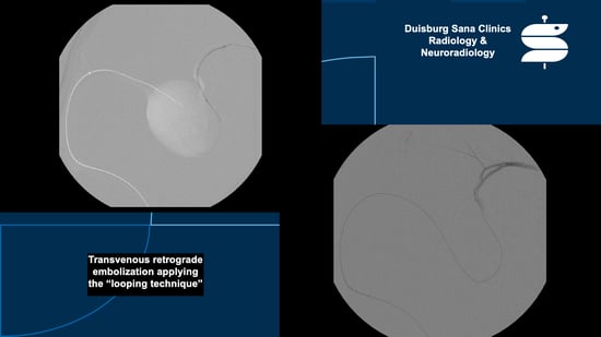
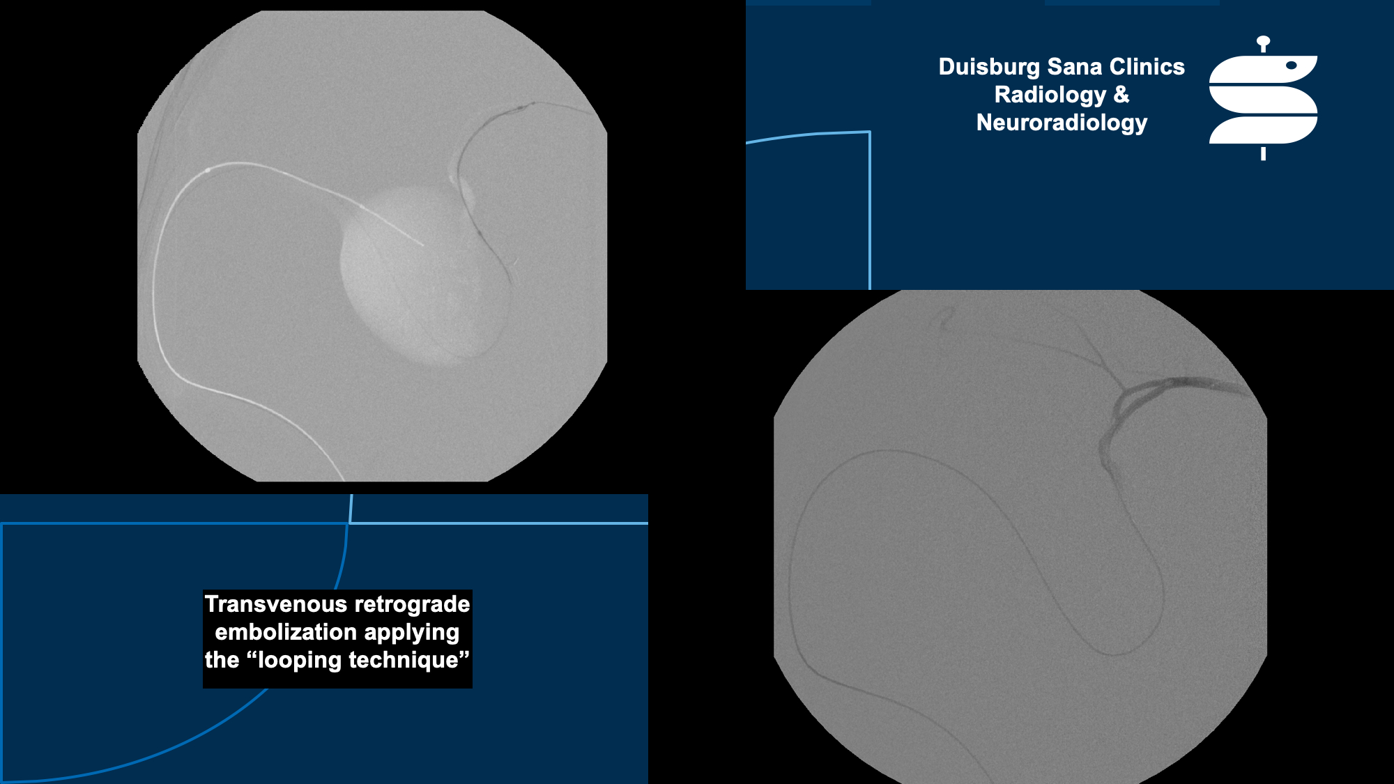
4.5.2. “Neurointerventional Treatment-Hemodynamic Stabilization with Embolization”

Nowadays, modern and innovative techniques are available for neurointerventional treatment of VGM. A list of materials that can be used, such as sheaths, catheters, microcatheters, coils, and liquid embolics, can be found in a book chapter [52]. In addition, the same publication presents the embolization strategy, taking into account the complex VGM angioarchitecture and ethical aspects of treatment [52]. Based on almost 30 years of experience of the first author, we are allowed to present selected technical aspects of embolization in the form of two cases: repeated combined transarterial and transvenous embolization using the “kissing microcatheter technique” (Figure 4) and single-stage transvenous retrograde embolization using the “looping technique” and the “wedging technique” (Figure 5) [29,52,53].

5. Conclusions

Neurointerventional treatment represents the essential pillar in the interdisciplinary management of patients with VGM, although standardization is lacking, based on the results of the structured review. As complementary treatments, pediatric critical care is mandatory and includes pre-, peri-, and post-neurointerventional medical hemodynamic stabilization. Neurosurgery and radiotherapy currently have no roles as first-line treatments due to high procedure-related morbidity and mortality and/or lack of efficacy.

Author Contributions

Study conception and design: all authors; acquisition of data, F.B., M.S.-H., D.G., F.B.N., S.S., M.S., C.F. and C.M.S.; analysis of data, F.B., R.L., C.F., V.K., S.S., and C.M.S.; interpretation of data, all authors; drafting of the manuscript, F.B., M.S.-H., F.B.N., and C.M.S.; critical revision of the manuscript, all authors. All authors have read and agreed to the published version of the manuscript.

Funding

The authors declare that no funding was involved.

Institutional Review Board Statement

Not required, as this review is a summary of previously published data and not a publication of original data.

Informed Consent Statement

The authors approved publication.

Data Availability Statement

Not applicable.

Acknowledgments

For all those who guide us in the effort to help others.

Conflicts of Interest

F.B. declares the following competing interest: shareholder of the company Bentley InnoMed GmbH, Hechingen, Germany. All other authors declare that they have no competing interests.

References

  1. Brassel, F.; Meila, D.; Papke, K. Vascular interventions in the head and neck region. part 2: Procedures for vessel occlusion. Radiologe 2011, 51, 519–533. [Google Scholar] [CrossRef] [PubMed]
  2. Mortazavi, M.M.; Griessenauer, C.J.; Foreman, P.; Bavarsad Shahripour, R.; Shoja, M.M.; Rozzelle, C.J.; Tubbs, R.S.; Fisher, W.S.; Fukushima, T. Vein of Galen Aneurysmal Malformations: Critical Analysis of the Literature with Proposal of a New Classification System. J. Neurosurg. Pediatr. 2013, 12, 293–306. [Google Scholar] [CrossRef] [PubMed]
  3. Steinheil, S.O. Über einen Fall von Varix aneurysmaticusim Bereichder Gehirngefässe; Dissertationsschrift; F. Fromme: Würzburg, Germany, 1895. [Google Scholar]
  4. Raybaud, C.A.; Strother, C.M.; Hald, J.K. Aneurysms of the Vein of Galen: Embryonic Considerations and Anatomical Features Relating to the Pathogenesis of the Malformation. Neuroradiology 1989, 31, 109–128. [Google Scholar] [CrossRef]
  5. Gailloud, P.; O’riordan, D.P.; Burger, I.; Lehmann, C.U. Confirmation of Communication between Deep Venous Drainage and the Vein of Galen after Treatment of a Vein of Galen Aneurysmal Malformation in an Infant Presenting with Severe Pulmonary Hypertension. AJNR Am. J. Neuroradiol. 2006, 27, 317–320. [Google Scholar]
  6. Recinos, P.F.; Rahmathulla, G.; Pearl, M.; Recinos, V.R.; Jallo, G.I.; Gailloud, P.; Ahn, E.S. Vein of Galen Malformations: Epidemiology, Clinical Presentations, Management. Neurosurg. Clin. N. Am. 2012, 23, 165–177. [Google Scholar] [CrossRef]
  7. Khullar, D.; Andeejani, A.M.I.; Bulsara, K.R. Evolution of Treatment Options for Vein of Galen Malformations. J. Neurosurg. Pediatr. 2010, 6, 444–451. [Google Scholar] [CrossRef]
  8. Yaşargil, M.G.; Yaşargil, M.G. AVM of the Brain, Clinical Considerations, General and Special Operative Techniques, Surgical Results, Nonoperated Cases, Cavernous and Venous Angiomas, Neuroanesthesia; 191 Tables; Microneurosurgery; Thieme [u.a.]: Stuttgart, Germany, 1988; ISBN 978-3-13-693501-9. [Google Scholar]
  9. Lasjaunias, P. Vascular Diseases in Neonates, Infants and Children: Interventional Neuroradiology Management; Springer: Berlin/Heidelberg, Germany, 1997; ISBN 978-3-662-10740-9. [Google Scholar]
  10. Florian, I.S. Springer Nature Switzerland AG; Cham, Switzerland, 2020. [Google Scholar]
  11. Sivasankar, R.; Limaye, U.S.; Wuppalapati, S.; Shrivastava, M. Endovascular Management of Vein of Galen Aneurysmal Malformations: A Retrospective Analysis over a 15-Year Period. J. Vasc. Interv. Neurol. 2019, 10, 23–29. [Google Scholar] [PubMed]
  12. Brevis Nuñez, F.; Dohna-Schwake, C. Epidemiology, Diagnostics, and Management of Vein of Galen Malformation. Pediatr. Neurol. 2021, 119, 50–55. [Google Scholar] [CrossRef] [PubMed]
  13. Jea, A.; Bradshaw, T.J.; Whitehead, W.E.; Curry, D.J.; Dauser, R.C.; Luerssen, T.G. The High Risks of Ventriculoperitoneal Shunt Procedures for Hydrocephalus Associated with Vein of Galen Malformations in Childhood: Case Report and Literature Review. Pediatr. Neurosurg. 2010, 46, 141–145. [Google Scholar] [CrossRef]
  14. Hassan, T.; Nassar, M.; Elghandour, M. Vein of Galen Aneurysms: Presentation and Endovascular Management. Pediatr. Neurosurg. 2010, 46, 427–434. [Google Scholar] [CrossRef]
  15. Berenstein, A.; Fifi, J.T.; Niimi, Y.; Presti, S.; Ortiz, R.; Ghatan, S.; Rosenn, B.; Sorscher, M.; Molofsky, W. Vein of Galen Malformations in Neonates: New Management Paradigms for Improving Outcomes. Neurosurgery 2012, 70, 1207–1213, discussion 1213–1214. [Google Scholar] [CrossRef] [PubMed]
  16. Howarth, R.A.; Reisner, A.; Chern, J.J.; Hayes, L.L.; Burns, T.G.; Berenstein, A. Neurocognitive Improvements Following Endovascular Repair of Vein of Galen Malformation in a Child. J. Neurosurg. Pediatr. 2015, 15, 197–202. [Google Scholar] [CrossRef] [PubMed]
  17. Hoang, S.; Choudhri, O.; Edwards, M.; Guzman, R. Vein of Galen Malformation. Neurosurg. Focus 2009, 27, E8. [Google Scholar] [CrossRef]
  18. Chow, M.L.; Cooke, D.L.; Fullerton, H.J.; Amans, M.R.; Narvid, J.; Dowd, C.F.; Higashida, R.T.; Halbach, V.V.; Hetts, S.W. Radiological and Clinical Features of Vein of Galen Malformations. J. Neurointerv. Surg. 2015, 7, 443–448. [Google Scholar] [CrossRef]
  19. Brinjikji, W.; Krings, T.; Murad, M.H.; Rouchaud, A.; Meila, D. Endovascular Treatment of Vein of Galen Malformations: A Systematic Review and Meta-Analysis. AJNR Am. J. Neuroradiol. 2017, 38, 2308–2314. [Google Scholar] [CrossRef] [PubMed]
  20. Lasjaunias, P.L.; Chng, S.M.; Sachet, M.; Alvarez, H.; Rodesch, G.; Garcia-Monaco, R. The Management of Vein of Galen Aneurysmal Malformations. Neurosurgery 2006, 59, S184–S194, discussion S3–S13. [Google Scholar] [CrossRef]
  21. Jones, B.V.; Ball, W.S.; Tomsick, T.A.; Millard, J.; Crone, K.R. Vein of Galen Aneurysmal Malformation: Diagnosis and Treatment of 13 Children with Extended Clinical Follow-Up. AJNR Am. J. Neuroradiol. 2002, 23, 1717–1724. [Google Scholar]
  22. Liberati, A.; Altman, D.G.; Tetzlaff, J.; Mulrow, C.; Gotzsche, P.C.; Ioannidis, J.P.; Clarke, M.; Devereaux, P.J.; Kleijnen, J.; Moher, D. The PRISMA Statement for Reporting Systematic Reviews and Meta-Analyses of Studies That Evaluate Healthcare Interventions: Explanation and Elaboration. BMJ 2009, 339, b2700. [Google Scholar] [CrossRef]
  23. Sommer, C.M.; Pieper, C.C.; Offensperger, F.; Pan, F.; Killguss, H.J.; Köninger, J.; Loos, M.; Hackert, T.; Wortmann, M.; Do, T.D.; et al. Radiological Management of Postoperative Lymphorrhea. Langenbecks Arch. Surg. 2021, 406, 945–969. [Google Scholar] [CrossRef]
  24. Alektoroff, K.; Papanagiotou, P. Vein of Galen aneurysmal malformation. Die Radiologie 2022, 62, 671–674. [Google Scholar] [CrossRef]
  25. Reddy, R.; Lucke-Wold, B. Primer of Vein of Galen Malformation Management. J. Pediatr. Heath Care Med. 2022, 5, 30–34. [Google Scholar]
  26. Cory, M.J.; Durand, P.; Sillero, R.; Morin, L.; Savani, R.; Chalak, L.; Angelis, D. Vein of Galen Aneurysmal Malformation: Rationalizing Medical Management of Neonatal Heart Failure. Pediatr. Res. 2022, 93, 39–48. [Google Scholar] [CrossRef] [PubMed]
  27. See, A.P.; Wilkins-Haug, L.E.; Benson, C.B.; Tworetzky, W.; Orbach, D.B. Percutaneous Transuterine Fetal Cerebral Embolisation to Treat Vein of Galen Malformations at Risk of Urgent Neonatal Decompensation: Study Protocol for a Clinical Trial of Safety and Feasibility. BMJ Open 2022, 12, e058147. [Google Scholar] [CrossRef] [PubMed]
  28. Fifi, J.T.; Bazil, M.J.; Matsoukas, S.; Shigematsu, T.; Sorscher, M.; Berenstein, A. Evolution of Transvenous Embolization in Vein of Galen Malformation: Case Series and Review of the Literature. J. Neurointerv. Surg. 2022, 14, neurintsurg-2022-019121. [Google Scholar] [CrossRef] [PubMed]
  29. Meila, D.; Hannak, R.; Feldkamp, A.; Schlunz-Hendann, M.; Mangold, A.; Jacobs, C.; Papke, K.; Brassel, F. Vein of Galen Aneurysmal Malformation: Combined Transvenous and Transarterial Method Using a “Kissing Microcatheter Technique”. Neuroradiology 2012, 54, 51–59. [Google Scholar] [CrossRef]
  30. Lasjaunias, P.; Berenstein, A.; Ter Brugge, K.G. Surgical Neuroangiography: Vol. 3: Clinical and Interventional Aspects in Children, 2nd ed.; Springer: Berlin/Heidelberg, Germany, 2007; ISBN 978-3-540-41681-4. [Google Scholar]
  31. Borthne, A.; Carteret, M.; Baraton, J.; Courtel, J.; Brunelle, F. Vein of Galen Vascular Malformations in Infants: Clinical, Radiological and Therapeutic Aspect. Eur. Radiol. 1997, 7, 1252–1258. [Google Scholar] [CrossRef]
  32. Heuer, G.G.; Gabel, B.; Beslow, L.A.; Stiefel, M.F.; Schwartz, E.S.; Storm, P.B.; Ichord, R.N.; Hurst, R.W. Diagnosis and Treatment of Vein of Galen Aneurysmal Malformations. Childs Nerv. Syst. 2010, 26, 879–887. [Google Scholar] [CrossRef]
  33. Wagner, K.M.; Ghali, M.G.Z.; Srinivasan, V.M.; Lam, S.; Johnson, J.; Chen, S.; Kan, P. Vein of Galen Malformations: The Texas Children’s Hospital Experience in the Modern Endovascular Era. Oper. Neurosurg. 2019, 17, 286–292. [Google Scholar] [CrossRef]
  34. McSweeney, N.; Brew, S.; Bhate, S.; Cox, T.; Roebuck, D.J.; Ganesan, V. Management and Outcome of Vein of Galen Malformation. Arch. Dis. Child. 2010, 95, 903–909. [Google Scholar] [CrossRef]
  35. Berenstein, A.; Paramasivam, S.; Sorscher, M.; Molofsky, W.; Meila, D.; Ghatan, S. Vein of Galen Aneurysmal Malformation: Advances in Management and Endovascular Treatment. Neurosurgery 2019, 84, 469–478. [Google Scholar] [CrossRef]
  36. Winn, H.R.; Youmans, J.R. (Eds.) Youmans Neurological Surgery, 5th ed.; W.B. Saunders: Philadelphia, PA, USA, 2004; ISBN 978-0-7216-8291-4. [Google Scholar]
  37. Hansen, D.; Kan, P.T.; Reddy, G.D.; Mohan, A.C.; Jea, A.; Lam, S. Pediatric Knowledge Update: Approach to the Management of Vein of Galen Aneurysmal Malformations in Neonates. Surg. Neurol. Int. 2016, 7, S317–S321. [Google Scholar] [CrossRef]
  38. Fullerton, H.J.; Aminoff, A.R.; Ferriero, D.M.; Gupta, N.; Dowd, C.F. Neurodevelopmental Outcome after Endovascular Treatment of Vein of Galen Malformations. Neurology 2003, 61, 1386–1390. [Google Scholar] [CrossRef] [PubMed]
  39. Johnston, I.H.; Whittle, I.R.; Besser, M.; Morgan, M.K. Vein of Galen Malformation: Diagnosis and Management. Neurosurgery 1987, 20, 747–758. [Google Scholar] [CrossRef]
  40. Norman, M.G.; Becker, L.E. Cerebral Damage in Neonates Resulting from Arteriovenous Malformation of the Vein of Galen. J. Neurol. Neurosurg. Psychiatry 1974, 37, 252–258. [Google Scholar] [CrossRef] [PubMed]
  41. Hoffman, H.J.; Chuang, S.; Hendrick, E.B.; Humphreys, R.P. Aneurysms of the Vein of Galen. Experience at the Hospital for Sick Children, Toronto. J. Neurosurg. 1982, 57, 316–322. [Google Scholar] [CrossRef] [PubMed]
  42. Ciricillo, S.F.; Edwards, M.S.; Schmidt, K.G.; Hieshima, G.B.; Silverman, N.H.; Higashida, R.T.; Halbach, V.V. Interventional Neuroradiological Management of Vein of Galen Malformations in the Neonate. Neurosurgery 1990, 27, 22–27, discussion 27–28. [Google Scholar] [CrossRef]
  43. Gupta, A.K.; Rao, V.R.K.; Varma, D.R.; Kapilamoorthy, T.R.; Kesavadas, C.; Krishnamoorthy, T.; Thomas, B.; Bodhey, N.K.; Purkayastha, S. Evaluation, Management, and Long-Term Follow up of Vein of Galen Malformations. J. Neurosurg. 2006, 105, 26–33. [Google Scholar] [CrossRef] [PubMed]
  44. Payne, B.R.; Prasad, D.; Steiner, M.; Bunge, H.; Steiner, L. Gamma Surgery for Vein of Galen Malformations. J. Neurosurg. 2000, 93, 229–236. [Google Scholar] [CrossRef]
  45. Triffo, W.J.; Bourland, J.D.; Couture, D.E.; McMullen, K.P.; Tatter, S.B.; Morris, P.P. Definitive Treatment of Vein of Galen Aneurysmal Malformation with Stereotactic Radiosurgery. J. Neurosurg. 2014, 120, 120–125. [Google Scholar] [CrossRef]
  46. Watban, J.A.; Rodesch, G.; Alvarez, H.; Lasjaunias, P. Transarterial Embolization of Vein of Galen Aneurysmal Malformation after Unsuccessful Stereotactic Radiosurgery. Report of Three Cases. Childs Nerv. Syst. 1995, 11, 406–408. [Google Scholar] [CrossRef]
  47. Choque-Velasquez, J.; Colasanti, R.; Muhammad, S.; Chioffi, F.; Hernesniemi, J. Vascular Lesions of the Pineal Region: A Comprehensive Review of the Therapeutic Options. World Neurosurg. 2022, 159, 298–313. [Google Scholar] [CrossRef]
  48. Duran, D.; Karschnia, P.; Gaillard, J.R.; Karimy, J.K.; Youngblood, M.W.; DiLuna, M.L.; Matouk, C.C.; Aagaard-Kienitz, B.; Smith, E.R.; Orbach, D.B.; et al. Human Genetics and Molecular Mechanisms of Vein of Galen Malformation. J. Neurosurg. Pediatr. 2018, 21, 367–374. [Google Scholar] [CrossRef] [PubMed]
  49. Goumans, M.-J.; Liu, Z.; ten Dijke, P. TGF-Beta Signaling in Vascular Biology and Dysfunction. Cell Res. 2009, 19, 116–127. [Google Scholar] [CrossRef] [PubMed]
  50. Giorgi, L.; Durand, P.; Morin, L.; Miatello, J.; Merchaoui, Z.; Lambert, V.; Boithias, C.; Senat, M.V.; Stos, B.; Maurey, H.; et al. Management and Outcomes of Neonatal Arteriovenous Brain Malformations with Cardiac Failure: A 17 Years’ Experience in a Tertiary Referral Center. J. Pediatr. 2020, 218, 85–91.e2. [Google Scholar] [CrossRef] [PubMed]
  51. Giesinger, R.E.; Elsayed, Y.N.; Castaldo, M.P.; McNamara, P.J. Targeted Neonatal Echocardiography-Guided Therapy in Vein of Galen Aneurysmal Malformation: A Report of Two Cases with a Review of Physiology and Approach to Management. AJP Rep. 2019, 9, e172–e176. [Google Scholar] [CrossRef] [PubMed]
  52. Brassel, F.; Kobba, S.; Sommer, C.M. Vein of Galen Aneurysmal Malformation (VGAM); Chapter 23 in Region Lesions; Springer Nature Switzerland AG: Cham, Switzerland, 2020. [Google Scholar]
  53. Brassel, F. Lecture: Approach—Vein of Galen Malformations; World AVM Congress 2022; Val d’Isère, France, 2022. [Google Scholar]
Figure 1. Graphical Representation of Outcome Quality. Note: points awarded for each of the six dimensions on the ordinate; 0 points: percentage value cannot be derived from the study; 1 point: lowest and second lowest percentage values of the eight included studies; 2 points: third lowest or fourth lowest percentage value of the eight included studies; 3 points: fifth lowest or sixth lowest percentage value of the eight included studies; 4 points: seventh lowest or eighth lowest percentage value of the eight included studies; the total score for each study was calculated as the sum of the points awarded for each of the six dimensions.
Jvd 02 00018 g001
Figure 2. Limited Comparability of the included Studies for Methodological Reasons. Note: percentage values plotted on the ordinate (if the percentage value cannot be derived from the study, a placeholder with a percentage value of −10% is used); six dimensions (first transformation step): 1: percentage value for patients younger than 1 month of age at the time of neurointerventional treatment; 2: percentage value for patients with severe heart failure as a dominant symptom; 3: percentage value for patients with highly complex VGM angioarchitecture (VGM Yasargil types IVa–IVc or VGM Lasjaunias type 1 “choroidal”); 4: percentage value for neurointerventional treatments without procedure-related complications; 5: percentage value for patients with neurologically normal or quasi-normal outcome; 6: percentage of patients that survived. In order to enable the best possible comparison and to meaningfully weight outcome quality by taking into account the specific patient and treatment characteristics, the semiquantitative multidimensional scoring system was introduced.
Jvd 02 00018 g002
Figure 3. Pediatric Critical Care–Medical Hemodynamic Stabilization with Prostaglandin E1. Note: hemodynamic instability in a 1-day-old patient with VGM Lasjaunias Type 1 “choroidal” documented with echocardiography and ultrasonography; (A) suprasystemic increased right ventricular pressure as a sign of right ventricular decompensation; (B) insufficiency of the tricuspid valve as an indirect sign of increased pulmonary arterial pressure; (C) patent ductus arteriosus botalli with predominant right-to-left shunt; (D) retrograde flow in the descending aorta (so-called “aortic steal effect”).
Jvd 02 00018 g003
Figure 4. Neurointerventional Treatment–Hemodynamic Stabilization with Repeated Combined Transarterial and Transvenous Embolization in a 1-day-old Patient with VGM Lasjaunias Type 1 “Choroidal”. Note: repeated combined transarterial and transvenous embolization (“kissing microcatheter technique”) with subsequent hemodynamic stabilization and neurological improvement; (A,B) MRI before embolization showing multiple arteriovenous fistulas and malformations; (CE) angiography of the arterial feeders (so-called “choroido-thalamic collateral network”); (FJ) repeated combined transarterial and transvenous embolization from different strategic targets with coils and/or EVOH; (KP) unremarkable MRI at 6-month follow-up.
Jvd 02 00018 g004
Figure 5. Neurointerventional Treatment–Hemodynamic Stabilization with One-stage Transvenous Embolization in a 4-month-old Patient with VGM Lasjaunias Type 2 “Mural”. Note: one-stage transvenous retrograde embolization (“looping technique” and “wedging technique”) with subsequent neurological improvement; (A,B) MRI before embolization showing single arteriovenous fistulas and venous outflow via the dilated MPV; (CE) angiography of the arterial feeders (posterior pericallosal artery); (FI) one-stage transvenous retrograde embolization from one strategic target with coils; (JL) angiography after embolization with confirmation of obliteration of the arteriovenous fistula and complete elimination of the shunt volume; (MP) unremarkable MRI at 23-month follow-up.
Jvd 02 00018 g005
Table 1. VGM Yasargil Types I–IVc.
TypeArterial FeedersVenous Outflow
I
(arteriovenous fistula only)
pericallosal artery and P3 segment of the posterior cerebral arteryinternal cerebral veins and atrial veins
II
(arteriovenous fistula only)
P1 segment of the posterior cerebral arterydilated MPV and (potentially) internal cerebral veins and atrial veins
III
(arteriovenous fistula only)
Pericallosal artery, P3 segment of the posterior cerebral artery, P1 segment of the posterior cerebral artery, and thalamoperforating arteriesdilated MPV and (potentially) internal cerebral veins and atrial veins
IVa
(thalamic arteriovenous malformation)
P1 segment of the posterior cerebral artery and thalamoperforating arteriesdilated MPV
IVb
(mesencephalic arteriovenous malformation)
posterior communicating artery, P1 segment of the posterior cerebral artery, and P3 segment of the posterior cerebral arterydilated MPV and (potentially) internal cerebral veins and atrial veins
IVc
(mesodiencephalic arteriovenous malformation and arteriovenous fistula)
posterior communicating artery and P1 segment of the posterior cerebral artery (malformation component)
pericallosal artery and P3 segment of the posterior cerebral artery (fistula component)
dilated MPV
Note: according to [2,8]. MPV: Median Prosencephalic Vein of Markowski; the (at least) partial absence of termini technici in connection with the anatomical terminology used appears problematic.
Table 2. VGM Lasjaunias Types 1 “Choroidal” and 2 “Mural”.
TypeArterial FeedersVenous Outflow
1 “Choroidal”anterior choroidal artery, posterior choroidal artery, anterior cerebral artery, thalamoperforating arteries, and/or collicular and quadrigeminal arteriesventral portion of the MPV
2 “Mural”collicular or quadrigeminal artery and/or posterior choroidal arteryinferior and lateral portions of the MPV
Note: according to [2,9]. MPV: Median Prosencephalic Vein of Markowski; the (at least) partial absence of termini technici in connection with the anatomical terminology used appears problematic.
Table 3. Bicêtre Neonatal Evaluation Score for Quantifying the Severity of Illness.
ScoringHeart FunctionBrain FunctionLung FunctionLiver FunctionKidney Function
5 Pointsnormalnormalnormal--
4 Pointsoverload, no medical treatmentsubclinical isolated EEG abnormalitiestachypnea, does finish bottle--
3 Pointsfailure; stable with medical treatmentnon-convulsive treatment, neurologic signstachypnea, does not finish bottleno hepatomegaly, normal hepatic functionsnormal
2 Pointsfailure; unstable with medical treatmentisolated convulsionassisted ventilation, normal saturation IO2F < 25%Hepatomegaly,
Normal hepatic functions
transient anuria
1 Pointassisted ventilation necessaryseizuresassisted ventilation, normal saturation IO2F > 25%moderate or transient hepatic insufficiencyunstable diuresis with treatment
0 Pointsrefractory to medical treatmentpermanent neurological signsassisted ventilation, desaturationabnormal coagulation, elevated enzymesanuria
Note: IO2F: Inspiratory Oxygen Fraction; a total score of <8: no neurointerventional treatment due to severely affected patients with an a priori very poor prognosis; a total score of 8–12: emergency neurointerventional treatment with the goal of immediate hemodynamic stabilization due to failure of all other treatments; a total score of >12: staged neurointerventional treatment at no earlier than 5 months of age after effective conservative treatment with stabilization.
Table 4. Clinical Background and Patient Characteristics.
StudyProportion of Patients Undergoing
Neurointerventional Treatment
Age of Patients
(Percentage of Patients Younger than 1 Month of Age at the Time of Neurointerventional Treatment *)
Clinical Indications
(Percentage of Patients with Severe Heart Failure as a Dominant Symptom)
Study
“New York”
2004–2015
45/451 month–>5 years
(0%)
heart failure; pulmonary arterial hypertension; macrocephalus; hydrocephalus; headache; pulsatile dilated facial veins; cognitive decline; seizures
(0%)
Study
“London”
2003–2008
28/331 day–18 months
(n.s.)
heart failure; macrocephalus; seizures; vomiting; unnatural gait
(58%)
Study
“Houston”
2002–2018
16/18n.s.
(67%)
heart failure; seizures; motor deficits; dilated scalp veins; hydrocephalus; headache/nausea
(50%)
Study
“Duisburg”
2001–2010
14/141 day–17 months
(57%)
heart failure; macrocephalus; hydrocephalus; seizures; cerebral ischemia
(57%)
Study
“Mumbai”
1998–2012
26/261 day–18 years
(4%)
heart failure/dyspnea on feeding; macrocephalus; developmental delay of neurocognitive functioning; seizures; failure to thrive; dilated scalp veins; visual disturbances; focal neurological deficits; headache
(4%)
Study
“Philadelphia”
1994–2007
11/131 day–31 months
(55%)
heart failure; seizures; cerebral ischemia; intracranial hemorrhage; leukomalacia; cerebral atrophy
(45%)
Study
“Le Kremlin-Bicêtre”
1981–2002
216 1/317<1 month–16 years 1
(38% 1)
heart failure; macrocephalus; hydrocephalus; seizures; mental retardation; cerebral atrophy; sinus thrombosis; pial reflux 1
(n.s. 1)
Study
“Paris”
1988–1994
13/141 month–5.5 years
(38%)
heart failure; hydrocephalus; streaming skull murmur; dilated facial veins; cerebral atrophy; visual disturbances (nystagmus, strabismus, papilledema); seizures; developmental delay
(21%)
Note: * related to the number of patients and not to the total number of procedures; n.s.: not specified; 1 performed in Le Kremlin-Bicêtre (and not performed in the other centers in this multicentric study); further details can be obtained from the first author.
Table 5. Technical Results of Neurointerventional Treatment.
StudyVGM Angioarchitecture
(Classification)
Treatment Technique
(Embolic Material)
Degree of VGM Obliteration *Procedure-Related Complication Rate
Study
“New York”
2004–2015
highly complex: 73% 1
(VGM Lasjaunias type 1 “choroidal”)
transarterial and/or transvenous
(glue/iodized oil, tantalum, EVOH, coils in exceptional cases)
complete: 82%, partial: 13%
n.s.: 4%
11% 2
Study
“London”
2003–2008
highly complex: 61% 1
(VGM Lasjaunias type 1 “choroidal”)
transarterial
(glue, presumably together with iodized oil)
complete: 39%, partial: 54%
n.s.: 7%
43% 2
Study
“Houston”
2002–2018
highly complex: 78% 1
(VGM Lasjaunias type 1 “choroidal”)
transarterial
(glue, presumably together with iodized oil, EVOH, coils, balloon)
complete: 20%, partial: 80%24% 3
Study
“Duisburg”
2001–2010
highly complex: 86% 1
(VGM Lasjaunias type 1 “choroidal”)
combined transarterial and transvenous
(coils)
complete: 57% > 90%: 21%
50%: 21%
9% 3
Study
“Mumbai”
1998–2012
highly complex: 42% 1
(VGM Lasjaunias type 1 “choroidal”)
transarterial
(glue/tantalum mixture, glue/iodized oil mixture, EVOH)
n.s.31%2
Study
“Philadelphia”
1994–2007
highly complex: 62% 1
(VGM Lasjaunias type 1 “choroidal”)
transarterial
(glue, presumably together with iodized oil, coils)
n.s.36% 2
Study
“Le Kremlin-Bicêtre”
1981–2002
n.s. 3transarterial and/or transvenous 3
(glue/tantalum/iodized oil mixture3)
>90%: 55% 4
50–90%: 39% 4
<50%: 6% 4
17% 2,4
Study
“Paris”
1988–1994
highly complex: 0% 1
(VGM Yasargil types I–III)
transarterial or transvenous
(glue/iodized oil mixture, nylon filament, coils)
n.s.n.s.
Note: * rounding error for total value < 100%, because the decimal place was not taken into account; EVOH: Ethylene Vinyl Alcohol Co-Polymer; n.s.: not specified; 1 related to the total number of patients included and not to the number of patients who received neurointerventional treatment; 2 related to the number of patients who received neurointerventional treatment and not to the total number of procedures; 3 related to the total number of procedures and not to the number of patients (used because of missing patient-related data in the original work); 4 performed in Le Kremlin-Bicêtre (and not performed in the other centers in this multicentric study); further details can be obtained from the first the author.
Table 6. Clinical and Neurological Outcome after Neurointerventional Treatment.
StudyNeurologically Normal or Quasi-Normal
Neurologically Moderately Impaired
Neurologically Severely Impaired
Dead
Overall Survival RateFollow-Up Period
Study
“New York”
2004–2015
87%
9%
0%
4%
96% 1n.s.
Study
“London”
2003–2008
61% 1
0% 1
18% 1
7% 1
79% 1mean of 33 months
Study
“Houston”
2002–2018
67% 1
n.s. 1
17% 1
6% 1
94% 1mean of 38 months
Study
“Duisburg”
2001–2010
64%
14%
14%
7%
93% 1mean of 53 months
Study
“Mumbai”
1998–2012
85%
0%
4%
12%
88%n.s.
Study
“Philadelphia”
1994–2007
54% 1
8% 1
8% 1
15% 1
77% 1mean of 50 months
Study
“Le Kremlin-Bicêtre”
1981–2002
66% 2,3
14% 2,3
9% 2,3
11% 2,3
89% 2,3n.s.
Study
“Paris”
1988–1994
n.s
n.s
n.s.
29%
71% 1n.s.
Note: n.s.: not specified; 1 related to the total number of patients included and not to the number of patients who received neurointerventional treatment; 2 related to the number of patients who received neurointerventional treatment and not to the total number of patients included; 3 Neurointerventional treatment performed in Le Kremlin-Bicêtre (and not performed in the other centers in this multicentric study; further details can be obtained from the authors.
Table 7. Comparison of Outcome Quality of the Included Studies.
Study1 12 23 34 45 56 6Total ScoreFinal Study Ranking
Study
“New York”
2004–2015
11334416sole #3
Study
“London”
2003–2008
04211210shared #4
Study
“Houston”
2002–2018
43323419shared #1
Study
“Duisburg”
2001–2010
33442319shared #1
Study
“Mumbai”
1998–2012
11123210shared #4
Study
“Philadelphia”
1994–2007
32211110shared #4
Study
“Le Kremlin-Bicêtre”
1981–2002
20032310shared #4
Study
“Paris”
1988–1994
2210016sole #8
Note: final study ranking after two sequential transformation steps considering the introduced semiquantitative multidimensional scoring system; 1 patients younger than 1 month of age at the time of neurointerventional treatment; 2 patients with severe heart failure as a dominant symptom (e.g., high-output heart failure, congestive heart failure, or cyanotic heart failure); 3 patients with highly complex VGM angioarchitecture (e.g., VGM Yasargil types IVa–IVc or VGM Lasjaunias type 1 “choroidal”); 4 Neurointerventional treatment without procedure-related complications; 5 patients with normal or quasi-normal neurological outcome; 6 patients that survived; #: place in the ranking.
Disclaimer/Publisher’s Note: The statements, opinions and data contained in all publications are solely those of the individual author(s) and contributor(s) and not of MDPI and/or the editor(s). MDPI and/or the editor(s) disclaim responsibility for any injury to people or property resulting from any ideas, methods, instructions or products referred to in the content.

