
Juan Valverde de Amusco: Pioneering the Transfer of Post-Vesalian Anatomy

Abstract
:1. Context
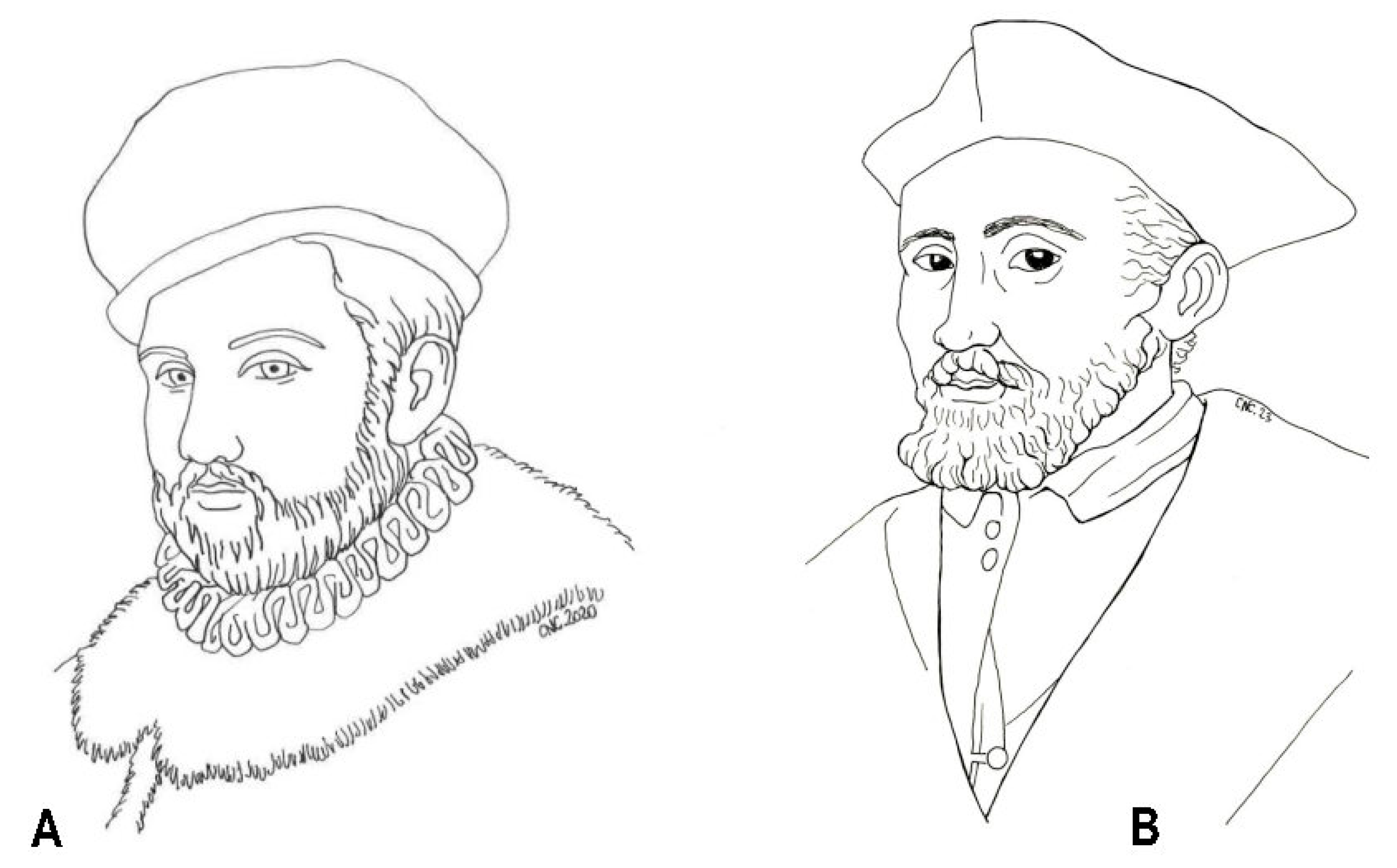
2. Life
3. Scientific Work
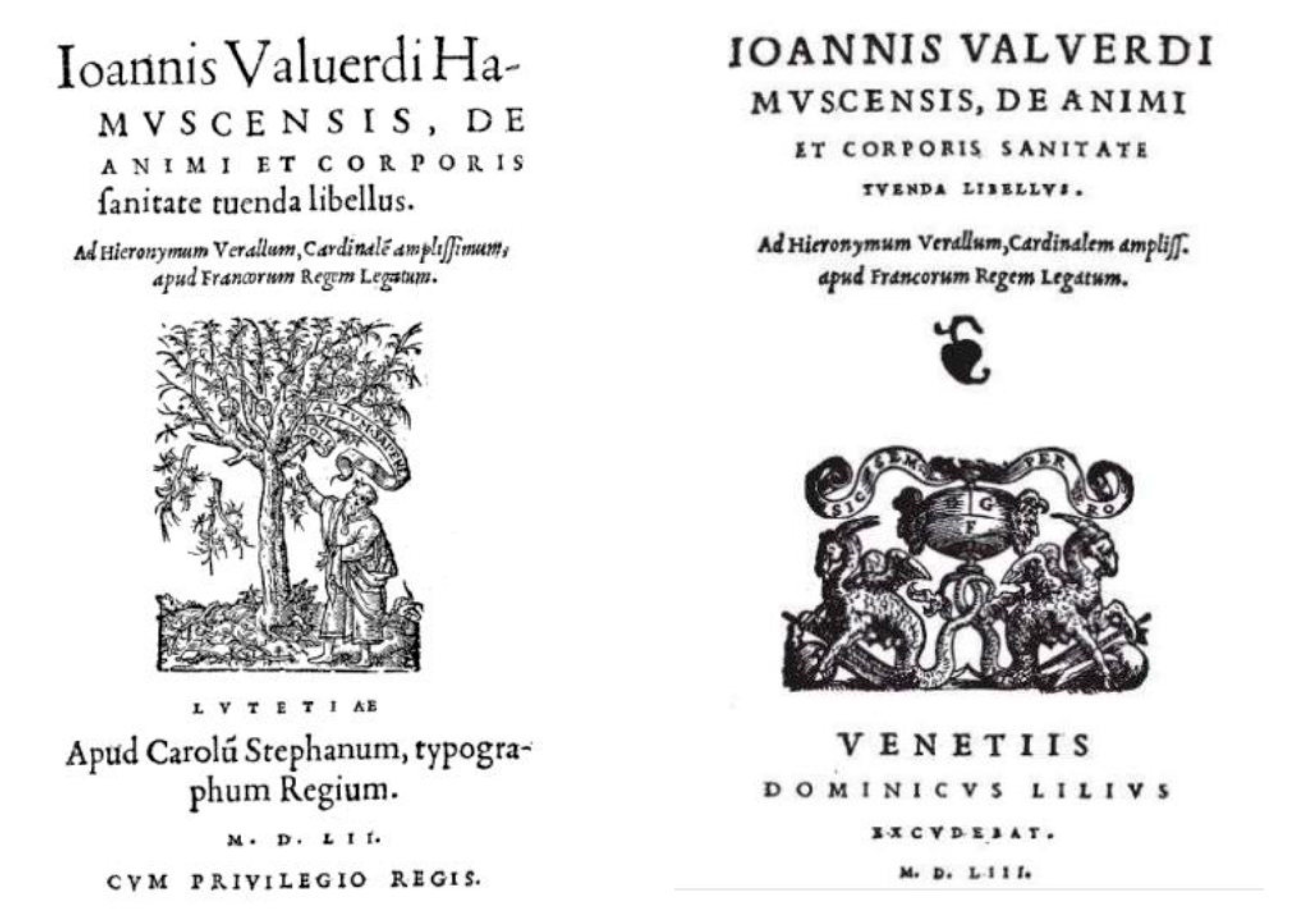
3.1. De animi et Corporis Sanitate Tuenda Libellus (“A Pamphlet on the Preservation of Mental and Physical Health”)
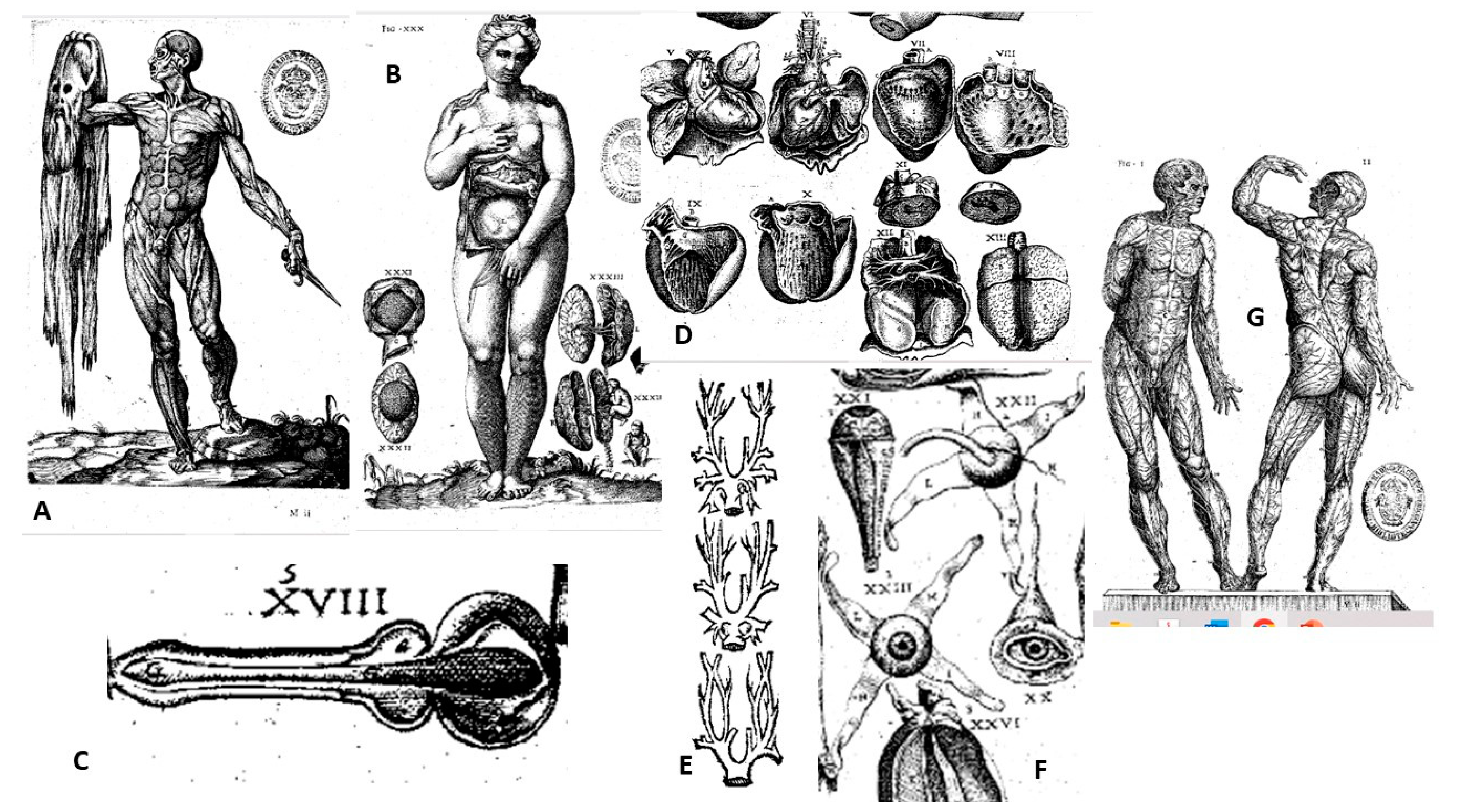
3.2. HISTORIA de la Composición del Cuerpo Humano (“HISTORY of the Composition of the Human Body”)
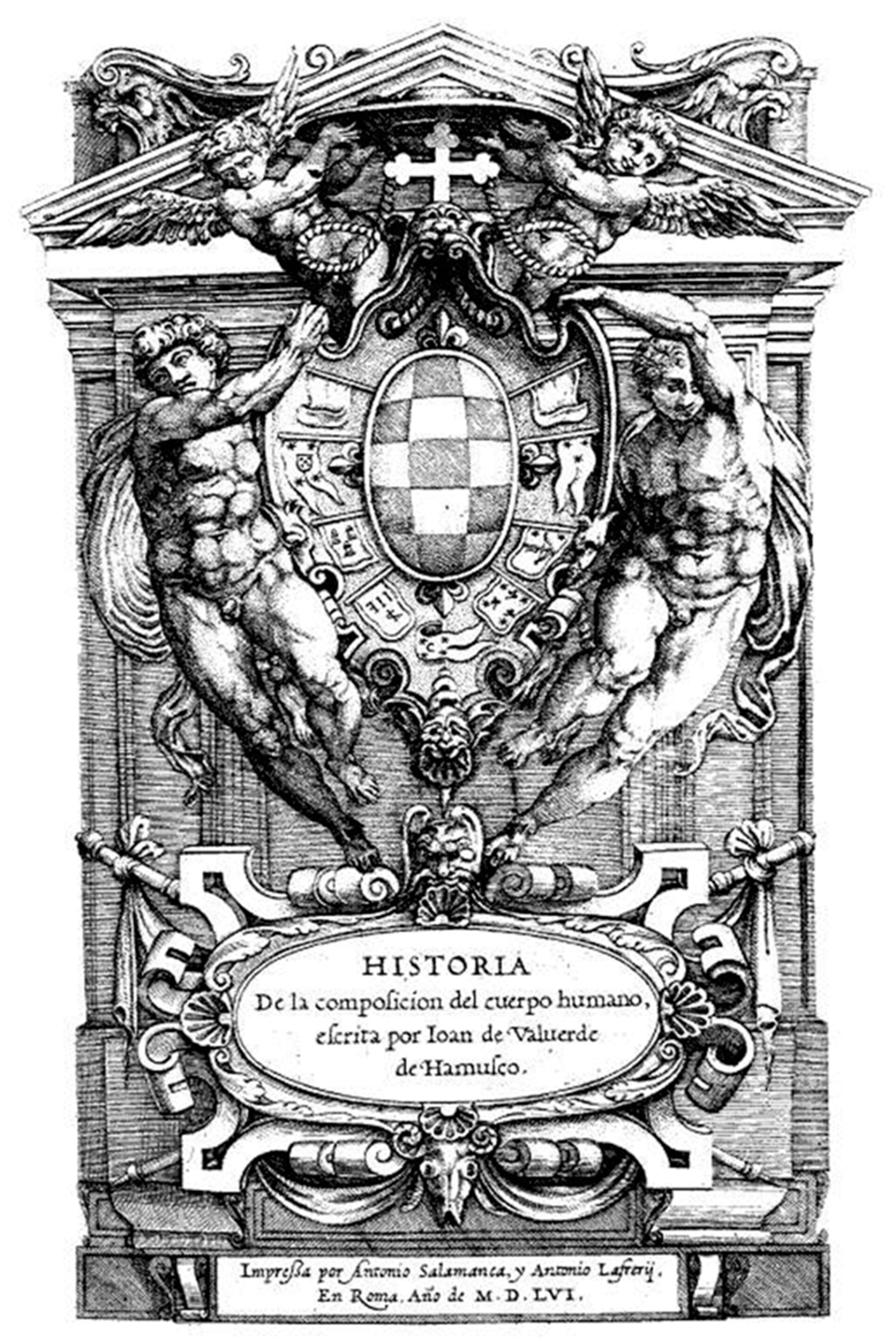
3.2.1. Princeps Edition
3.2.2. Diffusion of the Work
4. Reservations
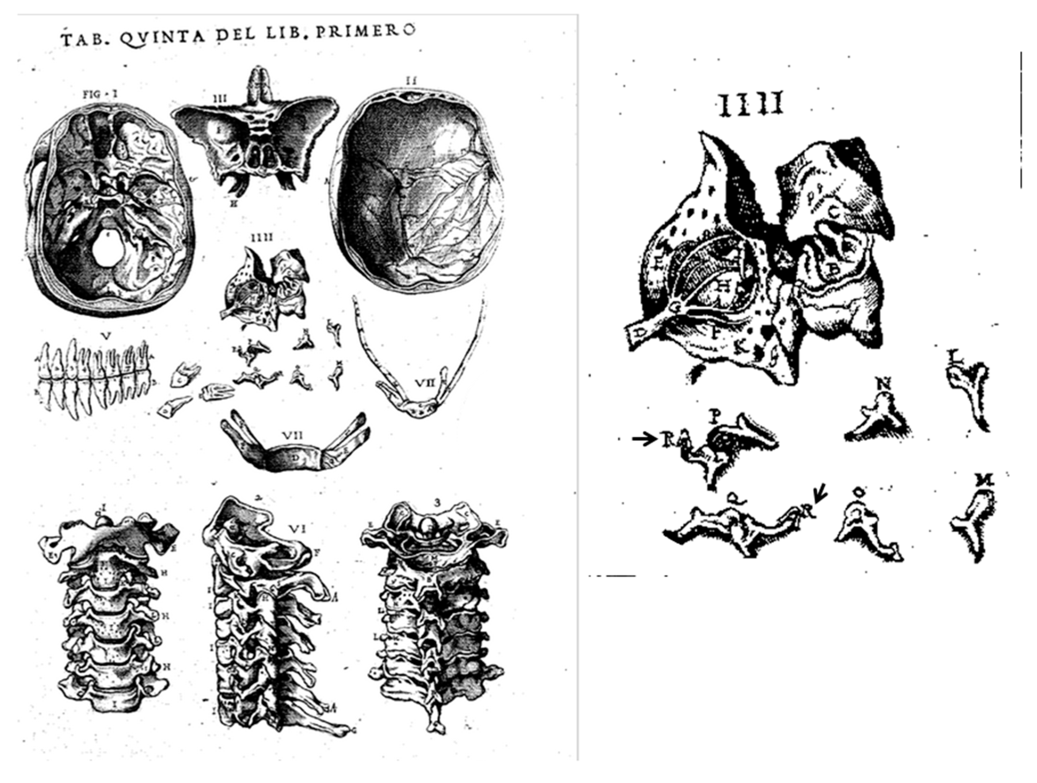
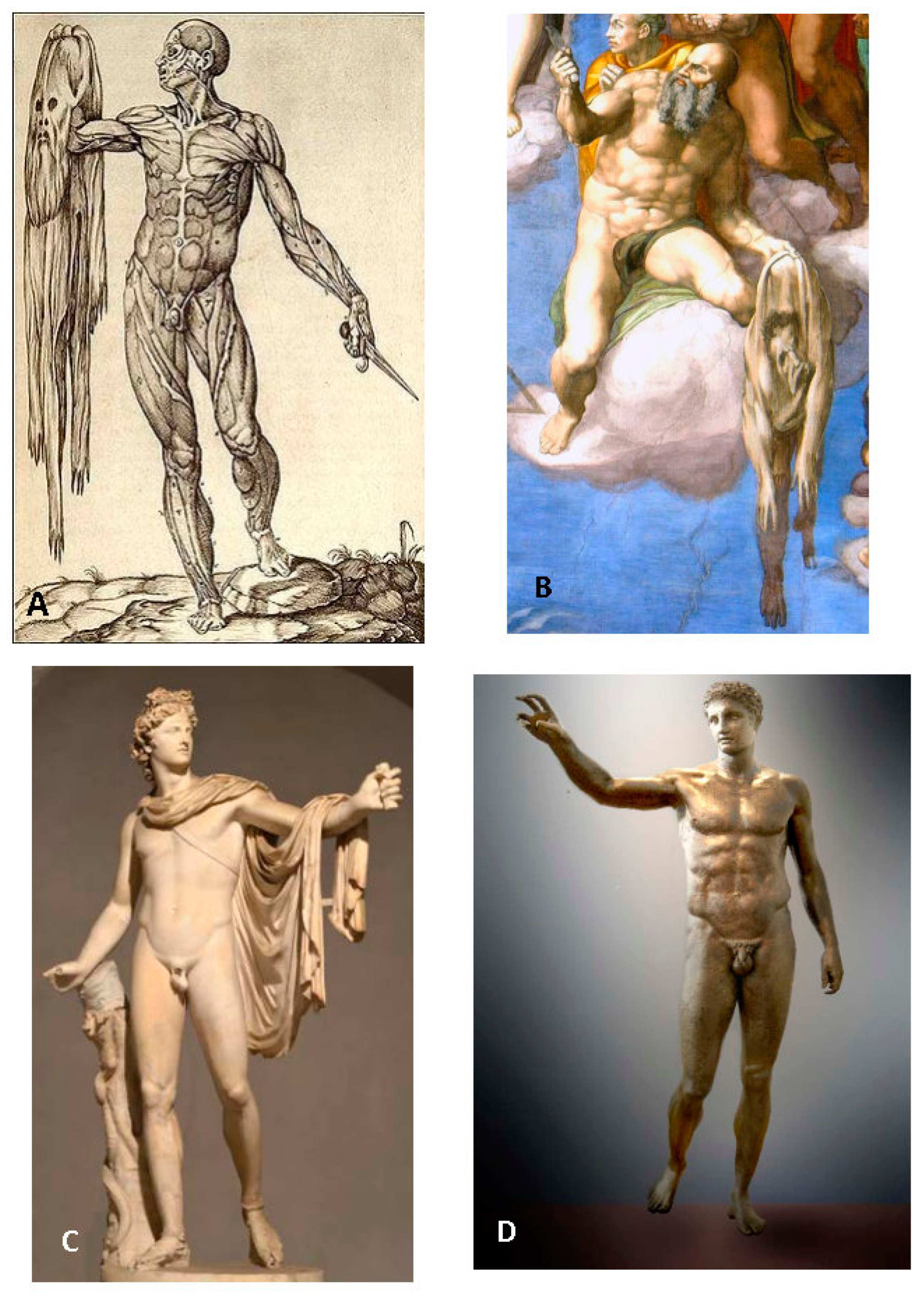
4.1. Illustrations
4.2. Artistic Authorship
4.3. Innovation or Plagiarism


5. Conclusions
6. Epilogue
Author Contributions
Funding
Institutional Review Board Statement
Informed Consent Statement
Data Availability Statement
Acknowledgments
Conflicts of Interest
List of Abbreviations (of Book Titles)
References
- Arráez-Aybar, L.; Bueno-López, J.; Raio, N. Toledo School of Translators and their influence on anatomical terminology. Ann. Anat. 2015, 198, 21–33. [Google Scholar] [CrossRef] [PubMed]
- Laín-Entralgo, P. Historia de la Medicina; Salvat Editores: Barcelona, Spain, 1978; Volume 4. [Google Scholar]
- Ghosh, S.K. Human cadaveric dissection: A historical account from ancient Greece to the modern era. Anat. Cell Biol. 2015, 48, 153–169. [Google Scholar] [CrossRef]
- O’Malley, C.D. Los saberes morfológicos en el renacimiento. La Anatomía. In Historia Universal de la Medicina; Laín-Entralgo, P., Ed.; Salvat Editores: Barcelona, Spain, 1972; pp. 43–77. [Google Scholar]
- Singer, C.J. Beginnings of Academic Practical Anatomy. In History and Bibliography of Anatomic Illustration; Choulant, L., Ed.; Hafner: Lawrence, MA, USA, 1952. [Google Scholar]
- Lassek, A.M. Human Dissection: Its Drama and Struggle; Ch. Thomas: Springfield, IL, USA, 1958; p. 74. [Google Scholar]
- Robinson, J.O. The barber-surgeons of London. Arch. Surg. 1984, 119, 1171–1175. [Google Scholar] [CrossRef]
- O’Malley, C.D. Andrés Laguna and his’ Anatomica methodus’. Physis 1963, 5, 65–69. [Google Scholar]
- Romero Reveron, R. Andreas Vesalius (1514–1564): Fundador de la Anatomía Humana Moderna. Int. J. Morphol. 2007, 25, 847–850. [Google Scholar] [CrossRef]
- Laín-Entralgo, P. From galen to magnetic resonance: History of medicine in Latin America. J. Med. Philos. 1996, 21, 571–591. [Google Scholar] [CrossRef] [PubMed]
- Andretta, E. Juan Valverde, or Building a “Spanish Anatomy” in 16th Century Rome. 2009, p. 13. Available online: http://hdl.handle.net/1814/12094 (accessed on 4 September 2023).
- Andretta, E. Rome Ville anatomique. In Roma Medica: Anatome d’un systéme médical au XVIe siècle; Andretta, E., Ed.; École française de Rome: Rome, Italy, 2011; pp. 499–557. [Google Scholar]
- Hernández-Morejón, A. Historia Bibliográfica de la Medicina Española; Viuda de Jordan e Hijos: Madrid, Spain, 1842–1852. [Google Scholar]
- Jimenez, F.A. The first autopsy in the New World. Bull. N. Y. Acad. Med. 1978, 54, 618–619. [Google Scholar]
- López-Piñero, J.M. Historia de la Medicina Universal; Ajuntament de València: :Valencia, Spain, 2010; p. 1840. [Google Scholar]
- Schulze-Schneider, I. La leyenda Negra de España: Propaganda en la Guerra de Flandes (1566–1584); Editorial Complutense: Madrid, Spain, 2009. [Google Scholar]
- Elliott, J.H. Imperial Spain 1469–1716; Penguin Books Limited: London, UK, 2002; p. 448. [Google Scholar]
- Arráez-Aybar, L.; Bueno-López, J. Antonio Gimbernat y Arbós: An anatomist-surgeon of the Enlightenment (in the 220th anniversary of his “A new method of operating the crural hernia”). Clin. Anat. 2013, 26, 800–809. [Google Scholar] [CrossRef]
- Riera-Palmero, J. Valverde y la anatomia del Renacimiento. In Historia de la Composición del Cuerp Humano. Juan Valverde de Hamusco (1501–1583); Riera-Palmero, J., Ed.; Universidad de Valladolid: Valladolid, Spain, 1981; p. 344. [Google Scholar]
- Riera-Climent, C.; Riera-Palmero, J. Biografía de Luis de Lucena. Llull Rev. Soc. Española Hist. Cienc. Técnicas 2005, 28, 551–562. [Google Scholar]
- Guerra, F. Juan Valverde de Amusco: Evidence of the identification of this portrait, claimed to be that of Vesalius, with a reapraisal of this work. Clio Méd. 1967, 2, 339–363. [Google Scholar]
- Ongaro, G. L’insegnamento clinico di Giovan Battista da Monte (1489–1551): Una revisione critica. Physis Riv. Internazionale Stor. Della Sci. 1994, 31, 357–369. [Google Scholar]
- Grendler, P.F. The Universities of the Italian Renaissance; The Johns Hopkins University Press: Baltimore, MD, USA, 2002; pp. 341–342. [Google Scholar]
- Tosoni, P. Della Anatomia degli Antichi e Della Scuola Anatomica Padovana, Memoria; Dalla Tipogr. del Seminario: Padova, Italy, 1844. [Google Scholar]
- Singer, C.J. To Vesalius on the fourth centenary of his De Humani Corporis Fabrica. J. Anat. 1943, 77 Pt 4, 261–265. [Google Scholar]
- Riera-Palmero, J. Juan Valverde de Amusco y la Medicina del Renacimiento; Universidad de Valladolid: Valladolid, Spain, 1986. [Google Scholar]
- Albarracín-Teulón, A. Los orígenes de la fisiología moderna. In Historia Universal de la Medicina; Laín-Entralgo, P., Ed.; Salvat Editores: Barcelona, Spain, 1973; pp. 78–85. [Google Scholar]
- Cunningham, A. The Anatomical Renaissance: The Resurrection of the Anatomical Projects of the Ancients; Scolar Press: London, UK, 1997; p. 292. [Google Scholar]
- Gratta, R.D.; Volpi, G.; Ruta, L. Acta Graduum Academiae Pisanae: 1543–1599; Gruppo di ricerca dell’Università di Pisa: Pisa, Italy, 1980; p. 6. [Google Scholar]
- Premuda, L. Storia dell’ Iconografia Anatomica; Ciba Edizioni: Milano, Italy, 1993. [Google Scholar]
- Ghosh, S.K. Evolution of illustrations in anatomy: A study from the classical period in Europe to modern times. Anat. Sci. Educ. 2015, 8, 175–188. [Google Scholar] [CrossRef]
- Hernández-Mansilla, J.M. Juan Valverde de Amusco 1525–1588 c.a. y la vocación por la anatomía en el Renacimiento hispanoitaliano. Med. Hist. 2015, 4, 22–34. [Google Scholar]
- Amelang, J.S. Exchanges between Italy and Spain: Culture and Religion. In Spain in Italy Politics, Society and Religion 1500–1700; Dandelet, T.J., Marino, J.A., Eds.; Brill: Leiden, Netherlands, 2007; p. 422. [Google Scholar]
- Hernández-Mansilla, J.M. La idea de Hombre en Juan Valverde Amusco. Ph.D. Thesis, Complutense University of Madrid, Madrid, Spain, 2014. [Google Scholar]
- Fernández-Ruiz, C. Historia de la Medicina Palentina. Publicaciones Inst. Tello Téllez Men. 1959, 20, 1–139. [Google Scholar]
- Barona-Vilar, J.L. Sobre Medicina y Filosofía Natural en el Renacimiento; Universitat de València: Valencia, Spain, 1993; Volume 3, p. 238. [Google Scholar]
- Castiglioni, A. Historia de la Medicina; Salvat Editores: Barcelona, Spain, 1941; p. 906. [Google Scholar]
- López-Piñero, J.M. Ciencia y Técnica en la Sociedad Española de los Siglos XVI y XVII; Editorial Labor: Barcelona, Spain, 1979; p. 511. [Google Scholar]
- Cátedra, P.M.; López-Vidriero, M.L.; Andrés-Escapa, P.; Domingo-Malvadi, A.C.L.; Rodríguez-Montederramo, J.L. «Juan de Valverde de Amusco, ‘Vivae imagines partium corporis humani…’», Fortuna de España. Textos españoles e imprenta europea (siglos XV-XVIII). Centro Virtual Cervantes. Available online: http://cvc.cervantes.es/obref/fortuna/expo/ciencia/cien022.htm (accessed on 4 September 2023).
- Valle-Inclán, C.d. El léxico anatómico de Bernardino Montaña de Monserrate y de Juan de Valverde. Asclepio 1949, 1, 121–188. [Google Scholar]
- Laín-Entralgo, P. El Cuerpo Humano: Oriente y Grecia Antigua; Biblioteca Virtual Miguel de Cervantes: Alicante, Spain, 2012. [Google Scholar]
- Hurst, J.W.; Fye, W.B. Realdo Colombo. Clin. Cardiol. 2002, 25, 135. [Google Scholar] [CrossRef]
- Sandoval-Gutiérrez, J.L. Nafis y Servet: Padres de la circulación pulmonar. Arch. Cardiol. México 2023, 93, 380. [Google Scholar] [CrossRef] [PubMed]
- Navarro, F.A. Servet. Rev. Española Cardiol. 2023, 76, 671. [Google Scholar] [CrossRef]
- Arráez-Aybar, L.; Navia-Álvarez, P.; Fuentes-Redondo, T.; Bueno-López, J. Thomas Willis, a pioneer in translational research in anatomy (on the 350th anniversary of Cerebri anatome). J. Anat. 2015, 226, 289–300. [Google Scholar] [CrossRef] [PubMed]
- López-Piñero, J.M. El Grabado en la Ciencia Hispánica; Editorial CSIC: Madrid, Spain, 1987; p. 140. [Google Scholar]
- Moreno Torres, A. Aproximación al Léxico de la Anatomía y de la Urología en Romance en el Siglo XVI; Universidad de Murcia: Murcia, Spain, 2000; p. 1632. [Google Scholar]
- García-Jáuregui, C. Aproximación al léxico anatómico del Renacimiento. Cuad. Inst. Hist. Leng. 2008, 1, 93–110. [Google Scholar] [CrossRef]
- López-Valverde, A.; de Diego, R.G.; De Vicente, J. Oral anatomy in the sixteenth century: Juan Valverde de Amusco. Br. Dent. J. 2013, 215, 141–143. [Google Scholar] [CrossRef]
- Alberti-López, L. La Anatomía y los Anatomistas Españoles del Renacimiento; CSIC: Madrid, Spain, 1948; pp. 81–130. [Google Scholar]
- López de Letona, C. El ojo en la historia de la composición del cuerpo humano (1556)(I). Arch. Soc. Española Oftalmol. 2005, 80, 117–118. [Google Scholar]
- Martín-Araguz, A.; Bustamante-Martínez, C.; Toledo-León, D.; López-Gómez, M.; Moreno-Martínez, J. La neuroanatomía de Juan Valverde de Amusco y la medicina renacentista española. Rev. Neurol. 2001, 32, 788–797. [Google Scholar] [CrossRef] [PubMed]
- Whitmore, I. Terminologia anatomica: New terminology for the new anatomist. Anat. Rec. (New Anat.) 1999, 257, 50–53. [Google Scholar] [CrossRef]
- Kachlik, D.; Baca, V.; Bozdechova, I.; Cech, P.; Musil, V. Anatomical terminology and nomenclature: Past, present and highlights. Surg. Radiol. Anat. 2008, 30, 459–466. [Google Scholar] [CrossRef] [PubMed]
- Romero-Reveròn, R. The first human anatomys book in Spanish: Libro de la anathomía del hombre written by Bernardino Montaña de Monserrate. Vesalius Acta Int. Hist. Med. 2019, 25, 8–18. [Google Scholar]
- Arráez-Aybar, L. Anatomy in the pages of Don Quixote. Interciencia 2006, 31, 690–694. [Google Scholar]
- Meyer, A.W.; Wirt, S.K. The Amuscan Illustrations. Bull. Hist. Med. 1943, 14, 667–687. [Google Scholar]
- Voet, L. The Golden Compasses; A history and evaluation of the printing and publishing activities of the Officina Plantiniana at Antwerp; Vangendt: Amsterdam, Netherlands, 1969–1972; Volume 2, p. 273. [Google Scholar]
- Voet, L.; Voet-Grisolle, J. The Plantin Press (1555–1589): A Bibliography of the Works Printed and Published by Christopher Plantin at Antwerp and Leiden; Van Hoeve: Amsterdam, Neterlands, 1980–1983; p. 3026. [Google Scholar]
- Van Hee, R.; Lowis, S. David van mauden (+/−1538 +/− 1597), “sworn medical doctor and surgical prelector of antwerp”, and his book on anatomy. Acta Chir. Belg. 2006, 106, 130–135. [Google Scholar] [CrossRef]
- Torres-Pérez, J.M.; González-Martín, R.; San-Julián-Arrupe, T. Anatome Corporis Humani. Universidad de Navarra. Available online: http://www.unav.es/biblioteca/fondoantiguo/hufaexp01/hufaexp01p01.html (accessed on 20 May 2016).
- Huard, P.; Imbault-Huart, M.-J. Andrés Vesalio: Iconografía Anatómica (Fabrica, Epitome, Tabula Sex); Laboratoros Beecham: Barcelona, Spain, 1983; p. 254. [Google Scholar]
- Post, C.R. A history of Spanish Painting; Harvard University Press: Cambridge, MA, USA, 1947; Volume 9, p. 931. [Google Scholar]
- Carducho, V. Dialogos de la Pintura sv Defensa, Origen, Esencia, Definicion, Modos y Diferencias; Francisco Martinez: Madrid, Spain, 1633. [Google Scholar]
- Barcia-Pavón, A.M.d. Catálogo de la Colección de Dibujos Originales de la Biblioteca Nacional de Madrid; Tipografía de la Revista de Arch., Bibli. y Museos: Madrid, Spain, 1906; p. 962. [Google Scholar]
- Martín-González, J. Precisiones sobre Gaspar Becerra. Arch. Español Arte 1969, 42, 327–356. [Google Scholar]
- Kleňhová, K. Juan Valverde de Hamusco v Kontextu Renesanční Filozofie a Medicíny; Západočeská Univerzita: Pilsen, Czech Republic, 2011. [Google Scholar]
- Barcia-Goyanes, J.J. El Mito de Vesalio; Universitat de València: Valencia, Spain, 1994; p. 214. [Google Scholar]
- Burggraeve, A. Histoire de l’Anatomie Physiologique, Pathologique et Philosophique: Avec un Exposé des Principales Découvertes de Cette Science Depuis Son Origine Jusqu’à Nos Jours; Ch. Chanteaud et Cie: Paris, France, 1880; p. 648. [Google Scholar]
- Singer, C.J. The Evolution of Anatomy: A Short History of Anatomical and Physiological Discovery to Harvey; Kegan Paul Trench Trubne: New York, NY, USA, 1925. [Google Scholar]
- Chinchilla-Piqueras, A. Anales Históricos de la Medicina en General, y Biográfico-Bibliográfico de la Española en Particular: 6 Vols. In Tribus Relig; Imprenta de López y Cía.: Valencia, Spain, 1841–1846. [Google Scholar]
- Eloy, N.F. Dictionnaire Historique de la Médecine Ancienne et Moderne. Ou Mémoires Disposés en Ordre Alphabétique Pour Servir à l’Histoire de Cette Science, et à Celle des Médecins, Anatomistes, Botanistes, Chirurgiens et Chymistes de Toutes Nations; Hoyois: Mons, Belgium, 1778; Volume 4, p. 626. [Google Scholar]
- Sprengel, K.P.J. Versuch Einer Pragmatischen Geschichte der Arzneykunde; Gebauerschen Buchhandlung: Halle, Germany, 1807; Volume 3, p. 62. [Google Scholar]
- O’Malley, C.D. Andreas Vesalius of Brussels, 1514–1564; University of California Press: Berkeley, CA, USA, 1964; p. 483. [Google Scholar]
- Berger, J.G.v. Physiologia Medica Sive De Natura Humana Liber Bipartitvs. Iterum in Lucem Prodit Cura Frider. Christiani Cregut….Cujus Dissertatio De Anthropologia Ejusque Praecipuis Tam Antiquis Quam Modernis Scriptoribus Introducionis Loco Praemittitur; Stock & Schilling: Francofurti (Frankfurt), Germany, 1737. [Google Scholar]
- Haller, A.v. Bibliotheca Anatomica; Orell, Gessner, Fussli & Co.: Zurich, Switzerland, 1774. [Google Scholar]
- Broussais, C. Atlas Historique et Bibliographique de la Médecine; ou, Histoire de la Médecine, Composée de Tableaux sur l’Histoire de l’Anatomie, de la Physiologie, de l’Hygiène, de la Chirurgie, de l’Obstétrique, de la Matière Médicale, de la Pharmacie, de la Médecine Légale, et de la Police Médicale, et de la Bibliographie, 2nd ed.; J.B. Baillière: Beauvais, France, 1834. [Google Scholar]
- Bahşi, İ.; Bahşi, A. “Teşrih-ül Ebdan ve Tercümânı Kıbale-i Feylesûfan”: The first illustrated anatomy handwritten textbook in Ottoman-Turkish medicine. Surg. Radiol. Anat. 2019, 41, 1135–1146. [Google Scholar] [CrossRef] [PubMed]
- Skandalakis, J.E.; Mirilas, P. Plagiarism. Arch. Surg. 2004, 139, 1022–1024. [Google Scholar] [CrossRef] [PubMed]
- Zimmerman, L. Surgery. In Medicine in Seventeenth Century England: A Symposium Held at UCLA in Honor of CD O’Malley; Debus, A.G., Ed.; University of California Press: Berkeley, CA, USA, 1974; pp. 46–69. [Google Scholar]
- Persaud, T.V. A history of Anatomy: The Post-Vesalian Era; Charles C. Thomas Publisher: New York, NY, USA, 1997. [Google Scholar]
- Skaarup, B.O. Anatomy and Anatomists in Early Modern Spain; Ashgate Publishing, Ltd.: Farnham, UK, 2015; p. 306. [Google Scholar]
- Kemp, M. Style and non-style in anatomical illustration: From Renaissance Humanism to Henry Gray. J. Anat. 2010, 216, 192–208. [Google Scholar] [CrossRef] [PubMed]
- Barcia-Goyanes, J.J.; Evans, N.R. Notes on the historical vocabulary of neuroanatomy. Hist. Psychiatry 1995, 6 Pt 4, 471–482. [Google Scholar] [CrossRef]
- Booker, L.D.; Bontis, N.; Serenko, A. The relevance of knowledge management and intellectual capital research. Knowl. Process Manag. 2008, 15, 235–246. [Google Scholar] [CrossRef]
- Parchman, M. Diffusion, Dissemination and Implementation: What Is the Difference. Available online: https://dcricollab.dcri.duke.edu/sites/NIHKR/KR/GR-Slides-10-09-15.pdf (accessed on 4 September 2023).


| Year | Author | Title (Place of Publication: Publisher) |
|---|---|---|
| 1535 | Andrés Laguna (c. 1510–c. 1559) | Anatomica methodus seu de sectioni humani corporis contemplatio (París: Ludouicum Cyaneum) |
| 1542 | Luis Lobera de Ávila (c. 1480–c. 1551) | Libro de Anatomía, es primera parte de “Remedio de cuerpos humanos y silva de experiencias y otras cosas utilísimas” (Alcalá de Henares: Juan Brocar) |
| 1549 | Pedro Jimeno (c. 1515–c. 1551) | Dialogus de re medica, compendiaria ratione, praeter quaedam alia, universam anatomem humani corporis perstringens (Valencia: Juan Mey) |
| 1551 | Bernardino Montaña de Monserrate (c. 1480–c. 1558) | Libro de la Anathomía del hombre (Valladolid: Sebastián Martínez) |
| 1555 | Luis Collado (c. 1520–c. 1589) | Cl. Galeni Pergameni Liber de Ossibus ad tyrones… enarrationibus illustratus (Valencia: Juan Mey) |
| 1556 | Juan Valverde de Amusco (c. 1525–c. 1587) | HISTORIA de la composición del cuerpo humano (Roma: Antonio Martínez de Salamanca y Antoine Lafréry) |
| 1559 | Alfonso Rodríguez de Guevara (c. 1520–c. 1587) | In pluribus ex iis quibus Galenus impugnatur ab Andrea Vesalio Bruxelensi in de constructione et usu partium corporis humani, defensio: et nonnullorum quae in anatome deficero videbantur supplementum. (Coimbra: Juan Barreiro) |
| Year | Language | Title | Print House/Publisher | Place |
|---|---|---|---|---|
| 1556 *1 | Spanish | HISTORIA de la composición del cuerpo humano | A. Martínez de Salamanca and A. Lafréry | Rome |
| 1559 *2 | Italian | Anatomia del corpo umano | Nicolò Bevilacqua | Venice |
| 1560 | “ | “ | Giunta | “ |
| 1586 | “ | “ | “ | “ |
| 1596 | “ | “ | “ | “ |
| 1606 | “ | “ | “ | “ |
| 1607 | “ | “ | “ | “ |
| 1608 | “ | “ | “ | “ |
| 1657 | “ | “ | “ | “ |
| 1682 | “ | “ | Giunta/Niccolò Pezzana | “ |
| 1589 *3 | Latin | Anatome corporis humani | Michele Colombo | Venice |
| 1607 | “ | “ | “ | “ |
| 1566 *4 | Latin | Viuæ imágenes partium corporis humaniæreis formis expressæ | Christophe Plantin | Antwerp |
| 1572 | “ | “ | “ | |
| 1579 | “ | “ | “ | |
| 1568 | Dutch | Anatomie, oft levende beelden vande deelen des menschelicken lichaems: met de verclaringhe van dien, inde Neder-duytsche spraecke | Christophe Plantin | Antwerp |
| 1583 | “ | “ | “ | |
| 1583 | Dutch | Bedieninghe der anatomien | David van Mauden | Antwerp |
| 1646 | “ | “ | “ | “ |
| Book | Valverde’s “HISTORIA…” | Vesalius’ “…Fabrica…” |
|---|---|---|
| First | Huesos y ternillas (bones and cartilages) | Bones and joints |
| Second | Ligamentos y músculos (ligaments and muscles) | Ligaments, muscles, and integumentum |
| Third | Miembros de la digestión y la generación (digestive and reproductive organs) | Veins, arteries, and glands |
| Fourth | Miembros de la vida (life members (lungs and heart)) | Nerves and spinal cord |
| Fifth | Miembros necesarios al movimiento y sentido (members needed to sense and movement; encephalon) | Organs of nutrition and generation |
| Sixth | Venas y arterias (veins and arteries) | Heart and associated organs |
| Seventh | Nervios (cranial and spinal nerves) | Encephalon |
Disclaimer/Publisher’s Note: The statements, opinions and data contained in all publications are solely those of the individual author(s) and contributor(s) and not of MDPI and/or the editor(s). MDPI and/or the editor(s) disclaim responsibility for any injury to people or property resulting from any ideas, methods, instructions or products referred to in the content. |
© 2023 by the authors. Licensee MDPI, Basel, Switzerland. This article is an open access article distributed under the terms and conditions of the Creative Commons Attribution (CC BY) license (https://creativecommons.org/licenses/by/4.0/).
Share and Cite
Arráez-Aybar, L.-A.; Reblet, C.; Bueno-López, J.L. Juan Valverde de Amusco: Pioneering the Transfer of Post-Vesalian Anatomy. Anatomia 2023, 2, 450-471. https://doi.org/10.3390/anatomia2040033
Arráez-Aybar L-A, Reblet C, Bueno-López JL. Juan Valverde de Amusco: Pioneering the Transfer of Post-Vesalian Anatomy. Anatomia. 2023; 2(4):450-471. https://doi.org/10.3390/anatomia2040033
Chicago/Turabian StyleArráez-Aybar, Luis-Alfonso, Concepción Reblet, and José Luis Bueno-López. 2023. "Juan Valverde de Amusco: Pioneering the Transfer of Post-Vesalian Anatomy" Anatomia 2, no. 4: 450-471. https://doi.org/10.3390/anatomia2040033