Article

The Prevalence of Leptospira Serovars in African Giant Pouched Rats (Cricetomys spp.) from the Ngorongoro Conservation Area, Tanzania

by
Prisca N. Kahangwa
1,2,3,*,
Amani S. Kitegile
2,
Robert S. Machang’u
4,
Ginethon G. Mhamphi
1,2,5 and
Abdul S. Katakweba
1,5
1
Africa Centre of Excellence for Innovative Rodent Pest Management and Biosensor Technology Development (ACE IRPM & BTD), Sokoine University of Agriculture, Morogoro P.O. Box 3110, Tanzania
2
Department of Wildlife Management, Sokoine University of Agriculture, Morogoro P.O. Box 3073, Tanzania
3
Department of Marketing, University of Dar es Salaam Business School, Dar es Salaam P.O. Box 35046, Tanzania
4
Department of Microbiology, Parasitology and Biotechnology, St. Francis University College of Health and Allied Sciences, Ifakara P.O. Box 175, Tanzania
5
Institute of Pest Management, Sokoine University of Agriculture, Morogoro P.O. Box 3110, Tanzania
*
Author to whom correspondence should be addressed.
Zoonotic Dis. 2024, 4(1), 37-48; https://doi.org/10.3390/zoonoticdis4010005
Submission received: 13 October 2023 / Revised: 20 November 2023 / Accepted: 22 November 2023 / Published: 23 January 2024

Abstract

:

Simple Summary

This study has investigated the presence of Leptospira bacteria in urine, kidney, and blood samples from African giant pouched rats (Cricetomys spp.) in the Ngorongoro Conservation Area (NCA), Tanzania. The giant rat is an important maintenance host of leptospires; understanding their abundance and distribution in this environment is important to help prevent the transmission of the bacteria from rodents to other animals and humans co-existing in the area. This study demonstrates that Cricetomys spp. in the NCA are infected with the spirochete, and are hence a potential threat to public health. This study calls for an enhanced awareness of the presence of Leptospira spp. in Cricetomys spp. and calls for a one-health approach to protect both humans and animals from the pathogens.

Abstract

Leptospirosis, also known as Weil’s disease, is a febrile tropical disease of humans and diverse animals. The maintenance hosts of the infectious pathogen, Leptospira spp., are primarily rodents, while other warm-blooded animals and some reptiles are secondary or transient hosts of this pathogen. African giant pouched rats (Cricetomys spp.) have been identified to be important maintenance hosts of pathogenic leptospires in the tropical and subtropical regions of the world. This study assessed the seroprevalence of Leptospira spp. in the African giant pouched rats of the Ngorongoro Conservation Area (NCA), Tanzania, where there is close human, domestic animal, and wildlife interaction. A total of 50 African giant pouched rats were sampled between July 2020 and December 2021. Blood sera were screened for specific leptospiral antibodies using a microscopic agglutination test (MAT), while urine and kidney tissues were examined for the pathogen and pathogen-specific genes using cultures and polymerase chain reactions (PCR), respectively. The pathogen detection varied from 0% in cultures to 6% via the MAT and 20% via PCR. The Fisher exact test was applied to compare positive cases detected through the diagnostic tests, and showed a significant difference in the indirect and direct detection of Leptospira serovars via the MAT and PCR. We conclude that pathogenic Leptospira serovar are found in the NCA and recommend that the NCA authority raises awareness of the existence of the Leptospira serovar in giant African pouched rats, and possibly other rodents. The NCA should initiate appropriate management strategies, including the guided disposal of household garbage, which is the major attractant of rodents to residential areas. Where necessary, the NCA should carry out limited rodent control and periodic monitoring of the pathogen carrier (rodent) populations.

Graphical Abstract

1. Introduction

Leptospirosis, also known as Weil’s disease, is a zoonotic disease with significant morbidity and high mortality rates whenever outbreaks occur. The average annual morbidity and mortality rates worldwide are estimated to be 14.77 and 0.84 cases per 100,000, respectively [1]. Leptospirosis is a febrile neglected tropical zoonotic disease with worldwide distribution in diverse mammalian hosts, including rodents, shrews, cattle, and wildlife [2,3,4]. Maintenance hosts are typically asymptomatic; however, accidental hosts, such as humans, may experience a variety of clinical manifestations, including serious pulmonary hemorrhage, hepatic and renal failure, and even death [5]. Rodents are the primary reservoir hosts for various Leptospira serovars, including pathogenic strains. They maintain and spread leptospires in the environment, which can lead to human and other animal infections through contact with contaminated soil, water, and food sources. Several rodent species, such as Rattus spp., Mus musculus, Bandicota bengalensis, Bandicota indica, and Cricetomys spp., are known to carry different pathogenic leptospiral serovars and exhibit host specificity for certain serovars, such as Icterohaemorrhagiae, Copenhageni, Ballum, Lora, Qunjian, and Australis, among others. Mus musculus, Rattus spp., and Bandicota spp. have been identified as hosts to several Leptospira spp. strains, including L. borgpetersenii, L. kirschneri, and L. interrogans, in research conducted in many parts of the world, like the US, Malaysia, Bangladesh, and Australia [6,7,8,9]. L. borgpetersenii has been detected in Mus musculus and L. interrogans in Rattus spp. in Angola [10]. L. interrogans and L. kirschneri are carried by Mus populations in Kenya [11]. Cricetomys spp. carry different Leptospira spp. strains such as L. interrogans serogroup Ballum and serogroup Australis in Tanzania [12,13,14]. Leptospira serovar colonize the renal tubules and urogenital tracts of the host; from there, they are excreted with urine into the environment [15]. Leptospira can stay for several months in wet conditions [16]. Contaminated water bodies and soils are, therefore, major sources of Leptospira bacteria infecting humans and susceptible animals [17]. The disease manifests asymptomatically in maintenance hosts; however, accidental hosts, such as humans, may experience a variety of clinical manifestations, including serious pulmonary hemorrhage, hepatic and renal failure, and even death [18]. Diagnostic tools that are commonly employed for leptospirosis include the microscopic agglutination test (MAT), considered the reference serological standard assay, and the enzyme-linked immunosorbent assay (ELISA) [19]. The MAT is the reference method for serological diagnosis, and is primarily used in epidemiological studies, as it can identify the most prevalent leptospiral serovars or serogroups [20]. While cross-reactivity can occur, the MAT provides valuable information on the epidemiology of leptospires. The test detects both IgM and IgG antibodies, making it useful for epidemiological studies [21].
Polymerase chain reactions (PCRs) are the standard molecular diagnostic tests for leptospirosis. They are favored for their speed, reliability, and ability to detect early infections and distinguish between pathogenic and saprophytic leptospiral serovars [22].
Cricetomys spp., commonly known as giant rats, are acknowledged as essential maintenance and reservoir hosts of pathogenic leptospires due to their burrowing behavior and preference for domestic garbage dump sites. They may carry and shed various leptospiral serovars, contributing to the environmental contamination of habitats shared with other mammals, including humans, domestic animals, and wildlife [2,23,24]. According to recent studies [25,26], most species of the genus Leptospira—family Leptospiraceae—are saprophytic. Habitats that humans and domestic animals share with rodents for food and shelter, particularly those that are close to human habitation, improper waste management sites, and wet environments, have been identified as significant sources of Leptospira infection for humans and domestic animals [27]. Several studies on Leptospira seropositivity in rodents have been carried out in many districts in Tanzania, including Katavi, Morogoro, Kilimanjaro, and Tanga [13,14,28,29]. A study in central Tanzania recommended the investigation of the effect of climatic change on rodent populations known to be Leptospira carriers [17]. The prevalence of Leptospira in important maintenance hosts has, however, been under-studied in areas such as the NCA, which is one of the world’s most iconic natural conservations with a unique, unrestricted interaction between humans, livestock and wildlife. Habitats that humans and domestic animals share with rodents for food and shelter, particularly close to human habitation, improper waste management sites, and wet environments, have been identified as significant sources of Leptospira infection for humans and domestic animals [30].
Therefore, understanding the presence of the Leptospira serovars circulating in their natural reservoir hosts in the NCA is important for creating awareness of the leptospires pathogen that will help to minimize the risks of transmission among the animals and humans living in the area. Furthermore, investigating leptospira serovars in Cricetomys spp. is crucial for understanding the prevalence of leptospira serovars among the natural and incidental reservoirs.
In this study, we report the presence and distribution of the African giant pouched rat in the NCA, and the prevalent pathogenic leptospiral serovars in these rats. The study hypothesizes that leptospiral serovars are prevalent in Cricetomys spp. in the NCA forests and around human settlements, including tourist hotels. To support this hypothesis, researchers collected blood, urine, and kidney tissue samples from Cricetomys spp. for leptospiral isolation, using culture and serology by microscopic agglutination test (MAT). In addition, DNA from the kidney tissues was examined for leptospira-specific genes using polymerase chain reaction (PCR). Understanding the presence of Leptospira in these rodents is not only essential for their conservation but also for understanding the risk of disease transmission from rodents to other animals that coexist within the NCA, as it has been known that this area is shared between wildlife, livestock, and humans.

2. Materials and Methods

2.1. Study Sites

This study was carried out in Ngorongoro Conservation Area (NCA), located in the Arusha region, northern Tanzania, situated between longitude 34°52′ and 35°58′ E and latitude 2°30′ and 3°38′ S. The NCA is a unique environment with a multiple land-use system that allows wildlife conservation, community farming, the grazing of livestock by pastoralists and tourism [31,32]. The conservation area has moist and misty conditions, with annual temperatures ranging from 35 °C in February to 2 °C in June/July [33]. Rainfall is seasonal and variable, ranging from 400 to 600 mm/year in the arid lowland plains in the west, while in the Eastern Highland forested areas, rainfall varies from 1000 to 1200 mm/year [34]. The NCA has a bimodal seasonal variability with two wet and two dry seasons. The wet periods are from October to December and March to May. A short dry season extends from January to February while a long dry period is between June and September [35].

2.2. Rodent Trapping and Sampling

Purposive convenient sampling was carried out in which rodents were sampled at the time of capture [36]. Traps were set around the human settlement and in four selected vegetated areas (forest, woodland, bushland, and grassland). These sites were selected as they were suitable for rodent harborage. Five line transects of 100 m each were created. For each transect 10 traps (60 × 15 × 170 cm) (Havahart® collapsible cage trap, Easy Technology Solutions Limited, Auckland, New Zealand) were set 10 m apart, making a total of 50 traps for each of the four types of vegetation [37]. The transects were set parallel to each other and the distance from one transect to another was 25 m [38]. A total of 50 traps were randomly set around human settlements (staff homes and tourist lodges) [39]. Each trap was baited with a mixture of avocado, ripe bananas, and green maize, left overnight, and inspected once daily every following morning before 08:00 h. The trapping was carried out between July 2020 and December 2021 for six sessions at each site, each session lasting 10 consecutive nights.
The captured rodents were humanely killed by excess inhalation of halothane-soaked cotton wool. Immediately after death, cardiac puncture was performed after treating the ventral part of the animals with 70% ethanol. Blood was placed in 2.5 mL microcentrifuge tubes for serum preparation. The collected blood was allowed to clot, and thereafter centrifuged at 4000 rpm for 10 min. The obtained serum was stored at −20 °C before the MAT test. After blood collection, each rodent was aseptically dissected using sterilized scissors and forceps then urine from the urinary bladder was collected using a sterile syringe and needle. Thereafter, the kidney was aseptically sectioned and a piece was placed into a sterile 5 mL glass tube containing 200 µL of phosphate-buffered saline for culture. A duplicate piece was placed into a microtube of 2.5 mL containing absolute ethanol for molecular analysis.

2.3. Detection of Leptospiral Antibodies by Microscopic Agglutination Test

The MAT, which is the serological gold standard assay in leptospirosis diagnosis, was used to detect antibodies against Leptospira serovar in the Cricetomys spp. serum [40]. Briefly, live leptospiral antigens of serovars Sokoine, Hebdomadis, Grippotyphosa, Lora, and Pomona were used. These serovars were selected based on previous studies performed to identify the circulating serovars in the northern part of Tanzania [28,41]. Sokoine has been isolated from cattle and rodents, Hebdomanis has been found in cattle, Grippotyphosa has been isolated from cattle, Lora has been isolated from rodents and Pomona has been isolated in cattle and buffaloes [14,17,42]. Hebdomanis, Grippotyphosa, and Pomona are found in cattle, and they have been included to check their availability in Cricetomys spp. Since in NCA, livestock, wildlife and humans interact closely, there is a possibility of transmission of Leptospira serovars.
The antigens were prepared in Ellinghausen and McCullough–Johnson and Harris (EMJH) [2] culture medium and checked for purity under a dark field microscope. Antigens with a density of 300 × 108 leptospires/mL and aged no more than 10 days were used in the test. The test was conducted by initially diluting 10 µL of serum with 90 µL of phosphate-buffered saline (PBS), pH 7.0, in a microtiter plate, followed by a serial doubling dilution into dilutions of 1:10, 1:20 1:40, and 1:80. Then, 50 µL of each antigen was added to the wells containing the diluted serum samples to achieve final dilutions of 1:20, 1:40, 1:80, and 1:160. For each serovar, a positive and negative control was set. The contents of the plates were homogenized briefly by manual shaking and then incubated at 30 °C for 2 h. The observation was performed under a dark field microscope (ZEISS, Cambridge, UK) at ×20 magnification. The agglutination titers were then evaluated and the cut-off titers were established visually as the point (titer) at which at least 50% of the leptospires exhibited agglutination compared to the negative control.

2.4. Molecular Characterization of Leptospira Serovars

2.4.1. DNA Extraction

DNA extraction of 50 kidney tissue samples was undertaken following the manufacturer’s instructions (Zymo research kit—Quick-DNA Miniprep Plus Kit, Zymo Research Corporation, Irvine, CA, USA). Samples were prepared prior to extraction by exposing them to air (while still in cryovials to prevent contamination) to thaw. This was followed by rinsing with double-distilled water to ensure that no preservatives remained that could inhibit the action of reagents during extraction. After extraction, the DNA was introduced to a nanodrop spectrophotometer for quantification and then stored at −20 °C [3].

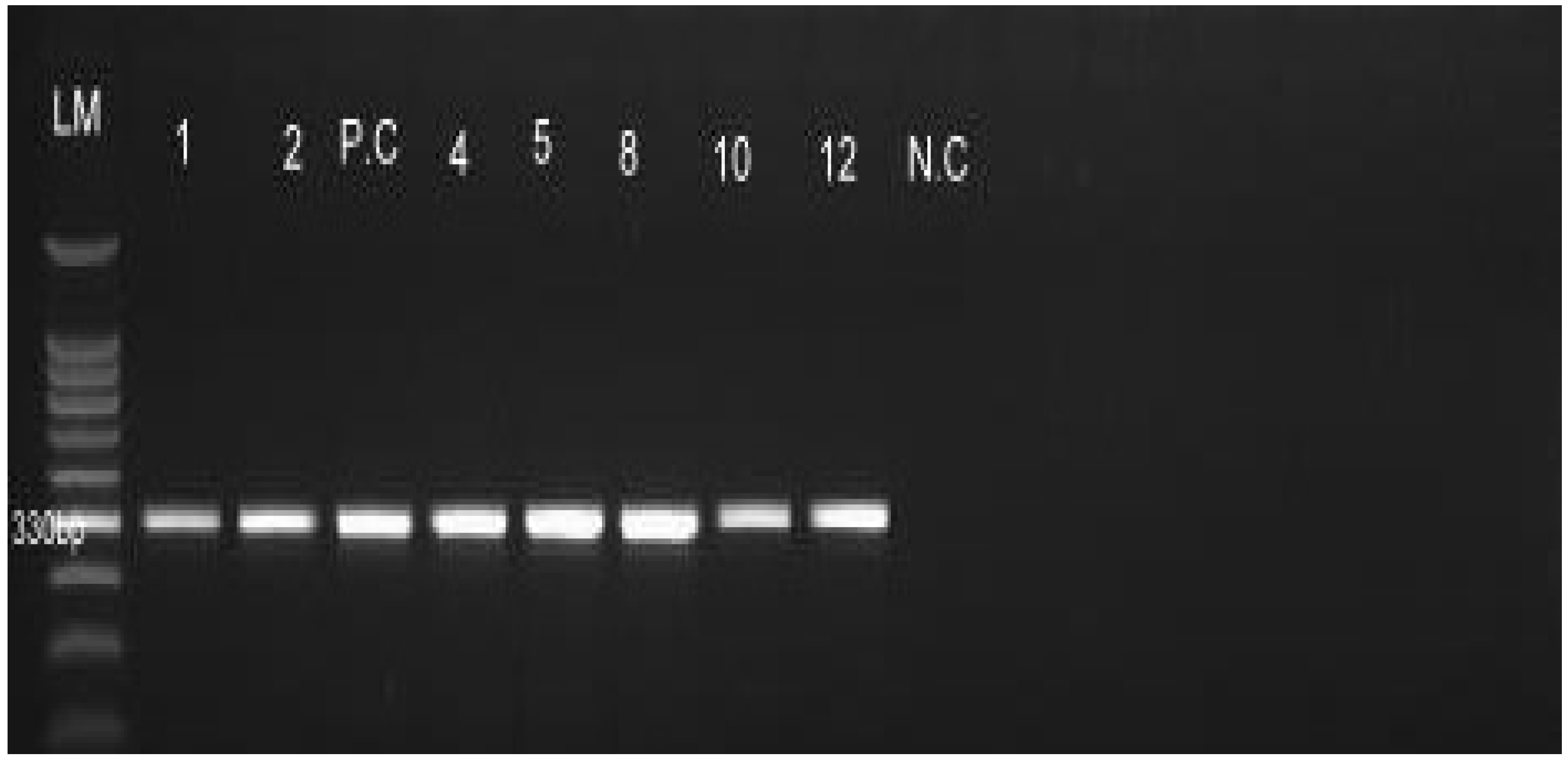
2.4.2. PCR Amplification, Gel Electrophoresis and Visualization

A 25 ul reaction mixture was prepared, consisting of 12.5 µL of Green Master mix (New England Biolabs, Ipswich, MA, USA), 0.5 µL (10 µM) each of Lepat 1 (5′-GAG-TCT-GGG-ATA-ACT-TT-3′) and Lepat 2 (5′-TCA-CAT-CG(YT)-GCT-TAT-TTT-3′); 8.5 µL of nuclease-free water and 3 µL of DNA template [43]. PCR amplification involved specific primers targeting a 330 bp region of the 16 S rRNA gene (rrs) that was subjected to initial denaturation of the DNA at 93 °C for 3 mn, followed by 35 PCR cycles; final denaturation at 93 °C for 1 mn and primer annealing at 48 °C for 1 mn, followed by initial elongation at 72 °C for 1 mn and final elongation at 72 °C for 10 mn [2]. The PCR products were then loaded on 1.5% agarose gel, stained with GRGreen Nucleic Acid Stain (Excellgen, Inc., Rockville, MA, USA), and visualized on a Gel DocTM (Bio-Rad, Hercules, CA, USA) [44].

2.5. Culture

Urine and kidney homogenates were used for culturing, in which two drops of urine were placed in a 10 mL tube containing a selective growth inhibitor and 5 mL Fletcher medium (200 µg/mL) of 5-Fluorouracil, and then incubated at 30 °C. A piece of kidney tissue, previously placed into tubes containing 1 mL of PBS, was then immediately aseptically crushed using a sterile glass Pasteur pipette to obtain a homogenate. Then, after this, two drops of the homogenates were inoculated into a 10 mL tube containing Fletcher’s medium as was undertaken for the urine (above) within 10 min, then they were incubated together at 30 °C for a minimum of 8 weeks while being examined weekly for leptospires growth by dark field microscopy [45,46].

2.6. Data Analysis

Microsoft Office Excel® 2016 was used for storing data and IBM SPSS version 26 (2019) was used for descriptive and inferential analyses [47]. The Fisher exact test was used to associate seroprevalence with trapping site and season. The confidence interval was set at a 95% limit and a p-value less than 0.05 was considered statistically significant [48].

3. Results

3.1. Abundance of Cricetomys spp. in Selected Sites Ngorongoro Conservation Area

A total of 50 Cricetomys spp. was captured, of which 49 were from near human settlements and one was found in the densely forested area TrS4 (Table 1, Figure 1). No captures were made in the woodland (TrS 2), bushland (TrS 3), or grassland (TrS 1). There was a significant difference in the numbers of captures between male and female Cricetomys spp., with male abundance (M = 4.71, S.D = 0.89) being greater than female (M = 2.43, S.D = 0.81) (t(6) = 3.77, p = 0.009) (Table 1).

3.2. Detection of Leptospira Serovar

The detection of Leptospira in samples by MAT and PCR, and in cultures of urine and kidney tissues, varied broadly between the three techniques. Via MAT, 3 of the 50 samples (6%) tested positive (Table 2), of which 2 were positive for Lora (L. interrorgans serogroup Australis) and 1 for Sokoine (L. kirschneri serogroup Icterohaemorrhagiae). Via PCR, 10 out of the 50 (20%) kidney tissue samples were found to be positive for leptospiral genes (Figure 2). One sample showed positivity for either MAT or PCR. There was a significant difference between positive and negative cases between the methods (Fisher exact test, p < 0.05). No Leptospira serovar were detected via culturing from the kidney tissue or urine samples—5 culture samples out of 100 (5%) were contaminated and hence lost during the procedure.

3.3. Distribution of Leptospira Serovars in Cricetomys spp.

The detection of Leptospira varied with the site of capture. All leptospira-positive Cricetomys spp. were captured near human settlements, including the tourist lodge (Table 3). Of the areas where Leptospira serovar were detected, three sites—Pongo, Kongoni, and Ngorongoro Wildlife Lodge—showed relatively high detections, while Viewpoint had one positive sample (Table 3). There was no significant difference in seroprevalence between the sites where Leptospira spp. was detected (Fisher exact test, p = 0.57). Sites that were littered with garbage had a higher seroprevalence of Leptospira spp. than the other sites (Table 3). Of the sites, Lerai was closest to a water pump, while the Lemala and Forest sites were close to streams. Both Lemala and Forest captures had no Leptospira detected in them, and had no dump sites in their vicinity.

3.4. Influence of Season on Seroprevalence of Leptospira spp. in Cricetomys Rats in the NCA

There was no significant difference in the seroprevalence of Leptospira spp. in Cricetomys rats between the dry and wet seasons (Fisher exact test, p = 0.18).

4. Discussion

To our knowledge, this is the first study reporting on the seroprevalence of Leptospira spp. in Cricetomys spp. in the NCA. The study shows that Cricetomys spp. were prevalent in the forest vegetation in the NCA where there was adequate cover for these rats against predation. In addition, the relatively high humidity in this area is favorable for the survival of Cricetomys rats in terms of foraging and nesting burrows [38]. The forest is located in the highlands of the Ngorongoro Crater and receives relatively more rainfall than the other vegetated environments [49,50].
There was a variation in the results obtained using the different laboratory diagnostic tests. Via culturing, it was not possible to demonstrate live leptospires, indicating that culturing was not sensitive enough to detect Leptospira in the kidney tissues or urine samples, possibly due to contamination by faster-growing microorganisms, since leptospires are slow-growing microorganisms [51], often due to the long period of incubation required to achieve the growth of Leptospira [24,52]. Furthermore, this classical detection method does not take into account the existence of viable but uncultivatable organisms, which is evident in many other bacteria but not Leptospira [53,54,55].
MAT detected more Leptospira serovar antibodies in the serum than culturing from kidney and urine samples. This is different to the findings of other studies that showed that the culture method had a higher sensitivity than MAT [3,54] but comparatively lower sensitivity compared to PCR. The argument over which is the most reliable method for the diagnosis of leptospirosis is not a recent issue [56]. Nevertheless, MAT remains the gold standard for Leptospira diagnosis in epidemiological studies because it detects the presence of IgM and IgG antibodies. The replacement of MAT is difficult because it is cost-efficient and in addition offers an abundance of information [20].
Serovar Lora (Australis serogroup) showed a higher seroprevalence in the Cricetomys spp. than serovar Sokoine (Icterohaemorrhagiae serogroup), although previous studies have demonstrated serovar Lora to be more common in smaller rodents [13,14]. Serovar Sokoine was originally isolated in cattle; this is a contributing factor to its lower seroprevalence in Cricetomys spp. [57]. This implies that serovar Lora could be the most prevalent serovar circulating in Cricetomys spp. in the Ngorongoro Conservation Area.
The seroprevalence of Leptospira spp. in African giant pouched rats in the NCA was 24%, which is higher than the seroprevalence values of 8.4% and 20.29% reported elsewhere [13,14] in Tanzania. This might be due to climatic differences between the study areas [34], such as extended rainy periods and high humidity in the NCA compared to Morogoro and Katavi, where the other studies were conducted. Additionally, this study’s sample size was relatively smaller than those in earlier studies conducted elsewhere in Tanzania [13,14].
In this study, the seroprevalence of Leptospira spp. was almost the same in each trapping site. However, there was some variations among the habitats sampled. For instance, sites closer to human settlements detected high seroprevalence compared to other sites. This is argued to be due to the poor management of refuse by humans, a situation that attracts rats including the nocturnal African giant pouched rat, and allows for high levels of interaction that lead to leptospira contamination in the environment. The contamination increases the probability of infection in other Cricetomys spp. that come to forage in the garbage sites [58]. According to other studies [59,60], infected rodents can remain carriers of leptospires for life. Cricetomys spp. individuals that were negative for Leptospira serovar (Table 2) were trapped in sites with no garbage dumps in their vicinity, although they were near water bodies (water pumps and streams). This suggests that the presence of garbage in the vicinity is a major factor that attracts rodents, including maintenance hosts of the pathogen, which in turn contaminate the environment [53]. Moreover, these sites featured a wet environment, which by itself did not contribute to the presence of the leptospires.
The seroprevalence of Leptospira spp. was higher in the dry than in the wet season. This is striking because Leptospira serovar survive best in more moist and cool surroundings. However, the forest vegetation of the NCA is characterized by relatively low temperatures and high humidity during the dry season [35,50]. This implies that the environmental condition in NCA supports the survival of Leptospira and its maintenance host.

Implications and Limitations

Findings on the presence of the Leptospira bacteria circulating in the NCA have a significant implication for the local communities, staff, and tourists in the NCA. These bacteria can be transmitted to humans, wildlife, and livestock in the NCA. Leptospirosis diseases caused by the Leptospira bacteria can cause a decrease in productivity, and severe cases cause death in humans, wildlife and livestock.
Several limitations were met when conducting this research, including a small sample size because of the nature of the study area, which is a conservation area, and limited serovars were tested. We focused only on the prevalent serovars circulating in the region, as well as host species.

5. Conclusions

It is concluded that Cricetomys spp. are reservoirs of the Leptospira serovar in the NCA, and are more abundant in the vicinity of human settlements where diverse food attractants are available.
Residents and tourists in the NCA are, therefore, at risk of being infected upon coming into contact with water bodies, soils, and vegetation contaminated with urine of Cricetomys spp. Therefore, to avoid infection, environmental sanitation should be emphasized around human settlements, and awareness of leptospirosis and its transmission should be imparted to the NCA population by public health staff of the conservation area. Rodent control should be carried out strictly where necessary, and under the supervision of conservationists to minimize ecosystem disturbances in the NCA.
Common serogroups in Cricetomys spp. have been demonstrated in this study. There is a high probability of the Leptospira serovars detected spilling over into domestic and wild animals. The findings of the study also highlight the possibility of transmission from host species to humans due to their close proximity within the conservation area. Hence, the serovars identified in this study from the NCA can be used in the future in the MAT antigen panel to detect Leptospira serovars in the humans and domestic and wild animals that co-exist in the NCA.
We recommend that human activities in the NCA should be restricted, and conservation strategies should be promoted, since the circulation of Leptospira serovars is influenced by the presence of human settlements. Reduced interaction between humans and rodents is safer.
Further studies can be performed on other rodent species, wildlife, humans and livestock in the NCA to understand their role as maintenance and accidental hosts of Leptospira serovars.

Author Contributions

Conceptualization, P.N.K. and A.S.K. (Amani S. Kitegile); methodology, P.N.K. and G.G.M.; software, P.N.K.; validation, P.N.K., A.S.K. (Abdul S. Katakweba) and R.S.M.; formal analysis, P.N.K. and R.S.M.; investigation, P.N.K., A.S.K. (Amani S. Kitegile) and G.G.M.; resources, P.N.K. and G.G.M.; data curation, P.N.K.; writing—original draft preparation, P.N.K., A.S.K. (Abdul S. Katakweba), R.S.M.; writing—review and editing, P.N.K., A.S.K. (Amani S. Kitegile), G.G.M., R.S.M. and A.S.K. (Abdul S. Katakweba); visualization, P.N.K. and R.S.M.; supervision, A.S.K. (Amani S. Kitegile) and A.S.K. (Abdul S. Katakweba). All authors have read and agreed to the published version of the manuscript.

Funding

This research was funded by the Government of the United Republic of Tanzania and the World Bank, through the Africa Centre of Excellence for Innovative Rodent Pest Management and Bio-sensor Technology Development (ACE IRPM and BTD), Project No 5799-TZ at Sokoine University of Agriculture (SUA).

Institutional Review Board Statement

The ethical clearance for conducting this study was granted by the Institutional Review Board of Sokoine University of Agriculture (SUA/ADM/R.1/8/571, dated 17 January 2020). Permission to capture and use wild rodents was issued by the Tanzania Commission of Science and Technology (COSTECH), permit no. 2020-16A-NA-2020-132, and the Tanzania Wildlife Research Institute (TAWIRI), reference number TWRI/RS/57“A”/88 dated 10 June 2020. Animals were handled in compliance with the “Animal Research: Reporting In Vivo Experiments” (ARRIVE guidelines).

Informed Consent Statement

Not applicable.

Data Availability Statement

Data presented in this study are available upon request from the corresponding author.

Acknowledgments

In memory of the Late Georgies Mgode, we acknowledge his significant contribution to the writing of the proposal and research design.

Conflicts of Interest

The authors declare no conflict of interest.

References

  1. Costa, F.; Hagan, J.E.; Calcagno, J.; Kane, M.; Torgerson, P.; Martinez-Silveira, M.S.; Stein, C.; Abela-Rider, B.; Ko, A.I. Global Morbidity and Mortality of Leptospirosis: A Systematic Review. PLoS Negl. Trop. Dis. 2015, 9, e0003898. [Google Scholar] [CrossRef] [PubMed]
  2. Machangu, R.S.; Mgode, G.F.; Assenga, J.; Mhamphi, G.; Weetjens, B.; Cox, C.; Hartskeerl, R.A. Serological and Molecular Characterization of Leptospira Serovar Kenya from Captive African Giant Pouched Rats (Cricetomys gambianus) from Morogoro Tanzania. FEMS Immunol. Med. Microbiol. 2004, 41, 117–121. [Google Scholar] [CrossRef]
  3. Mgode, G.F.; Mhamphi, G.; Katakweba, A.; Paemelaere, E.; Willekens, N.; Leirs, H.; Hartskeerl, R.A. Pcr Detection of Leptospira DNA in Rodents and Insecti-Vores from Tanzania. Belg. J. Zool. 2005, 135, 17–19. [Google Scholar]
  4. Mgode, G.F.; Mbugi, H.A.; Mhamphi, G.G.; Ndanga, D.; Nkwama, E.L. Seroprevalence of Leptospira Infection in Bats Roosting in Human Settlements in Morogoro Municipality in Tanzania. Tanzan. J. Health Res. 2014, 16, 23–28. [Google Scholar] [CrossRef] [PubMed]
  5. Evangelista, K.V.; Coburn, J. Leptospira as an Emerging Pathogen: A Review of Its Biology, Pathogenesis and Host Immune Responses. Future Microbiol. 2010, 5, 1413–1425. [Google Scholar] [CrossRef] [PubMed]
  6. Hamond, C.; LeCount, K.; Browne, A.S.; Anderson, T.; Stuber, T.; Hicks, J.; Camp, P.; Fernandes, L.G.V.; Van Der Linden, H.; Goris, M.G.A.; et al. Concurrent Colonization of Rodent Kidneys with Multiple Species and Serogroups of Pathogenic Leptospira. Appl. Environ. Microbiol. 2023, 89, e01204-23. [Google Scholar] [CrossRef]
  7. Pui, C.F.; Bilung, L.M.; Apun, K.; Su’ut, L. Diversity of Leptospira Spp. in Rats and Environment from Urban Areas of Sarawak, Malaysia. J. Trop. Med. 2017, 2017, 3760674. [Google Scholar] [CrossRef]
  8. Krijger, I.M.; Ahmed, A.A.A.; Goris, M.G.A.; Groot Koerkamp, P.W.G.; Meerburg, B.G. Prevalence of Leptospira Infection in Rodents from Bangladesh. Int. J. Environ. Res. Public. Health 2019, 16, 2113. [Google Scholar] [CrossRef]
  9. Bedoya-Pérez, M.A.; Westman, M.E.; Loomes, M.; Chung, N.Y.N.; Knobel, B.; Ward, M.P. Pathogenic Leptospira Species Are Present in Urban Rats in Sydney, Australia. Microorganisms 2023, 11, 1731. [Google Scholar] [CrossRef]
  10. Fortes-Gabriel, E.; Carreira, T.; Vieira, M.L. First Isolates of Leptospira Spp., from Rodents Captured in Angola. Am. J. Trop. Med. Hyg. 2016, 94, 955–958. [Google Scholar] [CrossRef]
  11. Halliday, J.E.B.; Knobel, D.L.; Allan, K.J.; de C Bronsvoort, B.M.; Handel, I.; Agwanda, B.; Cutler, S.J.; Olack, B.; Ahmed, A.; Hartskeerl, R.A.; et al. Urban Leptospirosis in Africa: A Cross-Sectional Survey of Leptospira Infection in Rodents in the Kibera Urban Settlement, Nairobi, Kenya. Am. J. Trop. Med. Hyg. 2013, 89, 1095–1102. [Google Scholar] [CrossRef] [PubMed]
  12. Machang’u, R.; Mgode, G.F.; Asenga, J.; Mhamphi, G.; Hartskeerl, R.; Goris, M.; Verhagen, R. Characterisation of Leptospira Isolates from Captive Giant African Pouched Rats, Cricetomys gambianus. Rats Mice People Rodent Biol. Manag. 2003, 40–43. [Google Scholar] [CrossRef]
  13. Assenga, J.A.; Matemba, L.E.; Muller, S.K.; Mhamphi, G.G.; Kazwala, R.R. Predominant Leptospiral Serogroups Circulating among Humans, Livestock and Wildlife in Katavi-Rukwa Ecosystem. PLoS Negl. Trop. Dis. 2015, 9, e0003607. [Google Scholar] [CrossRef] [PubMed]
  14. Mgode, G.F.; Machang’u, R.S.; Mhamphi, G.G.; Katakweba, A.; Mulungu, L.S.; Durnez, L.; Belmain, S.R. Leptospira Serovars for Diagnosis of Leptospirosis in Humans and Animals in Africa: Common Leptospira Isolates and Reservoir Hosts. PLoS Negl. Trop. Dis. 2015, 9, e0004251. [Google Scholar] [CrossRef]
  15. Mwachui, M.A.; Crump, L.; Hartskeerl, R.; Zinsstag, J.; Hattendorf, J. Environmental and Behavioural Determinants of Leptospirosis Transmission: A Systematic Review. PLoS Negl. Trop. Dis. 2015, 9, e0003843. [Google Scholar] [CrossRef]
  16. Biscornet, L.; Révillion, C.; Jégo, S.; Lagadec, E.; Gomard, Y.; Le Minter, G.; Rocamora, G.; Guernier-Cambert, V.; Mélade, J.; Dellagi, K.; et al. Predicting the Presence of Leptospires in Rodents from Environmental Indicators Opens Up Opportunities for Environmental Monitoring of Human Leptospirosis. Remote Sens. 2021, 13, 325. [Google Scholar] [CrossRef]
  17. Mgode, G.F.; Mhamphi, G.G.; Massawe, A.W.; Machang’u, R.S. Leptospira Seropositivity in Humans, Livestock and Wild Animals in a Semi-Arid Area of Tanzania. Pathogens 2021, 10, 696. [Google Scholar] [CrossRef]
  18. Adler, B.; Adler, B. Leptospira and Leptospirosis; Current Topics in Microbiology and Immunology; Springer: Berlin/Heidelberg, Germany, 2015. [Google Scholar]
  19. Budihal, S.; Perwez, K. Leptospirosis Diagnosis: Competancy of Various Laboratory Tests. J. Clin. Diagn Res. 2014, 8, 199–202. [Google Scholar] [CrossRef]
  20. Guedes, I.B.; Souza, G.O.D.; Castro, J.F.D.P.; Cavalini, M.B.; De Souza Filho, A.F.; Heinemann, M.B. Usefulness of the Ranking Technique in the Microscopic Agglutination Test (MAT) to Predict the Most Likely Infecting Serogroup of Leptospira. Front. Vet. Sci. 2021, 8, 654034. [Google Scholar] [CrossRef]
  21. Eugene, E.J.; Handunnetti, S.M.; Wickramasinghe, S.A.; Kalugalage, T.L.; Rodrigo, C.; Wickremesinghe, H.; Dikmadugoda, N.; Somaratne, P.; De Silva, H.J.; Rajapakse, S. Evaluation of Two Immunodiagnostic Tests for Early Rapid Diagnosis of Leptospirosis in Sri Lanka: A Preliminary Study. BMC Infect. Dis. 2015, 15, 319. [Google Scholar] [CrossRef]
  22. Sharma, B.; Thille, K.; Rametta, N.; Sharma, R. Detection of Leptospira Spp. Using Polymerase Chain Reaction Technique from Kidney of Rattus Norvegicus from Grenada, West Indies. Int. J. One Health 2019, 5, 81–85. [Google Scholar] [CrossRef]
  23. Engeman, R.; Woolard, J.W.; Perry, N.D.; Witmer, G.; Hardin, S.; Brashears, L.; Smith, H.; Muiznieks, B.; Constantin, B. Rapid Assessment for a New Invasive Species Threat: The Case of the Gambian Giant Pouched Rat in Florida. Wildl. Res. 2006, 33, 439. [Google Scholar] [CrossRef]
  24. Durnez, L.; Eddyani, M.; Mgode, G.F.; Katakweba, A.; Katholi, C.R.; Machang’u, R.R.; Kazwala, R.R.; Portaels, F.; Leirs, H. First Detection of Mycobacteria in African Rodents and Insectivores, Using Stratified Pool Screening. Appl. Environ. Microbiol. 2008, 74, 768–773. [Google Scholar] [CrossRef] [PubMed]
  25. Vincent, A.T.; Schiettekatte, O.; Goarant, C.; Neela, V.K.; Bernet, E.; Thibeaux, R.; Ismail, N.; Mohd Khalid, M.K.N.; Amran, F.; Masuzawa, T.; et al. Revisiting the Taxonomy and Evolution of Pathogenicity of the Genus Leptospira through the Prism of Genomics. PLoS Negl. Trop. Dis. 2019, 13, e0007270. [Google Scholar] [CrossRef]
  26. Benacer, D.; Woh, P.Y.; Zain, S.N.M.; Amran, F.; Thong, K.L. Pathogenic and Saprophytic Leptospira Species in Water and Soils from Selected Urban Sites in Peninsular Malaysia. Microbes Environ. 2013, 28, 135–140. [Google Scholar] [CrossRef] [PubMed]
  27. Ricardo, T.; Previtali, M.A.; Signorini, M. Meta-Analysis of Risk Factors for Canine Leptospirosis. Prev. Vet. Med. 2020, 181, 105037. [Google Scholar] [CrossRef]
  28. Allan, K.J.; Halliday, J.E.; Moseley, M.; Carter, R.W.; Ahmed, A.; Goris, M.G.; Maze, M.J. Assessment of Animal Hosts of Pathogenic Leptospira in Northern Tanzania. PLoS Negl. Trop. Dis. 2018, 12, e0006444. [Google Scholar] [CrossRef]
  29. Swai, E.S.; Schoonman, L. A Survey of Zoonotic Diseases in Trade Cattle Slaughtered at Tanga City Abattoir: A Cause of Public Health Concern. Asian Pac. J. Trop. Biomed. 2012, 2, 55–60. [Google Scholar] [CrossRef]
  30. Ricardo, T.; Jacob, P.; Chiani, Y.; Schmeling, M.F.; Cornejo, P.; Ojeda, A.A.; Teta, P.V.; Vanasco, N.B.; Previtali, M.A. Seroprevalence of Leptospiral Antibodies in Rodents from Riverside Communities of Santa Fe, Argentina. PLoS Negl. Trop. Dis. 2020, 14, e0008222. [Google Scholar] [CrossRef]
  31. Swanson, L.A. Ngorongoro Conservation Area: Spring of Life. Master’s Thesis, University of Pennsylavia, Philadelphia, PA, USA, 9 January 2007. [Google Scholar]
  32. MNRT (Ministry of Natural Resources and Tourism). The Wildlife Policy of Tanzania; Dar es Salaam. 1998. Available online: https://www.maliasili.go.tz/assets/pdfs/THE_WILDLIFE_POLICY_OF_TANZANIA_2007_(RE).pdf (accessed on 8 May 2023).
  33. Masao, C.A.; Revocatus, M.; Hussein, S. Will Ngorongoro Conservation Area Remain a World Heritage Site amidst Increasing Human Footprint? Int. J. Biodivers. Conserv. 2015, 7, 394–407. [Google Scholar] [CrossRef]
  34. Lawuo, Z.A.; Mbasa, B.; Mnyawi, S. Persistence of Land Conflicts between Maasai Community and Ngorongoro Conservation Area Authority (NCAA) in Ngorongoro Conservation Area (NCA). Int. J. Innov. Sci. Res. 2014, 5, 154–161. [Google Scholar]
  35. Żaba, J.; Gaidzik, K. The Ngorongoro Crater as the Biggest Geotouristic Attraction of the Gregory Rift (Northern Tanzania, Africa)—Geotouristic Valorization, Touristic Development and Hazard. Geotourism/Geoturystyka 2011, 24–25, 47. [Google Scholar] [CrossRef]
  36. Udechukwu, C.C.; Kudi, C.A.; Abdu, P.A.; Abiayi, E.A.; Orakpoghenor, O. Prevalence of Leptospira Interrogans in Wild Rats (Rattus norvegicus and Cricetomys gambianus) in Zaria, Nigeria. Heliyon 2021, 7, e05950. [Google Scholar] [CrossRef] [PubMed]
  37. Thomas, S.M.; Soka, G.E.; Mulungu, L.S.; Makonda, F.B.S. Spatial-Temporal Variations in Dietary Consumption of Two Dominant Rodent Species (Rhabdomys dilectus and Lophuromys acquilus) on Mount Kilimanjaro, Tanzania. Diversity 2022, 14, 659. [Google Scholar] [CrossRef]
  38. Yadok, G.B. Aspects of the Behaviour of African Giant Pouched Rats (Cricetomys Sp. Nov) Which Impacts Seed Dispersal of Large-Seeded Tree Species in a West African Montane Forest Landscape. Ph.D. Thesis, University of Canterbury, Christchurch, New Zealand, 2018. [Google Scholar]
  39. Kalemba, L.N.; Morgan, C.N.; Nakazawa, Y.J.; Mauldin, M.R.; Malekani, J.M.; Doty, J.B. Activity Patterns and Burrowing Ecology of the Giant Pouched Rat (Cricetomys emini) in Tshuapa Province, D.R. Congo. Mammalia 2022, 86, 562–569. [Google Scholar] [CrossRef]
  40. Goris, M.G.A.; Hartskeerl, R.A. Leptospirosis Serodiagnosis by the Microscopic Agglutination Test. Curr. Protoc. Microbiol. 2014, 32, Unit 12E.5. [Google Scholar] [CrossRef] [PubMed]
  41. Motto, S.K.; Shirima, G.M.; De Clare Bronsvoort, B.M.; Cook, E.A.J. Epidemiology of Leptospirosis in Tanzania: A Review of the Current Status, Serogroup Diversity and Reservoirs. PLoS Negl. Trop. Dis. 2021, 15, e0009918. [Google Scholar] [CrossRef] [PubMed]
  42. Guedes, I.B.; de Souza, G.O.; de Paula Castro, J.F.; Cavalini, M.B.; de Souza Filho, A.F.; Maia, A.L.P.; Dos Reis, E.A.; Cortez, A.; Heinemann, M.B. Leptospira Interrogans Serogroup Pomona Strains Isolated from River Buffaloes. Trop. Anim. Health Prod. 2021, 53, 194. [Google Scholar] [CrossRef]
  43. Murgia, R.; Riquelme, N.; Baranton, G.; Cinco, M. Oligonucleotides Specific for Pathogenic and Saprophytic Leptospira Occurring in Water. FEMS Microbiol. Lett. 2006, 148, 27–34. [Google Scholar] [CrossRef]
  44. Stoddard, R.A.; Gee, J.E.; Wilkins, P.P.; McCaustland, K.; Hoffmaster, A.R. Detection of Pathogenic Leptospira Spp. through TaqMan Polymerase Chain Reaction Targeting the LipL32 Gene. Diagn. Microbiol. Infect. Dis. 2009, 64, 247–255. [Google Scholar] [CrossRef]
  45. Faine, S. Guidelines for the Control of Leptospirosis; WHO Offset Publication; Obtainable from WHO Publication Centre USA; World Health Organization: Geneva, Switzerland; Albany, NY, USA, 1982. [Google Scholar]
  46. Freitas, J.C.D.; Silva, F.G.D.; Oliveira, R.C.D.; Delbem, Á.C.B.; Müller, E.E.; Alves, L.A.; Teles, P.S. Isolation of Leptospira Spp from Dogs, Bovine and Swine Naturally Infected. Ciênc. Rural 2004, 34, 853–856. [Google Scholar] [CrossRef]
  47. Zuur, A.F.; Ieno, E.N.; Elphick, C.S. A Protocol for Data Exploration to Avoid Common Statistical Problems. Methods Ecol. Evol. 2010, 1, 3–14. [Google Scholar] [CrossRef]
  48. Pallant, J. SPSS Survival Manual: A Step by Step Guide to Data Analysis Using IBM SPSS; McGraw-Hill Education: Berkshire, UK, 2020. [Google Scholar]
  49. Niboye, E.P. Vegetation Cover Changes in Ngorongoro Conservation Area from 1975 to 2000: The Importance of Remote Sensing Images. Open Geogr. J. 2010, 3, 15–27. [Google Scholar] [CrossRef]
  50. Hanby, J.; Bygott, D. Ngorongoro Conservation Area: Guide Book; Regal Press Ltd.: Nairobi, Kenya, 2004. [Google Scholar]
  51. Riediger, I.N.; Stoddard, R.A.; Ribeiro, G.S.; Nakatani, S.M.; Moreira, S.D.R.; Skraba, I.; Biondo, A.W.; Reis, M.G.; Hoffmaster, A.R.; Vinetz, J.M.; et al. Rapid, Actionable Diagnosis of Urban Epidemic Leptospirosis Using a Pathogenic Leptospira lipL32-Based Real-Time PCR Assay. PLoS Negl. Trop. Dis. 2017, 11, e0005940. [Google Scholar] [CrossRef] [PubMed]
  52. Cole, J.R.; Sulzer, C.R.; Pursell, A.R. Improved Microtechnique for the Leptospiral Microscopic Agglutination Test. Appl. Microbiol. 1973, 25, 976–980. [Google Scholar] [CrossRef] [PubMed]
  53. Bierque, E.; Thibeaux, R.; Girault, D.; Soupé-Gilbert, M.-E.; Goarant, C. A Systematic Review of Leptospira in Water and Soil Environments. PLoS ONE 2020, 15, e0227055. [Google Scholar] [CrossRef] [PubMed]
  54. Webster, J.P.; Ellis, W.A.; Macdonald, D.W. Prevalence of Leptospira Spp. in Wild Brown Rats (Rattus Norvegicus) on UK Farms. Epidemiol. Infect. 1995, 114, 195–201. [Google Scholar] [CrossRef]
  55. Li, L.; Mendis, N.; Trigui, H.; Oliver, J.D.; Faucher, S.P. The Importance of the Viable but Non-Culturable State in Human Bacterial Pathogens. Front. Microbiol. 2014, 5, 258. [Google Scholar] [CrossRef]
  56. Hathaway, S.C.; Blackmore, D.K.; Marshall, R.B. Leptospirosis in Free-Living Species in New Zealand. J. Wildl. Dis. 1981, 17, 489–496. [Google Scholar] [CrossRef]
  57. Mgode, G.F.; Machang’u, R.S.; Goris, M.G.; Engelbert, M.; Sondij, S.; Hartskeerl, R.A. New Leptospira Serovar Sokoine of Serogroup Icterohaemorrhagiae from Cattle in Tanzania. Int. J. Syst. Evol. Microbiol. 2006, 56, 593–597. [Google Scholar] [CrossRef]
  58. Krystosik, A.; Njoroge, G.; Odhiambo, L.; Forsyth, J.E.; Mutuku, F.; LaBeaud, A.D. Solid Wastes Provide Breeding Sites, Burrows, and Food for Biological Disease Vectors, and Urban Zoonotic Reservoirs: A Call to Action for Solutions-Based Research. Front. Public Health 2020, 7, 405. [Google Scholar] [CrossRef] [PubMed]
  59. Bharti, A.R.; Nally, J.E.; Ricaldi, J.N.; Matthias, M.A.; Diaz, M.M.; Lovett, M.A.; Levett, P.N.; Gilman, R.H.; Willig, M.R.; Gotuzzo, E.; et al. Peru-United States Leptospirosis Consortium. Leptospirosis: A Zoonotic Disease of Global Importance. Lancet Infect. Dis. 2003, 3, 757–771. [Google Scholar] [CrossRef] [PubMed]
  60. Leonard, F.C.; Quinn, P.J.; Ellis, W.A.; O’Farrell, K. Association between Cessation of Leptospiruria in Cattle and Urinary Antibody Levels. Res. Vet. Sci. 1993, 55, 195–202. [Google Scholar] [CrossRef] [PubMed]
Figure 1. Distribution of Cricetomys spp. in Ngorongoro Conservation Area.
Zoonoticdis 04 00005 g001
Figure 2. PCR product of DNA from kidney samples of Cricetomys spp. with Lepat 1 and Lepat 2 primers. LM = ladder marker; 1, 2, 4, 5, 8, 10 and 12 = positive samples; P.C = positive control; N.C = negative control; bp = base pair.
Zoonoticdis 04 00005 g002
Table 1. Distribution of Cricetomys spp. in NCA trap sites.
SiteAbundanceMaleFemale
Kongoni1486
Ngorongoro Wildlife lodge633
Pongo1174
Viewpoint505
Lerai752
Lemala642
Forest110
TOTAL502822
Table 2. Antibody titers of rodents (n = 50) tested with MAT including six Leptospira serovars.
Titer
Leptospira serovar1:201:401:160
Sokoine100
Lora011
Hebdomadis000
Canicola000
Pomona00 0
Table 3. Trapping sites of African giant pouched rat in Ngorongoro Conservation Area.
SITETotal CapturedPositive% PositivePresence/Absence of Dump SitePresence/Absence of Water Body
NCA Wildlife lodge636%PresentAbsent
Viewpoint512%PresentAbsent
Pongo1136%PresentAbsent
Kongoni1436%PresentAbsent
Lerai724%PresentPresent
Lemala600%AbsentPresent
Forest100%AbsentPresent
TOTAL501224%
Disclaimer/Publisher’s Note: The statements, opinions and data contained in all publications are solely those of the individual author(s) and contributor(s) and not of MDPI and/or the editor(s). MDPI and/or the editor(s) disclaim responsibility for any injury to people or property resulting from any ideas, methods, instructions or products referred to in the content.

