Review

Organoid Models and Next-Generation Sequencing for Bone Marrow and Related Disorders

1
Department of Clinical Pharmacology and Toxicology, University Hospital Zurich, University of Zurich, 8006 Zurich, Switzerland
2
School of Chemical and Process Engineering, University of Leeds, Leeds LS2 9JT, UK
3
Novogene Europe, 25 Science Park, Milton, Cambridge CB4 0FW, UK
4
Leeds Institute of Rheumatic and Musculoskeletal Medicine, University of Leeds, Leeds LS9 7JT, UK
*
Author to whom correspondence should be addressed.
These authors contributed equally to the manuscript.
Organoids 2023, 2(3), 123-139; https://doi.org/10.3390/organoids2030010
Submission received: 26 April 2023 / Revised: 23 May 2023 / Accepted: 8 June 2023 / Published: 1 July 2023

Abstract

:
Challenges to the musculoskeletal system negatively impact the quality of life of people suffering from them, leading to pain, a decline in mobility, genetic alterations, and potential disorders. The bone marrow (BM) forms an integral part of the musculoskeletal system responsible for erythropoiesis and optimal survival of the various immune and stem cells within the BM. However, due to its dynamic and complex three-dimensional (3D) structure, replicating the BM physiologically in traditional two-dimensional (2D) cell culture settings is often challenging, giving rise to the need for 3D in vitro models to better dissect the BM and its regeneration. Several researchers globally have been investigating various approaches to define an appropriate 3D model for their research. Organoids are novel preclinical models that provide a 3D platform for several tissues and have been analysed using next-generation sequencing (NGS) to identify new molecular pathways at the genetic level. The 3D in vitro models and organoids are increasingly considered important platforms for precision medicine. This review outlines the current knowledge of organoid and 3D in vitro models for the BM. We also discuss different types of 3D models which may be more adaptable for the BM. Finally, we critically review the NGS techniques used for such models and the future combination of these techniques.

1. Introduction

Musculoskeletal disabilities pose a substantial burden, affecting billions of individuals worldwide. In a study by Safiri et al., over 1.3 billion prevalent cases and 138.7 million disability-adjusted life years were reported to be attributable to musculoskeletal disorders alone [1]. Common diseases, such as osteoarthritis (OA), rheumatoid arthritis (RA), osteoporosis (OP), and bone-associated malignancies, have a notorious reputation for negatively influencing the lives of those who suffer from them. Bone regeneration presents significant medical complexities in treating degenerative diseases and fracture repair. Consequently, addressing these conditions remains a formidable challenge globally.
The bone marrow (BM) plays a crucial role in the musculoskeletal system, residing within the rigid bone structure and exhibiting a complex three-dimensional (3D) architecture composed of various cell types. The highly dynamic microenvironment, often referred to as the BM niche [2,3,4], fosters the optimal conditions for the survival and function of all specialised cell types. The BM serves as the site for haematopoiesis (blood formation) and a reservoir for stem cells, including haematopoietic stem cells (HSCs) and mesenchymal stem cells (MSCs). HSCs differentiate into immune cells, and MSCs into bone, fat, and cartilage [5,6]. However, the onset of the aforementioned conditions severely impairs the functional capacity of the BM.
Biomedical research aims to elucidate the aforementioned complexities of the BM, which is critical for treating and repairing bone-related pathologies. Nevertheless, this undertaking remains markedly arduous. Self-organisation is essential within this intricate tissue. Conventional in vitro modelling has utilised diverse cell types cultivated as monolayers on two-dimensional (2D) substrates, yet translating and applying these biological insights to in vivo BM scenarios presents considerable challenges. The issue stems from the 2D models’ incapacity to accurately simulate the authentic physiological behaviour of cells and their microenvironment in contrast to the native tissues within BM in vivo [7]. The absence of appropriate physical stimuli and the presence of cellular stresses hinder essential cellular cues and pivotal cell-matrix interactions, attenuating the cells’ ability to ‘self-organise’ and subsequently constraining vertical cell proliferation, forming monolayers. Physically, the cells within the skeletal system are under shear force, compression, and tension essential for their functioning, which are difficult to replicate in 2D. Thus, the inherent limitation of employing 2D structures obstructs our ability to examine vital cellular behaviour within the BM under healthy and pathological states [8,9].
Bone defects or bone-related diseases are particularly damaging to bone regeneration, and several animal models have been attempted to understand these conditions better. Among them, surgically induced models [10] and mouse models [10,11,12] are commonly used to analyse growth factors such as progranulin and bone morphogenetic protein-2 (BMP-2), as well as to investigate therapeutic strategies, particularly for delayed bone healing due to non-union [10,13,14,15]. Even though mice are most widely used for fracture repair [16], being much smaller in size than humans, is not always the preferred choice for bone-related in vivo investigations. Ovine [17,18] and porcine models [19,20] have more comparable mechanical properties when it comes to bone diseases [19,21], and thus, these large animals are better preclinical models for bone-related conditions. However, the animal and experimental conditions vary depending on the defect type, size, and task feasibility [22]. Therefore, the effect of drug therapies and the identification of any underlying molecular mechanism may be investigated, but only to a certain extent [10].
Nonetheless, animal models demonstrate disparities in outcomes relative to the human body, and a drug’s pharmacokinetics and pharmacodynamics (PK-PD) exhibit significant variation between species. Consequently, when extrapolating data from animal studies to humans, it is not uncommon for drugs that were successful in animal trials to fail in clinical trials [23,24]. In vivo experiments are resource-intensive and time-consuming. From an ethical perspective, such experimental setups require the animals to be euthanised at the selected time to facilitate observation and data collection. Therefore, this has given a massive push towards the ‘3R’ principle, that is, ‘Replacement’ (replace animal study where possible), ‘Reduction’ (reduce the number of animals being used), and ‘Refinement’ (to refine the protocol using animal study most ethically) of any study involving animals. These constraints have prompted the pursuit of alternative technologies that more closely represent the entire tissue’s complexity in vitro [4]. The 3D in vitro models, including spheroids, organoids, and engineered tissues, have become indispensable for approximating in vivo conditions and have made significant strides in biomedical and pharmaceutical research. Organoids are novel preclinical models that recapitulate complete or partial characteristics of their native organs and surpass 2D cell models’ ability to mimic complex spatiotemporal development, regeneration, and disease processes (Figure 1). A growing body of evidence suggests that organoids are becoming a significant platform for the future of medicine and precision medicine, especially in the case of several cancers [25,26,27].
A deeper examination of the BM, its functionality, alongside the pathogenesis of various diseases is intimately connected to the characteristics of each cell type within the BM. A technique that could provide large quantities of data at the genetic level would facilitate exploring mechanisms responsible for conditions within the BM. Traditional messenger RNA- or protein-based methodologies frequently fail to elucidate the contributions of rare cell types, such as subpopulations of HSCs and MSCs, short-lived progenitors, and circulating tumour cells. This limitation hinders their application in studies investigating organ development and diseases. The single-cell RNA sequencing (scRNA-seq) technique offers precisely that and is a novel approach for assessing gene expression variability at the individual cell level [28].
This review article aims to evaluate the current understanding of organoids and other 3D models investigated for BM over the last decade. We address the need for organoid and 3D platform development to gain deeper insights into BM, particularly in BM disorders. Furthermore, we examine the genetic landscape of these 3D models and highlight the benefits of utilising a synergistic approach by combining scRNA-seq and organoid technology to model bone and associated diseases.

2. The Multifaceted BM

The BM forms a critical part of the musculoskeletal system and is home to two types of stem cells, as outlined earlier—MSCs and HSCs. It serves as the residence for several immune cells and growth factors and provides the appropriate environment for blood formation or hematopoiesis. Its complexity resides within its intricate 3D microarchitecture that accommodates blood vessels for enhanced vascularity [3] within its hard bone exterior. Recent studies have indicated the presence of a ‘dynamic and heterogenous molecular landscape’ within the BM niche, which is further evidence of the ever-elusive BM niche [4,29]. The niche is further organised based on structural and biological properties such as endosteal and perivascular niches [30]. The endosteal niche includes skeletal cells that include the pre-osteoblasts, osteoblasts, osteocytes, and osteoclasts. In contrast, the perivascular niche contains various cells from the mesenchymal and hematopoietic cell lineages, along with endothelial and nerve cells [6].
The scope of this article does not encompass the varied cell lineages within the BM, their specific functions, and the recent advancements in the BM literature. Nevertheless, these aspects have been thoroughly discussed in several other publications [31,32] and our prior work [28]. The BM’s complexity stems from its ability to accommodate diverse cell types and growth factors, interlinked through a complex network of extracellular matrix (ECM) components such as collagen, lamin, and fibronectin [33]. Notably, the BM creates an optimal environment for communication and ‘cross-talk’ between these cell types, a process that plays a crucial role in maintaining bone remodeling. Given the intricate nature of the BM, as characteried by the aforementioned factors, replicating its complexity in conventional 2D in vitro models thus proves to be a formidable challenge.

3. Organoids and 3D In Vitro Models

The limitations of 2D cultures include but are not limited to (i) flattening or elongation of cells, (ii) the formation of cell junctions, (iii) poor differentiation, (iv) significant difference in the expression of genes and phenotypes, and (v) unnatural proliferation rates [33,34]. Overcoming these limitations can result in enhanced tissue culture properties that can modify the cellular fate. Recapitulating in vivo conditions can be achieved using 3D models and engineered tissue culture systems. The 3D models allow a broader range of physiological parameters to be present under laboratory culture conditions. They permit self-organisation, signalling gradients, and biochemical forces to encourage the development of specific organoids by influencing cell behaviour. Self-organisation has already been demonstrated in skin, mammary gland, muscle, and bone-reaggregation studies [9]. The self-aggregation properties of cells resulted in the creation of spheroids where the spheroid size could be controlled via the volume of cell suspension utilised, exhibiting better outcomes than 2D or static cultures [33]. These features further impact gene expression and production of particular proteins, thus enabling the development of diverse cell phenotypes, i.e., heterogeneous cell populations [1], better replicating physiological conditions in vitro.
There are generally two types of 3D cultural models: (i) anchorage-dependent and (ii) anchorage-independent, formed using specialised 3D platforms [33,35]. Studies have shown that adding biomaterials improved cell morphology, enhanced cell proliferation and differentiation, and improved response to stimuli [8,11]. Supplementing biomaterials structured as scaffolds or matrices directs cell growth creating specific designs [8,9]. Any culture model must mimic the natural tissue environment, ensuring that the essential interactions between cells and the microenvironment, waste removal, and nutrient and gas exchanges occur. Anchorage-dependent models often utilise variable structures with architectures ranging from simple to complex scaffolds fabricated with multiple layers. The cells require attachment to a surface or ECM to grow, survive, proliferate, and differentiate. Cells usually exhibit contact inhibition, meaning they stop dividing when they contact neighbouring cells, maintaining a monolayer or 3D structure. The scaffold selection depends on the target tissue for repair; thus, it can be tailored to enhance physiochemical properties depending upon the blended scaffold composition.
Microfluidic devices, a subset of 3D models in the anchorage-dependent model spectrum (Figure 2), have propelled the organ-on-a-chip paradigm during the preceding decade [36,37]. These models have been utilised to emulate physiological conditions of bone and the BM [38,39] and to predict pharmacological responses in various ailments, including, infectious diseases [40], neoplastic conditions and hepatic disorders [41], and most contemporarily, COVID-19 [42]. These devices facilitate microscale fluid manipulation and incorporate this function with tissue engineering methodologies, thereby simulating physiological conditions observed within Homo sapiens. Consequently, this platform is of considerable value for pharmaceutical evaluation and anticipating alterations within an organ’s microenvironment [43].
Bioprinting, specifically 3D assemblies, has aided in creating complex structures; however, the main barrier has been developing adequate vasculature, challenging in vivo and clinical translation applications [8]. Anchorage-independent models allow cells to aggregate, form ECM, and create spheres and organoids. The 3D anchorage-independent model techniques include (i) low attachment vessel [13], (ii) magnetic levitation [8,14], (iii) hanging-drop technique [15], and (iv) use of magnetic forces [16]. These have been summarised in Figure 2.
Organoids are not strictly classified as anchorage-independent models. They are 3D cell culture models derived from stem cells or organ-specific progenitor cells that can self-organise and differentiate into organ-like structures that resemble the in vivo organ structurally and functionally [44]. While organoids do not require attachment to a solid surface like traditional 2D cell cultures, they are not considered entirely anchorage-independent as their growth and differentiation rely on the interactions with the surrounding matrix, mimicking the in vivo environment [45,46]. Organoids are grown in a specialised culture medium, often containing a supportive ECM-like component, such as Matrigel or hydrogels, facilitating cell attachment, growth, and differentiation. The ECM-like component provides essential signals and a supportive framework for cell–cell and cell–matrix interactions, which are crucial for organoid development and organisation [46].
In summary, organoids represent a unique category of 3D cell culture models that do not strictly fall under the classification of anchorage-dependent or anchorage-independent models. Instead, they occupy an intermediate position, requiring a supportive matrix for their growth and differentiation but not attaching to a solid surface such as traditional anchorage-dependent cultures. Technical advances and a robust understanding of biological interactions have renewed the attention to 3D cell culture systems reproducing features of organs and tissue. The last decade has witnessed the development and optimisation of sophisticated with a focus on mimicking physiological and pathological conditions in vitro [21].

4. 3D Models and Organoids for the BM

4.1. Current In Vitro Models and Organoids

3D in vitro modelling is an important technique to investigate musculoskeletal conditions as it allows cell–cell and cell–ECM interactions in 3D, mimicking the complexity of physiological tissues [47]. The 3D culture models have been attempted for bone research, especially to represent bone diseases such as OA [48,49], OP [50,51], osteomyelitis [52,53], and cancer [53,54]. Such 3D culture models representing bone led to understanding the mechanism of action of factors such as irisin, an endogenous myokine secreted during sports, which has an anabolic effect [55]. These models are frequently generated from induced pluripotent stem cells (iPSCs) [56,57,58], HSCs [59,60,61], or BM-MSCs [60,62]. In a study by Raic et al., primary human HSCs and MSCs were isolated to produce a 3D in vitro model of the BM to characterise postoperative implant-associated osteomyelitis. The cytotoxic effect of biofilms produced by two isolated bacterial strains and strains from titanium washers was assessed, showing a 2-fold increase in caspase-3 expression in both HSCs and MSCs in response to strain-specific biofilms.
Self-assembling skeletal organoids [63,64] have been prepared from cell lines [65] and isolated primary chondrocytes, osteoblasts, and osteoclasts, representing the multicellular interactions present in the native tissue. Cells were isolated from bone, cartilage, or joints to imitate OA in 3D culture settings. Human primary chondrocytes, isolated from joints of patients suffering from OA, were cultured to form 3D neo-cartilage pellets. Risk genes associated with cartilage damage were analysed after exposure to mechanical stress. The observed changes in gene expression correlated to gene expression profiles from patients experiencing OA. Akiva et al. prepared a woven bone organoid by directly inducing human bone MSCs to differentiate into a 3D self-renewing co-culture of osteoblasts and osteocytes, wherein the osteocytes were embedded within the collagen matrix [66], representing tremendous potential towards replicating osteogenesis in vitro.
The 3D culture models of bone cancer are produced by culturing cancer cell lines alone or combined with other tissue-relevant cells [31,33,34]. Moreover, 3D culture platforms resembling bone cancer [30], i.e., sarcoma [31], myeloma [32], or lymphoma [33], are commonly used to investigate cancer formation, progression, and metastases. Simulating human myeloma bone disease in vitro was achieved through staggered co-culturing of osteoblasts, osteoclasts, and multiple myeloma cells in 3D bone organoids [67]. First, osteoblasts were cultured, leading to increased hydroxyapatite (HA) production, alkaline phosphatase, and calcium, which are molecules secreted during ossification. Adding osteoclasts enhanced the model’s resorptive and remodelling capability, which was detected through gene expression analysis. Finally, introducing multiple myeloma cells reduced the amount of HA while increasing protein levels responsible for bone resorption. To model lymphoma [68], OCI-LY18 or NU-DUL-1 (B-cell lymphoma cell lines) were seeded on a scaffold, where the cells adhered and formed a 3D culture. The role of breast cancer metastasis in BM was assessed using 3D co-cultures of healthy or OP bone pieces re-colonised with breast cancer cells (MCF-cell line). The study revealed that when affected by OP, breast cancer cells induced the secretion of pro-inflammatory cytokines. These findings support the application of 3D bone models to study bone characteristics and response to drugs [69].
Following the new US legislation in 2022, 3D organoid and spheroid cultures gained renewed importance, as the U.S. Food and Drug Administration (FDA) will reduce the requirement of preclinical evaluation of new medicines in animals for drug approval [70]. This extreme change in drug safety regulation puts 3D cultures in the spotlight for improved drug response prediction and safety/efficacy profile determination, being a promising alternative for bridging the preclinical to clinical validation gap [71]. The BM is sensitive to drug-induced damage because of its intricate architecture and proliferative capacity. Therefore, using versatile skeletal organoids to study bone and cartilage tissue development and model joint inflammatory disease and regeneration is of utmost interest. The effect of an A2AR stimulation on skeletogenic differentiation and joint regeneration has been evaluated in specified 3D skeletal organoids [63]. The treatment with an A2AR agonist increased the expression of RUNX2, while the treatment with an A2AR antagonist reduced the expression of SOX9. No alterations in ALP activity were detected. In co-cultured organoids, however, the ALP activity increased. Current 3D bone models were validated for studying bone development, disease, and drug discovery [72]. However, most 3D bone models are currently being used in cancer research.
Visconti et al. evaluated the effect of drugs from various drug classes, i.e., lenalidomide, alendronate, anti-dickkopf-related protein-1 (anti-DKK1), and anti-sclerostin, comparing 3D-normal bone-like fragments and 3D-myeloma bone disease models. They quantified HA and type 1 collagen C- telopeptide (CTX-1) levels as a measure of restored bone formation, and their results demonstrated a significant dose-dependent reduction in the 3D-myeloma bone disease model [67]. In drug development pipelines, 3D cell cultures of SaOS-2 and HOS (human osteosarcoma cell lines) cells and patient-derived cultures resembling osteosarcoma in vitro were treated with first and second-line osteosarcoma therapy, i.e., doxorubicin and cisplatin. Results revealed that HOS cultures responded comparably to doxorubicin treatment as patient-derived cultures. Chondroblast cultures, however, reacted similarly to SaOS-2 cultures. In alignment with this, doxorubicin-induced apoptosis in a lymphoma 3D model [68], mainly in added germinal centre B-cell (GCB)-derived OCI-LY18 cells. Although these models exhibit considerable diversity and simplicity, they have facilitated the groundwork for advancing knowledge pertaining to both physiological and pathological states within bone tissue. However, these models have not substantially contributed to understanding bone regeneration, as they lack a critical element—the spatial organisation of the BM. Currently, this aspect is being investigated through the utilisation of scaffolds, which will be elaborated upon in the following section.

4.2. Scaffolds for Bone Regeneration

Scaffolds for bone regeneration have garnered substantial interest due to their potential applications in addressing various osseous defects and disorders. In tissue engineering and regenerative medicine, scaffolds function as temporary 3D structures facilitating cellular attachment, proliferation, and differentiation, ultimately directing neo-tissue formation [73,74]. Numerous materials have been investigated to develop bone scaffolds, encompassing natural polymers, synthetic polymers, ceramics, and composites [75,76]. Natural polymers, such as collagen, chitosan, and silk fibroin, present advantages in terms of biocompatibility and bioactivity but may lack the required mechanical properties for load-bearing applications [76,77]. Synthetic polymers, including polylactic acid (PLA), polyglycolic acid (PGA), and polycaprolactone (PCL), provide tunable degradation rates and mechanical properties but may exhibit limited bioactivity [78]. Materials inspired by nature, such as biomorphic wood [79] and decellularised bone [80], may provide better adhesion and bioactivity but may need enhancing physicochemical and mechanical properties. Thus fabrication of a scaffold for bone tissue engineering applications is multifactorial, and the properties of an ideal scaffold can be challenging to achieve [81].
Ceramics, such as HA, dicalcium dihydrate (DCPD) [77], and tricalcium phosphate (TCP) [78], demonstrate exceptional biocompatibility, osteoconductivity, and mechanical properties analogous to the natural bone; however, their inherent brittleness restricts their applicability, in some instances [77,82]. Composite materials, comprising two or more constituents, can offer enhanced mechanical properties and bioactivity by capitalising on the advantages of each component material [83,84]. Recent advancements in scaffold fabrication techniques, encompassing electrospinning, lyophilisation, and 3D printing, facilitate the generation of structures with precisely regulated porosity, pore dimensions, fibre size/diameter, and interconnected architecture, which are imperative for cellular infiltration, nutrient and waste exchange, as well as vascularisation [85]. Conversely, 3D constructs synthesised via bioprinting methodologies, including laser-assisted, microextrusion-based, and inkjet bioprinting [27], offer the capacity for customisation in accordance with specific requirements. Moreover, incorporating growth factors or bioactive molecules into scaffolds can further augment their osteoinductive properties, promoting bone regeneration [86].
Traditional bone scaffolds present multiple constraints in bone tissue engineering and regenerative medicine; they often inadequately reproduce the structural, mechanical, and biological properties of native bone tissue, thereby limiting their efficacy in promoting regeneration and comprehending intricate bone processes. Many scaffolds insufficiently support angiogenesis, essential for regeneration, resulting in compromised nutrient and oxygen delivery, waste removal, and integration between the scaffold and host tissue [77]. Conventional scaffolds may not optimally facilitate cellular infiltration, migration, and homing, leading to non-uniform distribution and impeded integration with the host tissue [87].
Additionally, scaffold degradation rates, by-products of scaffold degradation, and mechanical properties might not coincide with natural bone healing, potentially causing implant failure or necessitating further surgeries [77,88]. Specific scaffolds, particularly synthetic ones, could elicit adverse immune responses or demonstrate poor biocompatibility, provoking inflammation, fibrous encapsulation, or implant rejection [89]. Scaffold fabrication may entail intricate, labour-intensive processes, posing challenges for scaling up in clinical applications and maintaining consistency between batches. These constraints underscore the need for alternative strategies, such as organoids, to advance bone tissue engineering and regenerative medicine more effectively.
Osteoconductive grafts have been found to treat minor bone defects; however, when defects are more significant, they require gap filling and vascularisation, challenging the current bone models [78]. Recently, researchers have created titanium alloy plates combined with hollow polymer tubes, which can be seeded with autologous bone particles and decellularised ECM to enhance the osteogenic potential [90]. Cartilage engineering has been achieved via spheroid culture techniques [91], while magnetic levitation allowed cultured cells to mix with magnetic nanoparticles and reduced spheroid necrosis. Additionally, BM-MSCs cells were found to maintain their properties while remaining quiescent, which is particularly important for subsequent clinical use [33,92].
Developing 3D models and bone scaffolds that mimic bone in tandem with recreating realistic scenarios to include factors such as genetics and gene ontology (GO), disease, hormones, nutrition, and metastases will be the next focus for enhanced BM models. The regeneration of bone has been extensively researched and has led to therapeutic progression in tissue engineering. Discussing the materials and methods involved in this process is beyond the scope of this article, but it has been discussed at length in other relevant publications [93,94,95]. Genetics and gene expression are one of the main factors used to investigate changes in diseases. Studies investigating prostate cancer bone metastases and tumour-induced bone diseases (TIBD) have reported variations in the expression of genes when compared in 2D than in 3D [96,97], further outlining the need for 3D in vitro models.

5. The Genetic Landscape in Organoids and 3D In Vitro Models

RNA-seq technology permits fast profiling with an in-depth investigation of any species’ transcriptome. Considerably, this approach presents a plethora of advantages over traditional gene expression experiments, such as microarray analysis [98]. For example, the capability of RNA-seq to discover and pinpoint larger quantities of differentially expressed genes (DEGs) surpasses the abilities of alternative methods, thereby allowing for deeper insight into molecular mechanisms [99]. A clear demonstration of this was carried out through the research by Zhao et al. RNA-seq was directly compared with microarray analyses of T cells. While both datasets did show similar results, RNA-seq was highly effective in detecting low-abundance transcripts, aiding the identification of critical isoforms and genetic variants. Additionally, the technical issues born from non-specific hybridisation and limited detection of individual probes was avoided, as RNA-seq does not depend on a pre-designed sequence probe for detection [100]. Conversely, one could argue that while RNA-seq is generally more effective, the cost, data storage, and analysis pose barriers and challenges to its employment [100].
Due to its rising popularity among scientists, it is no surprise that NGS has increasingly been used for genetic analyses of samples from 3D in vitro models, including those aimed at bone regeneration. In a recent study by Wu et al., RNA-seq was employed to delve deeper into macrophages’ response mechanisms to their scaffold’s degradation products [101]. Their findings indicated that the phagocytosis of these degradation products spurred oxidative stress and nudged macrophages toward an inflammatory state (M1). In a different investigation, Guerrero et al. evaluated two distinct types of bone substitute constructs for their application in the early phases of bone healing, using RNA-seq and histology as tools for comparison [102]. The results demonstrated that while both constructs exhibited robust expression of genes linked to cell adhesion, migration, and adaptive immune responses, one construct demonstrated elevated expression of genes involved with ossification, bone development, and angiogenesis. RNA-seq was thus instrumental in probing and furnishing evidence for the construct more suitable for bone repair. The subsequent section delves into Next-Generation Sequencing (NGS), its different forms, and its application in 3D in vitro models and organoids.
NGS of somatic or germline mutations, particularly in cancer, has faced significant challenges due to the limited availability of targeted therapies corresponding to these alterations [103,104]. In this context, 3D in vitro models such as organoids offer an approach to conducting functional assay experiments. Over the last decade, the advancement and widespread adoption of single-cell (SC) sequencing have mitigated some limitations associated with ‘conventional mRNA or protein-based methodologies’ [105]. Traditional methods have been largely ineffective in elucidating the contributions of less abundant cell types and their roles within the specific organ microenvironments, thereby hindering a comprehensive understanding of organ development and disease. Examining genetic variation at the single-cell level can compare distinct cell types and tissue states (e.g., tumour versus healthy), revealing gene expression-based disparities at single-cell resolution.
The employment of SC sequencing across multiple molecular disciplines has evolved into a dynamic tool to study the genetic profiles of specific cell types, also enabling the characterisation of lineage development [103]. Transcriptome data from diverse cell types aid scientists in obtaining a more comprehensive understanding of what shapes a specific cell type, specific cell functions, and how the expression of this transcriptome, and hence protein, reflects or contributes to disease.
Over the past decade, 10X Genomics and BD Biosciences have emerged as leading suppliers of library preparation kits for SC sequencing. The 10X technique uses microfluidic partitioning to capture single cells, followed by barcoding and generating cDNA libraries [104]. More specifically, this method entails mixing single cells with reverse transcription reagents, gel beads with the barcoded oligonucleotides, and oil on a microfluidic chip to form vesicles named Gel Beads in Emulsion (GEMs). The GEMs are fundamentally single-cell emulsion droplets pooled for downstream reactions to create libraries ready to sequence. Post-sequencing, the reads are mapped back to each corresponding single cell [104]. On the other hand, BD’s technique is based on bead/microwell cartridges enabling the capture of a wide range of single cells, coupled with an imaging device for both sample and workflow quality control [105]. Both of these techniques have been summarised below in Figure 3.
Psaila et al. developed novel methodologies in 2022, engineering human iPSC-derived organoids with homology to human BM [106], thereby advancing target discovery, validation, and translation. Myeloproliferative diseases tend to present in the later decades of life and are associated with advancing age within the BM [107,108,109]. The authors employed scRNA-seq to confirm organoid homology to the myelopoietic BM by detecting mesenchymal elements and myeloid cells. Further, to demonstrate that the generated organoids sustained the engraftment of healthy and malignant haematopoietic cells from human donors, cryopreserved cells derived from 15 organoids were processed using Chromium Single-Cell 3′ library and Gel Bead Kits v3.1 (10X Genomics) [104,106,110]. The scope of this work will not only reduce the reliance on animal models of BM disease but species-specific target identification allows for a clinically relevant ex vivo organoid and 3D in vitro model encompassing primary patient cells, thus enabling clinical translation.
Previously, RNA-seq has been used to validate the effect of silicon (Si)-based biomaterial scaffolds; the authors reported that data generated from RNA-seq and GO analysis revealed biological processes involved in bone and cartilage development, validating the role of their Si-based scaffolds in the potential regeneration of both these tissues [111]. More recently, in 2022, Ji et al. compared three typical 3D scaffolds for their applications in bone healing [112]. Among other techniques, they used RNA-seq to identify DEGs up or down-regulated in the three types of scaffolds to identify the characteristics specific to each scaffold.
Alongside the possibility of comparing the genomic landscape of cells from different types of 3D platforms, RNA-seq has also provided data to identify novel cellular populations/sub-populations that contributed towards further understanding epithelial stem cell populations in prostate cancer organoid models [113]. Of high relevance to the 3D in vitro model/organoid genetic landscape, Ma et al. proposed and executed the generation of ‘Organoid DB’, a comprehensive database of transcriptome-based organoid data primarily obtained by various experiments using NGS methodology, which manually collected information including human and mouse-derived organoid samples and primary tissues, cell lines and xenografts, facilitating imperative comparisons with organoids [114].

6. Challenges and Future Directions

Although 3D models offer more significant advantages than 2D and animal models, several challenges have been associated with the development of BM 3D/organoid models. The challenges include the lack of uniformity in organoid morphology and the cell-type ratios for fabrication, impacting the nutrient supply and waste elimination within the 3D model and also influencing the functionality and maturation of the cell populations involved variably due to the lack of uniformity. These drawbacks add to the current technical shortcomings of attaining vascularisation within these models and have been shown to cause cells to reside away from the inactive 3D matrix, which usually lacks vascularisation [33,59].
While recent research of innovative liquefied micro-capsule and compartment models is promising toward better replicating the BM [115,116], further research is needed to validate the reproducibility and applicability of such models. Guidelines have been released but are not implemented sufficiently to become a reference as a scientific gold standard yet. A sophisticated standardisation will ensure reproducibility and accurate data acquisition and interpretation by implementing control criteria and performance standards. Moreover, at the moment, no consensus and no methodological or pharmaceutical standardisation [117,118,119,120] have been put in place regulating the use and data analysis for drug testing on organoids. For 3D organoids mimicking musculoskeletal conditions, ‘scalable’ procedures and minimal regulatory requirements are yet to be formalised.
Regarding NGS for 3D models, numerous studies have employed SC analysis for looking at cerebral and liver organoids, demonstrating similar gene expression profiles to the foetal neocortex and foetal liver. Inter-organoid variability has been demonstrated through the lack of control over multilineage differentiation, cell culture media optimisations, and the reliability of developed cell fates are not often quantitatively measured compared to their counterparts in vivo. Thus, one challenge that arises is the practicality and viability of comparisons of drug testing using organoids [121,122,123]. Directly sequencing and subsequent bioinformatics analyses to decipher the latter would be one way to address the aforementioned issue.
Despite the challenges, we believe that 3D models and organoids will be the future of disease modelling, drug testing, and precision medicine [124,125,126]. Apart from contributing towards the 3Rs of animal studies (replacement, reduction, and refinement), they will pave the way for enhanced strategies and various applications, as indicated in Figure 4. We also foresee 3D scaffolds as a 3D model for the ‘ideal’ bone organoid as they can provide the hard base that remains integral for bone repair and remodelling.
By directly addressing bone conditions and disorders, investigation of various organoid models such as the OP models, bone defect models, bone tumour models, bone malformation models, and bone osteomyelitis models can provide an enhanced platform for further underpinning the mechanisms underlying each of these conditions [127]. Once the models can partially mimic the physiological conditions, NGS can be used to confirm the genetic landscape of the physiological conditions and explore and predict therapeutic responses. The combination of organoid and NGS technology has already been attempted to investigate breast cancer organoids [128] and retinal organoids [129]. As quoted by the creators of ‘OrganoidDB’, similar platforms could not only ‘facilitate a better understanding of organoids’ but additionally seek to improve current organoid culture protocols to model organoids that ‘fully recapitulate the structure of the modelled organs’ [96]. Therefore, future work combining the 3D model for bone and the high sensitivity of NGS would be extremely valuable to healthcare research.

Author Contributions

Conceptualization, P.G.; software, M.R., N.I., S.P., H.E.O. and P.G.; validation, M.R., N.I., S.P., H.E.O. and P.G.; resources, P.G.; writing—original draft preparation, M.R., N.I., S.P. and P.G.; writing—review and editing, M.R., N.I., S.P., H.E.O. and P.G.; visualisation, M.R., N.I., S.P., H.E.O. and P.G.; funding acquisition, P.G. All authors have read and agreed to the published version of the manuscript.

Funding

This work received no external funding. APC was funded by Organoids, MDPI for this invited review article.

Institutional Review Board Statement

Not applicable.

Informed Consent Statement

Not applicable.

Data Availability Statement

Not applicable.

Conflicts of Interest

The authors declare no conflict of interest.

References

  1. Safiri, S.; Kolahi, A.A.; Cross, M.; Hill, C.; Smith, E.; Carson-Chahhoud, K.; Mansournia, M.A.; Almasi-Hashiani, A.; Ashrafi-Asgarabad, A.; Kaufman, J. Prevalence, deaths, and disability-adjusted life years due to musculoskeletal disorders for 195 countries and territories 1990–2017. Arthritis Rheumatol. 2021, 73, 702–714. [Google Scholar] [CrossRef] [PubMed]
  2. Méndez-Ferrer, S.; Michurina, T.V.; Ferraro, F.; Mazloom, A.R.; MacArthur, B.D.; Lira, S.A.; Scadden, D.T.; Ma’ayan, A.; Enikolopov, G.N.; Frenette, P.S. Mesenchymal and haematopoietic stem cells form a unique bone marrow niche. Nature 2010, 466, 829–834. [Google Scholar] [CrossRef] [PubMed] [Green Version]
  3. Chen, J.; Hendriks, M.; Chatzis, A.; Ramasamy, S.K.; Kusumbe, A.P. Bone vasculature and bone marrow vascular niches in health and disease. J. Bone Miner. Res. 2020, 35, 2103–2120. [Google Scholar] [CrossRef] [PubMed]
  4. Wolock, S.L.; Krishnan, I.; Tenen, D.E.; Matkins, V.; Camacho, V.; Patel, S.; Agarwal, P.; Bhatia, R.; Tenen, D.G.; Klein, A.M. Mapping distinct bone marrow niche populations and their differentiation paths. Cell Rep. 2019, 28, 302–311.e305. [Google Scholar] [CrossRef] [Green Version]
  5. Jones, E.; McGonagle, D. Human bone marrow mesenchymal stem cells in vivo. Rheumatology 2008, 47, 126–131. [Google Scholar] [CrossRef] [Green Version]
  6. Morrison, S.J.; Scadden, D.T. The bone marrow niche for haematopoietic stem cells. Nature 2014, 505, 327–334. [Google Scholar] [CrossRef] [Green Version]
  7. Fatehullah, A.; Tan, S.H.; Barker, N. Organoids as an in vitro model of human development and disease. Nat. Cell Biol. 2016, 18, 246–254. [Google Scholar] [CrossRef] [Green Version]
  8. Moysidou, C.-M.; Barberio, C.; Owens, R.M. Advances in Engineering Human Tissue Models. Front. Bioeng. Biotechnol. 2021, 8, 620962. [Google Scholar] [CrossRef]
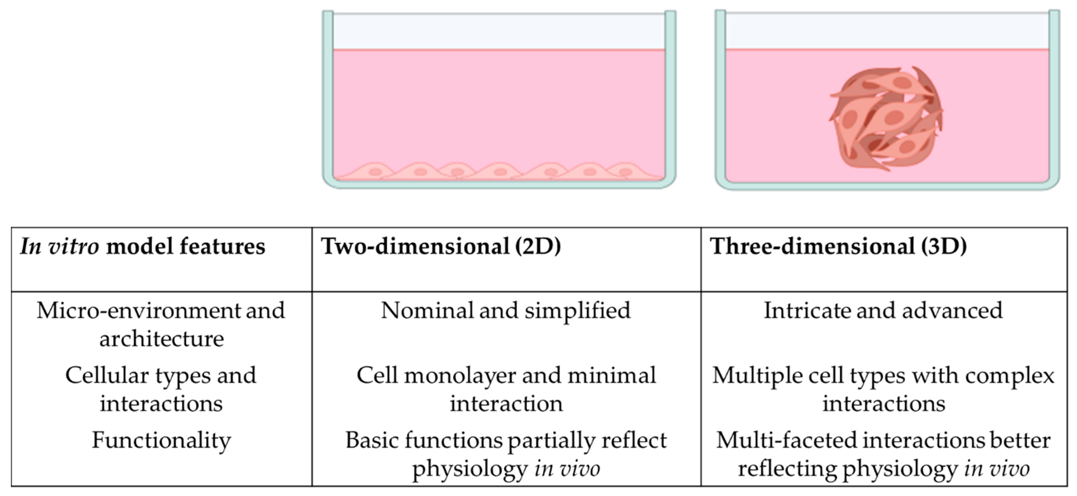
  9. Duval, K.; Grover, H.; Han, L.-H.; Mou, Y.; Pegoraro, A.F.; Fredberg, J.; Chen, Z. Modeling physiological events in 2D vs. 3D cell culture. Physiology 2017, 32, 266–277. [Google Scholar] [CrossRef] [Green Version]
  10. Ning, B.; Zhao, Y.; Buza Iii, J.A.; Li, W.; Wang, W.; Jia, T. Surgically-induced mouse models in the study of bone regeneration: Current models and future directions (Review). Mol. Med. Rep. 2017, 15, 1017–1023. [Google Scholar] [CrossRef] [Green Version]
  11. Kleinschmidt, K.; Ploeger, F.; Nickel, J.; Glockenmeier, J.; Kunz, P.; Richter, W. Enhanced reconstruction of long bone architecture by a growth factor mutant combining positive features of GDF-5 and BMP-2. Biomaterials 2013, 34, 5926–5936. [Google Scholar] [CrossRef]
  12. Zhao, Y.-P.; Tian, Q.-Y.; Liu, C.-J. Progranulin deficiency exaggerates, whereas progranulin-derived Atsttrin attenuates, severity of dermatitis in mice. FEBS Lett. 2013, 587, 1805–1810. [Google Scholar] [CrossRef] [Green Version]
  13. Zhao, Y.-P.; Tian, Q.-Y.; Frenkel, S.; Liu, C.-J. The promotion of bone healing by progranulin, a downstream molecule of BMP-2, through interacting with TNF/TNFR signaling. Biomaterials 2013, 34, 6412–6421. [Google Scholar] [CrossRef] [Green Version]
  14. Ben-David, D.; Srouji, S.; Shapira-Schweitzer, K.; Kossover, O.; Ivanir, E.; Kuhn, G.; Müller, R.; Seliktar, D.; Livne, E. Low dose BMP-2 treatment for bone repair using a PEGylated fibrinogen hydrogel matrix. Biomaterials 2013, 34, 2902–2910. [Google Scholar] [CrossRef]
  15. Frescaline, G.; Bouderlique, T.; Mansoor, L.; Carpentier, G.; Baroukh, B.; Sineriz, F.; Trouillas, M.; Saffar, J.-L.; Courty, J.; Lataillade, J.-J. Glycosaminoglycan mimetic associated to human mesenchymal stem cell-based scaffolds inhibit ectopic bone formation, but induce angiogenesis in vivo. Tissue Eng. Part A 2013, 19, 1641–1653. [Google Scholar] [CrossRef] [Green Version]
  16. Gao, H.; Huang, J.; Wei, Q.; He, C. Advances in Animal Models for Studying Bone Fracture Healing. Bioengineering 2023, 10, 201. [Google Scholar] [CrossRef]
  17. Pobloth, A.-M.; Johnson, K.A.; Schell, H.; Kolarczik, N.; Wulsten, D.; Duda, G.N.; Schmidt-Bleek, K. Establishment of a preclinical ovine screening model for the investigation of bone tissue engineering strategies in cancellous and cortical bone defects. BMC Musculoskelet. Disord. 2016, 17, 111. [Google Scholar] [CrossRef] [Green Version]
  18. Barton, K.I.; Heard, B.J.; Sevick, J.L.; Martin, C.R.; Shekarforoush, S.M.; Chung, M.; Achari, Y.; Frank, C.B.; Shrive, N.G.; Hart, D.A. Posttraumatic osteoarthritis development and progression in an ovine model of partial anterior cruciate ligament transection and effect of repeated intra-articular methylprednisolone acetate injections on early disease. Am. J. Sport. Med. 2018, 46, 1596–1605. [Google Scholar] [CrossRef]
  19. Cone, S.G.; Warren, P.B.; Fisher, M.B. Rise of the pigs: Utilization of the porcine model to study musculoskeletal biomechanics and tissue engineering during skeletal growth. Tissue Eng. Part C Methods 2017, 23, 763–780. [Google Scholar] [CrossRef]
  20. Mäkelä, T.; Takalo, R.; Arvola, O.; Haapanen, H.; Yannopoulos, F.; Blanco, R.; Ahvenjärvi, L.; Kiviluoma, K.; Kerkelä, E.; Nystedt, J. Safety and biodistribution study of bone marrow–derived mesenchymal stromal cells and mononuclear cells and the impact of the administration route in an intact porcine model. Cytotherapy 2015, 17, 392–402. [Google Scholar] [CrossRef]
  21. Sparks, D.S.; Saifzadeh, S.; Savi, F.M.; Dlaska, C.E.; Berner, A.; Henkel, J.; Reichert, J.C.; Wullschleger, M.; Ren, J.; Cipitria, A. A preclinical large-animal model for the assessment of critical-size load-bearing bone defect reconstruction. Nat. Protoc. 2020, 15, 877–924. [Google Scholar] [CrossRef] [PubMed]
  22. McGovern, J.A.; Griffin, M.; Hutmacher, D.W. Animal models for bone tissue engineering and modelling disease. Dis. Model. Mech. 2018, 11, dmm033084. [Google Scholar] [CrossRef] [PubMed] [Green Version]
  23. Greek, R.; Menache, A.; Rice, M.J. Animal models in an age of personalized medicine. Pers. Med. 2012, 9, 47–64. [Google Scholar] [CrossRef] [PubMed] [Green Version]
  24. Seifirad, S.; Haghpanah, V. Inappropriate modeling of chronic and complex disorders: How to reconsider the approach in the context of predictive, preventive and personalized medicine, and translational medicine. EPMA J. 2019, 10, 195–209. [Google Scholar] [CrossRef] [Green Version]
  25. Lo, Y.-H.; Karlsson, K.; Kuo, C.J. Applications of organoids for cancer biology and precision medicine. Nat. Cancer 2020, 1, 761–773. [Google Scholar] [CrossRef]
  26. El Harane, S.; Zidi, B.; El Harane, N.; Krause, K.-H.; Matthes, T.; Preynat-Seauve, O. Cancer Spheroids and Organoids as Novel Tools for Research and Therapy: State of the Art and Challenges to Guide Precision Medicine. Cells 2023, 12, 1001. [Google Scholar] [CrossRef]
  27. Sereti, E.; Papapostolou, I.; Dimas, K. Pancreatic Cancer Organoids: An Emerging Platform for Precision Medicine? Biomedicines 2023, 11, 890. [Google Scholar] [CrossRef]
  28. Ganguly, P.; Toghill, B.; Pathak, S. Aging, bone marrow and next-generation sequencing (NGS): Recent advances and future perspectives. Int. J. Mol. Sci. 2021, 22, 12225. [Google Scholar] [CrossRef]
  29. Tikhonova, A.N.; Dolgalev, I.; Hu, H.; Sivaraj, K.K.; Hoxha, E.; Cuesta-Domínguez, Á.; Pinho, S.; Akhmetzyanova, I.; Gao, J.; Witkowski, M. The bone marrow microenvironment at single-cell resolution. Nature 2019, 569, 222–228. [Google Scholar] [CrossRef]
  30. Nelson, M.R.; Roy, K. Bone-marrow mimicking biomaterial niches for studying hematopoietic stem and progenitor cells. J. Mater. Chem. B 2016, 4, 3490–3503. [Google Scholar] [CrossRef]
  31. Reagan, M.R.; Rosen, C.J. Navigating the bone marrow niche: Translational insights and cancer-driven dysfunction. Nat. Rev. Rheumatol. 2016, 12, 154–168. [Google Scholar] [CrossRef]
  32. Wang, H.; Leng, Y.; Gong, Y. Bone marrow fat and hematopoiesis. Front. Endocrinol. 2018, 9, 694. [Google Scholar] [CrossRef]
  33. Ashammakhi, N.; GhavamiNejad, A.; Tutar, R.; Fricker, A.; Roy, I.; Chatzistavrou, X.; Hoque Apu, E.; Nguyen, K.-L.; Ahsan, T.; Pountos, I.; et al. Highlights on Advancing Frontiers in Tissue Engineering. Tissue Eng. Part B Rev. 2021, 28, 633–664. [Google Scholar] [CrossRef]
  34. Costa, E.C.; Moreira, A.F.; de Melo-Diogo, D.; Gaspar, V.M.; Carvalho, M.P.; Correia, I.J. 3D tumor spheroids: An overview on the tools and techniques used for their analysis. Biotechnol. Adv. 2016, 34, 1427–1441. [Google Scholar] [CrossRef]
  35. Ho, W.J.; Pham, E.A.; Kim, J.W.; Ng, C.W.; Kim, J.H.; Kamei, D.T.; Wu, B.M. Incorporation of multicellular spheroids into 3-D polymeric scaffolds provides an improved tumor model for screening anticancer drugs. Cancer Sci. 2010, 101, 2637–2643. [Google Scholar] [CrossRef]
  36. Bhatia, S.N.; Ingber, D.E. Microfluidic organs-on-chips. Nat. Biotechnol. 2014, 32, 760–772. [Google Scholar] [CrossRef]
  37. Caplin, J.D.; Granados, N.G.; James, M.R.; Montazami, R.; Hashemi, N. Microfluidic organ-on-a-chip technology for advancement of drug development and toxicology. Adv. Healthc. Mater. 2015, 4, 1426–1450. [Google Scholar] [CrossRef] [Green Version]
  38. Sieber, S.; Wirth, L.; Cavak, N.; Koenigsmark, M.; Marx, U.; Lauster, R.; Rosowski, M. Bone marrow-on-a-chip: Long-term culture of human haematopoietic stem cells in a three-dimensional microfluidic environment. J. Tissue Eng. Regen. Med. 2018, 12, 479–489. [Google Scholar] [CrossRef]
  39. Kefallinou, D.; Grigoriou, M.; Boumpas, D.T.; Gogolides, E.; Tserepi, A. Fabrication of a 3D microfluidic cell culture device for bone marrow-on-a-chip. Micro Nano Eng. 2020, 9, 100075. [Google Scholar] [CrossRef]
  40. Wang, Y.; Wang, P.; Qin, J. Microfluidic organs-on-a-chip for modeling human infectious diseases. Acc. Chem. Res. 2021, 54, 3550–3562. [Google Scholar] [CrossRef]
  41. Regmi, S.; Poudel, C.; Adhikari, R.; Luo, K.Q. Applications of microfluidics and organ-on-a-chip in cancer research. Biosensors 2022, 12, 459. [Google Scholar] [CrossRef]
  42. Satta, S.; Rockwood, S.J.; Wang, K.; Wang, S.; Mozneb, M.; Arzt, M.; Hsiai, T.K.; Sharma, A. Microfluidic organ-chips and stem cell models in the fight against COVID-19. Circ. Res. 2023, 132, 1405–1424. [Google Scholar] [CrossRef] [PubMed]
  43. Li, Z.; Hui, J.; Yang, P.; Mao, H. Microfluidic Organ-on-a-Chip System for Disease Modeling and Drug Development. Biosensors 2022, 12, 370. [Google Scholar] [CrossRef] [PubMed]
  44. de Souza, N. Organoids. Nat. Methods 2018, 15, 23. [Google Scholar] [CrossRef]
  45. Giobbe, G.G.; Crowley, C.; Luni, C.; Campinoti, S.; Khedr, M.; Kretzschmar, K.; De Santis, M.M.; Zambaiti, E.; Michielin, F.; Meran, L.; et al. Extracellular matrix hydrogel derived from decellularized tissues enables endodermal organoid culture. Nat. Commun. 2019, 10, 5658. [Google Scholar] [CrossRef] [Green Version]
  46. Kim, S.; Min, S.; Choi, Y.S.; Jo, S.-H.; Jung, J.H.; Han, K.; Kim, J.; An, S.; Ji, Y.W.; Kim, Y.-G.; et al. Tissue extracellular matrix hydrogels as alternatives to Matrigel for culturing gastrointestinal organoids. Nat. Commun. 2022, 13, 1692. [Google Scholar] [CrossRef]
  47. Caddeo, S.; Boffito, M.; Sartori, S. Tissue engineering approaches in the design of healthy and pathological in vitro tissue models. Front. Bioeng. Biotechnol. 2017, 5, 40. [Google Scholar] [CrossRef] [Green Version]
  48. Liu, Y.; Peng, L.; Li, L.; Huang, C.; Shi, K.; Meng, X.; Wang, P.; Wu, M.; Li, L.; Cao, H. 3D-bioprinted BMSC-laden biomimetic multiphasic scaffolds for efficient repair of osteochondral defects in an osteoarthritic rat model. Biomaterials 2021, 279, 121216. [Google Scholar] [CrossRef]
  49. Lin, Y.-Y.; Kuan, C.-Y.; Chang, C.-T.; Chuang, M.-H.; Syu, W.-S.; Zhang, K.-L.; Lee, C.-H.; Lin, P.-C.; Dong, G.-C.; Lin, F.-H. 3D-Cultured Adipose-Derived Stem Cell Spheres Using Calcium-Alginate Scaffolds for Osteoarthritis Treatment in a Mono-Iodoacetate-Induced Rat Model. Int. J. Mol. Sci. 2023, 24, 7062. [Google Scholar] [CrossRef]
  50. Bai, H.; Cui, Y.; Wang, C.; Wang, Z.; Luo, W.; Liu, Y.; Leng, Y.; Wang, J.; Li, Z.; Liu, H. 3D printed porous biomimetic composition sustained release zoledronate to promote osteointegration of osteoporotic defects. Mater. Des. 2020, 189, 108513. [Google Scholar] [CrossRef]
  51. Hejazi, F.; Ebrahimi, V.; Asgary, M.; Piryaei, A.; Fridoni, M.J.; Kermani, A.A.; Zare, F.; Abdollahifar, M.-A. Improved healing of critical-size femoral defect in osteoporosis rat models using 3D elastin/polycaprolactone/nHA scaffold in combination with mesenchymal stem cells. J. Mater. Sci. Mater. Med. 2021, 32, 27. [Google Scholar] [CrossRef]
  52. Parente, R.; Possetti, V.; Schiavone, M.L.; Campodoni, E.; Menale, C.; Loppini, M.; Doni, A.; Bottazzi, B.; Mantovani, A.; Sandri, M. 3D co-cultures of osteoblasts and Staphylococcus aureus on biomimetic bone scaffolds as a tool to investigate the host–pathogen interface in osteomyelitis. Pathogens 2021, 10, 837. [Google Scholar] [CrossRef]
  53. Zhu, C.; He, M.; Sun, D.; Huang, Y.; Huang, L.; Du, M.; Wang, J.; Wang, J.; Li, Z.; Hu, B. 3D-Printed multifunctional polyetheretherketone bone scaffold for multimodal treatment of osteosarcoma and osteomyelitis. ACS Appl. Mater. Interfaces 2021, 13, 47327–47340. [Google Scholar] [CrossRef]
  54. Sitarski, A.M.; Fairfield, H.; Falank, C.; Reagan, M.R. 3D tissue engineered in vitro models of cancer in bone. ACS Biomater. Sci. Eng. 2018, 4, 324–336. [Google Scholar] [CrossRef]
  55. Posa, F.; Zerlotin, R.; Ariano, A.; Cosola, M.D.; Colaianni, G.; Fazio, A.D.; Colucci, S.; Grano, M.; Mori, G. Irisin Role in Chondrocyte 3D Culture Differentiation and Its Possible Applications. Pharmaceutics 2023, 15, 585. [Google Scholar] [CrossRef]
  56. Kawai, S.; Sunaga, J.; Nagata, S.; Nishio, M.; Fukuda, M.; Kamakura, T.; Sun, L.; Jin, Y.; Sakamoto, S.; Watanabe, A. 3D osteogenic differentiation of human iPSCs reveals the role of TGFβ signal in the transition from progenitors to osteoblasts and osteoblasts to osteocytes. Sci. Rep. 2023, 13, 1094. [Google Scholar] [CrossRef]
  57. Jeon, O.H.; Panicker, L.M.; Lu, Q.; Chae, J.J.; Feldman, R.A.; Elisseeff, J.H. Human iPSC-derived osteoblasts and osteoclasts together promote bone regeneration in 3D biomaterials. Sci. Rep. 2016, 6, 26761. [Google Scholar] [CrossRef] [Green Version]
  58. Janagama, D.; Hui, S.K. 3-D Cell Culture Systems in Bone Marrow Tissue and Organoid Engineering, and BM Phantoms as In Vitro Models of Hematological Cancer Therapeutics—A Review. Materials 2020, 13, 5609. [Google Scholar] [CrossRef]
  59. Langhans, S.A. Three-dimensional in vitro cell culture models in drug discovery and drug repositioning. Front. Pharmacol. 2018, 9, 6. [Google Scholar] [CrossRef]
  60. Raic, A.; Riedel, S.; Kemmling, E.; Bieback, K.; Overhage, J.; Lee-Thedieck, C. Biomimetic 3D in vitro model of biofilm triggered osteomyelitis for investigating hematopoiesis during bone marrow infections. Acta Biomater. 2018, 73, 250–262. [Google Scholar] [CrossRef]
  61. Munoz-Garcia, J.; Jubelin, C.; Loussouarn, A.; Goumard, M.; Griscom, L.; Renodon-Cornière, A.; Heymann, M.-F.; Heymann, D. In vitro three-dimensional cell cultures for bone sarcomas. J. Bone Oncol. 2021, 30, 100379. [Google Scholar] [CrossRef] [PubMed]
  62. Jalilian, E.; Massoumi, H.; Bigit, B.; Amin, S.; Katz, E.A.; Guaiquil, V.H.; Anwar, K.N.; Hematti, P.; Rosenblatt, M.I.; Djalilian, A.R. Bone marrow mesenchymal stromal cells in a 3D system produce higher concentration of extracellular vesicles (EVs) with increased complexity and enhanced neuronal growth properties. Stem Cell Res. Ther. 2022, 13, 425. [Google Scholar] [CrossRef] [PubMed]
  63. Abraham, D.M.; Herman, C.; Witek, L.; Cronstein, B.N.; Flores, R.L.; Coelho, P.G. Self-assembling human skeletal organoids for disease modeling and drug testing. J. Biomed. Mater. Res. Part B Appl. Biomater. 2022, 110, 871–884. [Google Scholar] [CrossRef] [PubMed]
  64. Sieberath, A.; Della Bella, E.; Ferreira, A.M.; Gentile, P.; Eglin, D.; Dalgarno, K. A comparison of osteoblast and osteoclast in vitro co-culture models and their translation for preclinical drug testing applications. Int. J. Mol. Sci. 2020, 21, 912. [Google Scholar] [CrossRef] [Green Version]
  65. Vernon, A.R.; Pemberton, R.M.; Morse, H.R. A novel in vitro 3D model of the human bone marrow to bridge the gap between in vitro and in vivo genotoxicity testing. Mutagenesis 2022, 37, 112–129. [Google Scholar] [CrossRef]
  66. Akiva, A.; Melke, J.; Ansari, S.; Liv, N.; van der Meijden, R.; van Erp, M.; Zhao, F.; Stout, M.; Nijhuis, W.H.; de Heus, C. An organoid for woven bone. Adv. Funct. Mater. 2021, 31, 2010524. [Google Scholar] [CrossRef]
  67. Visconti, R.J.; Kolaja, K.; Cottrell, J.A. A functional three-dimensional microphysiological human model of myeloma bone disease. J. Bone Miner. Res. 2021, 36, 1914–1930. [Google Scholar] [CrossRef]
  68. Ceccato, J.; Piazza, M.; Pizzi, M.; Manni, S.; Piazza, F.; Caputo, I.; Cinetto, F.; Pisoni, L.; Trojan, D.; Scarpa, R. A bone-based 3D scaffold as an in-vitro model of microenvironment–DLBCL lymphoma cell interaction. Front. Oncol. 2022, 12, 947823. [Google Scholar] [CrossRef]
  69. Suhito, I.R.; Kim, T.-H. Recent advances and challenges in organoid-on-a-chip technology. Organoid 2022, 2, e4. [Google Scholar] [CrossRef]
  70. .Han, J.J. FDA modernization act 2.0 allows for alternatives to animal testing. Artif. Organs 2023, 47, 449–450. [Google Scholar] [CrossRef]
  71. Mittal, R.; Woo, F.W.; Castro, C.S.; Cohen, M.A.; Karanxha, J.; Mittal, J.; Chhibber, T.; Jhaveri, V.M. Organ-on-chip models: Implications in drug discovery and clinical applications. J. Cell. Physiol. 2019, 234, 8352–8380. [Google Scholar] [CrossRef] [Green Version]
  72. León, I.E.; Cadavid-Vargas, J.F.; Resasco, A.; Maschi, F.; Ayala, M.A.; Carbone, C.; Etcheverry, S.B. In vitro and in vivo antitumor effects of the VO-chrysin complex on a new three-dimensional osteosarcoma spheroids model and a xenograft tumor in mice. JBIC J. Biol. Inorg. Chem. 2016, 21, 1009–1020. [Google Scholar] [CrossRef]
  73. Verrier, S.; Alini, M.; Alsberg, E.; Buchman, S.; Kelly, D.; Laschke, M.; Menger, M.; Murphy, W.; Stegemann, J.; Schütz, M. Tissue engineering and regenerative approaches to improving the healing of large bone defects. Eur. Cell. Mater. 2016, 32, 87–110. [Google Scholar] [CrossRef]
  74. Perez, J.R.; Kouroupis, D.; Li, D.J.; Best, T.M.; Kaplan, L.; Correa, D. Tissue Engineering and Cell-Based Therapies for Fractures and Bone Defects. Front. Bioeng. Biotechnol. 2018, 6, 105. [Google Scholar] [CrossRef] [Green Version]
  75. Qu, H.; Fu, H.; Han, Z.; Sun, Y. Biomaterials for bone tissue engineering scaffolds: A review. RSC Adv. 2019, 9, 26252–26262. [Google Scholar] [CrossRef]
  76. Filippi, M.; Born, G.; Chaaban, M.; Scherberich, A. Natural polymeric scaffolds in bone regeneration. Front. Bioeng. Biotechnol. 2020, 8, 474. [Google Scholar] [CrossRef]
  77. Iqbal, N.; Braxton, T.M.; Anastasiou, A.; Raif, E.M.; Chung, C.K.Y.; Kumar, S.; Giannoudis, P.V.; Jha, A. Dicalcium Phosphate Dihydrate Mineral Loaded Freeze-Dried Scaffolds for Potential Synthetic Bone Applications. Materials 2022, 15, 6245. [Google Scholar] [CrossRef]
  78. Pereira, H.F.; Cengiz, I.F.; Silva, F.S.; Reis, R.L.; Oliveira, J.M. Scaffolds and coatings for bone regeneration. J. Mater. Sci. Mater. Med. 2020, 31, 27. [Google Scholar] [CrossRef] [Green Version]
  79. Ganguly, P.; El-Jawhari, J.J.; Vun, J.; Giannoudis, P.V.; Jones, E.A. Evaluation of Human Bone Marrow Mesenchymal Stromal Cell (MSC) Functions on a Biomorphic Rattan-Wood-Derived Scaffold: A Comparison between Cultured and Uncultured MSCs. Bioengineering 2022, 9, 1. [Google Scholar] [CrossRef]
  80. Chen, G.; Lv, Y. Decellularized bone matrix scaffold for bone regeneration. Decellularized Scaffolds Organog. Methods Protoc. 2018, 1577, 239–254. [Google Scholar]
  81. Roseti, L.; Parisi, V.; Petretta, M.; Cavallo, C.; Desando, G.; Bartolotti, I.; Grigolo, B. Scaffolds for bone tissue engineering: State of the art and new perspectives. Mater. Sci. Eng. C 2017, 78, 1246–1262. [Google Scholar] [CrossRef] [PubMed]
  82. Maté-Sánchez de Val, J.E.; Mazón, P.; Calvo-Guirado, J.L.; Ruiz, R.A.D.; Ramírez Fernández, M.P.; Negri, B.; Abboud, M.; De Aza, P.N. Comparison of three hydroxyapatite/β-tricalcium phosphate/collagen ceramic scaffolds: An in vivo study. J. Biomed. Mater. Res. Part A 2014, 102, 1037–1046. [Google Scholar] [CrossRef] [PubMed]
  83. Yıldızhan, Ş.; Çalık, A.; Ozcanli, M.; Serin, H. Bio-composite materials: A short review of recent trends, mechanical and chemical properties, and applications. Eur. Mech. Sci. 2018, 2, 83–91. [Google Scholar] [CrossRef] [Green Version]
  84. Kumari, S.; Katiyar, S.; Darshna; Anand, A.; Singh, D.; Singh, B.N.; Mallick, S.P.; Mishra, A.; Srivastava, P. Design strategies for composite matrix and multifunctional polymeric scaffolds with enhanced bioactivity for bone tissue engineering. Front. Chem. 2022, 10, 1051678. [Google Scholar] [CrossRef]
  85. Ansari, A.I.; Sheikh, N.A. A Review of Bone Regeneration Mechanisms and Bone Scaffold Fabrication Techniques (Conventional and Non-Conventional). J. Inst. Eng. (India) Ser. C 2022, 103, 1485–1513. [Google Scholar] [CrossRef]
  86. Safari, B.; Davaran, S.; Aghanejad, A. Osteogenic potential of the growth factors and bioactive molecules in bone regeneration. Int. J. Biol. Macromol. 2021, 175, 544–557. [Google Scholar] [CrossRef]
  87. Hodge, J.; Quint, C. The improvement of cell infiltration in an electrospun scaffold with multiple synthetic biodegradable polymers using sacrificial PEO microparticles. J. Biomed. Mater. Res. Part A 2019, 107, 1954–1964. [Google Scholar] [CrossRef]
  88. Ho-Shui-Ling, A.; Bolander, J.; Rustom, L.E.; Johnson, A.W.; Luyten, F.P.; Picart, C. Bone regeneration strategies: Engineered scaffolds, bioactive molecules and stem cells current stage and future perspectives. Biomaterials 2018, 180, 143–162. [Google Scholar] [CrossRef]
  89. Sadtler, K.; Wolf, M.T.; Ganguly, S.; Moad, C.A.; Chung, L.; Majumdar, S.; Housseau, F.; Pardoll, D.M.; Elisseeff, J.H. Divergent immune responses to synthetic and biological scaffolds. Biomaterials 2019, 192, 405–415. [Google Scholar] [CrossRef]
  90. Lee, Y.J.; Ryu, Y.H.; Lee, S.J.; Moon, S.-H.; Kim, K.J.; Jin, B.J.; Lee, K.-D.; Park, J.K.; Lee, J.W.; Lee, S.-J.; et al. Bone Regeneration with 3D-Printed Hybrid Bone Scaffolds in a Canine Radial Bone Defect Model. Tissue Eng. Regen. Med. 2022, 19, 1337–1347. [Google Scholar] [CrossRef]
  91. Kronemberger, G.S.; Dalmônico, G.M.; Rossi, A.L.; Leite, P.E.C.; Saraiva, A.M.; Beatrici, A.; Silva, K.R.; Granjeiro, J.M.; Baptista, L.S. Scaffold-and serum-free hypertrophic cartilage tissue engineering as an alternative approach for bone repair. Artif. Organs 2020, 44, E288–E299. [Google Scholar] [CrossRef]
  92. Anil-Inevi, M.; Yilmaz, E.; Sarigil, O.; Tekin, H.C.; Ozcivici, E. Single cell densitometry and weightlessness culture of mesenchymal stem cells using magnetic levitation. Stem Cell Nanotechnol. Methods Protoc. 2020, 2125, 15–25. [Google Scholar]
  93. Ganguly, P.; Jones, E.; Panagiotopoulou, V.; Jha, A.; Blanchy, M.; Antimisiaris, S.; Anton, M.; Dhuiège, B.; Marotta, M.; Marjanovic, N. Electrospun and 3D printed polymeric materials for one-stage critical-size long bone defect regeneration inspired by the Masquelet technique: Recent Advances. Injury 2022, 53, S2–S12. [Google Scholar] [CrossRef]
  94. Stevens, B.; Yang, Y.; Mohandas, A.; Stucker, B.; Nguyen, K.T. A review of materials, fabrication methods, and strategies used to enhance bone regeneration in engineered bone tissues. J. Biomed. Mater. Res. Part B Appl. Biomater. 2008, 85, 573–582. [Google Scholar] [CrossRef]
  95. Polo-Corrales, L.; Latorre-Esteves, M.; Ramirez-Vick, J.E. Scaffold design for bone regeneration. J. Nanosci. Nanotechnol. 2014, 14, 15–56. [Google Scholar] [CrossRef] [Green Version]
  96. Fitzgerald, K.A.; Guo, J.; Tierney, E.G.; Curtin, C.M.; Malhotra, M.; Darcy, R.; O’Brien, F.J.; O’Driscoll, C.M. The use of collagen-based scaffolds to simulate prostate cancer bone metastases with potential for evaluating delivery of nanoparticulate gene therapeutics. Biomaterials 2015, 66, 53–66. [Google Scholar] [CrossRef]
  97. Dadwal, U.C.; Merkel, A.R.; Page, J.M.; Kwakwa, K.A.; Kessler, M.; Rhoades, J.A. 3D bone morphology alters gene expression, motility, and drug responses in bone metastatic tumor cells. Int. J. Mol. Sci. 2020, 21, 6913. [Google Scholar] [CrossRef]
  98. Conesa, A.; Madrigal, P.; Tarazona, S.; Gomez-Cabrero, D.; Cervera, A.; McPherson, A.; Szcześniak, M.W.; Gaffney, D.J.; Elo, L.L.; Zhang, X. A survey of best practices for RNA-seq data analysis. Genome Biol. 2016, 17, 13. [Google Scholar] [CrossRef] [Green Version]
  99. Rao, M.S.; Van Vleet, T.R.; Ciurlionis, R.; Buck, W.R.; Mittelstadt, S.W.; Blomme, E.A.; Liguori, M.J. Comparison of RNA-Seq and microarray gene expression platforms for the toxicogenomic evaluation of liver from short-term rat toxicity studies. Front. Genet. 2019, 9, 636. [Google Scholar] [CrossRef] [Green Version]
  100. Zhao, S.; Fung-Leung, W.-P.; Bittner, A.; Ngo, K.; Liu, X. Comparison of RNA-Seq and microarray in transcriptome profiling of activated T cells. PLoS ONE 2014, 9, e78644. [Google Scholar] [CrossRef]
  101. Wu, H.; Wei, X.; Liu, Y.; Dong, H.; Tang, Z.; Wang, N.; Bao, S.; Wu, Z.; Shi, L.; Zheng, X. Dynamic degradation patterns of porous polycaprolactone/β-tricalcium phosphate composites orchestrate macrophage responses and immunoregulatory bone regeneration. Bioact. Mater. 2023, 21, 595–611. [Google Scholar] [CrossRef] [PubMed]
  102. Guerrero, J.; Maevskaia, E.; Ghayor, C.; Bhattacharya, I.; Weber, F.E. Influence of Scaffold Microarchitecture on Angiogenesis and Regulation of Cell Differentiation during the Early Phase of Bone Healing: A Transcriptomics and Histological Analysis. Int. J. Mol. Sci. 2023, 24, 6000. [Google Scholar] [CrossRef] [PubMed]
  103. Lin, M.; Gao, M.; Cavnar, M.J.; Kim, J. Utilizing gastric cancer organoids to assess tumor biology and personalize medicine. World J. Gastrointest. Oncol. 2019, 11, 509–517. [Google Scholar] [CrossRef] [PubMed]
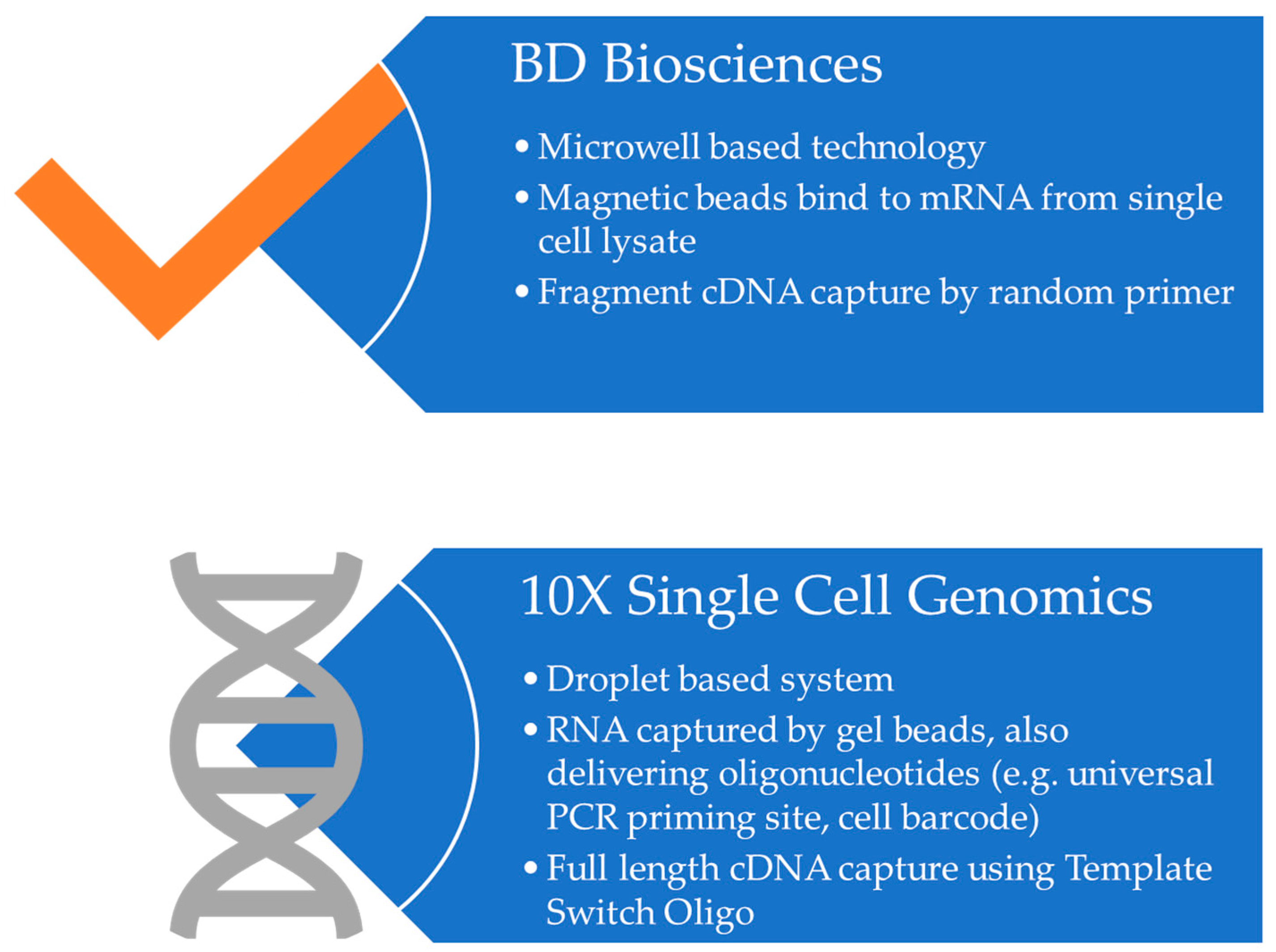
  104. Gao, C.; Zhang, M.; Chen, L. The Comparison of Two Single-cell Sequencing Platforms: BD Rhapsody and 10x Genomics Chromium. Curr. Genom. 2020, 21, 602–609. [Google Scholar] [CrossRef] [PubMed]
  105. Ulbrich, J.; Lopez-Salmeron, V.; Gerrard, I. BD Rhapsody™ Single-Cell Analysis System Workflow: From Sample to Multimodal Single-Cell Sequencing Data. In Single Cell Transcriptomics; Methods in Molecular Biology; Springer: Berlin/Heidelberg, Germany, 2023; Volume 2584. [Google Scholar] [CrossRef]
  106. Khan, A.O.; Rodriguez-Romera, A.; Reyat, J.S.; Olijnik, A.-A.; Colombo, M.; Wang, G.; Wen, W.X.; Sousos, N.; Murphy, L.C.; Grygielska, B.; et al. Human Bone Marrow Organoids for Disease Modeling, Discovery, and Validation of Therapeutic Targets in Hematologic Malignancies. Cancer Discov. 2023, 13, 364–385. [Google Scholar] [CrossRef]
  107. Pathak, S.; Rowczenio, D.; Lara-Reyna, S.; Kacar, M.; Owen, R.; Doody, G.; Krause, K.; Lachmann, H.; Doffinger, R.; Newton, D.; et al. Evidence of B Cell Clonality and Investigation Into Properties of the IgM in Patients With Schnitzler Syndrome. Front. Immunol. 2020, 11, 569006. [Google Scholar] [CrossRef]
  108. Pathak, S.; Rowczenio, D.M.; Owen, R.G.; Doody, G.M.; Newton, D.J.; Taylor, C.; Taylor, J.; Cargo, C.; Hawkins, P.N.; Krause, K.; et al. Exploratory study of MYD88 L265P, rare NLRP3 variants and clonal hematopoiesis prevalence in patients with Schnitzler’s Syndrome. Arthritis Rheumatol. 2019, 71, 2121–2125. [Google Scholar] [CrossRef]
  109. Rowczenio, D.M.; Pathak, S.; Arostegui, J.I.; Mensa-Vilaro, A.; Omoyinmi, E.; Brogan, P.; Lipsker, D.; Scambler, T.; Owen, R.; Trojer, H.; et al. Molecular genetic investigation, clinical features, and response to treatment in 21 patients with Schnitzler syndrome. Blood 2018, 131, 974–981. [Google Scholar] [CrossRef]
  110. Wang, X.; He, Y.; Zhang, Q.; Ren, X.; Zhang, Z. Direct Comparative Analyses of 10X Genomics Chromium and Smart-seq2. Genom. Proteom. Bioinform. 2021, 19, 253–266. [Google Scholar] [CrossRef]
  111. Bunpetch, V.; Zhang, X.; Li, T.; Lin, J.; Maswikiti, E.P.; Wu, Y.; Cai, D.; Li, J.; Zhang, S.; Wu, C. Silicate-based bioceramic scaffolds for dual-lineage regeneration of osteochondral defect. Biomaterials 2019, 192, 323–333. [Google Scholar] [CrossRef]
  112. Ji, C.; Qiu, M.; Ruan, H.; Li, C.; Cheng, L.; Wang, J.; Li, C.; Qi, J.; Cui, W.; Deng, L. Transcriptome analysis revealed the symbiosis niche of 3D scaffolds to accelerate bone defect healing. Adv. Sci. 2022, 9, 2105194. [Google Scholar] [CrossRef]
  113. McCray, T.; Moline, D.; Baumann, B.; Vander Griend, D.J.; Nonn, L. Single-cell RNA-Seq analysis identifies a putative epithelial stem cell population in human primary prostate cells in monolayer and organoid culture conditions. Am. J. Clin. Exp. Urol. 2019, 7, 123. [Google Scholar]
  114. Ma, Q.; Tao, H.; Li, Q.; Zhai, Z.; Zhang, X.; Lin, Z.; Ni Kuang, N.; Pan, J. OrganoidDB: A comprehensive organoid database for the multi-perspective exploration of bulk and single-cell transcriptomic profiles of organoids. Nucleic Acids Res. 2023, 51, D1086–D1093. [Google Scholar] [CrossRef]
  115. Correia, C.R.; Bjørge, I.M.; Zeng, J.; Matsusaki, M.; Mano, J.F. Liquefied Microcapsules as Dual-Microcarriers for 3D+ 3D Bottom-Up Tissue Engineering. Adv. Healthc. Mater. 2019, 8, 1901221. [Google Scholar] [CrossRef]
  116. Oliveira, C.S.; Nadine, S.; Gomes, M.C.; Correia, C.R.; Mano, J.F. Bioengineering the human bone marrow microenvironment in liquefied compartments: A promising approach for the recapitulation of osteovascular niches. Acta Biomater. 2022, 149, 167–178. [Google Scholar] [CrossRef]
  117. Wang, H.; Brown, P.C.; Chow, E.C.; Ewart, L.; Ferguson, S.S.; Fitzpatrick, S.; Freedman, B.S.; Guo, G.L.; Hedrich, W.; Heyward, S. 3D cell culture models: Drug pharmacokinetics, safety assessment, and regulatory consideration. Clin. Transl. Sci. 2021, 14, 1659–1680. [Google Scholar] [CrossRef]
  118. Marinucci, M.; Ercan, C.; Taha-Mehlitz, S.; Fourie, L.; Panebianco, F.; Bianco, G.; Gallon, J.; Staubli, S.; Soysal, S.D.; Zettl, A. Standardizing Patient-Derived Organoid Generation Workflow to Avoid Microbial Contamination From Colorectal Cancer Tissues. Front. Oncol. 2022, 11, 5605. [Google Scholar] [CrossRef]
  119. Kim, J.; Koo, B.-K.; Knoblich, J.A. Human organoids: Model systems for human biology and medicine. Nat. Rev. Mol. Cell Biol. 2020, 21, 571–584. [Google Scholar] [CrossRef]
  120. Vives, J.; Batlle-Morera, L. The challenge of developing human 3D organoids into medicines. Stem Cell Res. Ther. 2020, 11, 72. [Google Scholar] [CrossRef] [Green Version]
  121. Cahan, P.; Li, H.; Morris, S.A.; da Rocha, E.L.; Daley, G.Q.; Collins, J.J. CellNet: Network biology applied to stem cell engineering. Cell 2014, 158, 903–915. [Google Scholar] [CrossRef] [Green Version]
  122. Radley, A.H.; Schwab, R.M.; Tan, Y.; Kim, J.; Lo, E.K.W.; Cahan, P. Assessment of engineered cells using CellNet and RNA-seq. Nat. Protoc. 2017, 12, 1089–1102. [Google Scholar] [CrossRef] [PubMed] [Green Version]
  123. Tan, Y.; Cahan, P. SingleCellNet: A Computational Tool to Classify Single Cell RNA-Seq Data Across Platforms and Across Species. Cell Syst. 2019, 9, 207–213.e2. [Google Scholar] [CrossRef] [PubMed]
  124. Lensink, M.A.; Jongsma, K.R.; Boers, S.N.; Noordhoek, J.J.; Beekman, J.M.; Bredenoord, A.L. Responsible use of organoids in precision medicine: The need for active participant involvement. Development 2020, 147, dev177972. [Google Scholar] [CrossRef] [PubMed] [Green Version]
  125. Zhou, Z.; Cong, L.; Cong, X. Patient-derived organoids in precision medicine: Drug screening, organoid-on-a-chip and living organoid biobank. Front. Oncol. 2021, 11, 5625. [Google Scholar] [CrossRef] [PubMed]
  126. Papaccio, F.; Cabeza-Segura, M.; Garcia-Micò, B.; Tarazona, N.; Roda, D.; Castillo, J.; Cervantes, A. Will Organoids Fill the Gap towards Functional Precision Medicine? J. Pers. Med. 2022, 12, 1939. [Google Scholar] [CrossRef]
  127. Chen, S.; Chen, X.; Geng, Z.; Su, J. The horizon of bone organoid: A perspective on construction and application. Bioact. Mater. 2022, 18, 15–25. [Google Scholar] [CrossRef]
  128. Liu, Y.; Gan, Y.; AiErken, N.; Chen, W.; Zhang, S.; Ouyang, J.; Zeng, L.; Tang, D. Combining Organoid Models with Next-Generation Sequencing to Reveal Tumor Heterogeneity and Predict Therapeutic Response in Breast Cancer. J. Oncol. 2022, 2022, 9390912. [Google Scholar] [CrossRef]
  129. Agarwal, D.; Kuhns, R.; Dimitriou, C.N.; Barlow, E.; Wahlin, K.J.; Enke, R.A. Bulk RNA sequencing analysis of developing human induced pluripotent cell-derived retinal organoids. Sci. Data 2022, 9, 759. [Google Scholar] [CrossRef]
Figure 1. Comparison between 2D and 3D in vitro models.
Organoids 02 00010 g001
Figure 2. Types of in vitro models.
Organoids 02 00010 g002
Figure 3. Comparative Analysis of Single Cell Sequencing Approaches from BD Biosciences and 10X Genomics—These two predominant methodologies facilitate the isolation and subsequent barcoding of individual cells’ transcriptomes, thereby permitting an in-depth investigation of cellular gene expression heterogeneity within complex biological systems.
Organoids 02 00010 g003
Figure 4. Fabrication (indicated in green) and various applications (indicated in yellow and blue) of 3D models for bone and bone marrow.
Organoids 02 00010 g004
Disclaimer/Publisher’s Note: The statements, opinions and data contained in all publications are solely those of the individual author(s) and contributor(s) and not of MDPI and/or the editor(s). MDPI and/or the editor(s) disclaim responsibility for any injury to people or property resulting from any ideas, methods, instructions or products referred to in the content.

