Organoids 2024, 3(2), 67-82; https://doi.org/10.3390/organoids3020006 - 04 Apr 2024
Abstract
►
Show Figures
For over 150 years, researchers have studied the (patho)physiology of the endocrine pancreas and devised treatment options for diabetes mellitus (DM). However, no cure has been developed so far. In dogs, diabetes mellitus type 1 (T1DM) is the most common presentation. Treatment consists
[...] Read more.
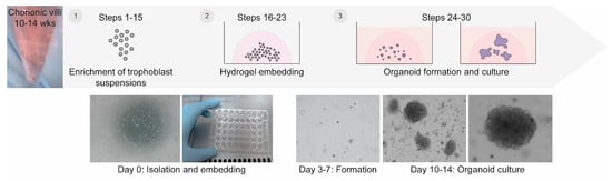
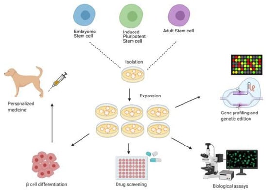
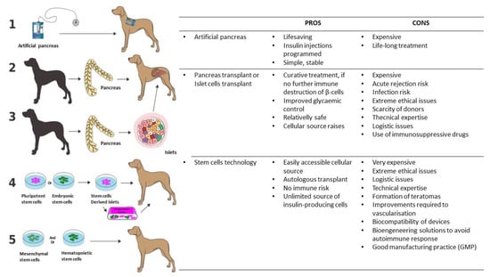
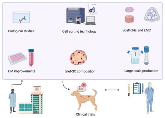
For over 150 years, researchers have studied the (patho)physiology of the endocrine pancreas and devised treatment options for diabetes mellitus (DM). However, no cure has been developed so far. In dogs, diabetes mellitus type 1 (T1DM) is the most common presentation. Treatment consists of twice daily insulin injections, monitored by spatial blood glucose measurements. Even though dogs were instrumental in the discovery of insulin and islet transplantations, the treatment in diabetic dogs has remained unchanged for decades. Providing twice daily insulin injections is demanding for both owners and dogs and may result in hypoglycaemic events, creating the need for new treatment strategies. Novel regenerative medicine-based tools, such as improved β-cell culture protocols and artificial devices, have sparked hope for a cure. In human medicine, emerging technologies such as the transplantation of insulin-producing β-cells, generated by stem cell differentiation, with or without an encapsulation device, are currently tested in phase I/II clinical trials. As the pathogenesis of T1DM is remarkably similar between humans and dogs, novel treatment methods could be implemented in canine medicine. This review briefly summarises the physiology of the canine endocrine pancreas and the pathophysiology of canine DM before exploring current and possible future treatment options for canine DM.
Full article
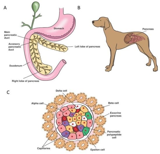
Figure 1

