Review

Secondary Syphilis: Pathophysiology, Clinical Manifestations, and Diagnostic Testing

Paul L. Foster School of Medicine, Texas Tech University Health Sciences Center El Paso, 5001 El Paso Dr., El Paso, TX 79905, USA
*
Author to whom correspondence should be addressed.
Venereology 2023, 2(2), 65-75; https://doi.org/10.3390/venereology2020006
Submission received: 27 December 2022 / Revised: 18 February 2023 / Accepted: 28 March 2023 / Published: 11 April 2023

Abstract

:
The subspecies T. pallidum pallidum is the cause of the most infamous sexually and congenitally transmitted disease, syphilis. This disease has an estimated incidence of six million infections every year. Multiple studies have noted that the prevalence of syphilis has been steadily increasing worldwide in recent decades, especially among MSMs and HIV-positive patients. Clinically, syphilis presents in four stages with multiple different clinical manifestations. In this paper, we examine the current literature to determine the history and progression, pathogenesis, clinical features, and testing of secondary syphilis. Secondary syphilis is a stage of the disease with the most exuberant local and systemic clinical manifestations. The basis of the pathogenesis of SS underscores the unique mechanisms by which Treponema pallidum utilizes to escape immune recognition while simultaneously induces inflammation. SS can affect multiple organ systems and become more than just an STD. The most common presentation of secondary syphilis is rash, which manifests as a copper-colored maculopapular lesion on the trunk, palms, and soles. Although the RPR, VDRL, and FTA-ABS tests are perhaps the most commonly used diagnostic tools for syphilis and make up the traditional and reverse algorithms, there are other methods, including morphology and immunohistochemistry.

1. Syphilis: More Than just an STI

Several subtypes of the spiral-shaped bacterium, Treponema pallidum, have transmitted diseases for centuries. The subspecies T. pallidum pallidum is the cause of the most infamous sexually and congenitally transmitted disease, syphilis. This disease has an estimated incidence of six million infections every year, with an overwhelming number of cases residing in endemic, low-income countries as well as in populations of men who have sex with men (MSMs) [1]. Worldwide, a disproportionate number of MSMs contract syphilis (402.0 cases of primary and secondary syphilis per 100,000 men) compared to women (10.8 cases of primary and secondary syphilis per 100,000 females) [2,3]. In 2021, the CDC reported a major increase in the number of syphilis up to 51.5 cases per 100,000 people across all populations (including MSMs, women, men who have sex with women, and men with unknown sexual partners), a number that had previously been in decline for thirty years in the United States [4]. A number of factors have contributed to this worrying rise in syphilis cases in the United States, many of which can be attributed to the COVID-19 pandemic. Aside from the CDC’s Division of Sexually Transmitted Disease (STD) Prevention reporting a dramatic decrease in the amount of resources dedicated to venereal disease screening and treatment, almost half of the laboratories had reported disruptions of service, with staff reassignment and testing or treatment shortages, as the pandemic raged on in 2020 [5]. Factors related to the coordination of public health services and private insurers, alongside other socio-economic barriers, have further led to more patients in the U.S. to suffer from preventable and treatable venereal diseases, such as syphilis [6].
The previous observed decline in the number of individuals living with syphilis was probably a consequence of public health efforts to combat HIV in the early 2000s through education on STD transmission. It has been theorized that the subsequent widespread increase in syphilis after the 2000s could be due to the relaxation of public health and educational efforts after the widespread usage of antiretroviral therapy and the emergence of new sublineages, which are resistant to traditional macrolide therapy [7]. Multiple studies have noted that the prevalence of syphilis has been steadily increasing worldwide in recent decades, especially among MSMs and HIV-positive patients [8,9,10]. These populations in developing countries comprise up to 90% of new syphilis diagnoses each year; however, due to the economies of scale and issues with infrastructure in these countries, testing and treatment have been difficult [11].
Sexually active women comprise a population that has a high rate of contracting STIs, such as syphilis. A worrying side effect of STI transmission in reproductive-age females, besides primary sexual transmission of syphilis, is the ability for this disease to be vertically transmitted during pregnancy. The rate of vertical transmission of syphilis during its primary and secondary stages remains high at 60–100%, a worrying statistic for mothers in endemic areas and areas with low prenatal care [12]. Despite syphilis being a preventable and treatable illness, the WHO still posits congenital syphilis as the second leading cause of stillbirths worldwide (reported by at least 78 countries), with an incidence of 3.2% in pregnant women [13]. Congenital syphilis is also on the rise in the U.S., with the CDC reporting more than 2677 cases per 100,000 newborns in 2021, a drastic increase from 334 cases per 100,000 newborns in 2012. The number of cases has gradually increased even before the COVID-19 pandemic, with a percent increase of 24.05% in cases from 2020 to 2021 [4].
Clinically, syphilis presents in four stages with multiple different clinical manifestations [14]. Each progressive stage of syphilis, apart from its latent stage, brings physiological changes to the human body. The primary and tertiary stages of syphilis have been well defined: the primary stage is generally composed of chancres in the genitals, breasts, or mucocutaneous regions, while the tertiary stage is characterized as systemic dysfunction, particularly in the cardiovascular and neurological systems. The secondary stage of syphilis is commonly mistaken for other diseases due to its clinical presentation and its high rate of coinfection with HIV [15,16]. The presentation of secondary syphilis (SS) as a maculopapular rash may include differential diagnoses or misdiagnoses, such as Rocky Mountain spotted fever, while oral lesions may be misdiagnosed as herpes; both differential diagnoses are viruses with completely different treatment methodologies [17].
Given the surging menace of syphilis, we herein provide a comprehensive review on secondary syphilis with the hope of bringing awareness to practicing physicians and other healthcare providers. For this paper, we searched the PubMed repository for peer-reviewed literature, with a focus on the literature released within the past five years (2017–2022). We also examined current literature to determine the history and progression, pathogenesis, clinical features, and testing of secondary syphilis.

2. Pathogenesis of Secondary Syphilis

SS is a stage of the disease with the most exuberant local and systemic clinical manifestations [14,18]. The basis of the pathogenesis of SS underscores the unique mechanisms by which Treponema pallidum utilizes to escape immune recognition while simultaneously induces inflammation. T. pallidum utilizes adherence mechanisms to bind to epithelia and underlying fibronectin, as well as laminin; after dissemination through the bloodstream, the spirochete invades tissues via inter-junctional penetration [19,20], which allows it to escape to the skin. In secondary syphilis, spirochetes are found within the epidermis and superficial dermis [21].
The clinical manifestations of SS result from the local inflammatory response elicited by syphilis spirochetes replicating within tissues [20]. The most common clinical manifestation of secondary syphilis is a disseminated cutaneous eruption [19]. As soon as these spirochetes reach a sufficient density in the skin, they trigger a local inflammatory response, making SS skin lesions clinically apparent [22]. T. pallidum provokes an intense inflammatory response in the skin, with the presence of various cytokines (IFN-γ and TNF-α), chemokines (CCL2 and CXCL10), macrophage and dendritic cell (DC) activation markers (CD40 and CD86), Fc-mediated phagocytosis receptors (FcγRI and FcγR3), and IFN-β and effector molecules associated with CD8 and NK cell cytotoxic responses [22]. A similar intense skin inflammatory process with activation of plasmacytoid DCs and production of type I IFN (IFN-α and IFN-β) is key to starting the development of a psoriatic plaque [23,24]. This may explain the psoriasiform appearance of SS lesions in some patients [25]. Although some other SS lesions present various degrees of inflammation, the vessels around these lesions typically show inflammatory proliferative changes and cellular infiltrates in their walls [25].
Less frequent sites of localized inflammation in SS are gastric, renal, and hepatic sites [19]. Ocular disease and meningitis are the manifestations of neurosyphilis, which are also infrequent in SS. Such presentations provide evidence that syphilis spirochetes are able to travel to the CSF and to the eyes, possibly through hematogenous dissemination [26]. Despite the overt inflammation, immune cells are inefficient in phagocytosing these spirochetes [22]. SS lesions take weeks to months to resolve, which is possibly linked to the unusually slow replication rate of T. pallidum. When leukocytes from the skin lesions of patients with SS are examined, there is a predominance of activated dermal plasmacytoid dendritic cells and activated CD4+ with a memory/effector phenotype [18].
Syphilis spirochetes not only fail to be cleared rapidly but can replicate and circulate in the midst of a prolific antibody response [20]. The basis of this antibody avoidance relies on these spirochetes’ molecular architecture as the antigen availability is limited, given that the outer membrane proteins are mainly attached to the inner leaflet of the outer membrane [20], without sufficient surface exposure. These antigens finally become available for immune recognition upon treatment, thereby explaining the development of the Jarisch–Herxheimer reaction, a transient intense inflammatory condition occurring within 24 h of antibiotic therapy [14].
The immune evasiveness of T. pallidum is also demonstrated by the fact that its infection does not lead to immunity against reinfection, and repeated episodes of syphilis may occur [14].

3. Clinical Features of Secondary Syphilis

Based on clinical features and laboratory tests, syphilis can be divided into four phases: latent, primary, secondary, and tertiary syphilis [27]. Among the four phases of syphilis, secondary syphilis is arguably the most crucial. Once syphilis has progressed to the secondary phase, treatment becomes vital to prevent progression into devastating tertiary syphilis.
Primary syphilis usually presents two to three weeks after the inoculation of one or multiple painless genital chancres at the site of inoculation (anal, oral, and genital), with or without local lymphadenopathy [28]. Its painless nature allows the genital chancres to go unnoticed and progress to secondary syphilis. In rare cases, the genital chancres may still be present with the appearance of secondary syphilis features [29]. SS presents four to eight weeks later and represents hematogenous dissemination of syphilis spirochetes [25].
Syphilis has been coined “the great imitator” based on the diverse clinical features of the secondary phase. SS can affect multiple organ systems and become more than just an STD. The most common presentation of secondary syphilis is rash, which manifests as a copper-colored maculopapular lesion on the trunk, palms, and soles. This description might not hold true in dark colored skin where rash is usually violaceous or hyperpigmented [25]. In such patients, skin lesions may go unnoticed, but rash in the palms and soles would still be evident. In addition, rash may vary in presentation and can erupt on the back, face, arms, legs, and genitalia. Other morphology of syphilis rash includes papulosquamous, annular, psoriasiform, pustular, and follicular lesions [30]. A rupiod presentation, i.e., well-demarcated, cone-shaped plaques with thick, dark, lamellate, and adherent crusts on the skin resembling oyster or limpet shells [31], is relatively rare. It usually manifests as skin lesions all over the body [32], but when confined, it can be misdiagnosed as verruca vulgaris [33]. Physicians are well trained to recognize the most common manifestations of rash in the palms and soles but may not be aware of the deviations from the classic presentation.
Depending on the morphology and the site of the rash, secondary syphilis may be often mistaken for other diseases. A maculopapular rash on the palms and soles can be confused for Rocky Mountain spotted fever, other rickettsioses, viral exanthem, or erythema multiforme [17,34]. If the rash is pustular and presents on the face, syphilis can masquerade as acne vulgaris [25]. SS lesions could also present as prominent scales that resemble psoriasis [27]. Other non-infectious conditions to consider are pityriasis rosea, drug eruption, lichen planus, psoriasis, and sarcoidosis [17].
The presence of condylomata lata may raise suspicion of secondary syphilis. Condyloma lata is a painless, flat, and well-demarcated grey-to-white plaque or papule that is usually present in the genital, perianal, or other intertriginous areas. Although condyloma lata can be diagnosed with nucleic acid amplification testing, biopsy may be needed to differentiate syphilitic condylomata lata from other lesions, such as human papillomavirus HPV-induced condyloma acuminata, malignant tumors, and genital herpes [35]. Biopsy shows numerous spirochetes with plasma cell infiltrate via immunostaining, making condylomata lata the most infectious skin lesions in syphilis based on the concentration of spirochetes in the exudated serum [36].
Oral lesions are not uncommon in SS, and the areas most affected are the mucosal surfaces of the upper and lower lips, tongues, and buccal mucosa of the cheeks [37]. Lesions usually present as slightly elevated oval plaques associated with a grey or white pseudomembrane. Other lesions are described as “snail track ulcers” characterized by multiple mucous patches that may coalesce to produce serpiginous lesions [38].
Other associated symptomatology includes lymphadenopathy, genital and oral mu- cus patches, and, less frequently, fever and alopecia [39]. Alopecia in syphilis usually presents as a “moth-eaten” pattern and is considered a pathognomonic manifestation of secondary syphilis. It presents in less than 10% of patients [40] and could be the only sign of syphilis infection [41]. Some patients present with diffuse alopecia or a combination of diffuse and moth-eaten patterns. Other conditions, such as alopecia areata, alopecia neoplastica, and trichotillomania, can mimic syphilis alopecia [40]. The presence of other cutaneous or mucosal findings can help rule out some of the other differential diagnoses.
Rare manifestations include hepatitis, hepatomegaly, splenomegaly, meningitis, peripheral neuropathy, deafness, periostitis, iridocyclitis, and arthritis [39]. Misdiagnosis can occur in the manifestations of neurosyphilis in tertiary syphilis and may be underreported given the lack of investigation of syphilis as a cause of uveitis or meningitis.
Ocular syphilis may present in any phase of syphilis, except in primary syphilis, and it accounts for about 5% of the cases in the United States [42]. The most common ocular manifestation of syphilis is uveitis, which is more common in patients with neurosyphilis and HIV infection. Panuveitis and posterior uveitis are the most common ocular manifestations [43]. Ocular syphilis can easily be misdiagnosed when it is the only disease manifestation, and such misdiagnosis can lead to ocular complications and blindness [44]. A suspicion of ocular syphilis (usually in someone with syphilis and eye symptoms) warrants an opthalmologic evaluation, where findings such as posterior placoid lesions, well-circumscribed discrete miliary lesions, and superficial creamy yellow precipitates point toward ocular syphilis [45]. According to the Center for Disease Control and Prevention, patients with ocular syphilis should receive a CSF evaluation and lumbar puncture. A diagnosis can be made with ocular findings and a positive treponemal test [45].
Syphilis hepatitis is a rare manifestation of SS, which is reported in less than 10% of patients [46]. Syphilis hepatitis involves hepatic dysfunction with evidence of treponemal infection. Like many forms of hepatitis, syphilis hepatitis can manifest as fatigue, icterus, abdominal pain, lymphadenopathy, arthralgia, and elevated liver enzymes (including alkaline phosphatase (ALP), aspartate transaminase (AST), alanine transaminase (ALT), gamma-glutamyltransferase (GGT), and total bilirubin). Syphilis hepatitis is usually mild and can be asymptomatic, with only slight elevations of liver enzymes and with or without hepatomegaly [47,48]. The diagnostic criteria for syphilitic hepatitis were proposed in 2004, and they include abnormal liver enzyme levels, serological evidence of syphilis infection, exclusion of other causes of hepatitis, and normalization of liver enzymes after treatment [49].
Syphilitic arthritis is another rare but possible presentation of SS. It manifests as the hematogenous spread of spirochetes to the joints. The joints affected include bilateral knees, hips, shoulders, and proximal interphalangeal (PIP) joints [50]. Although bone and joint pain can be present in more than 12% of patients with SS [30], arthritis and arthralgia are seen in only 2% of patients [39]. Patients present with tenderness, swelling, and a restricted range of motion in the affected joints. Joint pain is usually relieved by movement and exacerbated by heat, and most patients report that pain is most severe at night [51]. This constellation of symptoms can be mistaken for other forms of arthritis, such as rheumatoid arthritis. Like other presentations of syphilis, evidence of treponemal infection and therapeutic response to syphilis medications support the diagnosis [50].
Secondary syphilis excels at disguising as other diseases, so clinicians should always have this great imitator on their radar. The goal is to commence treatment before syphilis progresses to the tertiary phase, where damage can be permanent and mortality is significant.

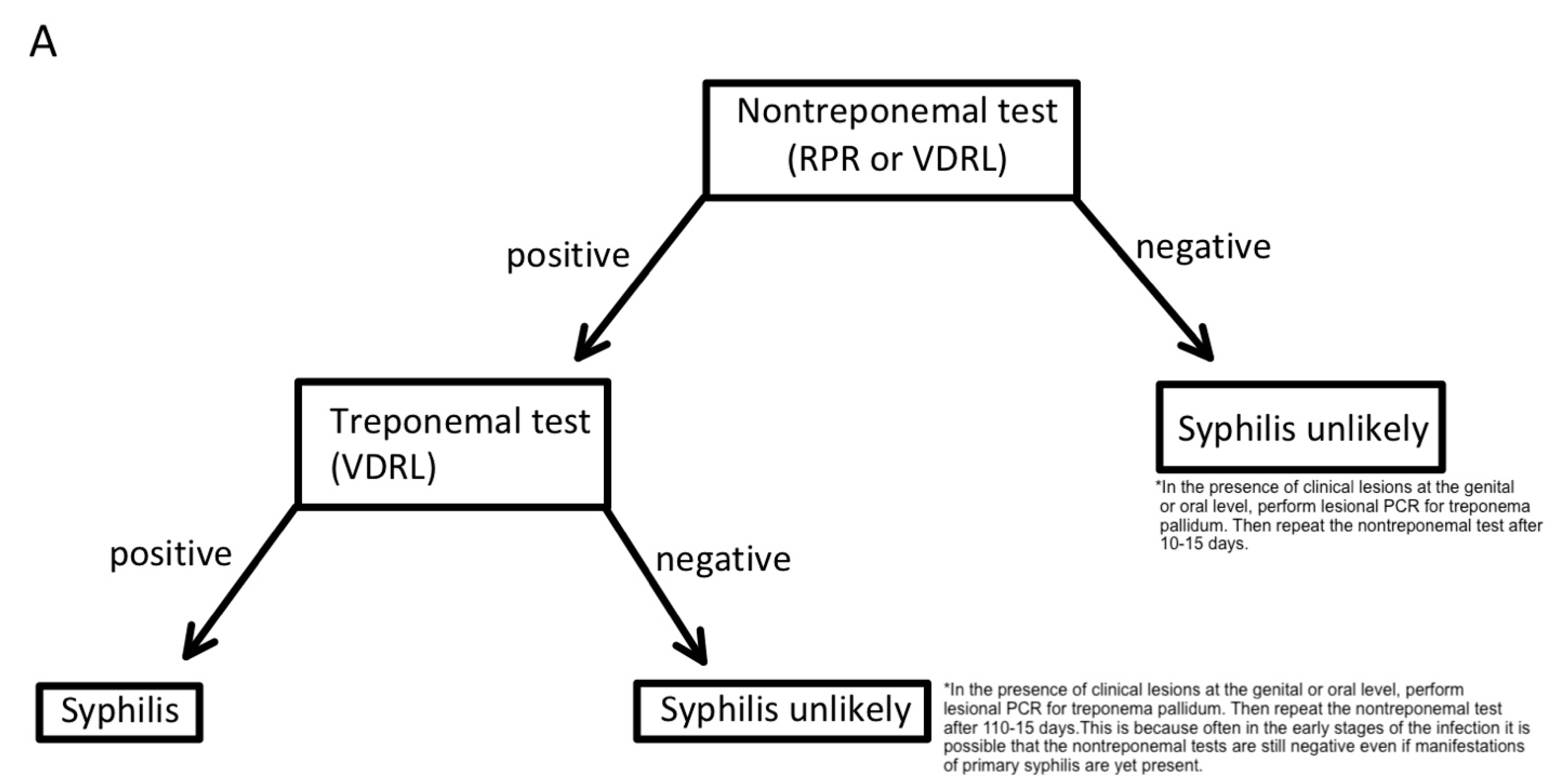
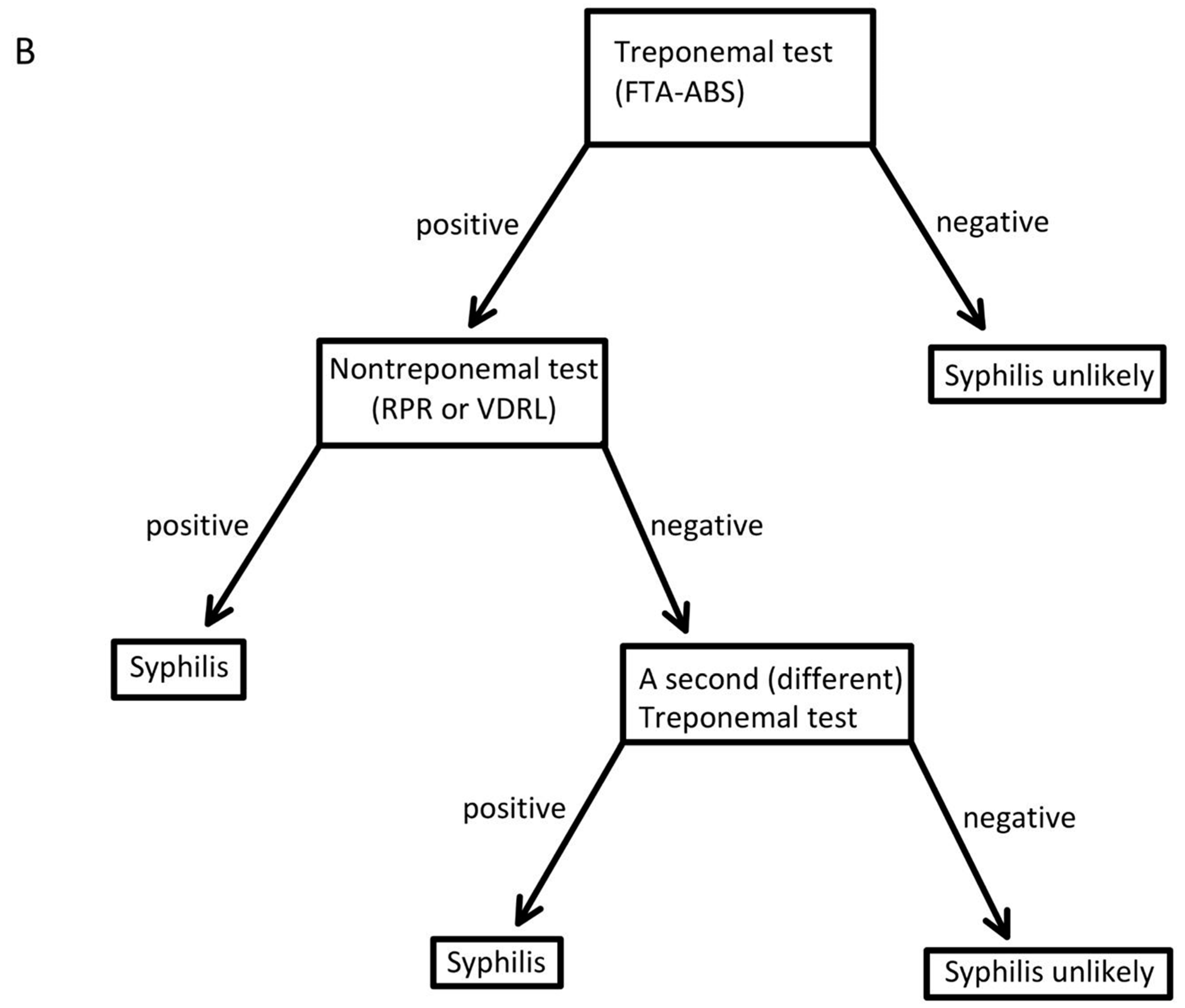
4. Testing for Syphilis

4.1. Serological Assays

Diagnostic testing for syphilis in high-risk individuals, regardless of whether or not they are symptomatic, is necessary as the disease is highly transmissible. The first serological test for syphilis was developed in 1906 as a complement fixation assay using antigens from human and monkey extracts rich in T. pallidum [52]. In current practice, serologic evaluation for antibodies to T. pallidum using nontreponemal testing (NTT) and treponemal testing (TT) is the gold standard for diagnosis [20].
NTTs include the rapid plasma reagin (RPR) test, the toluidine red unheated serum test (TRUST), and the Venereal Disease Research Laboratory (VRDL) test. All these tests measure levels of anti-lipid IgG or IgM antibodies produced in response to the lipoidal material released from damaged host cells or T. pallidum cardiolipin [53]. NTTs are routinely used in the initial screening of syphilis because they are rapid, inexpensive, and simple to operate [54]. Based on previous reports, the sensitivity of serum NTTs varies based on the classification of syphilis that is present. The sensitivity of NTTs is 62–78% for the diagnosis of primary syphilis, 97–100% for secondary syphilis, 82–100% for early latent syphilis, and 47–64% for tertiary syphilis, which is unsatisfactory [55]. Among current NTTs, the RPR and VDRL tests have additional clinical significance. The RPR test remains the classic index of clinical efficacy as declining titers may correlate with recovery, RPR titers may also decline without therapy [56]. The VDLR test is primarily used for testing CSF from patients suspected to suffer from neurosyphilis, and this test has high specificity although the sensitivity is unsatisfactory [57].
TTs include the fluorescent treponemal antibody-absorption (FTA-ABS) test, the T. pallidum particle agglutination (TP-PA) test, the T. pallidum microhemagglutination (TP-MHA) test, and treponema-specific enzyme immunoassay (EIA). These tests detect antibodies against T. pallidum proteins directly and are supposed to possess high sensitivity and specificity. A recent systematic review [58] aiming to answer what the sensitivity and specificity of the treponemal tests currently approved by the FDA are for the diagnosis of syphilis found that the TP-MHA and FTA-ABS are less sensitive for primary and secondary syphilis than the TP-PA. This study also found that the TP-PA is also the most specific manual treponemal assay [58], especially for secondary syphilis. The TP-PA is, therefore, the recommended assay among the manual treponemal tests. Regardless, treponemal assays based on EIA are often chosen for screening over conventional methods, such as the FTA-ABS or TP-PA, and even make up the diagnosing algorithm in countries such as Canada. This is due to the higher testing throughput and the objective interpretation of results [59]. Unfortunately, none of the TTs is helpful in evaluating treatment efficacy, and they cannot distinguish active stage from previously treated infection because treponemal antibodies in patients with secondary syphilis might persist throughout life [60].
Testing for syphilis infections can definitely aid in the diagnosis and treatment of the illness, but low- and middle-income countries tend to have higher rates of syphilis endemic to their populations due to the lack of access to funding for both testing and treatment [11]. It is important to establish an accurate and efficient diagnostic strategy for syphilis to combat the rise in prevalence.
In the past, the algorithm for diagnosing syphilis consisted of nontreponemal tests (e.g., the RPR or VDRL) for screening purposes, followed by a confirmatory treponemal test (e.g., the FT-ABS). However, in current practice, a paradigm shift has occurred, and this algorithm has been reversed to be secondary to the emergence of automated and rapid treponemal testing. It is now considered more cost-effective and widely accepted to evaluate a patient with clinical findings of syphilis using a treponemal test as the initial diagnostic tool [61]. This reverse algorithm helps in establishing a more accurate diagnosis and facilitates the identification of false biologic positives. Although the RPR, VDRL, and FTA- ABS are perhaps the most commonly used diagnostic tools for syphilis and make up the traditional (Figure 1A) and reverse algorithms (Figure 1B), there are other methods which we will further expand on. Some of these methods include morphology and immunohistochemistry.

4.2. Morphological Testing

Since T. pallidum is unfortunately very difficult to culture in vitro, culturing is therefore not a reliable method to diagnose secondary syphilis without the addition of dark-field microscopy (DFM) or serological testing [60]. Morphology is the science of the form or structure of organisms and can be used to diagnose certain diseases based on morphological characteristics. T. pallidum has a very specific spiral-like shape, consisting of a hard, tight, and deep helix, along with a characteristic motion that can assist in the recognition of this spirochete [60]. Morphological testing has been implemented to screen for primary syphilis in patients present with a chancre using only an optical DFM [62]. Morphological tests have been routinely adopted in many laboratories to detect chancre samples from patients because of its simplicity, low price, and rapidity. However, they do require well-trained laboratory personnel as the identification of T. pallidum can be affected by subjective interpretation [60]. However, DFM is not appropriate for detection in oral or rectal swabs because symbiotic treponema with high similarity may exist in these samples [20], limiting its efficacy in diagnosing secondary syphilis. Additionally, a negative result does not exclude the diagnosis of syphilis due to the possibility of very few organisms being involved. Due to the unsatisfactory specificity of DFM, direct fluorescent antibody staining for T. pallidum was developed to identify its presence in lesions or tissues [63]. DFA is as sensitive as DFM for the diagnosis of early syphilis with improved specificity, but a major limitation of this test is the availability of reliable specific anti-T. pallidum antibodies [54]. Due to all the reasons mentioned, DFM is rarely used for the diagnosis of secondary syphilis and is practically only used for confirmation of recurrent chancres or condyloma lata.

4.3. Nucleic Acid Amplification Testing

Given the difficulties in growing and observing T. pallidum. NAAT is a popular method to detect T. pallidum but is only performed on a biopsy, which still has low sensitivity and specificity due to low burden of organisms. Samples used for NAAT are generally taken from lesion sites, including genital, anal, and oral ulcers or surface rashes; tissue lesions; and mucosal erosions [60] present in secondary syphilis. Samples of superficial skin rashes, however, cannot undergo NAAT due to low bacterial burden. The target genes used for NAAT include polA, tpp47, bmp, 16S rRNA gene, tmpC, and tmpA, but most notably, polA, tpp47, and bmp are the main target genes [64]. Studies have shown that there are five different types of NAAT that could be used to diagnose syphilis, including routine PCR, nested PCR (nPCR), multiplex real-time PCR (mPCR), reverse transcription PCR (RT-PCR), and loop-mediated isothermal amplification (LAMP) assay [60,65,66]. A recent study evaluated the utility of genital ulcer real-time PCR multiplex assay for detecting T. pallidum, showing an overall sensitivity of 80%, a specificity of 98.8%, PPV of 98.8%, and NPV of 80.2% [65]. This study concluded that PRC multiplex assay is valuable in its ability to rapidly detect T. pallidum in the context of suspected SS lesions. Table 1 below compares the sensitivities and specificities of the major diagnostic methods used for secondary syphilis.

5. Conclusions

In conclusion, efficient diagnosis of syphilis is critical in controlling the transmission of this disease. The most widely adopted diagnostic tools in clinics are undoubtedly seroassays. However, despite remaining important diagnostic tools, seroassays have their limitations, such as lifelong positivity, probability of serafast status, and results that are incomprehensible [60]. This leads to the need for developing new diagnostic methods with high sensitivity and specificity. Despite NAAT playing a major role in primary syphilis due to the seroconversion of T. pallidum occurring around 3–6 weeks [61], in secondary syphilis, serological testing has a much higher sensitivity than NAAT. NAAT is generally used for samples obtained from mucosal lesion sites [60] present in secondary syphilis in patients with active infection. As a take-home message, the most favorable method for diagnosing secondary syphilis is serological testing combined with clinical features and a history of exposure. As technology advances, we are hopeful that new and improved methods for diagnosing syphilis will arise. The exploration of T. pallidum recombinant proteins and the application of NAAT are priorities of future studies, which will hopefully result in more effective methods for diagnosing and evaluating therapeutic approaches for syphilis [60].

6. Methodology

The literature search was conducted in PUBMED for English-language articles related to secondary syphilis in order to compose this updated review article. The initial search was limited to clinical trials, as well as systematic reviews and meta-analyses, related to the pathogenesis, clinical features, and diagnostic methods of secondary syphilis. The preliminary studies were selected using the following search algorithm keywords: (“secondary syphilis”, OR “T. pallidum” OR “Treponema pallidum” OR “pathogenesis of secondary syphilis” OR “clinical characteristics of secondary syphilis” OR “secondary syphilis testing”. There was no time period restriction, but emphasis was placed on finding the most recent and updated articles. Case reports and case series were not included unless they were the only studies providing evidence of a specific treatment or diagnostic strategy. The reference lists of identified articles were searched for additional relevant references.

Author Contributions

S.C., I.A., T.S. and J.C. wrote the manuscript. All authors have read and agreed to the published version of the manuscript.

Funding

This research received no external funding.

Institutional Review Board Statement

Not applicable.

Informed Consent Statement

Not applicable.

Data Availability Statement

Not applicable.

Conflicts of Interest

The authors declare no conflict of interest.

References

  1. Tsuboi, M.; Evans, J.; Davies, E.P.; Rowley, J.; Korenromp, E.L.; Clayton, T.; Chico, R.M. Prevalence of syphilis among men who have sex with men: A global systematic review and meta-analysis from 2000–20. Lancet Glob. Health 2021, 9, e1110–e1118. [Google Scholar] [CrossRef] [PubMed]
  2. Office of Disease Prevention and Health Promotion. Reduce the Syphilis Rate in Females—STI 03. Healthy People 2030. U.S. Department of Health and Human Services. 2023. Available online: https://health.gov/healthypeople/objectives-and-data/browse-objectives/sexually-transmitted-infections/reduce-syphilis-rate-females-sti-03 (accessed on 3 January 2023).
  3. Office of Disease Prevention and Health Promotion. Reduce the Syphilis Rate in Men Who Have Sex with Men—STI 05. Healthy People 2030. U.S. Department of Health and Human Services. 2023. Available online: https://health.gov/healthypeople/objectives-and-data/browse-objectives/sexually-transmitted-infections/reduce-syphilis-rate-men-who-have-sex-men-sti-05 (accessed on 3 January 2023).
  4. CDC. Preliminary 2021 STD Surveillance Data. 2022. Available online: https://www.cdc.gov/std/statistics/2021/default.htm (accessed on 3 January 2023).
  5. Wright, S.S.M.; Kreisel, K.M.; Hitt, J.C.M.; Pagaoa, M.A.M.; Weinstock, H.S.M.; Thorpe, P.G.M. Impact of the COVID-19 Pandemic on Centers for Disease Control and Prevention–Funded Sexually Transmitted Disease Programs. Sex. Transm. Dis. 2022, 49, e61–e63. [Google Scholar] [CrossRef] [PubMed]
  6. National Academies of Sciences, Engineering, and Medicine; Health and Medicine Division; Board on Population Health and Public Health Practice; Committee on Prevention and Control of Sexually Transmitted Infections in the United States. Sexually Transmitted Infections: Adopting a Sexual Health Paradigm; Crowley, J.S., Geller, A.B., Vermund, S.H., Eds.; National Academies Press: Washington, DC, USA, 2021. [CrossRef]
  7. Beale, M.A.; Marks, M.; Cole, M.J.; Lee, M.K.; Pitt, R.; Ruis, C.; Thomson, N.R. Global phylogeny of Treponema pallidum lineages reveals recent expansion and spread of contemporary syphilis. Nat. Microbiol. 2021, 6, 1549–1560. [Google Scholar] [CrossRef]
  8. Mao, C.; Wang, L.; Li, L.M. Historical perspective of progress and achievement on epidemiology in the past 70 years in China. Chin. J. Endem. 2019, 40, 1173–1179. [Google Scholar] [CrossRef]
  9. Gong, X.D.; Yue, X.L.; Teng, F.; Jiang, N.; Men, P.X. Syphilis in China from 2000 to 2013: Epidemiological trends and characteristics. Chin. J. Dermatol. 2014, 47, 310–315. Available online: https://pesquisa.bvsalud.org/portal/resource/pt/wpr-447025 (accessed on 3 January 2023).
  10. Kidd, S.; Torrone, E.; Su, J.; Weinstock, H. Reported Primary and Secondary Syphilis Cases in the United States: Implications for HIV Infection. Sex. Transm. Dis. 2018, 45, S42–S47. [Google Scholar] [CrossRef]
  11. Adawiyah, R.; Saweri, O.P.M.; Boettiger, D.C.; Applegate, T.L.; Probandari, A.; Guy, R.; Guinness, L.; Wiseman, V. The costs of scaling up HIV and syphilis testing in low- and middle-income countries: A systematic review. Heal. Policy Plan. 2021, 36, 939–954. [Google Scholar] [CrossRef] [PubMed]
  12. Remington, J.S. Infectious Diseases of the Fetus and Newborn Infant, 7th ed; Saunders/Elsevier: Philadelphia, PA, USA, 2011. [Google Scholar]
  13. WHO. Data on Syphilis. World Health Organization. 2020. Available online: https://www.who.int/data/gho/data/themes/topics/topic-details/GHO/data-on-syphilis (accessed on 3 January 2023).
  14. Tiecco, G.; Degli Antoni, M.; Storti, S.; Marchese, V.; Focà, E.; Torti, C.; Castelli, F.; Quiros-Roldan, E. A 2021 Update on Syphilis: Taking Stock from Pathogenesis to Vaccines. Pathogens 2021, 10, 1364. [Google Scholar] [CrossRef]
  15. Mahmud, S.; Mohsin, M.; Muyeed, A.; Hossain, S.; Islam, M.M.; Islam, A. Prevalence of HIV and Syphilis and Their Co-Infection among Men Having Sex with Men in Asia: A Systematic Review and Meta-Analysis. medRxiv 2021, 12, 21.21268191. Available online: https://www.medrxiv.org/content/10.1101/2021.12.21.21268191v1 (accessed on 3 January 2023).
  16. Sarigül, F.; Sayan, M.; Inan, D.; Deveci, A.; Ceran, N.; Çelen, M.K.; Çağatay, A.; Özdemir, H.; Kuşcu, F.; Karagöz, G.; et al. Current status of HIV/AIDS-syphilis co-infections: A retrospective multicentre study. Central Eur. J. Public Heal. 2019, 27, 223–228. [Google Scholar] [CrossRef] [Green Version]
  17. Klausner, J.D. The great imitator revealed: Syphilis. Top. Antivir. Med. 2019, 27, 71–74. [Google Scholar]
  18. Salazar, J.C.; Cruz, A.R.; Pope, C.D.; Valderrama, L.; Trujillo, R.; Saravia, N.G.; Radolf, J.D. Treponema pallidumElicits Innate and Adaptive Cellular Immune Responses in Skin and Blood during Secondary Syphilis: A Flow-Cytometric Analysis. J. Infect. Dis. 2007, 195, 879–887. [Google Scholar] [CrossRef] [PubMed] [Green Version]
  19. LaFond, R.E.; Lukehart, S.A. Biological Basis for Syphilis. Clin. Microbiol. Rev. 2006, 19, 29–49. [Google Scholar] [CrossRef] [Green Version]
  20. Peeling, R.W.; Mabey, D.; Kamb, M.L.; Chen, X.-S.; Radolf, J.D.; Benzaken, A.S. Primer: Syphilis. Nat. Rev. Dis. Prim. 2017, 3, 17073. [Google Scholar] [CrossRef] [PubMed]
  21. Trujillo, R.; Cervantes, J.; Hawley, K.L.; Cruz, A.R.; Babapoor, S.; Murphy, M.; Dadras, S.S.; Salazar, J.C. Inflammation and immune evasion coexist in Treponema pallidum–infected skin. JAAD Case Rep. 2018, 4, 462–464. [Google Scholar] [CrossRef]
  22. Cruz, A.R.; Ramirez, L.G.; Zuluaga, A.V.; Pillay, A.; Abreu, C.; Valencia, C.A.; Vake, C.L.; Cervantes, J.L.; Dunham-Ems, S.; Cartun, R.; et al. Immune Evasion and Recognition of the Syphilis Spirochete in Blood and Skin of Secondary Syphilis Patients: Two Immunologically Distinct Compartments. PLoS Negl. Trop. Dis. 2012, 6, e1717. Available online: https://journals.plos.org/plosntds/article?id=10.1371/journal.pntd.0001717 (accessed on 3 January 2023). [CrossRef] [PubMed]
  23. Gisondi, P.; Bellinato, F.; Girolomoni, G.; Albanesi, C. Pathogenesis of Chronic Plaque Psoriasis and Its Intersection with Cardio-Metabolic Comorbidities. Front. Pharmacol. 2020, 11, 117. [Google Scholar] [CrossRef]
  24. Rendon, A.; Schäkel, K. Psoriasis pathogenesis and treatment. Int. J. Mol. Sci. 2019, 20, 1475. [Google Scholar] [CrossRef] [Green Version]
  25. Baughn, R.E.; Musher, D.M. Secondary Syphilitic Lesions. Clin. Microbiol. Rev. 2005, 18, 205–216. [Google Scholar] [CrossRef] [PubMed] [Green Version]
  26. Wormser, G.P. Hematogenous dissemination in early Lyme disease. Wien. Klin. Wochenschr. 2006, 118, 634–637. [Google Scholar] [CrossRef]
  27. Yang, W.-J.; Hu, H.-H.; Yang, Y.; Li, J.-H.; Guo, H. Unusual erythematous plaque with white scales, a case of acquired syphilis in a child and literature review. BMC Infect. Dis. 2021, 21, 1–6. [Google Scholar] [CrossRef]
  28. Emma, C.; Andre, F.; Andrei, B. Secondary syphilis. Clevel. Clin. J. Med. 2017, 84, 510–511. [Google Scholar] [CrossRef]
  29. Krishnaswamy, M.; Arunprasath, P.; Rai, R. Synchronous primary and secondary syphilis—An uncommon presentation. Indian J. Sex. Transm. Dis. AIDS 2021, 42, 85–87. [Google Scholar] [CrossRef] [PubMed]
  30. Hira, S.K.; Patel, J.S.; Bhat, S.G.; Chilikima, K.; Mooney, N. Clinical Manifestations of Secondary Syphilis. Int. J. Dermatol. 1987, 26, 103–107. [Google Scholar] [CrossRef]
  31. Chung, H.J.; Marley-Kemp, D.; Keller, M. Rupioid psoriasis and other skin diseases with rupioid manifestations. Cutis 2014, 94, 119–121. Available online: https://www.ncbi.nlm.nih.gov/pubmed/25279472 (accessed on 3 January 2023).
  32. Liu, J.-W.; Ma, D.-L. Oyster Shell-Like Skin Lesions in a Young Man. Mayo Clin. Proc. 2021, 96, 1120–1121. [Google Scholar] [CrossRef] [PubMed]
  33. Yu, T.; Che, J.; Song, J.; Duan, X.; Yang, J. Annular rupioid secondary syphilis confined to the face. Int. J. Infect. Dis. 2022, 122, 644–646. [Google Scholar] [CrossRef]
  34. Ehlers, S.; Sergent, S.; Ashurst, J. Secondary Syphilis. Clin. Pract. Cases Emerg. Med. 2020, 4, 675–676. [Google Scholar] [CrossRef]
  35. Pourang, A.; Fung, M.A.; Tartar, D.; Brassard, A. Condyloma lata in secondary syphilis. JAAD Case Rep. 2021, 10, 18–21. [Google Scholar] [CrossRef]
  36. Tayal, S.; Shaban, F.; Dasgupta, K.; Tabaqchali, M.A. A case of syphilitic anal condylomata lata mimicking malignancy. Int. J. Surg. Case Rep. 2015, 17, 69–71. [Google Scholar] [CrossRef] [Green Version]
  37. Zawar, V.; Chuh, A. Mucous patches and arthralgia. J. R. Soc. Med. 2004, 97, 79–80. [Google Scholar] [CrossRef] [Green Version]
  38. Barbosa, P.L.F.; Silva, S.J.P.; Fonseca, O.M.T.; Francisco, D.A.; Darceny, Z.-B. Oral Manifestations of Secondary Syphilis. Int. J. Infect. Dis. 2015, 35, 40–42. [Google Scholar] [CrossRef] [Green Version]
  39. Mindel, A.; Tovey, S.J.; Timmins, D.J.; Williams, P. Primary and secondary syphilis, 20 years’ experience. 2. Clinical features. Sex. Transm. Infect. 1989, 65, 1–3. [Google Scholar] [CrossRef] [Green Version]
  40. Bi, M.Y.; Cohen, P.R.; Robinson, F.W.; Gray, J.M. Alopecia syphi- litica-report of a patient with secondary syphilis presenting as moth-eaten alopecia and a review of its common mimickers. Dermatol. Online J. 2009, 15, 6. [Google Scholar] [CrossRef] [PubMed]
  41. Cuozzo Daniel, W.; Benson Paul, M.; Sperling Leonard, C.; Skelton Henry, G. Essential syphilitic alopecia revisited. J. Am. Acad. Dermatol. 1995, 32 (5 PART 2), 840–843. [Google Scholar] [CrossRef]
  42. Paulraj, S.; Kumar, P.A.; Gambhir, H.S. Eyes As the Window to Syphilis: A Rare Case of Ocular Syphilis As the Initial Presentation of Syphilis. Cureus 2020, 12, e6998. [Google Scholar] [CrossRef] [PubMed] [Green Version]
  43. Gu, X.; Gao, Y.; Yan, Y.; Marks, M.; Zhu, L.; Lu, H.; Guan, Z.; Shi, M.; Ni, L.; Peng, R.; et al. The importance of proper and prompt treatment of ocular syphilis: A lesson from permanent vision loss in 52 eyes. J. Eur. Acad. Dermatol. Venereol. 2020, 34, 1569–1578. [Google Scholar] [CrossRef]
  44. Zhang, X.; Du, Q.; Ma, F.; Lu, Y.; Wang, M.; Li, X. Characteristics of syphilitic uveitis in northern China. BMC Ophthalmol. 2017, 17, 95. [Google Scholar] [CrossRef] [PubMed] [Green Version]
  45. Kaza, H.; Tyagi, M.; Pathengay, A.; Agrawal, H.; Behera, S.; Lodha, D.; Pappuru, R.R.; Basu, S.; Murthy, S. Clinical manifestations and outcomes of ocular syphilis in Asian Indian population: Analysis of cases presenting to a tertiary referral center. Indian J. Ophthalmol. 2020, 68, 1881–1886. [Google Scholar] [CrossRef]
  46. Shah, J.; Lingiah, V.; Niazi, M.; Olivo-Salcedo, R.; Pyrsopoulos, N.; Galan, M.; Shahidullah, A. Acute Liver Injury as a Manifestation of Secondary Syphilis. ACG Case Rep. J. 2021, 8, e00668. [Google Scholar] [CrossRef]
  47. Al Dallal, H.A.; Narayanan, S.; Alley, H.F.; Eiswerth, M.J.; Arnold, F.W.; Martin, B.A.; Shandiz, A.E. Case Report: Syphilitic Hepatitis–A Rare and Underrecognized Etiology of Liver Disease with Potential for Misdiagnosis. Front. Med. 2021, 8, 789250. [Google Scholar] [CrossRef] [PubMed]
  48. Lee, R.V.; Thornton, G.F.; Conn, H.O. Liver Disease Associated with Secondary Syphilis. New Engl. J. Med. 1971, 284, 1423–1425. [Google Scholar] [CrossRef]
  49. Alemam, A.; Ata, S.; Shaikh, D.; Leuzzi, B.; Makker, J. Syphilitic Hepatitis: A Rare Cause of Acute Liver Injury. Cureus 2021, 13, e14800. [Google Scholar] [CrossRef]
  50. Ao, X.; Chen, J.H.; Kata, P.; Kanukuntla, A.; Bommu, V.; Rothberg, M.; Cheriyath, P. The Great Impostor Did It Again: Syphilitic Arthritis. Cureus 2021, 13, e17344. [Google Scholar] [CrossRef]
  51. Williams, W.C.; Marion, G.S. Secondary syphilis presenting with arthritis, hepatitis, and glucose intolerance. J. Fam. Pr. 1987, 25, 509–511. [Google Scholar]
  52. Seña, A.C.; Zhang, X.-H.; Li, T.; Zheng, H.-P.; Yang, B.; Yang, L.-G.; Salazar, J.C.; Cohen, M.S.; Moody, M.A.; Radolf, J.D.; et al. A systematic review of syphilis serological treatment outcomes in HIV-infected and HIV-uninfected persons: Rethinking the significance of serological non-responsiveness and the serofast state after therapy. BMC Infect. Dis. 2015, 15, 1–15. [Google Scholar] [CrossRef] [PubMed] [Green Version]
  53. Gao, K.; Shen, X.; Lin, Y.; Zhu, X.-Z.; Lin, L.-R.; Tong, M.-L.; Xiao, Y.; Zhang, H.-L.; Liang, X.-M.; Niu, J.-J.; et al. Origin of Nontreponemal Antibodies During Treponema pallidum Infection: Evidence From a Rabbit Model. J. Infect. Dis. 2018, 218, 835–843. [Google Scholar] [CrossRef]
  54. Morshed, M.G.; Singh, A.E. Recent Trends in the Serologic Diagnosis of Syphilis. Clin. Vaccine Immunol. 2014, 22, 137–143. [Google Scholar] [CrossRef] [PubMed] [Green Version]
  55. Tuddenham, S.; Katz, S.S.; Ghanem, K.G. Syphilis Laboratory Guidelines: Performance Characteristics of Nontreponemal Antibody Tests. Clin. Infect. Dis. 2020, 71, S21–S42. [Google Scholar] [CrossRef]
  56. Lin, L.-R.; Zhu, X.-Z.; Liu, D.; Liu, L.-L.; Tong, M.-L.; Yang, T.-C. Are nontreponemal tests suitable for monitoring syphilis treatment efficacy? Evidence from rabbit infection models. Clin. Microbiol. Infect. 2020, 26, 240–246. [Google Scholar] [CrossRef]
  57. Marra, C.M.; Tantalo, L.C.; Maxwell, C.L.; Ho, E.L.; Sahi, S.K.; Jones, T. The Rapid Plasma Reagin Test Cannot Replace the Venereal Disease Research Laboratory Test for Neurosyphilis Diagnosis. Sex. Transm. Dis. 2012, 39, 453–457. [Google Scholar] [CrossRef] [Green Version]
  58. Park, I.U.; Tran, A.; Pereira, L.; Fakile, Y. Sensitivity and Specificity of Treponemal-specific Tests for the Diagnosis of Syphilis. Clin. Infect. Dis. 2020, 71, S13–S20. [Google Scholar] [CrossRef]
  59. Binnicker, M.J.; Jespersen, D.J.; Rollins, L.O. Treponema-Specific Tests for Serodiagnosis of Syphilis: Comparative Evaluation of Seven Assays. J. Clin. Microbiol. 2011, 49, 1313–1317. [Google Scholar] [CrossRef] [Green Version]
  60. Luo, Y.; Xie, Y.; Xiao, Y. Laboratory Diagnostic Tools for Syphilis: Current Status and Future Prospects. Front. Cell. Infect. Microbiol. 2021, 10, 574806. [Google Scholar] [CrossRef]
  61. Henao-Martínez, A.F.; Johnson, S.C. Diagnostic tests for syphilis: New tests and new algorithms. Neurol. Clin. Pr. 2014, 4, 114–122. [Google Scholar] [CrossRef] [Green Version]
  62. Wolgemuth, C.W. Flagellar motility of the pathogenic spirochetes. Semin. Cell Dev. Biol. 2015, 46, 104–112. [Google Scholar] [CrossRef] [Green Version]
  63. Ito, F.; Hunter, E.F.; George, R.W.; Pope, V.; A Larsen, S. Specific immunofluorescent staining of pathogenic treponemes with a monoclonal antibody. J. Clin. Microbiol. 1992, 30, 831–838. [Google Scholar] [CrossRef] [Green Version]
  64. Xiao, Y.; Xie, Y.; Xu, M.; Liu, S.; Jiang, C.; Zhao, F.; Zeng, T.; Liu, Z.; Yu, J.; Wu, Y. Development and Evaluation of a Loop-Mediated Isothermal Amplification Assay for the Detection of Treponema pallidum DNA in the Peripheral Blood of Secondary Syphilis Patients. Am. J. Trop. Med. Hyg. 2017, 97, 1673–1678. [Google Scholar] [CrossRef] [Green Version]
  65. Grange, P.A.; Jary, A.; Isnard, C.; Burrel, S.; Boutolleau, D.; Touati, A.; Bébéar, C.; Saule, J.; Martinet, P.; Robert, J.-L.; et al. Use of a Multiplex PCR Assay To Assess the Presence of Treponema pallidum in Mucocutaneous Ulcerations in Patients with Suspected Syphilis. J. Clin. Microbiol. 2021, 59, e01994-20. [Google Scholar] [CrossRef]
  66. Wang, C.; Cheng, Y.; Liu, B.; Wang, Y.; Gong, W.; Qian, Y.; Zhou, P. Sensitive detection of Treponema pallidum DNA from the whole blood of patients with syphilis by the nested PCR assay. Emerg. Microbes Infect. 2018, 7, 83. [Google Scholar] [CrossRef] [Green Version]
  67. Satyaputra, F.; Hendry, S.; Braddick, M.; Sivabalan, P.; Norton, R. The Laboratory Diagnosis of Syphilis. J. Clin. Microbiol. 2021, 59, e00100-21. [Google Scholar] [CrossRef]
Figure 1. Algorithm for diagnostic syphilis tests. traditional algorithms (A); reverse algorithms (B).
Venereology 02 00006 g001aVenereology 02 00006 g001b
Table 1. Sensitivity and specificity of the major diagnostic methods used for secondary syphilis.
TestSensitivitySpecificityReference
Nontreponemal tests
VDRL100%96–99%[67]
RPR100%98%[67]
Treponemal tests
FTA-ABS92.8–100% 87.0–100%[58]
MHA-TP90–100% 90–100%[58]
TP-PA100% 99.6–100%[58]
EIA96.9% 94.7%[59]
Nucleic acid amplification test from Genital swabs
mPCR79%99.2%[65]
nPCR88%100%[22]
Sensitivities and specificities of different diagnostic methods for diagnosing secondary syphilis.
Disclaimer/Publisher’s Note: The statements, opinions and data contained in all publications are solely those of the individual author(s) and contributor(s) and not of MDPI and/or the editor(s). MDPI and/or the editor(s) disclaim responsibility for any injury to people or property resulting from any ideas, methods, instructions or products referred to in the content.

